Master thesis
Dacapo regulates axonal transport through the
modulation of microtubule acetylation
A master’s thesis submitted in fulfillement of the requirements for the
degree of Master in Biomedical Sciences
2016-2017
Université de Liège - GIGA Neurosciences
Laboratory of the Molecular Regulation of Neurogenesis
Author
Romain Le Bail
Supervisors
Giovanni Morelli
Ivan Gladwyn-Ng
Promoter
Laurent Nguyen
1 Acknowledgments
I would like to express my sincere gratitude to Laurent Nguyen gave me the opportunity to work in this welcoming, enriching and warm environment and for that I am greatly thankful. His precious input on the structure of the manuscript were extremely helpful and I am looking forward to pursuing my journey in his lab
Loads of gratefulness to Giovanni Morelli and Ivan Gladwyn-Ng for their relentless supervision over the course of my master’s thesis. Our many talks and your diligent reading of the manuscript were enlightening and taught me more than I could have hoped for. Thank you for all the time and effort you’ve dedicated to my technical and scientific formation with such a casual and friendly attitude. Ivan, our informal talks about astrophysics, philosophy, theology (you too Chris !), history, management were eye and mind opening. Giovanni, thank you for your constant input on how to be a better researcher but most of all thank you for making me feel like a PhD pokemon trainer !
Over the course of my master’s thesis, I’ve had the opportunity to meet many interesting and kind hearted people. I would like to give my warm appreciation to Sébastien, Gulistan, Céline, Sophie, Laura, Ron, Fanny for the many fun and relaxing coffee breaks but also all the memorable random chats or singalongs that made these few months some of the best of my life. Special dedication to Senseibastien for his friendship and support at all times, I may have lost some bets but at least I have not lost my will !
Very warm thanks to the Nguyen and Malgrange groups as a whole for their technical and intellectual help throughout the master. In addition to that, you made the work environment feel like home with the barbecues, squash games and your general enthousiasm. I cannot thank all of you enough for making this enriching experience a fun and thrilling adventure !
Enormous thanks to my office mates Vincent, Sonia and Marine. You have made the office a cheerful place but also a temple of statistics to which I was lucky enough to be initiated. My philosophical discussions with Sonia, although not academic, were also profitable and I hope we can come to a conclusion as to the meaning of life at some point !
2 Abstract
Neurons are highly polarized cells with a long axon extending from the soma to reach distant targets. The axon carries action potentials that convey signals to distant cells by releasing neurotransmitters at the synapse. This synaptic input is at the foundation of neurotransmission but requires constant energy and protein supply. The axon and synapses therefore rely on the synthetic and recycling abilities of the cell-soma as well as on an active transport system called axonal transport. Anterograde transport ensures the delivery of newly synthesized proteins, lipids, RNA and organelles from the cell soma to the axon to maintain pre-synaptic activity. Conversely, retrograde transport removes aging proteins and organelles from the distal axon while delivering neurotrophic signals to the cell-soma. Cargos are driven in both directions but are also directed to specific sub-cellular compartments by cargo-specific mechanisms of regulation that rely on the diversity among microtubule tracks, motors or scaffolding proteins. p27kip1 was originally discovered as a cell-cycle inhibitor and then emerged as a multifunctional protein with roles extending beyond cell-cycle regulation such as microtubule binding, promotion of microtubule polymerization and regulation of microtubule acetylation. These non-canonical functions of p27kip1 led to the hypothesis that this protein could be involved in the regulation of axonal transport. Drosophila Melanogaster is a prime model for the investigation of axonal transport and we therefore studied the mechanisms regulating axonal transport by focusing on dacapo, the drosophila ortholog of p27kip1. We show that dacapo depleted larval motoneurons display anterograde and retrograde slowdown in the velocity of mitochondria and synaptic vesicles, concomitant with a reduction of microtubule acetylation levels. Although no synaptic morphological defect was highlighted at the neuromuscular junction, dacapo knockdown animals exhibited locomotor behavior defects at the larval and adult stage. Restoring physiological tubulin acetylation with a histone deacetylase 6 (HDAC6) inhibitor subsequently ameliorated the transport velocity of mitochondria and synaptic vesicles and rescued the motor defects. Together our results highlight dacapo as a regulator of microtubule acetylation which subsequently modulates the transport velocity of mitochondria and synaptic vesicles along microtubules. The rescue of locomotor behavior defects by HDAC6 inhibition suggests that physiological axonal transport may be required to ensure proper synaptic function in dacapo knockdown larvae, independently of synaptic morphology changes.
3 Résumé
Les neurones sont des cellules fortement polarisées de par leur axone qui s’étend depuis le soma afin d’atteindre des cibles distantes. L’axone conduit les potentiels d’actions qui transmettent les signaux en libérant des neurotransmetteurs à la synapse. Ce mode de signalisation constitue le fondement de la neurotransmission mais requiert un ravitaillement constant d’énergie et de protéines. L’axone et les synapses dépendent donc de la capacité du soma à synthétiser des protéines et à les recycler mais aussi d’un système de transport actif appelé transport axonal. Le transport antérograde assure la distribution de protéines néo-synthétisées, de lipides, d’ARN et d’organelles depuis le soma jusqu’à l’axone afin de maintenir l’activité pré-synaptique. A l’inverse, le transport rétrograde élimine les protéines et organelles vieillissant tout en délivrant au soma des signaux neurotrophiques. Les cargos transitent dans les deux directions mais sont aussi dirigés vers des sous compartiments cellulaires par des mécanismes de régulation spécifiques aux cargos qui reposent sur la diversité que montrent les microtubules, les moteurs ou les protéines d’échafaudage.
p27kip1 a été décrite comme un inhibiteur du cycle cellulaire mais est maintenant considérée comme une protéine multifonctionnelle avec des rôles tels que la fixation aux microtubules, leur acétylation ou la promotion de leur polymérisation. Ces fonctions non-canoniques de p27kip1 ont mené à l’hypothèse que cette protéine pourrait être impliquée dans la régulation du transport axonal. Drosophila Melanogaster est un modèle de choix pour l’investigation du transport axonal et par conséquent nous avons étudié les mécanismes régulant le transport en se focalisant sur dacapo, l’orthologue de p27kip1 chez la drosophile. Nous montrons que la
déplétion de dacapo dans les motoneurons de larve entraîne une diminution de la vélocité antérograde et rétrograde des mitochondries et vésicules synaptiques, concomitante avec une réduction des niveaux d’acétylation des microtubules. Bien qu’aucun défaut morphologique n’ait été mis en évidence à la jonction neuromusculaire, les animaux déplétés en dacapo présentaient des déficits locomoteurs au stade de larve et d’adulte. La restauration d’une acétylation physiologique par un inhibiteur d’histone désacétylase 6 (HDAC6) augmentait la vélocité des mitochondries et vésicules synaptiques tout en restaurant la locomotion. Nos résultats impliquent dacapo dans la régulation de l’acétylation des microtubules, laquelle module la vélocité des mitochondries et vésicules synaptiques. La restauration des déficits locomoteurs par l’inhibition d’HDAC6 suggère qu’un transport axonal physiologique puisse être nécessaire pour assurer la fonction synaptique chez les larves déplétées en dacapo, et ce indépendamment de changement morphologiques à la synapse.
4
1. INTRODUCTION ... 5
1.1THE REGULATION OF AXONAL TRANSPORT INVOLVES HETEROGENIC MOLECULAR MOTORS ... 6
1.1.1 The kinesin superfamily ... 6
1.1.2 Cytoplasmic dynein complexes ... 7
1.2MICROTUBULES: MALLEABLE RAILS SUPPORTING AXONAL TRANSPORT ... 7
1.2.1 Microtubule structure and dynamics ... 7
1.2.2 Microtubule diversity ... 8
1.3MICROTUBULE ACETYLATION ... 9
1.3.1 Tubulin deacetylases and acetyl-transferases ... 9
1.3.2 Biological functions of tubulin acetylation ... 10
1.4DACAPO/P27KIP1 MAY BE INVOLVED IN THE REGULATION OF AXONAL TRANSPORT ... 11
1.4.1 Dacapo/p27 regulate the cell cycle ... 12
1.4.2 p27 is a multifunctional protein with cell-cycle independent functions ... 12
1.5THE DROSOPHILA MELANOGASTER MODEL ... 13
1.5.1 Drosophila Melanogaster life cycle ... 13
1.5.2 Balancers and the bipartite UAS/GAL4 system... 14
1.5.3 The drosophila central nervous system ... 15
1.5.4 Drosophila as a model for axonal transport ... 15
1.5.5 Locomotor behavior assessment in drosophila ... 16
1.5.6 Drosophila display a remarkable homology with mammals ... 16
1.6AIM OF THE MASTER’S THESIS ... 17
2. MATERIAL AND METHODS ... 18
2.1DROSOPHILA MELANOGASTER WORK ... 18
2.1.1 Fly stocks ... 18
2.1.2 Culture conditions and husbandry ... 18
2.1.3 Crossings ... 18
2.2DRUG ADMINISTRATION ... 19
2.3LARVAL DISSECTION FOR IMMUNOSTAININGS OF MOTONEURONS OR NEUROMUSCULAR JUNCTION ... 19
2.4IMMUNOHISTOCHEMISTRY AND CONFOCAL IMAGING OF DISSECTED LARVAE ... 21
2.5PREPARATION OF THE LARVAE FOR TRANSPORT LIVE-IMAGING ... 21
2.6ANALYSIS AND STATISTICS ... 22
3. RESULTS ... 23
3.1DACAPO KNOCKDOWN SLOWS DOWN MITOCHONDRIA AND VESICULAR TRANSPORT... 23
3.2OPTIMIZATION OF DROSOPHILA LARVAE DISSECTION ... 24
3.2.1 Larval CNS isolation ... 24
3.2.2 Larval body dissection ... 24
3.3 DACAPO KNOCKDOWN RESULTS IN MARKEDLY REDUCED TUBULIN ACETYLATION ... 25
3.4HDAC6 INHIBITION IN DACAPO KNOCKDOWN RESCUES MICROTUBULE ACETYLATION AND AXONAL TRANSPORT VELOCITY ... 25
3.4.1 HDAC6 inhibition by tubastatin restores microtubule acetylation levels in dap-kd ... 25
3.4.2 Transport velocity is rescued with tubastatin treatment ... 26
3.5 DACAPO KNOCKDOWN RESULTS IN LOCOMOTOR DEFECTS WITHOUT ALTERATIONS OF THE NMJ ... 26
4. DISCUSSION ... 28
4.1DACAPO AS A REGULATOR OF MICROTUBULE ACETYLATION ... 28
4.1.1 Dacapo could modulate acetylation levels through microtubule stabilization ... 28
4.1.2 Dacapo may act as an upstream regulator of acetyltransferases and/or deacetylases ... 29
4.2HDAC6 INHIBITION MAY MODULATE AXONAL TRANSPORT INDEPENDENTLY FROM INCREASED MICROTUBULE ACETYLATION .. 30
4.3MECHANISMS COUPLING MICROTUBULE ACETYLATION TO TRANSPORT KINETICS ... 31
4.4LOCOMOTOR BEHAVIOR DEFECTS AND AXONAL TRANSPORT ... 33
4.5AXONAL TRANSPORT AND MICROTUBULE ACETYLATION IN NEURODEGENERATIVE DISEASES ... 33
5. ABBREVIATIONS ... 35
6. CONTRIBUTIONS AND SUPPLEMENTARY FIGURE ... 36
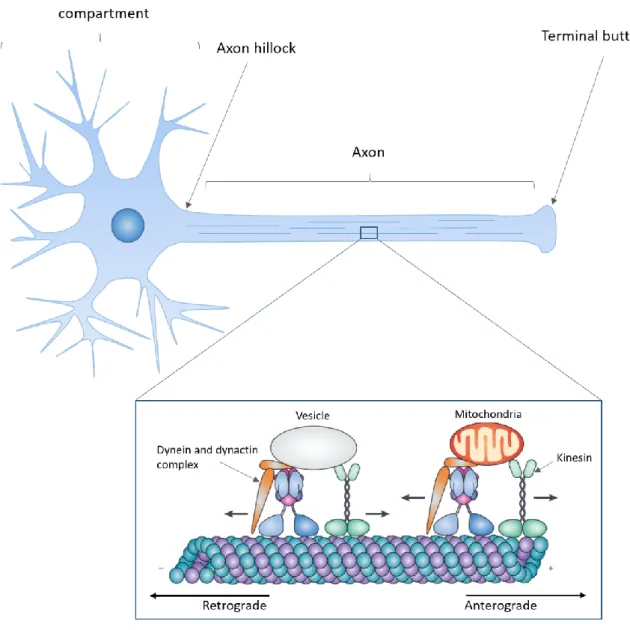
Figure 1.1: Axonal transport overview (adapted from Millecamps & Julien 2013)
Neurons are polarized cells with a dendritic arborescence and a single long axon emerging from the cell body. Microtubule tracks are distributed along the axon to support the transport of various cargos such mitochondria or proteins contained in vesicles. Anterograde transport refers to the delivery of cargos towards the axon termination which is powered by kinesin motors. Conversely, cargos transported towards the cell soma are propelled by cytoplasmic dynein in a process termed retrograde transport.
5 1. Introduction
Neurons are highly polarized cells due to their extended neurites emerging from the cell body. Dendrites emerge in a treelike fashion to receive inputs from other neurons while the axon extends over long distances to convey electrical signals generated at the axon hillock. The conduction of the axon potential and release of neurotransmitters at the synapse requires constant energy and protein supply. However, protein synthesis is restricted to the cell soma and dendrites while the axon lacks the required machinery for its high protein demands. The axon therefore relies on the synthetic abilities of the cell soma and the constant supply of these proteins by active transport, a process called axonal transport. Human motoneurons can have axons extending as far as one meter away from the cell body, highlighting the challenge that axonal transport must overcome (Twelvetrees et al., 2012).
The materials transported along the microtubule cytoskeleton are named cargos. They are propelled by kinesins and dynein motors that drive bidirectional axonal transport. Kinesins transport cargos in an anterograde manner, from the cell soma to the axon terminations to ensure the delivery of newly synthesized proteins, lipids, RNA and organelles to the axon. Conversely the retrograde movement of cargos from the axon to the cell body is mediated by dynein and is required for the degradation and recycling of these components (Twelvetrees et al., 2012). For their part, mitochondria are transported both in an anterograde and retrograde manner to adjust to energy demands (Saxton and Hollenbeck, 2012) (Figure 1.1).
Our current understanding of axonal transport supports a model in which heterogeneous cargo-specific patterns of motility ensures delivery in the destined sub-cellular compartments (Maday et al., 2014). This heterogeneity in the transport patterns and spatial delivery of cargos emerges from the diversity and fine tuning of motors, microtubules and cargos. One of the putative mechanism regulating the movement of cargos is microtubule acetylation. This post-translational modification of microtubules has been widely studied in the recent years (reviewed in Li and Yang, 2015; Sadoul and Khochbin, 2016). Although its functions remain elusive, promising studies suggest that acetylated microtubules modulate the speed and spatial delivery of the cargos they support in mammal embryonic primary cultures and drosophila larvae (Reed et al., 2006; Dompierre et al., 2007; Godena et al., 2014). Hence, I will summarize the current literature regarding axonal transport, with emphasis on the heterogeneity of the key players. I will then focus on microtubule acetylation and its role in the regulation of axonal transport, linking this post-translational modification to p27kip1, termed hereafter p27. Finally, I will introduce Drosophila Melanogaster as a model for physiological and biomolecular studies as well as axonal transport.
6 1.1 The regulation of axonal transport involves heterogenic molecular motors
The motility patterns of axonal transport can be subdivided in two main categories: slow axonal transport (reviewed in Roy, 2013) and fast axonal transport (reviewed in Maday et al., 2014). Organelles such as mitochondria or vesicles loaded with proteins are transported at fast speeds of about 400mm/day or ~ 4µm/s (Griffin et al., 1976). On the other hand, soluble proteins and neurofilament polymers are slowly transported at speeds averaging 1-10mm/day or ~ 1-10µm/s (Griffin et al., 1976). Slow axonal transport appears responsible for the trafficking of approximately three quarters of the proteins reaching synapses (Garner and Lasek, 1982; Roy, 2013). Nonetheless, the mechanisms of fast axonal transport have been extensively studied whereas slow axonal transport remains more enigmatic due to the difficulty in visualizing slow moving particles.
Breakthroughs in the field of confocal microscopy and molecular biology enabled the identification of more types of cargos and patterns of motility among fast moving organelles. Diverse cargos are transported in a compartment specific manner and at different speeds to meet specific cellular demands. This heterogeneity in motility patterns is thought to result from cargo-specific mechanisms of regulation reviewed in Maday et al., 2014. Motors but also to some extent microtubule tracks may be unique to their specific cargo. I will introduce these players while providing examples that illustrate how the diversity they display contributes to cargo-specific regulation.
1.1.1 The kinesin superfamily
Molecular motors consume ATP to actively drag cargos along the microtubule tracks. Kinesin-1, was the first protein that exhibited such properties (Vale et al., 1985) and has been determined as a driver of anterograde transport (Hirokawa et al., 1991). Later on, the microtubule-associated motor dynein was discovered (Paschal, 1987) and implicated in retrograde transport (Paschal and Vallee, 1987). Based on a database search of the human and mouse genome, 45 different kinesins have now been identified among which 38 are expressed in the mouse brain across all developmental stages (Miki et al., 2001). The expression of 20 different kinesins has also been highlighted in mature mouse hippocampal neurons cultured in vitro, illustrating the diversity of anterograde motors in neurons (Silverman et al., 2010).
Some kinesin isotypes are associated with specific cargos, which suggests cargo-specific mechanisms of transport mediated by kinesin isotypes. For instance, KIF1A, a member of the kinesin-3 family, is implicated in the transport of dense core vesicles destined to synapses (Lo et al., 2011). In the same study, Lo et al. showed that the transport of mitochondria is unaffected by the knockdown of KIF1A. Instead mitochondria transport relies mostly on KIF5A (Campbell
7 et al., 2014), KIF5B (Tanaka et al., 1998) and KIF5C (Kanai et al., 2000) of the kinesin-1 family and partially on KIF1Bα of the kinesin-3 family (Lo et al., 2011; Okada et al., 1995). Together these studies show that almost all kinesin isotypes are expressed in the brain across developmental stages but a wide diversity of kinesins are also expressed in a homogenous neuronal population. This particularity of nerve cells may reflect the need for a fine tuning of axonal transport, partially achieved by the cargo-specific binding of kinesin motors.
1.1.2 Cytoplasmic dynein complexes
Dynein is the motor driving retrograde transport and its function requires the dynein activator dynactin, a highly conserved multiprotein (Schroer, 2004). Dynactin binds to dynein and microtubules to initiate retrograde transport at the distal ends of microtubules (Moughamian and Holzbaur, 2012). Dynein itself is a complex for which the heavy chain provides the ATPase activity and binds to microtubules. The heavy chain of cytoplasmic dynein is encoded by a single gene and therefore, retrograde axonal transport relies exclusively on the heavy chain of cytoplasmic dynein to drive cargos towards the cell soma (Pfister, 2015). In addition to its role as the generator of stall force, the heavy chain of the dynein complex acts as a scaffold for the other subunits. The intermediate chain, the light intermediate chain and the three light chains bind together with two heavy chains to form the dynein complex (Trokter et al., 2012). While the heavy chain is encoded by a single gene, the other subunits display genomic diversity and all heterodimers combinations can be generated in vitro (Lo et al., 2006). The subunit diversity of dynein may thus be at the origin of cargo specific transport. For instance Mitchell et al., 2012 show that IC-2C and IC-2B, two intermediate chain isotypes, co-localize respectively with mitochondria and endosomes in rat cells.
1.2 Microtubules: malleable rails supporting axonal transport
Microtubules serve as tracks for axonal transport but have many other functions including scaffolding for cilia and flagella, chromosome segregation during mitosis, regulation of cell polarity and morphogenesis (Akhmanova and Steinmetz, 2015; Conde and Cáceres, 2009). Their role as the main support for axonal transport was discovered by Schnapp et al. in 1985 and since then, the dynamic nature and diversity of microtubule tracks emerge as modulators of axonal transport.
1.2.1 Microtubule structure and dynamics
Microtubule filaments consist of dimers of α and β tubulin that associate together in a non-covalent and dynamic way (reviewed in Akhmanova and Steinmetz, 2015). In the axon, all microtubules are uniformly polarized with their fast growing +end directed towards the axon
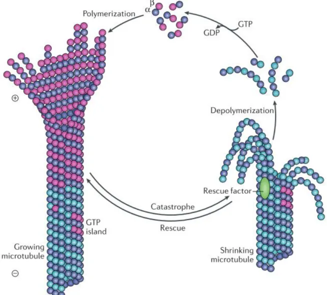
Figure 1.2: Microtubule dynamics (from Akhmanova & Steinmetz 2015)
Microtubules are constituted by dimers of α- and β-tubulin that polymerize predominantly at the + end of microtubules. GTP-bound tubulin dimers are incorporated into growing microtubules and GTP-hydrolysis occurs progressively so that a GTP-cap is maintained at the tip of growing microtubules. Microtubules are stabilized by GTP-bound tubulin whereas GDP-bound tubulin has the opposite effect. The presence of the GTP-cap ensures the stability of the growing microtubule but once the kinetics of GTP-hydrolysis outweighs the incorporation of new tubulin dimers, the GTP-cap is lost and the microtubule lattice becomes unstable. The fast depolymerization of microtubules is termed “catastrophe” and it can be “rescued” by GTP islets or rescue factors. The “rescue” transiently stabilizes microtubules to enable the incorporation of new tubulin dimers and therefore the formation of a new GTP-cap.
8 terminations (Stepanova et al., 2003). They alternate between phases of polymerization and depolymerization in a cyclic manner, a process termed “dynamic instability”, first coined by Mitchison and Kirschner, 1984. In short, GTP-bound β-tubulin stabilizes microtubules whereas GDP-bound β-tubulin changes the conformation of the dimer and induces strain on the microtubule’s lattice (Alushin et al., 2014). According to a conceptual model first described in (Carlier and Pantaloni, 1981), growing microtubules display a GTP cap that stabilizes their structure and enables them to grow further. As their length increases, GTP is hydrolyzed into GDP, which affects allosteric protofilaments interactions and destabilizes microtubular structure (Yajima et al., 2012). If the GTP cap is lost, the stability of the + end is compromised and the microtubule is prone to depolymerization. The fast depolymerization of microtubules is termed catastrophe and it can be rescued once the GTP cap is restored (Desai and Mitchison, 1997) (Figure 1.2).
Polymerizing microtubules bind a complex of plus-end interacting proteins or +TIPs which subsequently facilitates the initiation of retrograde transport through dynactin interaction with dynein (Moughamian et al., 2013). The very dynamic nature of microtubules may thus contribute to the regulation of transport in addition to the versatility it confers to these filaments.
1.2.2 Microtubule diversity
Interestingly, wide heterogeneity exists between microtubules due to many different tubulin isotypes (reviewed in Janke, 2014), post-translational modifications (PTMs) on tubulin residues (reviewed in Song and Brady, 2015) and a myriad of microtubule associated proteins (MAPs) (reviewed in Akhmanova and Steinmetz, 2015).
Tubulin isotypes are α- or β-tubulin proteins with small variations in their amino acid sequence that are encoded in a single organism (Ludueuna, 1993). They are found in many species including human and rodents (Little and Seehaus, 1988) but also drosophila (Rudolph et al., 1987) and they are expressed in a tissue specific manner. For instance, in vertebrates, while β1 is found in most tissues, β2 is enriched in the brain and β3 is specific to neurons (Sullivan, 1988). In vitro research has shown that different kinesin motor subtypes exhibit different velocities depending on the tubulin isotype composing the microtubule track (Sirajuddin et al., 2014). This supports a model in which microtubule rails each have a “tubulin code” giving specificity to the transport of cargoes.
MAPs constitute a complex interconnected network of proteins that bind to microtubules to exert diverse effects on their dynamic behavior (Akhmanova and Steinmetz, 2015). Some MAPs have been implicated in the regulation of sub-cellular specific delivery. For instance, CLIP-associating protein 2 (CLASP2) is a MAP that promotes the capture of microtubule +
9 ends at the synaptic membrane. This microtubule network organization subsequently drives the focal delivery of acetylcholine receptors by vesicles transported along the attached microtubule (Basu et al., 2015). Vershinin et al., 2007 also demonstrated a role of MAPs in the regulation of axonal transport in vitro by showing that the mainly axonal MAP tau reduces kinesin motors attachment rate. In the presence of tau, kinesin motors deploy less force and are more likely to dissociate from the microtubule.
In addition to tubulin isoforms and MAPs, PTMs of tubulin are emerging as decisive regulators of microtubule properties and function and as a major contributor to microtubules diversity. PTMs are covalent modifications of proteins that broaden the scope of protein functionalities. The most studied microtubule PTMs are detyrosination, glutamylation, glycylation, phosphorylation and acetylation. These modifications modulate microtubule functions and can occur differentially in specific sub-cellular compartments (Janke and Chloë Bulinski, 2011). Together, tubulin isoforms, MAPs and PTMs define microtubule identities at the tissue and cellular level to adjust microtubule function. As the aim of this master’s thesis was to shed light on the regulation of axonal transport by microtubule acetylation, we will now focus on this specific PTM.
1.3 Microtubule acetylation
Although most PTMs occur at the C-terminal domain of tubulins, which extends in the cytoplasm, acetylation occurs on the lysine 40 (Lys40) of α-tubulin. This amino-acid is located inside the lumen of microtubules, making α-tubulin acetylation the only microtubule PTM occurring inside the microtubule lattice (Howes et al., 2014). The degree of microtubule acetylation depends on the balance between acetyltransferase and deacetylase activity. In recent years, the main effectors of acetylation have been identified but their regulation and the biological role of acetylation remains elusive (reviewed in Li and Yang, 2015; Sadoul and Khochbin, 2016).
1.3.1 Tubulin deacetylases and acetyl-transferases
The deacetylation of tubulin is carried out by histone deacetylase 6 (HDAC6) (Hubbert et al., 2002; Zhang et al., 2003) and sirtuin 2 (SIRT2) (North et al., 2003) which act together to reverse Lys40 acetylation on tubulin. Even though HDAC6 and SIRT2 are part of the histone deacetylase family, only SIRT2 has the ability to deacetylate histones (Seidel et al., 2015). SIRT2 is predominantly cytoplasmic but is transported in the nucleus during mitosis to achieve its function as an histone deacetylase (North and Verdin, 2007). On the other hand, HDAC6 is observed in the nucleus of undifferentiated cells but relocates to the cytoplasm after
10 differentiation for many tissues including the brain (Chen et al., 2013). Both HDAC6 and SIRT2 display multiple functions due to the varied targets they deacetylate such as cortactin and heat shock protein (HSP)90 for HDAC6 or p53 and histone H4 for SIRT2 (reviewed in Harting and Knöll, 2010; Seidel et al., 2015).
The ARD1-NAT1 complex was the first reported microtubule acetylator in purified microtubule fractions (Ohkawa et al., 2008). Studies in mice and C. Elegans then described an α-tubulin acetyl-transferase activity by Elp3 of the Elongator complex (Creppe et al., 2009; Solinger et al., 2010a). Next followed the discovery of GCN5 (Conacci-Sorrell et al., 2010), an histone acetyl transferase. Finally, alpha-tubulin N-acetyltransferase 1 (αTAT1) was described as the main acetyl-transferase in mammals (Akella et al., 2010; Kim et al., 2013). αTAT1 is expressed across all tissues in embryonic and adult mice and αTAT1 knockout mice display no detectable microtubule acetylation in the brain (Kim et al., 2013). In C. Elegans, the depletion of αTAT1 or its paralog alpha-tubulin N-acetyltransferase 2 (αTAT2) resulted in diminished but detectable levels of microtubule acetylation while double null mutants exhibited no detectable α-tubulin acetylation in whole worm lysates (Shida et al., 2010). These findings indicate that αTAT1 in mammals, or αTAT1 and αTAT2 in C. Elegans are the main acetylators of microtubules while Elp3, GCN5 and ARD1-NAT1 may only be responsible for the regulation of tubulin acetylation or minor tubulin acetylation in vivo.
1.3.2 Biological functions of tubulin acetylation
The study of microtubule acetylation functions has become more accessible with the discovery of the main enzymes regulating tubulin acetylation levels. It is now possible to manipulate these enzymes to observe the physiological processes underlying microtubule acetylation (reviewed in Li and Yang, 2015; Sadoul and Khochbin, 2016).
Various studies have been carried out on the link between microtubule acetylation and stability with conflicting results that may be due to the diversity of cell types and models that have been used. Microtubules with a high turnover rate tend to be less acetylated than the stable ones in human fibroblasts (Webster and Borisy, 1989) and stable microtubules in the axons display high levels of acetylation (Cambray-Deakin and Burgoyne, 1987). These observations led to the hypothesis that tubulin acetylation stabilizes microtubules. However, non-acetylated stable microtubules or highly acetylated unstable microtubules have been observed in marsupial kidney cells and chick embryo fibroblasts respectively (Schulze et al., 1987). This suggests that microtubule acetylation is not a prerequisite for increased stability. In addition, HL-1 atrial myocytes exhibit a normal response to nocodazole, a drug inducing microtubule depolymerization, notwithstanding that their microtubule network is fully acetylated
11 (Belmadani et al., 2004). This suggests that even though acetylation is often correlated with aged microtubules in some cell types, there may not be a causative link between acetylation and microtubule stability.
Promising studies have now implicated the degree of α-tubulin acetylation in transport with particular emphasis on neurons. Reed et al., 2006 have demonstrated that microtubule acetylation increases kinesin-1 binding and moving speed. They also show that the cargo protein JNK-interacting protein 1 (JIP1) is transported by kinesin-1 along acetylated microtubule tracks towards a subset of neurite tips. This suggests a contribution of microtubule acetylation to sub-compartment specific delivery as well as a modulation of transport speed. When acetylation of microtubules was upregulated by a pharmacological inhibitor of HDAC6, the entire microtubule network exhibits high levels of acetylation and JIP1 then accumulates in all neurite tips (Reed et al., 2006). This remarkable observation suggests that only a subset of microtubules may be acetylated in the dendrites of hippocampal neurons thus serving as rails driving specific cargos to their destined subcellular spaces. In a model of Huntington’s disease, Dompierre et al., 2007 have shown that HDAC6 inhibition increases microtubule acetylation and subsequently increases the speed of vesicular transport. In a similar study conducted in a drosophila model of Parkinson’s disease, Godena et al., 2014 have shown that mutant leucine-rich repeat kinase 2 (LRRK2) interacted with deacetylated microtubules to inhibit axonal transport. Following upregulation of microtubule acetylation by HDAC6 inhibitors or αTAT1 overexpression, the affinity of LRRK2 for microtubules was diminished and the transport defect was rescued. Finally, HDAC6 inhibitors have also been used to rescue axonal transport defects in a mouse model of Charcot-Marie-Tooth disease (d’Ydewalle et al., 2011). This treatment remarkably rescued the locomotor behavior of the mutant mice, suggesting that rescuing axonal transport by increasing microtubule acetylation may be a putative therapeutic strategy for some neurodegenerative diseases (Millecamps and Julien, 2013). Taken together, these studies strongly suggest that microtubule acetylation may act to regulate the speed of intracellular trafficking but also the specificity of cargo transport through differential acetylation in the microtubule network of a single neuron.
1.4 Dacapo/p27kip1 may be involved in the regulation of axonal transport
The fine tuning of transport requires intricate molecular pathways that remain to be comprehensively understood. Studying the upstream regulators of transport such as the ones orchestrating microtubule acetylation would enable a broader understanding of axonal transport and promote the discovery of pharmacological targets to modulate transport in disease states.
Figure 1.3: p27kip1 is an intrinsically disordered multifunctional protein (Adapted from (Godin & Nguyen
2014))
A) p27 is an intrinsically disordered protein without a fixed three dimensional structure. p27 binds multiple targets through specific domains which subsequently induces the folding required to exert its function. The C-terminal part interacts with various proteins and bears the proline-rich domain necessary for p27 microtubule-associated function but not the ability to bind microtubules. Instead p27 appears to bind microtubules through both the N and C-terminal. The cell-cycle related functions of p27 rely on the N-terminal where the cyclin-dependent kinase (CDK) and cyclin binding sites are localized. The N-N-terminal part is also necessary for the stabilization of Neurogenin-2 (Ngn2) by p27, an activity that does not require the CDK and cyclin binding sites. To regulate the shuttling of p27 between the nucleus and cytoplasm, a putative nuclear localization signal (NLS) and nuclear export signal (NES) have been identified. B) Multiple functions of p27 have been unraveled in the context of corticogenesis. The canonical function
of p27 is to promote cell-cycle exit by associating with CDK/cyclin complexes to prevent G1-S phase transition. Independently from its cell-cycle dependent activities, p27 is able to regulate neuronal differentiation by stabilizing Ngn2, a proneuronal transcription factor. p27 also promotes neuronal migration in both interneurons and projection neurons by interacting with proteins involved in cytoskeleton remodeling. In interneurons and projection neurons, p27 inhibits RhoA leading to a reorganization of the actin cytoskeleton that is required to ensure the proper migration. In interneurons specifically, p27 controls migration by binding to microtubules and promoting their polymerization.
12 With this in mind, we aim to shed light on dacapo, the Drosophila Melanogaster homolog of p27kip1, hereafter named p27.
1.4.1 Dacapo/p27 regulate the cell cycle
p27 is a protein of the Cip/Kip family originally discovered as a cell cycle regulator (Toyoshima and Hunter, 1994). It inhibits the G1/S phase transition by regulating a broad range of cyclin-dependent kinases (CDKs) using its conserved CDK-inhibitory domain in the N-terminal (Lacy et al., 2004). Multiple cancers display mutations in p27 due to its role in cell cycle regulation, which justifies the large body of research related to p27 relation with cancer (Bencivenga et al., 2017).
In drosophila, the literature has mostly focused on dacapo as a cell cycle regulator that inhibits cyclin E/cdk2 complexes to arrest the G1 phase at a specific developmental stage (Lane et al., 1996). During the drosophila brain development, dacapo is specifically upregulated in postmitotic cells called ganglion cells to stop their proliferation (Colonques et al., 2011).
1.4.2 p27 is a multifunctional protein with cell-cycle independent functions
Proteins of the Cip/Kip family have now emerged as multifunctional proteins with cell cycle-independent functions such as transcriptional regulation, cell migration, cell fate determination or cytoskeletal dynamics (Besson et al., 2008). To our knowledge, non-canonical functions of dacapo have not yet been demonstrated in flies. However, the homology in the amino acid sequence of p27 and dacapo suggests a putative functional homology. p27 exerts its many functions by virtue of its intrinsically disordered nature (Bienkiewicz et al., 2002; Wang et al., 2011) (Figure 1.3A). This protein and the Cip/Kip paralogs lack a secondary or tertiary structure in physiological conditions. Instead they bind to their many targets through specific domains which subsequently induces the folding of the protein responsible for its activity (Yoon et al., 2012).
Non-canonical functions of p27 have been studied in the brain where this protein has been linked to the control of neuronal differentiation and migration during cortical development (reviewed in Godin and Nguyen, 2014) (Figure 1.3B). Cortical development involves a tight regulation of neuronal differentiation and migration by transcriptional programs and molecular signaling pathways. In mice, the genesis of the cortical layers occurs between E11 and E18 (Gupta et al., 2002). p27 is expressed throughout the developing mouse cortex, both in progenitors and postmitotic neurons (Itoh et al., 2007). The N-terminal of p27 has been shown to stabilize the transcription factor Neurogenin-2, crucial to trigger the switch between the progenitor and the neuronal state (Nguyen et al., 2006). Also, the C-terminal tail of p27 promotes neuronal migration in interneurons and projection neurons through a cooperative
Figure 1.4: Life cycle of the Drosophila Melanogaster (From http://www.creative-diagnostics.com/)
The life cycle of the drosophila comprises four stages: egg, larva, pupa and fly. 8 hours after eclosion, female adults will mate and shortly after they will lay eggs that hatch in a day. The larval stage lasts for an average of 5 days during which larvae pass through the stages of 1st instar, 2nd instar and 3rd instar. Once larval development is complete, animals metamorphose within a pupal case over the course of approximately 4 days. Adult flies then emerge from the case in a process termed eclosion thus allowing the cycle to repeat itself.
Figure 1.5: The GAL4/UAS system (from St Johnston 2002)
The GAL4/UAS system is a regulator of transcription in yeast that was adapted as a bipartite tool for targeted gene expression in drosophila. GAL4 is a transcription factor binding to the upstream activating sequence (UAS), a genomic enhancer. Fly lines expressing GAL4 under the control of a specific promoter can be generated to drive the expression of GAL4 in various cell and tissue types. Corresponding fly lines expressing a gene of interest (gene X) downstream of the UAS sequence can then be generated. The gene X is therefore transcriptionally silent until a fly line expressing GAL4 is crossed with the flies carrying the UAS enhancer. The resulting progeny will then express gene X in a transcriptional pattern reflected by the GAL4 expression pattern which depends on the promoter driving GAL4 expression.
14 in the pupal stage at the end of which eclosion occurs. Females are then sexually mature 8 hours after eclosion, thus allowing the cycle to repeat itself (Hales et al., 2015) (Figure 1.4).
1.5.2 Balancers and the bipartite UAS/GAL4 system
The most attractive characteristic of flies probably is the wide range of genetic tools that have been developed over the years. For the fly’s geneticist, the most precious of these tools are balancer chromosomes (Hales et al., 2015) and the UAS/GAL4 system (Brand and Perrimon, 1993).
For a genetic model such as flies, the ability to create a stable transgenic inbred stock and subsequently cross these stocks to generate an offspring with a known genotype is a prerequisite. Balancer chromosomes are the fundamental tool that enables both of these necessities. They carry a recessive lethal mutation in order to prevent the mutation of interest from being selected out other multiple crossings. Balancer chromosomes additionally carry a phenotypic marker to unambiguously identify the chromosomes inherited by each fly following a specific crossing (Greenspan, 1997; Hales et al., 2015).
The UAS/GAL4 system is a regulator of transcription in yeast that is used as a bipartite tool in transgenic drosophila to enable targeted gene expression (Brand and Perrimon, 1993). GAL4 is a transcription factor binding to the upstream activating sequence (UAS). Fly lines expressing GAL4 in a specific cell line or tissue can be generated using vectors in which GAL4 is cloned with an upstream specific promoter. A corresponding fly line expressing a gene of interest downstream of the enhancer UAS can then be generated. The transcription of the gene of interest therefore requires the concomitant expression of the GAL4 transcription factor. The absence of GAL4 in the fly line carrying the gene of interest maintains them in a transcriptionally silent state. The expression of the gene of interest is triggered by crossing the flies expressing GAL4 with the flies carrying the UAS enhancer. The resulting progeny will then express the gene of interest in a transcriptional pattern reflected by the GAL4 expression pattern which depends on the promoter driving GAL4 expression. Similarly to the fly life cycle, GAL4 activity depends on temperature. Minimal GAL4 activity is measured at 16°C while 29°C is the optimal balance between maximal GAL4 activity and minimal effects on fertility and viability (Duffy, 2002). It is therefore possible to adjust the expression level of the gene of interest or perform a conditional expression at a specific developmental stage by altering the rearing temperature (Duffy, 2002; Hales et al., 2015) (Figure 1.5).
Using these tools, fly stocks can be maintained, genotypes can be visually identified and sequences of interest can easily be expressed in specific cell types or tissues.
15
1.5.3 The drosophila central nervous system
The organization of the fly nervous system also makes it an attractive model. The central nervous system (CNS) of most insects including flies displays an outer layer or cortex containing cell bodies and an inner layer formed by multiple neuropiles. Neuropiles are dense regions of the brain containing numerous axons and dendrites to form circuits (Nassif et al., 2003). In insects, the brain is prolonged by a ventral nerve cord (VNC) organized in neuromeres which are parts of the CNS that process sensory inputs and control motor behavior for a single segment of the body (Niven et al., 2008). Networks in the brain and VNC are responsible for stereotyped sequences of movement called central pattern generators. In drosophila larvae, the feeding and crawling behavior are the two main stereotyped motor programs (Cardona et al., 2009). Interestingly, the crawling movement of drosophila larvae displayed right after hatching can still be observed when only the medial and posterior portion of the VNC is active, suggesting that the network responsible for the waves of peristaltic movements resides in this part of the nervous system. Brain inputs still seem crucial for goal directed behavior such as chemotaxis (Berni et al., 2012).
The VNC mirrors the arrangement of body segments so that motoneurons from anterior neuromeres contact muscles in the anterior part of the body whereas conversely, the most posterior muscles are innervated by motoneurons emerging from the posterior neuromeres of the VNC. In drosophila larvae, the interaction between motoneurons and muscles is known as the neuromuscular junction (NMJ) and it has been extensively studied over the years due to its simplicity, ease of study and robustness (Ruiz-Cañada and Budnik, 2006). Using presynaptic and/or postsynaptic markers, the synaptic morphology of the NMJ can be observed in a highly reproducible way by quantifying the number of synaptic boutons, their size or even fluorescence intensity (Johnson et al., 2009; Mao et al., 2014; Xiong et al., 2013).
1.5.4 Drosophila as a model for axonal transport
Drosophila models allow the study of gene mutations, overexpression and knockdown but physiological processes such as axonal transport can also be visualized with ease. Flies are thus widely used in the field of neurosciences to shed light on molecular mechanisms regulating axonal transport (Horiuchi et al., 2005) but also as models for neurodegenerative diseases (Lessing and Bonini, 2009).
Fly lines are readily accessible to express a recombinant green fluorescent protein (GFP) specifically in the mitochondria (Pilling et al., 2006) or synaptic vesicles (Zhang et al., 2002) of motoneurons. These lines can then be crossed to genetically manipulate the regulation of axonal transport and perform rescue experiments. Using these in vivo markers of cargoes,
16 axonal transport can be studied in 3rd instar larvae (Zala et al., 2013), in pupal brain explants (Medioni et al., 2015) but also in the wings of adult flies (Vagnoni and Bullock, 2016). The transparency of 3rd instar larvae and the anatomical configuration of the brain and motoneurons makes it a particularly attractive model for the study of axonal transport. The brain and the large axons of motoneurons are indeed juxtaposed to the ventral muscles of the larva transparent body, making it easy to record live-images in motoneurons using a confocal microscope.
1.5.5 Locomotor behavior assessment in drosophila
Neurodegenerative diseases are often linked with locomotor behavior defects, thus making the assessment of motor behavior primordial for the comprehensive understanding of a pathological state. The locomotor behavior of flies enables an easy and quantitative readout of motor function (Nichols et al., 2012) thus reinforcing its usefulness in the context of brain related studies.
The crawling behavior of larvae can be recorded and then analyzed to provide insights on the locomotor capacities following a pharmacological or genetic manipulation. The distance covered by the larvae as well as the number of peristaltic movements exhibited in a specific time span can be quantified. In adult flies, the climbing assay is a reproducible and easy readout of motor function. In this assay, adult flies are placed in transparent vials and mean height climbed in a specific time span is assessed. Flies with locomotor defects will tend to climb to lower heights that controls (Nichols et al., 2012).
Multiple studies have shown locomotor defects in some fly models of neurodegenerative diseases (Cragnaz et al., 2015; Lanson et al., 2011) with some highlighting motor behavior abnormalities linked with axonal transport defects (Janssens et al., 2014; Johnson et al., 2013).
1.5.6 Drosophila display a remarkable homology with mammals
Despite a smaller genome size, flies have orthologous genes for 80% of human disease-associated genes (Reiter, 2001) and all major signal transduction pathways are conserved between flies and humans (Perrimon et al., 2012). This remarkable homology between drosophila and mammals highlights flies as a fantastic tool for the study of gene function (Perrimon et al., 2016). In addition, the increased genome size in mammals reflects the genesis of multiple paralogs for various gene families with redundant and overlapping functions. The simpler genome of flies therefore facilitates the interpretation of loss of function studies (McGurk et al., 2015).
Relevant to my master’s thesis, αTAT1, αTAT2, Elp3, ARD1, GCN5, HDAC6 and SIRT2 all have orthologs in flies based on the Uniprot database. These proteins involved in microtubule acetylation were mostly studied in mammals and C. Elegans therefore less is known about the
17 mediators of acetylation in flies. So far, only αTAT1 and HDAC6 have been linked with the regulation of microtubule acetylation in drosophila (Godena et al., 2014).
1.6 Aim of the master’s thesis
Over the course of my master’s thesis, we studied the role of dacapo -the Drosophila Melanogaster ortholog of p27- as a modulator of axonal transport. Based on the previous literature, we identified p27 as a candidate for the regulation of axonal transport. Some MAPs have been shown to regulate axonal transport (Vershinin et al., 2007; Basu et al., 2015) and microtubule dynamics also contribute to the modulation of transport (Moughamian et al., 2013). In this sense, the discovery that p27 is able to bind to microtubules and promote microtubule polymerization in cortical interneurons is compelling (Godin et al., 2012). Interestingly also, in mouse fibroblasts, p27 depletion resulted in a decrease of acetylated α-tubulin while p27 overexpression conversely increased α-tubulin acetylation levels (Baldassarre et al., 2005). This suggests that p27 may contribute to the regulation of acetylation levels in microtubules. As stated before, microtubule acetylation plays an important role in the speed and specificity of cargo trafficking (Reed et al., 2006; Dompierre et al., 2007; d’Ydewalle et al., 2011; Godena et al., 2014) and as such, p27 may act as a regulator of axonal transport through the modulation of acetylation levels on microtubules.
We used transgenic fly lines expressing a specific dacapo RNAi in motoneurons to knockdown dacapo. We show that the velocity of GFP-tagged mitochondria was reduced following dacapo knockdown in 3rd instar larvae mtoneurons. Using immunostainings, we then confirmed that dacapo knockdown downregulates tubulin acetylation by quantifying the acetylation levels of microtubules in dacapo depleted motoneurons. To demonstrate a causal link between decreased microtubule acetylation and axonal transport defect in dacapo depleted larvae, we rescued physiological microtubule acetylation levels using pharmacological inhibition of HDAC6 as described in previous studies (Dompierre et al., 2007). We provide data showing that the transport defect is rescued consequently to the restoration of microtubule acetylation levels, suggesting that dacapo mediates its regulatory effect on axonal transport through the modulation of microtubule acetylation. Morelli et al. (submitted) have highlighted locomotor behavior defects in dacapo knockdown larvae and adult flies that are rescued by acute HDAC6 inhibition. We therefore performed immunostainings of the larval NMJ to determine whether this defect is caused by morphological alterations to the motoneurons and their synapses. We highlighted no defect in the number of synaptic buttons of dacapo knockdown larvae, therefore suggesting that the locomotor defects do not arise from developmental alterations in NMJ morphology but rather from a malfunction of the synaptic transmission.
Figure 2.1: Discrimination between Drosophila Melanogaster males, females and female virgins (from
http://flymove.uni-muenster.de)
From left to right a male (♂), female (♀) and virgin female (☿) drosophila under a dissection microscope. Males are on average smaller and darker than females. While females have a straight and round abdomen, the abdomen of males is thinner and slightly curved inwards. The easiest trait to distinguish males and females is the brown genitals on the postero-ventral side of males. In the 3 hours following eclosion, flies exhibit a dark spot named meconium in the upper left corner of the abdomen which corresponds to the remainings of the last meal as a larva. Since females start mating 8 hours after eclosion, the presence of the meconium is a guaranteed way to single out virgin females.
18 2. Material and methods
2.1 Drosophila Melanogaster work
2.1.1 Fly stocks
We used a D42-GAL4 > UAS-mitoGFP Drosophila Melanogaster that expresses a specific recombinant GFP displaying a mitochondrial import sequence. The expression of GFP is driven by the D42 promoter, which is subsequently localized in the mitochondria of motoneurons and salivary glands (Pilling et al., 2006). The D42-GAL4 > UAS-mitoGFP was crossed with specific lines expressing RNAi driven by the upstream activation sequence (UAS) (Table 2.1).
Fly line Origin
D42-GAL4 > UAS-mitoGFP Bloomington Drosophila Stock Center 42737
UAS-RNAi-Dacapo Kind gift from Pr. Perrimon
UAS-RNAi-Dacapo ; P(w+,dap1gm)III.1 (Dacapo overexpression)
Generated in the lab with P(w+,dap1gm)III.1 kindly received from Pr. Lehner (Meyer et al., 2002)
UAS-RNAi-ZPG Vienna Drosophila Resource Center CG10125
Table 2.1: Sources of the fly lines used in this thesis
2.1.2 Culture conditions and husbandry
Flies were reared in a standard culture medium adapted from Bloomington Drosophila Stock Center recipes (http://flystocks.bio.indiana.edu). The medium was prepared by mixing in hot water for approximately one hour: brewer yeast extract (Acros Organics 368080050), corn flour (Genesee Scientific #62100), agar (Sobigel SB12073) and the antimycotic methyl 4-hydroxybenzoate (Acros Organics 126965000) at a concentration of 2.5g/L. Once dense, the medium was poured into plastic vials and dried overnight. Foam caps were then used to close the vials which were kept at 4°C for optimal conservation or 20°C for experimental purposes. The fly lines were maintained at 25°C and 20°C; the former temperature was utilized to promote the production of D42-GAL4 > UAS-mitoGFP virgin females for crossing whereas the latter was employed for rigorous control of fly life-cycle and husbandry.
2.1.3 Crossings
Males and females were segregated based on their morphological differences as described in Figure 2.1. Males are on average smaller than females and their abdomen is thin and slightly curved inwards. Conversely the abdomen of females is straight and round (Greenspan, 1997).
19 The genital apparatus of males appear as a relatively large brown mass in the postero-ventral part of the abdomen while the female genitals are less noticeable (Greenspan, 1997). Virgin females were singled-out based on the presence of the meconium, a dark spot in the upper left corner of the abdomen which is visible up to 3 hours after eclosion (Greenspan, 1997). Since females do not mate in the first 8 hours following eclosion, the presence of the meconium guarantees the virginity of the females (Greenspan, 1997).
Balancer chromosomes carry a dominant marker mutation and are recessive lethal. Based on these properties, flies can only carry one or no balancer chromosome and they can be discriminated based on the dominant phenotypic marker expressed on the balancer chromosome (Hales et al., 2015b). Cyo flies carrying the balancer will exhibit curled wings (Cy marker) while TM6B flies, larvae and pupae are shorter and thicker than wild-types (Tubby marker). To obtain a homogenous heterozygote progeny from a crossing, it is required to cross homozygous virgin females with homozygous males. Flies carrying a balancer chromosome must therefore be excluded based on the phenotypic trait they display. Some of the fly lines used in Table 2.1 carried balancer chromosomes. To ensure the homogeneity of the progeny, only homozygous males carrying neither Cy or Tubby were crossed with D42-GAL4 > UAS-mitoGFP virgin females. Females and males were kept for a duration of 2 to 3 days at 25°C and then transferred in a new vial. The eggs laid over the course of this period were kept at 29°. This temperature has been determined as the optimal balance between maximal GAL4 activity and minimum effects on fertility and viability (Duffy 2002). 3rd instar larvae were collected 5 to 6 days after the start of the crossing and were used for immunohistochemistry or transport studies. Due to inter-individual variability, each larva was checked on an epifluorescent microscope before experiments to ensure that GFP was expressed.
2.2 Drug administration
When tubastatin treatment was required, tubastatin A 1mM (Sigma) or an equivalent amount of dimethyl sulfoxide (DMSO) were diluted in a 10% sucrose solution. Larvae were then incubated in the solution for 30min using a 24 well plate with 500µL per well. After incubation, larvae were dried properly and dissected immediately for immunohistochemistry or anesthetized for time-lapse imaging of transport.
2.3 Larval dissection for immunostainings of motoneurons or neuromuscular junction This protocol is adapted from (Brent et al. 2009) and (Devireddy et al. 2014).
Figure 2.2: Critical steps of larval dissection and CNS representation
(A) Critical steps of the larva dissection, in all frames the anterior part is up. In order, the larva is placed on the
dissection plate (1), it is then pinned at its anterior and posterior extremity (2). PBS is added and a hole is pierced adjacent to the posterior pin (3) and a longitudinal cut from the posterior to the anterior end is performed (4). The organs are floating in PBS (5), making it easier to remove those using fine forceps (6). When most organs have been removed, 4 pins are inserted to flatten the body walls (7) and the remaining organs are removed (8). (B) Magnification of the frame drawn in (A8). (C) Annotated version of (B) highlighting the brain lobes, ventral nerve cord and motoneurons. (D) Confocal image of a 3rd instar larva D42 > mitoGFP brain at 20x after dissection.
20 Dissection microscope (Leica M80) with incident illumination
Vannas iris scissors (Aesculap OC498R)
Two pairs of forceps (Fine Science Tools - Dumostar #55) Minutien insect pins (Fine Science Tools #26002-10)
Homemade Sylgard plate with small edges (Sylgard 184 Silicone Elastomer Kit from Dow Corning)
PBS buffer PFA 4%
10% sucrose solubilized in distilled water.
1mL of the sucrose solution at room temperature was added in each well of a 12-wells plate and 3rd instar larvae were segregated in the wells according to their genotype. The sucrose solution ensures that larvae have access to food while preventing them from crawling on the walls of the petri dish. Larvae were usually kept for a maximum of one hour in this solution before a new batch was collected. Once in the sucrose solution, the GFP expression is checked with a binocular epifluorescent microscope. The larvae exhibiting no GFP in the nervous system and salivary glands were excluded.
The description of the procedure is illustrated with 8 key frames in Figure 2.2A which are referred to only by their number in the following text. A larva was transferred from a well to the Sylgard plate with the ventral part of the body facing downwards to make the pharyngeal tubes apparent (1). An insect pin was then inserted through the posterior part of the body between the pharynges. A second pin was inserted in the anterior part between the mouth hooks to slightly stretch the larva and hold it in place (2). 100µL of PBS was added to submerge the larva in order to make internal organs float which facilitated their removal (5). Using a larger pin, a small hole was created by gently pulling on the skin that was pierced at the posterior end (3). A longitudinal cut along the midline was then performed starting from the posterior end to the anterior end (4). This step is critical as the integrity of the nervous system must be preserved. To avoid damaging motoneurons or the brain, the cut must be made just underneath the larval body wall. Organs (fat, salivary glands, intestine, pharyngeal tubes) were grossly removed with fine forceps (6) while avoiding strong pulling forces on the segmental nerves. 4 pins were then placed on the 4 extremities of the body wall to flatten the larva and stretch it in a horizontal and vertical manner (7). The remaining organs were then carefully removed to leave only the CNS and peripheral nerves (8). Higher magnifications of the brain revealed by the dissection are shown in Figure 2.2 B-D. The PBS was removed using a micropipette and 100µL of PFA 4% was added. After 20 minutes at room temperature, the PFA was removed and the larva was
21 washed 3 times with PBS. The pins were then gently pulled while holding the body of the larva to prevent ripping. The dissected larvae can either be used for immunostaining directly or they can be stored at 4°C for a few weeks in a plastic container filled with PBS.
2.4 Immunohistochemistry and confocal imaging of dissected larvae
Immunochemistry experiments were performed in glass dishes with a spherical bottom which makes it easy to remove and add liquid without damaging the larvae. The antibodies used were tubulin 1:150 from sheep (Cytoskeleton ATNO2), acetylated-tubulin 1:5000 from mouse (Sigma, T6793), horse raddish peroxidase (HRP) 1:1000 from rabbit (Sigma) and cysteine string protein (CSP) 1:10 from mouse, kindly gifted by S. Benzer. Secondary antibodies against the appropriate species were used with a 1:500 dilution.
Larvae were washed 3 times in PBS 0.3% Triton X-100 for 5 minutes and incubated for 30 minutes in the blocking solution of PBS 0.3% Triton X-100 and 1% bovine serum albumin (BSA). Larvae were incubated at 4° over-night in the primary antibodies diluted in the blocking solution. To prevent evaporation of the solution, the dish was covered with parafilm. Larvae were then washed 3 times in PBS 0.3% Triton X-100 for 5 minutes and incubated for 2 hours at room temperature in the secondary antibodies diluted in the blocking solution. Larvae were washed 3 times in PBS 0.3% Triton X-100 for 5 minutes and immediately mounted on glass slides with the nervous system facing upwards. A maximum of 3 larvae were mounted per slide using 100µL of the mounting medium Mowiol. The slides were kept at room temperature for a few hours for the Mowiol to dry and then stored at 4°C until imaging.
This protocol was used for tubulin/acetylated tubulin stainings. The protocol for CSP/HRP staining is identical but PBS 0.2% Triton X-100 was used at each step.
Larvae were imaged using a Nikon A1Ti inverted confocal microscope. For quantitative analysis, laser power, gain and offset were optimized to avoid surexposition while maintaining adequate sensitivity. The optimized settings were used throughout experiments to ensure samples were comparable. Imaging of tubulin/acetylated tubulin stainings was performed using the 60x oil-immersed objective while the 40x oil-immersed was used for CSP/HRP.
2.5 Preparation of the larvae for transport live-imaging
Larvae were collected in sucrose 10% and the GFP expression was checked as described before. A maximum of 3 larvae were then dried on absorbing paper and placed in a perforated falcon cap. The closed falcon was placed upside down in a sealed beaker containing paper soaked in ether. Larvae were kept in the ether fumes for 8 minutes, a duration optimized by Morelli et al. to ensure survival and efficient anesthesia. Each larva was then placed on a glass slide with the
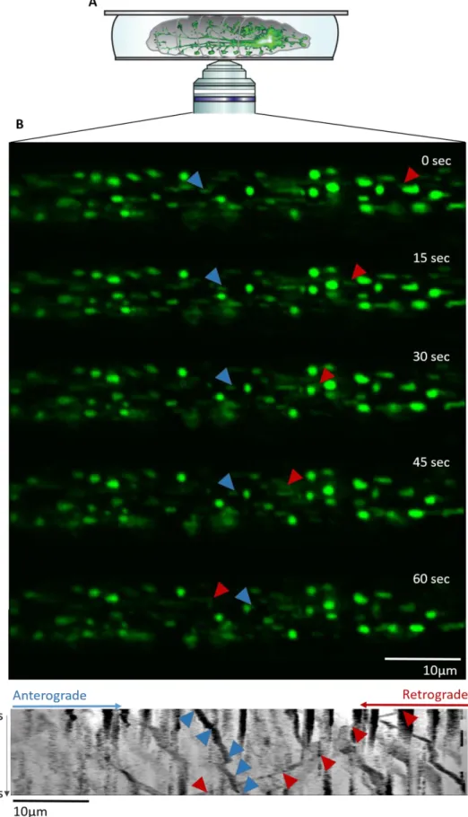
Figure 2.3: Live-imaging setup and illustration of mitochondria transport
(A) The anesthetized larva is mounted on a glass slide with a drop of 80% glycerol and a coverslip. The brain and
motoneurons can be seen through the transparent body wall using an inverted confocal microscope (from Morelli et al., in revision) (B) Five frames of a 1 minute movie recording GFP-tagged mitochondria in a motoneuron. Blue and red arrows track a single mitochondrion moving respectively in the anterograde and retrograde direction. (C) Kymograph generated by KymoToolBox. Time is shown on the y-axis and distance on the x-axis. The trajectories of the mitochondria marked with the arrows in (B) can be seen on the kymograph.
22 ventral side facing upwards. A drop of glycerol 80% was added and the larvae were flattened gently using a coverslip stuck with nail-polish (Figure 2.3A). The transport of mitochondria was then imaged using a Nikon A1Ti microscope in resonant mode at 600ms interval during 1 minute with the 60x oil-immersed objective. An example of the captured images is shown in Figure 2.3B. Larvae display slight variations in the intensity of GFP expression and therefore laser power, gain and offset were adjusted between animals to maximize the visibility of mitochondria. Three movies were recorded for each larva, ensuring that the time elapsed between anesthetization and imaging never exceeded 30 minutes.
2.6 Analysis and statistics
For tubulin/acetylated tubulin stainings, the fluorescence intensity of 10 motoneurons was quantified per larva using ImageJ. The ratio between tubulin and acetylated tubulin was then calculated and normalized to control.
For CSP/HRP stainings, two NMJs were imaged for segments 3 to 5, for a total of 6 NMJs per larva. Multiple images were taken in the Z plan to ensure that all buttons were imaged. A maximum intensity projection was then performed and the number of synaptic boutons per NMJ was counted using the ImageJ cell counter.
For time-lapse movies of mitochondria transport, slight movements of the larvae were first compensated using the StackReg ImageJ plugin. Kymographs were then generated and analyzed using the KymoToolBox plugin as shown in Pineda et al. 2009. The kymographs generated by this plugin report the position of mitochondria along the x axis and the time elapsed along the y axis (Figure 2.3C). The mitochondria trajectories can then be manually drawn to extract the speed and direction of their movement. Mitochondria with speeds inferior to 0.1µm/sec were considered as stationary and were thus excluded from the analysis.
Statistical analysis was performed using the GraphPad Prism 7.0 software. D’Agostino & Pearson, Shapiro-Wild and KS normality tests were performed for each analysis. If the data displayed a normal distribution, an unpaired t-test or one-way ANOVA with Bonferroni post-hoc test were performed. Conversely, if the distribution was not normal, a Kruskal-Wallis with Dunn’s post-hoc test were performed. The criterion for statistical significance was set at p < 0.05 with * = p<0,05, ** = p<0,01, *** = p<0,001, ****=p<0,0001.
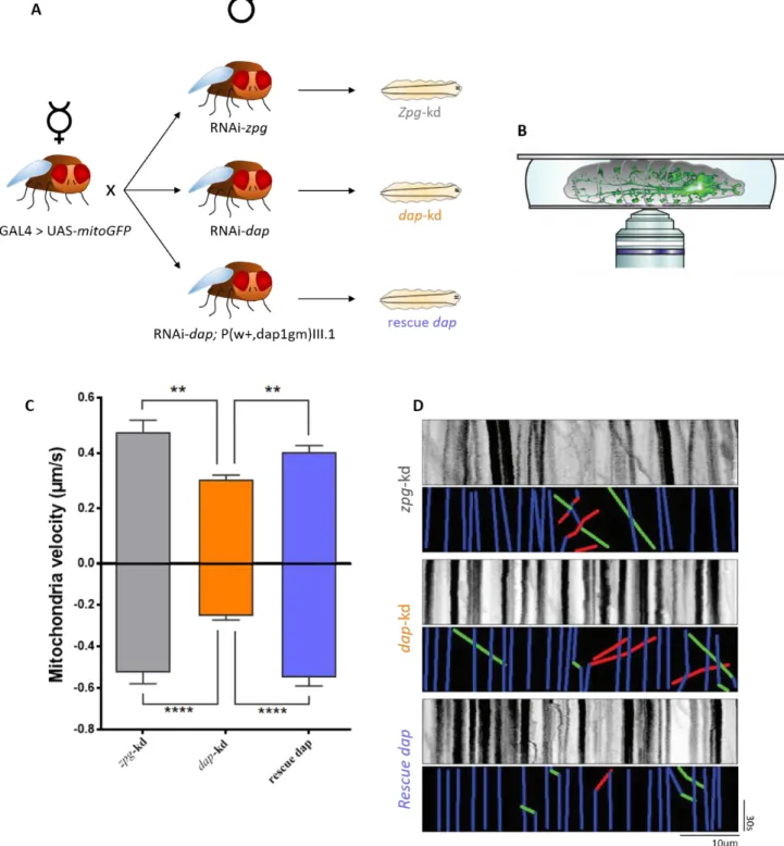
Figure 3.1: dacapo knockdown slows down mitochondria transport in vivo
(A) D42-GAL4 > UAS-mitoGFP virgin females were crossed with RNAi-zpg, RNAi-dap and RNAi-dap;
P(w+,dap1gm)III.1 males to respectively generate zpg-kd, dap-kd and rescue dap larvae. (B) 3rd instar larvae from these crossings were then anesthetized and mounted on glass slides for live-imaging of the GFP positive mitochondria. (C) Quantification of mitochondria velocity based on live-imaging of zpg-kd, dap-kd and rescue
dap larvae. Positive and negative values represent anterograde and retrograde movement respectively. Values are
mean ± standard error of the mean and were analyzed using the non-parametric Kruskal-Wallis test followed by Dunn post hoc-tests. ** p<0,01, ***p<0,001, **** p<0.0001. N (of mitochondria) is > 50 with at least 5 animals per condition. (D) Representative kymographs and their corresponding colored kymographs. Green, red and blue lines represent respectively anterograde, retrograde and stationary mitochondria.
23 3. Results
3.1 Dacapo knockdown slows down mitochondria and vesicular transport
p27, the mammalian ortholog of dacapo, has previously been shown to interact with microtubules and promote their polymerization (Godin et al., 2012) while in mouse fibroblasts, p27 depletion resulted in a decrease of acetylated α-tubulin (Baldassarre et al., 2005). Since a correlation between microtubule acetylation and the regulation of transport has been established in several studies (Dompierre et al., 2007; Godena et al., 2014; Reed et al., 2006), we tested whether the p27 fly ortholog dacapo is involved in the regulation of axonal transport.
D42-GAL4 > UAS-mitoGFP flies express a recombinant GFP displaying a mitochondrial import sequence thus enabling the transport of newly synthesized GFP to mitochondria (Rizzuto et al., 1995). The D42 promoter drives efficient expression of GAL4 specifically in motoneurons (Sanyal, 2009). We crossed D42-GAL4 > UAS-mitoGFP female virgins with the three lines listed in Table 1 to assess the consequences of dacapo knockdown on transport (Figure 3.1A). The efficiency of dacapo knockdown in dap-kd larvae as well as the rescue of its expression level in rescue dap has been shown by Morelli et al. (submitted). Zero Population Growth (zpg) is a gene only expressed in the germline of flies (Tazuke et al., 2002). The expression of a RNAi targeted towards zpg mRNA is therefore used as a control for brain related studies involving knock-down experiments (Zala et al., 2013).
Lines crossed with D42-GAL4 > mitoGFP Designation of the progeny
UAS-RNAi-zpg zpg-kd
UAS-RNAi-dacapo dacapo-kd
UAS-RNAi-dacapo; P(w+,dap1gm)III.1 rescue dap
Table 1: Crossings performed and their corresponding nomenclature.
We performed time-lapse imaging of mitochondria transport in anesthetized zpg-kd, dap-kd and rescue dap 3rd instar larvae. Time-lapse recordings of the moving GFP-expressing mitochondria in motoneurons were recorded through the body walls using a confocal microscope (Figure 3.1B). Both retrograde and anterograde transport of mitochondria had a markedly reduced velocity following dap knockdown (Figure 3.1C-3.1D) while genetic re-expression of dap restored retrograde and anterograde transport velocities. Additionally, data collected by Morelli et al. (unpublished) shows that synaptic vesicles marked with synaptotagmin-GFP display the same defects in anterograde and retrograde velocities (Supplementary Figure 3.1A-3.1B). Taken together, these results reveal that dacapo is implicated in the modulation of axonal transport for both mitochondria and synaptic vesicles in 3rd instar larvae.