Accepted Manuscript
Title: Q fever IN JApaN: an update REVIEW
Authors: Sarah Rebecca Porter, Guy Czaplicki, Jacques Mainil, Yoichiro Horii, Naoaki Misawa, Claude Saegerman
PII: S0378-1135(10)00544-4
DOI: doi:10.1016/j.vetmic.2010.11.017
Reference: VETMIC 5096
To appear in: VETMIC
Received date: 11-1-2010
Revised date: 2-11-2010
Accepted date: 9-11-2010
Please cite this article as: Porter, S.R., Czaplicki, G., Mainil, J., Horii, Y., Misawa, N., Saegerman, C., Q fever IN JApaN: an update REVIEW, Veterinary Microbiology (2010), doi:10.1016/j.vetmic.2010.11.017
This is a PDF file of an unedited manuscript that has been accepted for publication. As a service to our customers we are providing this early version of the manuscript. The manuscript will undergo copyediting, typesetting, and review of the resulting proof before it is published in its final form. Please note that during the production process errors may be discovered which could affect the content, and all legal disclaimers that apply to the journal pertain.
Accepted Manuscript
Q FEVER IN JAPAN: AN UPDATE REVIEW
1
Sarah Rebecca Porter1, Guy Czaplicki2, Jacques Mainil3, Yoichiro Horii4, Naoaki Misawa5, 2
Claude Saegerman1* 3
1: Research Unit in Epidemiology and Risk Analysis applied to Veterinary Sciences 4
(UREAR), Department of Infectious and Parasitic Diseases, Faculty of Veterinary Medicine, 5
University of Liege, Boulevard de Colonster 20, B42, 4000 Liege, Belgium 6
2: Association Régionale de Santé et d‟Identification Animales, Avenue Alfred Deponthière 7
40, B-4431 Loncin, Belgium 8
3: Laboratory of Bacteriology, Department of Infectious and Parasitic Diseases, Faculty of 9
Veterinary Medicine, University of Liege, Boulevard de Colonster 20, B43a, 4000 Liege, 10
Belgium 11
4: University of Miyazaki, Faculty of Agriculture, Department of Veterinary Science, 12
Laboratory of Veterinary Parasitic Diseases, 1-1 Gakuenkibanadainishi, Miyazaki 889-2192, 13
Japan 14
5: University of Miyazaki, Faculty of Agriculture, Department of Veterinary Science, 15
Laboratory of Veterinary Public Health, 1-1 Gakuenkibanadainishi, Miyazaki 889-2192, 16
Japan 17
18
* Corresponding author: Research Unit in Epidemiology and Risk Analysis applied to 19
Veterinary Sciences (UREAR), Department of Infectious and Parasitic Diseases, Faculty of 20
Veterinary Medicine, University of Liege, Boulevard de Colonster 20, B42, 4000 Liege, 21
Belgium; Tel.: +32 4 366 45 79; Fax: +32 4 366 42 61; E-mail: [email protected] 22
Accepted Manuscript
2324
ABSTRACT
25
As neglected zoonosis for many years, Q fever is now ubiquitous in Japan. Similarly to 26
elsewhere in the world, domestic animals are considered to be important reservoirs of the 27
causal agent, Coxiella burnetii, a resistant intracellular bacterium. Infected animals shed 28
bacteria in milk, feces, urine, vaginal mucous and birth products. Inhalation of bacteria 29
present in the environment is the main route of animal and human infection. Shedding of C. 30
burnetii in milk by domestic ruminants has a very limited impact as raw milk is seldom
31
ingested by the Japanese population. The clinical expression of Q fever in Japan is similar to 32
its clinical expression elsewhere. However clinical cases in children are more frequently 33
reported in this country. Moreover, C. burnetii is specified as one of the causative organisms 34
of atypical pneumonia in the Japanese Respiratory Society Guideline for the management of 35
community-acquired pneumonia. In Japan, C. burnetii isolates are associated with acute 36
illness and are mainly of moderate to low virulence. Cats are considered a significant source 37
of C. burnetii responsible for human outbreaks in association with the presence of infected 38
parturient cats. Since its recognition as a reportable disease in 1999, 7 to 46 clinical cases of Q 39
fever have been reported by year. The epidemiology of Q fever in Japan remains to be 40
elucidated and the exact modes of transmission are still unproven. Important further research 41
is necessary to improve knowledge of the disease itself, the endogenous hosts and reservoirs, 42
and the epidemiological cycle of coxiellosis in Japan. 43
Keywords: Q fever, Coxiella burnetii, zoonosis, Epidemiology, Clinical aspects, Animals,
44
Birds, Humans, Cats 45
46 47
Accepted Manuscript
TABLE OF CONTENTS 48 1. Introduction 49 2. Clinical expression 50 2.1. In humans 51 2.2. In farm animals 52 2.3. In pet animals 53 3. Epidemiological data 543.1. Prevalence of Q fever in animals 55
3.2. Prevalence of Q fever in the Japanese population 56
3.3. Isolation of C. burnetii from animals and humans 57
4. Diagnosis of Q fever and vaccination 58
5. Conclusions and perspectives 59
60
1. INTRODUCTION
61
Q fever was first described in 1937 (Derrick, 1937). Q fever is a ubiquitous neglected 62
zoonosis caused by a resistant intracellular bacterium, Coxiella burnetii (Derrick, 1937; 63
Mitscherlich & Marth, 1984; Babudieri, 1959; Maurin and Raoult, 1999; Rousset et al., 64
2009). Ignored for many years, Q fever is now thought to be ubiquitous in Japan since the 65
reservoirs are present throughout the country (Hirai & To, 1998). Similarly to elsewhere in 66
the world, domestic animals are considered to be important reservoirs of C. burnetii (Hirai & 67
To, 1998). However Q fever is known to infect a large variety of hosts, mammals (humans, 68
caprids, bovids, ovids, small rodents, dogs, and cats) but also birds, fish, reptiles and 69
arthropods (Hirai & To, 1998; Maurin & Raoult, 1999; Bildfell et al., 2000; Berri et al., 2007; 70
Rousset et al., 2007; Okimoto et al., 2007; Hartzell et al., 2008). It is a highly infectious 71
disease (Ormsbee et al., 1978). Infected mammals shed bacteria in milk, feces, urine, vaginal 72
Accepted Manuscript
mucous and very importantly in birth products. Inhalation of resistant bacteria present in the 73
environment is the main route of animal and human infection. The main characteristic of Q 74
fever is its clinical polymorphism (Kuroiwa et al., 2007; Hartzell et al., 2008; Million et al., 75
2009; Pape et al., 2009). After an incubation period of 1 to 3 weeks (Maurin & Raoult, 1999; 76
Watanabe & Takahashi, 2008), Q fever can cause either an acute or a chronic disease. 77
This manuscript describes the epidemiological situation of Q fever in Japan. The authors‟ 78
choice of country was based firstly on the peculiar role of cats and additionally of wild birds 79
in the epidemiology of Q fever in Japan and secondly the limited epidemiological 80
investigation and awareness of Q fever in Japan incited further research on the subject. 81 82 2. CLINICAL EXPRESSION 83 2.1. In humans 84
Since April 1999, the management of infection control and prevention in Japan has changed 85
drastically and Q fever was designated as a national reportable disease. Q fever occurs almost 86
all over the country. Under the revised surveillance system, clinical cases of Q fever have 87
been reported 7-46 cases since 1999 for a total of 127 million inhabitants (Mahara, 2006). The 88
clinical expression of Q fever in Japan is similar to its clinical expression elsewhere (Ejercito 89
et al., 1993; Htwe et al., 1993). In the acute form, infections can be totally asymptomatic or
90
can lead to self-limiting „influenza-like” illness, pneumonia or hepatitis (To et al., 1998b; 91
Sampere et al., 2003; Setiyono et al., 2005; Berri et al., 2007; Kuroiwa et al., 2007; Delsing 92
& Kullberg, 2008; Hartzell et al., 2008; Schimmer et al., 2008; Frangoulidis et al., 2009; 93
Million et al., 2009; Pape et al., 2009; Ughetto et al., 2009). Pneumonia is typically mild but 94
progression to acute distress syndrome can occur (Hartzell et al., 2008; Watanabe & 95
Takahashi, 2008). Indeed a study found Q fever to be involved in 2.5% of patients with an 96
Accepted Manuscript
acute infection/exacerbation of a chronic lower respiratory tract disease state (Okimoto et al., 97
2007). In Japan, pneumonia is a common clinical presentation of acute Q fever. The 98
prevalence of C. burnetii infection as causative agent for atypical pneumonia differs between 99
different countries. Indeed, To et al. (1996) found that 39.7% of Japanese patients with 100
atypical pneumonia were infected by C. burnetii. On the other hand, in the rural areas of Nova 101
Scotia, a province of Canada, only 20% of patients admitted to hospital with atypical 102
pneumonia were infected by Q fever (Marrie, 1990). In France, Tissot Dupont et al. (1992) 103
reported a prevalence of atypical pneumonia caused by C. burnetii of 45.8%. The variability 104
in prevalence between countries is most probably due to differences in incidence of Q fever in 105
domestic ruminants (goats, sheep, and cattle). On the other hand, variability in occurrence of 106
clinical illness could be explained by differences between local strains and their respective 107
virulence, and/or by physiological differences in the host (To et al., 1996). Alimentary habits 108
could also play a role. In France, for example, farmers and stock breeders are known to drink 109
unpasteurized milk. Moreover, 61.9% of French patients infected by Q fever presented 110
clinical signs of hepatitis (Tissot Dupont et al., 1992; Maurin & Raoult; 1999). However, 111
currently, bacterial genotype and not route of infection is thought to determine clinical 112
presentation. Furthermore transmission of infection by oral route remains a matter of debate 113
(Marmion & Stoker, 1958; Benson et al., 1963; Krumbiegel & Wisniewki, 1970; AFSSA, 114
2004; Dorko et al., 2008; Natale et al., 2009). 115
In Japan, C. burnetii isolates are mainly associated with acute illness and of moderate to low 116
virulence (Oda and Yoshiie, 1989; Hirai & To, 1998). C. burnetii is specified as one of the 117
causative organisms of atypical pneumonia in the Japanese Respiratory Society Guideline for 118
the management of community-acquired pneumonia (Okimoto et al., 2004; Watanabe & 119
Takahashi, 2008). 120
Accepted Manuscript
In Japan, on the contrary to other countries, clinical expression of the disease has frequently 121
been observed in children (Hirai & To, 1998) (Table I). The study by To et al. (1996) 122
suggested that Q fever was an important cause of atypical pneumonia in Japanese children. 123
Cases of hepatitis have also been reported and can potentially be fatal (Kuroiwa et al., 2007). 124
The difference in prevalence of infection in Japanese children compared to children from 125
other countries could be due to: (1) a more frequent clinical expression (as mentioned here 126
above) due to a different virulence of the bacterial strain or to a greater sensitivity of the host, 127
increasing the probability of diagnosis; (2) to a greater awareness of physicians of the 128
possibility of Q fever infection in atypical and/or non-specific clinical cases. 129
In pregnant women, clinical expression of Q fever, initially asymptomatic, consists in 130
abortions, intrauterine growth retardation, fetal and neonatal death, oligoamnios or premature 131
delivery (Peter et al., 1987; Numazaki et al., 2000; Desling & Kullberg, 2008; Hartzell et al., 132
2008; Schimmer et al., 2008; Vaidya et al., 2008b; Frangoulidis et al., 2009). Sporadically 133
other clinical signs have been reported (such as osteomyelitis, septic arthritis, pericarditis, 134
myocarditis, arteritis, hemolytic anemia, granulomatous hepatitis, lymphadenopathy, Guillain-135
Barré, optic neuritis, paralysis of the oculomotor nerve, meningitis, encephalitis, 136
polyradiculonevritis, peripheral neuropathy, cranial nerve deficiency, and exanthema) (Hirai 137
& To, 1998; Frangoulidis et al., 2009; Million et al., 2009; Pape et al., 2009). 138
In Japan, such as other countries, chronic infection leads commonly to endocarditis (Yuosa et 139
al., 1996). Chronic hepatitis, osteomyelitis, septic arthritis, interstitial lung disease (Berri et 140
al., 2007), and infection of aneurysm and vascular grafts (Delsing & Kullberg, 2008; Ughetto
141
et al., 2009) have also been reported in chronic cases of Q fever (Frangoulidis et al., 2009;
142
Pape et al., 2009). Individuals with underlying valvulopathy or other cardiovascular 143
abnormalities are predisposed to the development of endocarditis (Maurin & Raoult, 1999; 144
Kuroiwa et al., 2007; Delsing & Kullberg, 2008; Harzell et al., 2008; Million et al., 2009; 145
Accepted Manuscript
Pape et al., 2009; Ughetto et al., 2009). Furthermore, chronic fatigue syndrome has been 146
diagnosed in previously infected individuals (Berri et al., 2007; Million et al., 2009). 147
2.2. In farm animals
148
C. burnetii is widespread among cattle population in Japan (4.4 millions of heads). Bovine
149
coxiellosis is rarely an overt disease, except for reproductive disorders (such as abortion, 150
infertility, metritis and mastitis) in females likewise to other parts of the world (To et al., 151
1995, 1998a; Vaidya et al., 2008a). Although high rates of abortions are rarely observed in 152
cattle (Palmer et al., 1983), shedding of large quantities of germs remains a reality in the 153
absence of any clinical sign. A retrospective study by Bildfell et al. (2000) demonstrated that 154
C.burnetii only sporadically leads to abortion in cattle, but was significantly associated with
155
placentitis. Some studies have reported an increase in seroprevalence of Q fever in Japanese 156
cattle in recent years (Hirai & To, 1998). Cattle play an important role in maintaining 157
infection and in dispersing the organism in the environment (Beaudeau et al., 2006; Guatteo 158
et al., 2006; Rodolakis et al., 2007) They are one of the major reservoirs of C. burnetii in
159
Japan (To et al., 1998a). The controversy associated to transmission of Q fever through milk 160
ingestion is of minor importance for the Japanese population. Indeed shedding of C. burnetii 161
in milk has a very limited impact as raw milk is seldom ingested by the native population 162
(Okimoto et al., 2004). Raw milk is commonly pasteurized at 63°C for 30 minutes or more 163
therefore no problem is expected (Watanabe & Takahashi, 2008). 164
The rarity of sheep (10,000 heads) and goats (32,000 heads) populations renders these animals 165
non significant for the spread of the disease (Hirai & To, 1998). Q fever has not been reported 166
in pigs as yet but available data remains scarce (Hirai & To, 1998). 167
2.3. In pet animals
Accepted Manuscript
Dogs and cats have been found to be positive for C. burnetii by serology and bacteriology 169
throughout the Japanese territory (Hirai & To, 1998; Komiya et al., 2003). Nagaoka et al. 170
(1998) isolated C. burnetii in swabs of feline vaginal mucosa and suggested that the organism 171
could be associated with reproductive disorders or abortions in the feline species. Further 172
epidemiological study about the relationship between feline disorders and C. burnetii 173
infection are suggested by the authors (Nagaoka et al., 1998). Small human outbreaks of 174
coxiellosis associated with the presence of infected parturient cats have been reported in 175
several studies (Marrie et al., 1988a; Marrie et al., 1988b; Marrie et al., 1989; Pinsky et al., 176
1991). Cats are thus considered as a potential source for human infections in this country. 177
However premature conclusions must not be made and supplementary evidence of feline to 178
human transmission of Q fever is necessary with special attention to potential confounding 179
factors. Outbreaks associated to infected dogs have not yet been reported to our knowledge. 180
The dogs‟ role as reservoir of the pathogen remains poorly explored. 181
182
3. EPIDEMIOLOGICAL DATA
183
The epidemiology of Q fever in Japan remains to be elucidated and the exact modes of 184
transmission are still unproven (Hirai & To, 1998). The review by Hirai and To (1998) 185
attempted to explain the epidemiology of Q fever in Japan. Figure 1 illustrates their 186
hypotheses. Environment and ticks would be responsible for infection of domestic animals; 187
infected domestic animals hereafter leading to human infections. Transmission directly from 188
infected wild animals to humans would also be possible. Ticks could play a role in 189
transmission of disease from the environment to domestic animals. Tick transmission from 190
domestic and wild animals to humans (Hirai & To, 1998) is considered minor. 191
3.1. Prevalence of Q fever in animals
Accepted Manuscript
Hirai and To (1998) reported the seroprelavence of Q fever in domestic and wild animals 193
present in Japan (Table I). Several authors contributed to these estimations. In domestic 194
animals, cattle with reproductive disorders had the highest percentage of seroprevalence of 195
coxiellosis. A significant level of seropositivity was detected in wild animals but the results 196
must be interpreted with care as the sample size is often limited. Crows (seroprevalence of 197
36%) could be involved in transmission of C. burnetii from infected areas to non infected 198
areas. 199
In a study performed by To et al. (1998b) many wild birds were found to be seropositive 200
against C. burnetii by monoclonal antibody assay (MA). The polymerase chain reaction 201
(PCR) also used in this study confirmed the serological results by detecting the bacterial 202
DNA. Furthermore areas where infected livestock was present were associated with a higher 203
seroprevalence of Q fever in birds. The authors suggested that wild birds could be used as 204
indicators of foci of infection (To et al., 1996). Domestic birds were also found to be 205
seropositive for Q fever and capable of infecting humans (Hirai & To, 1998) rendering 206
additional investigation necessary, especially to determine the eventual role as natural 207
reservoir of C. burnetii (dejection, soil). In 2006, chicken products were highly suspected as 208
responsible for Q fever infections in humans (Muramatsu et al., 2006). C. burnetii was 209
detected in market eggs and mayonnaise (Tatsumi et al., 2006). Initially the probability that 210
the bacteria were alive was considered high (Tatsumi et al., 2006). However, the results of 211
further investigations remain non precise and incomplete. PCR-based detection of C. burnetii 212
DNA in dead bacterial fragments was reported but there are no reports demonstrating 213
contamination with viable bacteria (Watanabe & Takahashi, 2008). No data is available 214
concerning the extent of transmission of C. burnetii from wild animals and birds to humans 215
and domestic animals (Hirai & To, 1998). Its epidemiological importance, however, is 216
considered minor (Hirai & To, 1998). A previous study reported that four out of 11 Japanese 217
Accepted Manuscript
wild species had a prevalence of infection higher to 50%, two had a prevalence of infection 218
lesser to 50%, and five species were free from infection (Ejercito et al., 1993). Three 219
hypotheses could explain absence of infection in the five species: the species were either 220
isolated in a area free from Q fever, or they had an innate resistance to infection, or were false 221
negative animals due to a lack of sensitivity of the laboratory diagnostic method (Ejercito et 222
al., 1993). Further epidemiological studies are necessary to explain this apparent or real
223
resistance to infection. 224
To et al. (1998a) studied the seropositivity rate in herds of dairy cattle with reproductive 225
disorders in Japan. The three main reproductive problems studied were infertility, metritis and 226
mastitis. The rates of positivity were assessed by indirect immunofluorescence assay (IFA) 227
(with a distinction for phase 1 antibodies and phase 2 antibodies), by PCR (in sera and milk 228
samples) and by isolation (in milk samples). Phase 2 antibodies and phase 1 antibodies are 229
associated with acute and with chronic infections, respectively. The study showed that 60.4% 230
of the cows considered were seropositive by IFA towards phase 2 bacteria. In addition, 3.9% 231
and 24.6% were seropositive by PCR on sera and milk respectively. All PCR-positive samples 232
were confirmed by isolation. Whatever the laboratory method, a positive result was always 233
obtained. This study by To et al. (1998a) demonstrated that in herds with reproductive 234
disorders the prevalence of Q fever was far from nil. 235
In studies on feline seroprevalence, stray cats were found to have a higher incidence of 236
infection than domestic cats (Nagaoka et al., 1998; Komiya et al., 2003) (Table I). In this 237
species it is suspected that Q fever could be responsible for breeding disorders (Nagaoka et 238
al., 1998).
239
3.2. Prevalence of Q fever in the Japanese population
Accepted Manuscript
Table I reports the seroprevalence in humans in Japan (Hirai & To, 1998). It is interesting to 241
notice that many healthy humans are seropositive to Q fever. In this study, the seroprevalence 242
of infection was relatively high in children with respiratory disorders, flu-like symptoms, 243
atypical pneumonia and in adults with chronic respiratory disease (Hirai & To, 1998). Sample 244
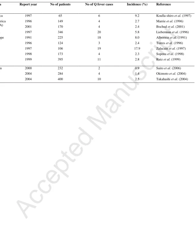
size is a problem for interpretation in certain categories of human beings. Table II reports the 245
estimated number of cases of community-acquired pneumonia in Europe and the USA from 246
1989 to 2001. In Japan, the incidence rate of Q fever has significantly increased between 2000 247
and 2004. However, Q fever has been underdiagnosed for many years in Japan and increased 248
awareness and recognition of the illness might be responsible for the increase observed in this 249
study (Watanabe & Takahashi, 2008). 250
3.3. Isolation of C. burnetii from animals and humans
251
Hirai and To (1998) reported the isolation rates of C. burnetii in different animals present in 252
Japan (Table I). Fetuses of healthy cattle had a high content in bacteria. Ticks of the Ixodes 253
order were significantly infected by C. burnetii. Indeed 75% of the sampled Ixodes ticks in the 254
Toyama prefecture were infected by the bacteria. 255
As mentioned previously, cats are considered a significant source of C. burnetii in Japan 256
(Marrie et al., 1988a; Marrie et al., 1988b; Marrie et al., 1989; Pinsky et al., 1991). In the 257
study by Nagaoka et al. (1998) bacteria were isolated from vaginal swabs of asymptomatic 258
cats as well as of cats with respiratory disorders, with fever or with fever and abortion. The 259
bacteria were also isolated in cats with atypical clinical manifestations (compared to human 260
clinical manifestations) such as peritonitis and mammary tumors. 261
Table I reports the isolation rates of C. burnetii in humans with various clinical signs (Hirai & 262
To, 1998). The rate of isolation in children is particularly high compared to those observed in 263
other parts of the world (To et al., 1996; Maurin & Raoult, 1999). Positive serologies, 264
Accepted Manuscript
occasionally associated to bacterium isolation, were a relative frequent finding in adults with 265
fever of unknown origin (Knockaert et al., 2003; Arnow and Flaherty, 1997; Hirschman, 266
1997; Lozano et al., 1996). In consequence, Q fever serology should be included in the 267
standard work-up of fever of unknown origin in Japan. To confirm these results a second 268
study with larger samples of humans would be necessary. 269
270
4. DIAGNOSIS OF Q FEVER AND VACCINATION
271
Q fever is rarely mentioned in Japanese medical text books and many physicians are unaware 272
of its existence (Watanabe and Takahashi, 2008). Similarly, Japanese veterinarians are 273
insufficiently informed about the risks associated to manipulations of infected animals or 274
infected biological matter (Abe et al., 2001). Thus the recognition of Q fever infections 275
remains limited throughout the country (Watanabe & Takahashi, 2008). Reported clinical 276
cases are rare with the first clinical case reported dating from 1989 (Watanabe & Takahashi, 277
2008). Increasing the physicians‟ awareness of the possibility of Q fever infections is essential 278
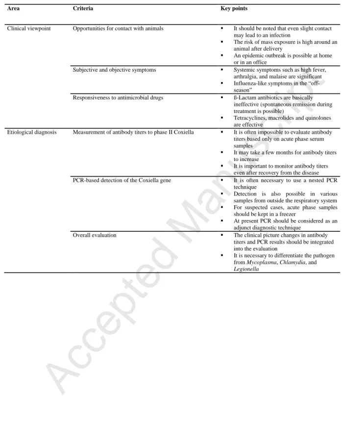
as rapid diagnosis is known to improve prognosis (To et al., 1996). Table III reports the 279
different aspects of the illness to facilitate diagnosis by a clinician (Watanabe & Takahashi, 280
2008). To reach definite diagnosis IFA, complement fixation test (CFT), enzyme linked 281
immunoassay (ELISA) and PCR are available (e.g., Field et al., 2000; Ughetto et al., 2009). 282
Imported IFA and ELISA kits present problems when used on Japanese individuals. Indeed, it 283
has been observed that the increase in IgM antibodies in many Japanese patients infected in 284
Japan is slow; whereas the increase in IgM antibodies is very rapid in Japanese patients 285
infected abroad. This suggests that Coxiella strains vary between different countries 286
(Watanabe & Takahashi, 2008). Moreover, the Japanese population might have a different 287
physiological response to infection compared to Caucasians. Currently, results obtained with 288
Accepted Manuscript
imported IFA and ELISA kits remain difficult to interpret. Furthermore, ELISA kits require a 289
retest with the standard IFA before evaluating a patient (Watanabe & Takahashi, 2008). In 290
conclusion, new rapid diagnostic tests specifically using the Japanese strain of C. burnetii are 291
indispensable. In addition, a larger number of Japanese institutions and laboratories should be 292
equipped with the diagnostic tests (Watanabe & Takahashi, 2008). Vaccination is uncommon 293
in Japan because of the limited recognition of the disease (Watanabe & Takahashi, 2008). 294
295
5. CONCLUSION AND PERSPECTIVES
296
Q fever is a newly discovered disease in Japan. Previously it was considered completely 297
absent. Knowledge of the illness is thus limited. Available epidemiological data consists 298
frequently of small samples of animals or humans rendering the interpretation poorly 299
accurate. The lack of knowledge of the epidemiological and geographical situation in certain 300
areas of the country also causes problems. The estimation of the prevalence or incidence of Q 301
fever is difficult due to the recent awareness of the illness, to the absence of previous data and 302
to seroprevalences estimated on sampled individuals that are not necessarily representative of 303
the endogenous population. Recently differential diagnoses are including Q fever and cases 304
are being diagnosed and reported. Important further research is however necessary to improve 305
knowledge of the disease itself, of the endogenous hosts and reservoirs (e.g., the role of 306
domestic birds should be more investigated), and of the epidemiological cycle of coxiellosis 307
in this country. Diagnostic tests must be improved to increase their sensitivity and avoid the 308
necessity of retesting. They must be adapted to the Japanese strain of bacteria and to the 309
Japanese conditions. The multidisciplinary approach needed would involve a large variety of 310
scientists. To this day, Q fever remains a challenge for the veterinary and medical profession. 311
Accepted Manuscript
6. REFERENCES
313
Abe, T., Yamaki, T., Hayakawa, T., Fukuda, H., Ito, Y., Kume, H., Komiya, T., Ishihara, K., 314
Hirai, K., 2001. A seroepidemiological study of the risks of Q fever infection in 315
Japanese veterinarians. Eur. J. Epidemiol. 17(11), 1029-1032. 316
AFSCA, 2004. Fièvre Q: Rapport sur l‟évaluation des risqué pour la santé publique et des 317
outils de gestion des risqué en élevage de ruminants. 1-88. 318
Albornoz, M.M.C., Heredia, J.H.P., Alvarez, J.S., Lopez, G.T., Palacios, R.H., Puente, A.R., 319
1991. Epidemiology of community-acquired pneumonia in the health area I from 320
Navarra. Med. Clin. (Barc) 97, 50-52. 321
Arnow, P.M., Flaherty, J.P., 1997. Fever of unknown origin. The Lancet 350, 575-580. 322
Babudieri, B., 1959. Q fever: a zoonosis. Adv. Vet. Sci. 5, 82-182. 323
Beaudeau, F., Guatteo, R., Seegers, H., 2006. Voies d‟excrétion de Coxiella burnetii par la 324
vache laitière: implication pour le dépistage et la maîtrise de l‟infection en élevage. 325
Epidémiol. Et santé anim. 49, 1-4. 326
Benson, W.W., Brock, D.W., Mather, J., 1963. Serologic analysis of a penitentiary group 327
using raw milk from a Q fever infected herd. Public Health Rep. 78, 707-710. 328
Berri, M., Rousset, E., Champion, J.L., Russo, P., Rodolakis, A., 2007. Goats may experience 329
reproductive failures and shed Coxiella burnetii at two successive parturitions after a 330
Q fever infection. Res. Vet. Sci. 83, 47-52. 331
Bildfell, R.J., Thomson, G.W., Haines, D.M., McEwen, B.J., Smart, N., 2000. Coxiella 332
burnetii infection is associated with placentitis in cases of bovine abortion. J. Vet.
333
Diagn. Invest. 12, 419-425. 334
Accepted Manuscript
Bochud, P.Y., Moser, F., Erard, P., Verdon, F., Studer, J.P., Villard, G., 2001. Community-335
acquired pneumonia. A prospective outpatient study. Medicine (Baltimore). 80, 75-87. 336
Delsing, C.E., Kullberg, B.J., 2008. Q fever in the Netherlands: a concise overview and 337
implications of the largest ongoing outbreak. Neth. J. Med. 66, 365-367. 338
Derrick, E.H., 1937. “Q” fever, a new fever entity: clinical features, diagnosis and laboratory 339
investigation. Med. J. Aust. 2, 281-299. 340
Dorko, E., Kalinova, Z., Weissova, T., Pilipcinec, E., 2008. Seroprevalence of antibodies to 341
Coxiella burnetii among employees of the Veterinary University in Kosice, eastern 342
Slovakia. Ann. Agric. Environ. Med. 15,119-124. 343
Ejercito, C.L., Cai, L., Htwe, K.K., Taki, M., Inoshima, Y., Kondo, T., Kano, C., Abe, S., 344
Shirota, K., Sugimoto, T., Yamaguchi, T., Fukushi, H., Minamoto, N., Kinjo, T., 345
Isogai, E., Hirai, K., 1993. Serological evidence of Coxiella burnetii infection in wild 346
animals in Japan. J. Wildl. Dis. 29, 481-484. 347
Field, P.R., Mitchell, J.L., Santiago, A., Dickeson, D.J., Chan, S.W., Ho, D.W., Murphy, 348
A.M., Cuzzubbo, A.J., Devine, P.L., 2000. Comparison of a commercial enzyme-349
linked imunosorbent assay with immunofluorescence and complement fixation tests 350
for detection of Coxiella burnetii (Q fever) immunoglobulin. M. J. Clin. Microbiol. 351
38, 1645-1647. 352
Frangoulidis, D., Rodolakis, A., Heiser, V., Landt, O., Splettstoesser, W., Meyer, H., 2009. 353
DNA microarray-chip based diagnosis of Q-fever (Coxiella burnetii). Clin. Microbiol. 354
Infect. Clin. Microbiol. Infect. 15(2), 165-166. 355
Gidding, H.F., Wallace, C., Lawrence, G.L., McIntyre, P.B., 2009. Australia's national Q 356
fever vaccination program. Vaccine 27, 2037-2041. 357
Accepted Manuscript
Guatteo, R., Beaudeau, F., Ledoux, D., Drean, E.l., Seegers, H., 2007. Risk of false-negative 358
results when delaying PCR from sampling for Coxiella burnetii detection in dairy 359
cows. Revue de Médecine Vétérinaire 158, 641-644. 360
Hartzell, J.D., Wood-Morris, R.N., Martinez, L.J., Trotta, R.F., 2008. Q fever: epidemiology, 361
diagnosis, and treatment. Mayo Clin. Proc. 83, 574-579. 362
Hirai, K., To, H., 1998. Advances in the understanding of Coxiella burnetii infection in Japan. 363
J. Vet. Med. Sci. 60, 781-790. 364
Hirai, K., 1999. Recent study of Q Fever. J. Vet. Med. Ass. 52, 77-83 (in Japanese). 365
Hirschman, J.V., 1997. Fever of unknown origin in adults. Clin. Infect. Dis. 24, 291-300. 366
Htwe, K.K., Amano, K., Sugiyama, Y., Yogami, K., Minamoto, N., Hashimoto, A., 367
Yamaguchi, T., Fukushi, H., Hirai, K., 1992. Seroepidemiology of Coxiella burnetii in 368
domestic and companion animals. Vet. Rec. 131, 490. 369
Htwe, K.K., Yoshida, T., Hayashi, S., Miyake, T., Amano, K., Morita, C., Yamaguchi, T., 370
Fukushi, H., Hirai, K., 1993. Prevalence of antibodies to Coxiella burnetii in Japan. J. 371
Clin. Microbiol. 31, 722-723. 372
Knockaert, D.C., Vanderschueren, S., Blockmans, D., 2003. Fever of unknown origin in 373
adults: 40 years on. Journal of Internal Medecine 253, 263-275. 374
Komiya, T., Sadamasu, K., Kang, M.I., Tsuboshima, S., Fukushi, H., Hirai, K., 2003. 375
Seroprevalence of Coxiella burnetii infections among cats in different living 376
environments. J. Vet. Med. Sci. 65, 1047-1048. 377
Accepted Manuscript
Koulla-Shiro, Kuaban, C., Belec, L., 1997. Microbial etiology of acute community-acquired 378
pneumonia in adult hospitalized patients in Yaounde-Cameroon. Clin. Microbiol. 379
Infect. 3, 180-186. 380
Krumbiegel, E.R., Wisniewski, H.J.,1970. Q fever in the Milwaukee area. II. Consumption of 381
infected raw milk by human volunteers. Arch. Environ. Health 21, 63-65. 382
Kuroiwa, Y., Oyanagi, R., Fuse, S., Mori, T., Ueno, H., Tsutsumi, H., 2007. Persistent 383
hepatitis and repeated wheezing in an infant. Q fever. Pediatr. Infect. Dis. J. 26, 763, 384
768-769. 385
Lieberman, D., Schleaffer, F, Boldur, I, Lieberman, D, Horowitz, S., Friedman, M.G., 386
Leiononen, M., Horovitz, O., Manor, E., Porath, A., 1996. Multiple pathogens in adult 387
patients admitted with community-acquired pneumonia: a one-year prospective study 388
of 346 consecutive patients. Thorax 51, 179-184. 389
Lieberman, D., Lieberman, D., Ben-Yaakov, M., Lazarovich, Z., Hoffman, S., Ohana, B., 390
Friedman, M.G., Dvoskin, B., Leinonen, M., Boldur, I., 2001. Infectious etiologies in 391
acute exacerbation of COPD. Diagn. Microbiol. Infect. Dis. 40, 95-102. 392
Lozano, F., Torre-Cisneros, T., Bascuñana, A., Polo, J., Viciana, P., García-Ordóñez, M.A., 393
Hernández-Quero, J., Márquez, M., Vergara, A., Díez, F., Pujol, E., Torres-Tortosa, 394
M., Pasquau, J., Hernández-Burruezo, J.J., Suárez, I., Grupo Andaluz para el Estudio 395
de las Enfermedades Infecciosas., 1996. Prospective evaluation of fever of unknown 396
origin in patients infected with the human immunodeficiency virus. Eur. J. Clin. 397
Microbiol. Infect. Dis. 15, 705-711. 398
Mahara, F., 2006. Rickettsioses in Japan and the Far East. Ann. N. Y. Acad. 1078, 60-73. 399
Accepted Manuscript
Marmoin, B.P., Stoker, M.G., 1958. The epidemiology of Q fever in Great Britain; an analysis 400
of the findings and some conclusions. Br. Med. J. 2, 809-816. 401
Marrie, T.J., Durant, H., Williams, J.C., Mintz, E., Waag, D.M., 1988a. Exposure to parturient 402
cats: a risk factor for acquisition of Q fever in Maritime Canada. J. Infect. Dis. 158, 403
101-108. 404
Marrie, T.J., MacDonald, A., Durant, H., Yates, L., McCormick, L., 1988b. An outbreak of Q 405
fever probably due to contact with a parturient cat. Chest 93, 98-103. 406
Marrie, T.J., Langille, D., Papukna, V., Yates, L., 1989. Truckin' pneumonia: an outbreak of 407
Q fever in a truck repair plant probably due to aerosols from clothing contaminated by 408
contact with newborn kittens. Epidemiol. Infect. 102, 119-127. 409
Marrie, T.J., 1990. Q Fever. The disease. CRC Press, Boca Raton, Florida, pp. 125-160. 410
Marrie, T.J., Peeling, R.W., Fine, M.J., Singer, D.E., Coley, C.M., Kapoor, W.N., 1996. 411
Ambulatory patients with community-acquired pneumonia the frequency of atypical 412
agents and clinical course. Am. J. Med. 101, 508-515. 413
Maurin, M., Raoult, D., 1999. Q fever. Clin. Microbiol. Rev. 12, 518-553. 414
Million, M., Lepidi, H., Raoult, D., 2009. Q fever: current diagnosis and treatment options. 415
Med. Mal. Infect. 39, 82-94. 416
Mitscherlich, E., Marth, E., H. 1984. Microbial survival in the environment. Bacteria and 417
rickettsiae important in human and animal health. Springer-Verlag, Düsseldorf, 418
Germany, pp. 802. 419
Accepted Manuscript
Morita, C., Katsuyama, J., Yanase, T., Ueno, H., Muramatsu, Y., Hondatsu, T., Koyama, H.? 420
1994. Seroepidemiological survey of Coxiella burnetii in domestic cats in Japan. 421
Microbiol. Immunol. 38, 1001-1003. 422
Muramatsu, Y., Hukuta, K., Satoh, S., Muramatsu, M., Nishimura, M., Nagahata, H., Ueno, 423
H., Morita, C., Tamura, Y., 2006. Seroepidemiologic survey of Coxiella burnetii and 424
attempt to detect Coxiella DNA in aged non-laying chickens in a prefecture of Japan 425
where poultry farming prospers. J. Vet. Med. Sci. 68, 1007-1008. 426
Nagaoka, H., Akiyama, M., Sugieda, M., Nisho, T., Akahane, S., Hattori, H., Ho, T., Fushi, 427
H., Hirai, K., 1996. Isolation of Coxiella burnetii from children with influenza-like 428
symptoms in Japan. Microbiol. Immunol. 40, 147-151. 429
Nagaoka, H., Sugieda, M., Akiyama, M., Nishina, T., Akahane, S., Fujiwara, K., 1998. 430
Isolation of Coxiella burnetii from the vagina of feline clients at veterinary clinics. J. 431
Vet. Med. Sci. 60, 251-252. 432
Natale, A., Busani, L., Comin, A., De Rui, S., Buffon, L., Nardelli, S., Marangon, S., Ceglie, 433
L., 2009. First report of bovine Q-fever in north-eastern Italy: preliminary results. 434
Clin. Microbiol. Infect. 15, 144-145. 435
Nguyen, S.A. V., To, H., Minamoto, N., Ogawa, M., Yamaguchi, T., Fukushi, H., Hirai, K., 436
1997. Evaluation of high-density agglutination test for Coxiella burnetii antibodies in 437
animals. Clin. Diagn. Lab. Immunol. 4, 679-680. 438
Numazaki, K., Ueno, H., Yokoo, K., Muramatsu, Y., Chiba, S., Morita, C., 2000. Detection of 439
serum antibodies to Bartonella henselae and Coxiella burnetii from Japanese children 440
and pregnant women. Microbes Infect. 2, 1431-1434. 441
Accepted Manuscript
Oda, H., Yoshiie, K., 1989. Isolation of Coxiella burnetii strain that has low virulence for 442
mice from a patient with acute Q fever. Microbiol. Immunol. 33, 969-973. 443
Okimoto, N., Asaoka, N., Osaki, K., Kurihara, T., Yamato, K., Sunagawa, T., Fujita, K., 444
Ohba, H., Nakamura, J., Nakada, K., 2004. Clinical features of Q fever pneumonia. 445
Respirology 9, 278-282. 446
Okimoto, N., Kibayashi, T., Mimura, K., Yamato, K., Kurihara, T., Honda, Y., Osaki, K., 447
Asaoka, N., Ohba, H., 2007. Coxiella burnetii and acute exacerbations/infections in 448
patients with chronic lung disease. Respirology 12, 619-621. 449
Ormsbee, R., Peacock, M., Gerloff, R., Tallent, G., Wike, D., 1978. Limits of Rickettsial 450
infectivity. Infect. Immun. 19, 239-245. 451
Palmer, N.C., Kierstead, M., Key, D.W., Williams, J.C., Peacock, M.G., Vellend, H., 1983. 452
Placentitis and abortion in goats and sheep in Ontario caused by Coxiella burnetii, 453
Can. Vet. J. 24, 60–61. 454
Pape, M., Mandraveli, K., Arvanitidou-Vagiona, M., Nikolaidis, P., Alexiou-Daniel, S., 2009. 455
Q fever in northern Greece: epidemiological and clinical data from 58 acute and 456
chronic cases. Clin. Microbiol. Infect. 15(2), 150-151. 457
Peter, O., Dupuis, G., Peacock, M.G., Burgdorferj W., 1987. Comparison of Enzyme-Linked 458
Immunosorbent Assay and complement fixation and Indirect Fluorescent-Antibody 459
Tests for detection of Coxiella burnetii antibody. J. Clin. Microbiol. 25, 1063-1067. 460
Pinsky, R.L., Fishbein, D.B., Greene, C.R., Gensheimer, K.F., 1991. An outbreak of cat-461
associated Q fever in the United States. J. Infect. Dis. 164, 202-204. 462
Accepted Manuscript
Rodolakis, A., Berri, M., Héchard, C., Caudron, C., Souriau, A., Bodier, C.C., Blanchard, B., 463
Camuset, P., Devillechaise, P., Natorp, J.C., Vadet, J.P., Arricau-Bouvery, N. 2007. 464
Comparison of Coxiella burnetii shedding in milk of dairy bovine, caprine, and ovine 465
herds. J. Dairy Sci. 90, 5352-5360. 466
Rousset, E., Duquesne, V., Russo, P., Thiéry, R., 2007, La fièvre Q : problématiques et 467
risques sanitaires. Bull. Acad. Vet. France 160, 107-114. 468
Rousset, E., Berri, M., Durand, B., Dufour, P., Prigent, M., Delcroix, T., Touratier, A., 469
Rodolakis, A., 2009. Coxiella burnetii shedding routes and antibody response after 470
outbreaks of Q fever-induced abortion in dairy goat herds. Appl. Environ. Microbiol. 471
75, 428-433. 472
Ruiz, M., Ewig, S., Marcos, M.A., Martinez, J.A., Arancibia, F., Mensa, J., Torres, A., 1999. 473
Etiology of community-acquired pneumonia: impact of age, comorbidity, and severity. 474
Am. J. Respir. Crit. Care Med. 160, 397-405. 475
Saito, A., Kohno, S., Matsushima, T., Watanabe, A., Oizumi, K., Yamaguchi, K., 2006. 476
Prospective multicenter study on the causative organisms of community-acquired 477
pneumonia in adults in Japan. J. Infect. Chemother. 12, 63-69. 478
Sampere, M., Font, B., Font, J., Sanfeliu, I., Segura, F., 2003. Q fever in adults: review of 66 479
clinical cases. Eur. J. Clin. Microbiol. Infect. Dis. 22, 108-110. 480
Schimmer, B., Morroy, G., Dijkstra, F., Schneeberger, P.M., Weers-Pothoff, G., Timen, A., 481
Wijkmans, C., van der Hoek, W., 2008. Large ongoing Q fever outbreak in the south 482
of The Netherlands, 2008. Euro. Surveill. 13. 483
Accepted Manuscript
Setiyono, A., Ogawa, M., Cai, Y., Shiga, S., Kishimoto, T., Kurane, I., 2005. New criteria for 484
immunofluorescence assay for Q fever diagnosis in Japan. J. Clin. Microbiol. 43, 485
5555-5559. 486
Sopena, N., Sabria-Leal, M., Pedro-Botet, M.L., Padilla, E., Dominguez, J., Morera, J., 487
Tudela, P., 1998. Comparative study of the clinical presentation of Legionella 488
pneumonia and other community-acquired pneumonias. Chest 113, 1195-1200. 489
Takahashi, H., Tokue, Y., Kikuchi, T., Kobayashi, T., Gomi, K., Goto, I., Shiraishi, H., 490
Fukushi, H., Hirai, K., Nukiwa, T., Watanabe, A., 2004. Prevalence of community-491
acquired respiratory tract infections associated with Q fever in Japan. Diagn. 492
Microbiol. Infect. Dis. 48, 247-252. 493
Tatsumi, N., Baumgartner, A., Qiao, Y., Yamamoto, I., Yamaguchi, K., 2006. Detection of 494
Coxiella burnetii in market chicken eggs and mayonnaise. Ann. N. Y. Acad. Sci.
495
1078, 502-505. 496
Tissot Dupont, H., Raoult, D., Brouqui, P., Janbon, F., Peyramond, D., Weiller, P.J., 497
Chicheportiche, C., Nezri, M., Poirier, R., 1992. Epidemiologic features and clinical 498
presentation of acute Q fever in hospitalized patients: 323 French cases. Am. J. Med. 499
93, 427-434. 500
To, H., Htwe, K.K., Yamasaki, N., Zhang, G. Q., Ogawa, M., Yamagichi, T., Fukushi, H., 501
Hirai, K., 1995. Isolation of Coxiella burnetii from dairy cattle and ticks, and some 502
characteristics of the isolates in Japan. Microbiol. Immunol. 39, 663-671. 503
To, H., Kako, N., Zhang, G.Q., Otsuka, H., Ogawa, M., Ochiai, O., Nguyen, S.V., 504
Yamaguchi, T., Fukushi, H., Nagaoka, N., Akiyama, M., Amano, K., Hirai, K., 1996. 505
Q fever pneumonia in children in Japan. J. Clin. Microbiol. 34, 647-651. 506
Accepted Manuscript
To, H., Htwe, K.K., Kako, N., Kim, H.J., Yamaguchi, T., Fukushi, H., Hirai, K., 1998a. 507
Prevalence of Coxiella burnetii infection in dairy cattle with reproductive disorders. J. 508
Vet. Med. Sci. 60, 859-861. 509
To, H., Sakai, R., Shirota, K., Kano, C., Abe, S., Sugimoto, T., Takehara, K., Morita, C., 510
Takashima, I., Maruyama, T., Yamaguchi, T., Fukushi, H., Hirai, K., 1998b. 511
Coxiellosis in domestic and wild birds from Japan. J. Wildl. Dis. 34, 310-316. 512
Torres, A., Dorca, J., Zalacain, R., Bello, S., EL-Elbiary, M., Molinos, L., Arévalo, M., 513
Blanquer, J., Celis, R., Iriberri, M., Prats, E., Fernandez, R., Irigaray, R., Serra, J., 514
1996. Community-acquired pneumonia in chronic obstructive pulmonary disease: a 515
Spanish multicenter study. Am. J. Respir. Crit. Care Med. 154, 1456-1461. 516
Ughetto, E., Gouriet, F., Raoult, D., Rolain, J.M., 2009. Three years experience of real-time 517
PCR for the diagnosis of Q fever. Clin. Microbiol. Infect. 15(2), 200-201. 518
Vaidya, V.M., Malik, S.V., Bhilegaonkar, K.N., Rathore, R.S., Kaur, S., Barbuddhe, S.B., 519
2008a. Prevalence of Q fever in domestic animals with reproductive disorders. Comp. 520
Immunol. Microbiol. Infect. Dis. 33(4), 307-321. 521
Vaidya, V.M., Malik, S.V.S., Simranpreet, K., Satish, K., Barbuddhe, S.B., 2008b. 522
Comparison of PCR, immunofluorescence assay, and pathogen isolation for diagnosis 523
of Q fever in humans with spontaneous abortions. J. Clin. Microbiol. 46, 2038-2044. 524
Watanabe A., Takahashi H., 2003. Epidemiology, diagnosis, and treatment of Q fever. 525
Japanese Journal of Chemotherapy 51(8), 461-469 (in Japanese). 526
Watanabe, A., Takahashi, H., 2008. Diagnosis and treatment of Q fever: attempts to clarify 527
current problems in Japan. J. Infect. Chemother. 14, 1-7. 528
Accepted Manuscript
529530
Yoshiie, K., Oda, H., Nagano, N., Matayoshi, S., 1991. Serological evidence that the Q fever 531
agent (Coxiella burnetii) has spread widely among dairy cattle. Microbiol. Immunol. 532
35, 577-581. 533
Yuasa, Y., Yoshiie, K., Takasaki, T., Yoshida, H., Oda, H., 1996. Retrospective survey of 534
chronic Q fever in Japan by using PCR to detect Coxiella burnetii DNA in paraffin-535
embedded clinical samples. J Clin Microbiol. 34(4), 824-827. 536
Zalacain, R., Talayero, N., Achotegui, V., Corral, J., Barrena, I., Sobradillo, V., 1997. 537
Community-acquired pneumonia. Reliability of the criteria for deciding ambulatory 538
treatment. Arch. Bronconeumol. 33, 74-79. 539
540 541
Accepted Manuscript
Figure and table legends
543
Figure 1. Epidemiology of Q fever in Japan (from Hirai and To, 1998)
544
Table I. Seroprevalence and isolation of Coxiella in animals and humans from Japan, through
545
1990 to 2008 (from Hirai and To, 1998; Hirai, 1999; Nagaoka et al., 1998 and various 546
sources) 547
548
Table II. Incidence of Q fever as a cause of community-acquired pneumonia in Japan,
549
Europe, Asia, Africa and America (from Watanabe and Takahashi, 2003 and 2008 and varied 550
sources) 551
552
Table III. Diagnostic points of acute Q fever (from Watanabe and Takahashi, 2008)
553 554
Accepted Manuscript
Table I. Seroprevalence and isolation of Coxiella in animals and humans from Japan, through
1990 to 2008 (from Hirai and To, 1998; Hirai, 1999; Nagaoka et al., 1998 and various sources)
Parameter Kingdom Species Number of
samples % of positive Reference Seroprevalence
Animal Healthy cattle 329 29.2 Yoshiie et al. (1991)
562 46.6 Htwe et al. (1992) 1501 25.4 Htwe et al. (1992) 619 16.9 Nguyen et al. (1997) Reproductive disorder cattle 102 84.3 Htwe et al. (1992)
166 78.9 To et al. (1995) 207 60.4 To et al. (1996)
Sheep 256 28.1 Htwe et al. (1992)
Goat 85 23.5 Htwe et al. (1992)
Dog 635 15 Htwe et al. (1992)
589 10.2 Nguyen et al. (1997) 81 9.9 Nagaoka et al. (1996)
301 16.6 Hirai (1999)
Cat 274 0 Htwe et al. (1992)
100 16 Morita et al. (1994) 150 15.3 Nguyen et al. (1997) 101 6.7 Nagaoka et al. (1996)
304 18.8 Hirai (1999)
Pig 396 0 Htwe et al. (1992)
Chicken 1589 2 To et al. (1996)
Quail 174 2.9 To et al. (1996)
Duck 158 2.2 To et al. (1996)
Bear 36 77.8 Ejercito et al. (1993)
Deer 133 61.7 Ejercito et al. (1993)
Hare 8 62.5 Ejercito et al. (1993)
Monkey 54 27.7 Ejercito et al. (1993)
Nutria 32 12.5 Ejercito et al. (1993)
Wild rodent 129 24.1 Hirai et al. (1998)
Crow 431 36 To et al. (1996)
Rock Dove 201 6 To et al. (1996)
Human Veterinarians 9 22.2 Yoshiie et al. (1991)
Healthy humans (adults) 60 3.3 Htwe et al. (1992)
275 22.2 Htwe et al. (1992)
Meat-processing workers 107 11.2 Htwe et al. (1992)
Adults with respiratory disorders (in general) 184 15.2 Htwe et al. (1992) Adults with atypical pneumonia 284 1.4 Okimoto et al. (2004)
120 4.17 Watanabe and Takahashi (2008) Children with flu-like symptoms 55 32.7 Nagaoka et al. (1996)
Children with atypical pneumonia 56 35.7 To et al. (1996)
58 46.55 Maurin and Raoult (1999) Hospitalized patients (adults) 3000 5.2 Nguyen et al. (1997)
Veterinary students 275 35.64 Htwe et al. (1993)
Adults with acute exacerbation of chronic respiratory disease
80 2.5 Okimoto et al. (2007)
Accepted Manuscript
Adults with acute exacerbation of COPD* 240 0.4 Lieberman et al. (2001) Isolation Animal Cattle with reproductive disorder (raw milk) 207 24.6 To et al. (1995)
Healthy cattle (raw milk) 47 36.3 Nagaoka et al. (1996)
Healthy cattle (fetus) 4 50.0 To et al. (1995)
Tick (Ixodes spp.) 15 26.7 To et al. (1995)
Dogs (sera) 5 100 To et al. (1996)
Cat (sera, uterus swabs) 5 100 To et al. (1996)
Human Acute Q fever (adults) 1 100 Oda et al. (1989)
Atypical pneumonia (children) 58 36.2 To et al. (1996)
Hospitalized patients (adults) 17 76.5 Hirai et al. (1997) Chronic Q feverendocarditis (adults)# 56 7.1 Yuosa et al. (1996) Legend: *: COPD: chronic obstructive pulmonary disease; #: Light microscopic observation.
Accepted Manuscript
Table II. Incidence of Q fever as a cause of community-acquired pneumonia in Japan,
Europe, Asia, Africa and America (from Watanabe and Takahashi, 2003 and 2008 and varied sources)
Area Report year No of patients No of Q fever cases Incidence (%) Reference
Africa 1997 65 6 9.2 Koulla-shiro et al. (1997)
America (USA)
1996 149 4 2.7 Marrie et al. (1996)
2001 170 4 2.4 Bochud et al. (2001)
Asia 1997 346 20 5.8 Lieberman et al. (1996)
Europe 1991 225 18 8.0 Albornoz et al. (1991)
1996 124 3 2.4 Torres et al. (1996) 1997 106 19 17.9 Zalacain et al. (1997) 1998 173 4 2.3 Sopena et al. (1998) 1999 395 11 2.8 Ruiz et al. (1999) Japan 2000 232 2 0.9 Saito et al. (2006) 2004 284 4 1.4 Okimoto et al. (2004) 2004 400 10 2.5 Takahashi et al. (2004) Table 2
Accepted Manuscript
Table III. Diagnostic points of acute Q fever (from Watanabe and Takahashi, 2008)
Area Criteria Key points
Clinical viewpoint Opportunities for contact with animals It should be noted that even slight contact may lead to an infection
The risk of mass exposure is high around an animal after delivery
An epidemic outbreak is possible at home or in an office
Subjective and objective symptoms Systemic symptoms such as high fever, arthralgia, and malaise are significant Influenza-like symptoms in the
“off-season”
Responsiveness to antimicrobial drugs ß-Lactam antibiotics are basically ineffective (spontaneous remission during treatment is possible)
Tetracyclines, macrolides and quinolones are effective
Etiological diagnosis Measurement of antibody titers to phase II Coxiella It is often impossible to evaluate antibody titers based only on acute phase serum samples
It may take a few months for antibody titers to increase
It is important to monitor antibody titers even after recovery from the disease PCR-based detection of the Coxiella gene It is often necessary to use a nested PCR
technique
Detection is also possible in various samples from outside the respiratory system For suspected cases, acute phase samples
should be kept in a freezer
At present PCR should be considered as an adjunct diagnostic technique
Overall evaluation The clinical picture changes in antibody titers and PCR results should be integrated into the evaluation
It is necessary to differentiate the pathogen from Mycoplasma, Chlamydia, and
Legionella