Université de Montréal
Comparison of ELISA protocols measuring HPV16 IgG
antibodies and evaluation of the association between
HPV16 seropositivity and HPV DNA detection
By
Andrea Trevisan
Department of social and preventive medicine School of Public Health
Dissertation submitted for the degree of Master in epidemiology
June 2018
Résumé
Bien que les infections cervicales au VPH soient très courantes, la séroconversion ne se produit pas toujours. Nous avons comparé deux protocoles basés sur deux dilutions sériques pour mesurer la séroréactivité du papillomavirus humain (VPH) de type 16 et avons étudié si la présence de l'ADN du VPH était associé à la séropositivité au VPH16. Nous avons également évalué si l'association était influencée par la co-infection avec d’autres types de VPH et par la charge virale.
Les données utilisées proviennent de femmes brésiliennes qui ont participées à l'étude de cohorte Ludwig-McGill portant sur l'histoire naturelle de l'infection du col de l’utérus par le VPH. Les protocoles de sérologie étaient basés sur des particules pseudo-virales (VLP) composés par les protéines L1 ou L1 et L2 qui sont, respectivement, les protéines principale et secondaire de la capside virale. Deux dilutions sériques ont aussi été utilisées, soient : 1:10 et 1:50. La séroréactivité au VPH16 a été exprimée en rapports d'absorbance normalisé (NAR). Le génotypage de l'ADN du VPH et la charge virale ont été évalués par des méthodes basées sur la PCR. La corrélation et la concordance entre les dilutions de chaque protocole (VLP L1 et L1+L2) ont été évaluées par la corrélation de Pearson (r) et la méthode de Bland-Altman, respectivement. La performance des différents protocoles a été comparée à l’aide de courbe ROC (receiver operating characteristic) en utilisant la présence de l'ADN de VPH16 comme étalon-or. La régression linéaire a été utilisée pour analyser l'association entre la séropositivité au VPH16 et la détection de l’ADN du VPH avec les deux protocoles. La présence de l’ADN du VPH a été analysée en fonction (1) des types spécifiques de VPH plus ou moins apparentés
au VPH16 et (2) l’infection VPH16 détectée seule ou en co-infection avec d’autres types de VPH.
Les modèles de régression linéaire présentés ci-haut ont aussi été utilisées sur l’ensemble de la cohorte testée avec le protocole VLP L1+L2 et dilution sérique 1:10. L'impact de l'âge en tant que facteur de confusion potentiel ou modificateur d'effet a été analysé dans ce modèle. Finalement, l’association entre la charge virale de VPH16 et la séroréactivité a été analysée à l’aide de la corrélation de Pearson.
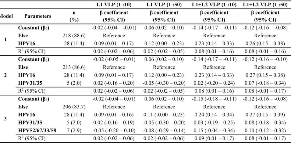
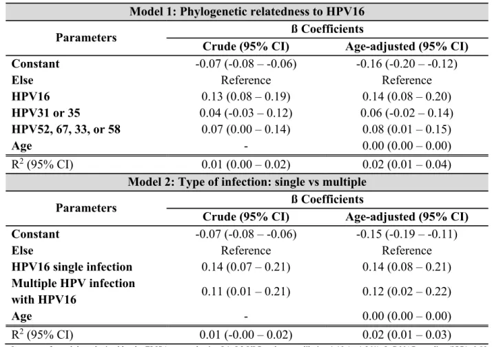
L’ampleur des différences de la moyenne des log10-NAR et les écart-types entre les dilutions sériques observées pour chaque protocole (VLP L1 et L1+L2) étaient, respectivement, -0,081 (0,123) et -0,026 (0,150) unités logarithmiques. Les NARs obtenus par les dilutions sériques utilisées (1:10 et 1:50) pour chaque protocole étaient fortement corrélés (r = 0,87 vs. 0,94, respectivement). Cependant, l'utilisation de VLP L1+L2 a augmenté la performance du test à détecter les anticorps IgG anti-VPH16 en particulier avec la dilution sérique 1:10 [l’aire sous la courbe ROC la plus élevée (IC 95%) = 0,7330 (0,6465 – 0,8495)]. Les modèles de régression ont montré que la séroréactivité au VPH16 n’étaient qu’associée à la présence de l’ADN du VPH16 et non pas aux autres types. Par exemple, les analyses avec le protocole VLP L1+L2 et la dilution sérique 1:10 ont montré que la séroréactivité au VPH16 était associée à la présence de l'ADN du VPH16, β (IC 95%) = 0,24 (0,14 – 0,34), et non pas aux VPH31 ou 35, β (IC 95%) = 0.03 (-0,19 – 0,25), ou VPH52, 67, 33 ou 58, β (IC 95%) = 0,15 (-0,04 – 0,34), comparativement aux femmes infectées par tout autre type de VPH ou négative.
Les analyses sur la cohorte entière avec le même protocole ont aussi montré que l’association entre la séroréactivité et l’ADN du VPH16 était similaire quand l’infection était présente seule ou en co-infection, β (IC 95%) = 0,14 (0,07 – 0,21) et β (IC 95%) = 0,11 (0,01 – 0,21), respectivement, comparativement à celles infectées par tout autre type de VPH ou négative. L’âge n’a pas été un facteur de confusion important et n’a pas été un modificateur d’effet dans l'analyse de l'ensemble de la cohorte. La charge virale du VPH16 n’a pas été corrélée avec la séroréactivité du VPH16, r (95% IC) = 0,04 (0,34 – 0,27); β (IC 95%) = -0,01 (-0,08 – 0,06). En conclusion, le protocole le plus fortement corrélé avec l’ADN du VPH-16 a été celui avec le VLP L1+L2 et la dilution sérique 1:10. Seule la présence de l'ADN du HPV16 a été associée à la séropositivité au HPV16 (pas d’autre type de HPV), et elle n'a pas été influencée par la co-infection ou la charge virale.
Mots-clés : séroréactivité au VPH16, ADN de VPH, particules pseudo-virales, test immuno-enzymatique, anticorps IgG
Abstract
Although cervical HPV infections are very common, seroconversion does not always occur. We compared two protocols based on two serum dilutions to measure human papillomavirus (HPV) type 16 seroreactivity and investigated if HPV DNA positivity was associated with HPV16 seropositivity. We also assessed if the association was influenced by co-infection with other HPV types and viral load.
The data used are from Brazilian women participating in the Ludwig-McGill cohort study on the natural history of cervical HPV infection. The serology protocols were based on virus-like particles (VLPs) composed by the L1 or L1 and L2 proteins which are, respectively, the major and minor viral capsid proteins. Two serum dilutions were used: 1:10 and 1:50. HPV16 seroreactivity was expressed as normalized absorbance ratio (NAR). HPV DNA genotyping and viral load were evaluated by PCR-based methods. Correlation and agreement between serum dilutions of each protocol (L1 and L1+L2 VLP) were assessed by Pearson’s correlation (r) and Bland-Altman method, respectively. The performance of the different protocols was compared using the receiver operating characteristic (ROC) curve using the presence of HPV16 DNA as the gold standard. Linear regression was used to analyze the association between HPV16 seropositivity and the detection of HPV DNA infection with both protocols. The presence of HPV DNA was analyzed based on (1) specific HPV types more or less related to HPV16 and (2) HPV16 infection detected alone or in co-infection with other HPV types.
The linear regression models presented above were also used on the entire cohort tested with VLP L1+L2 and serum dilution 1:10. The impact of age as a potential confounding
factor or effect modifier was analyzed in this model. Finally, the association between HPV16 viral load and seroreactivity was analyzed using Pearson correlation.
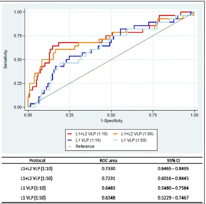
The magnitude of log10-NARs mean differences between serum dilutions and their standard deviations for each protocol (L1 and L1+L2 VLP) were -0,081 (0.123) and -0.026 (0.150) log units, respectively. The NARs obtained by the serum dilutions used (1:10 and 1:50) for each protocol were strongly correlated (r = 0.87 vs. 0.94, respectively). However, the use of L1+L2 VLPs increased the performance of the test to detect HPV16 IgG antibodies, especially with the 1:10 serum dilution [the highest ROC area (95% CI) = 0.7330 (0.6465 – 0.8495)]. The regression models showed that HPV16 seroreactivity was uniquely associated with the presence of HPV16 DNA and not with other HPV types. For example, the analyses with the protocol L1+L2 VLP and serum dilution 1:10 showed that HPV16 seroreactivity was associated with the presence of HPV16 DNA, β (95% CI) = 0.24 (0.14 - 0.34), and not to HPV31 or 35, β (95% CI) = 0.03 0.19 - 0.25), or HPV52, 67, 33 or 58, β (95% CI) = 0.15 (-0.04 - 0.34), compared to women infected with any other HPV type or negative.
The analysis of the entire cohort shows that the association between HPV16 seroreactivity and HPV16 DNA infection was similar when the infection was present alone or in co-infection, β (95% CI) = 0.14 (0.07 - 0.21) and β (95% CI) = 0.11 (0.01 - 0.21), respectively, compared to those infected with any other HPV type or negative. Age was not a significant confounder nor an effect modifier in the analysis of the entire cohort. The HPV16 viral load was not correlated with HPV16 seroreactivity, r (95% CI) = -0.04 (-0.34 – 0.27); β (95% CI) = -0.01 (-0.08 – 0.06). In conclusion, the protocol with the higher correlation with HPV 16 positivity was that with the L1+L2 VLP and serum dilution 1:10. Only the presence
of HPV16 DNA was associated with HPV16 seropositivity (no other HPV type), and it was not influenced by co-infection or viral load.
Keywords: HPV16 seroreactivity, HPV DNA infection, virus-like particles, Enzyme-linked immunosorbent assay, IgG antibodies
Table of contents
Résumé ... i
Abstract ... iv
Table of contents ... vii
List of tables ... ix
List of figures ... xi
List of acronyms ... xiii
List of abbreviations ... xvi
Acknowledgements ... xviii
Introduction ... 1
Chapter 1. Literature review ... 5
1.1. Papillomaviruses ... 5
1.1.1. Viral structure and classification ... 5
1.1.2. HPV life cycle and diagnosis ... 8
1.1.3. Natural history, prevalence and risk factors of HPV infections ... 9
1.1.4. An overview of the host immune response ... 13
1.1.4.1. Innate immunity ... 14
1.1.4.2. Adaptive immunity ... 14
1.1.4.2.1. Humoral immune response ... 15
1.1.4.3. Viral strategies to avoid host immune response ... 16
1.1.5. HPV seroepidemiology ... 17
1.1.5.1. Virus-like particles (VLP): antigens for serological assays ... 18
1.1.5.2. Main serological assays ... 19
1.1.5.3. Determinants of HPV16 seroreactivity ... 21
1.2. Relevance of the study ... 23
Chapter 2. Methodology ... 25
2.1. Objectives ... 25
2.2. The Ludwig-McGill Cohort Study ... 25
2.3.1. Study participants ... 26
2.3.2. Cervical specimens ... 27
2.3.3. HPV detection and genotyping ... 27
2.3.4. HPV serology ... 28
2.3.5. Viral load ... 30
2.3.6. Statistical analysis ... 31
2.3.7. Power estimation ... 34
2.4. Ethical considerations ... 35
2.5. Contribution to the Ludwig-McGill cohort study ... 36
Chapter 3. Manuscript ... 37
Chapter 4. Supplemental results ... 67
Chapter 5. Discussion ... 74
5.1. Results in light of the literature ... 74
5.2. Limits and strengths of the study ... 77
5.3. Potential threats to internal validity ... 78
5.4. Potential threats to external validity ... 82
Conclusion ... 82
References ... 84
Appendix I. Inclusion and exclusion criteria for the summary table of the literature review ... i
Appendix II. Summary table of the literature review ... iii
List of tables
In the manuscript
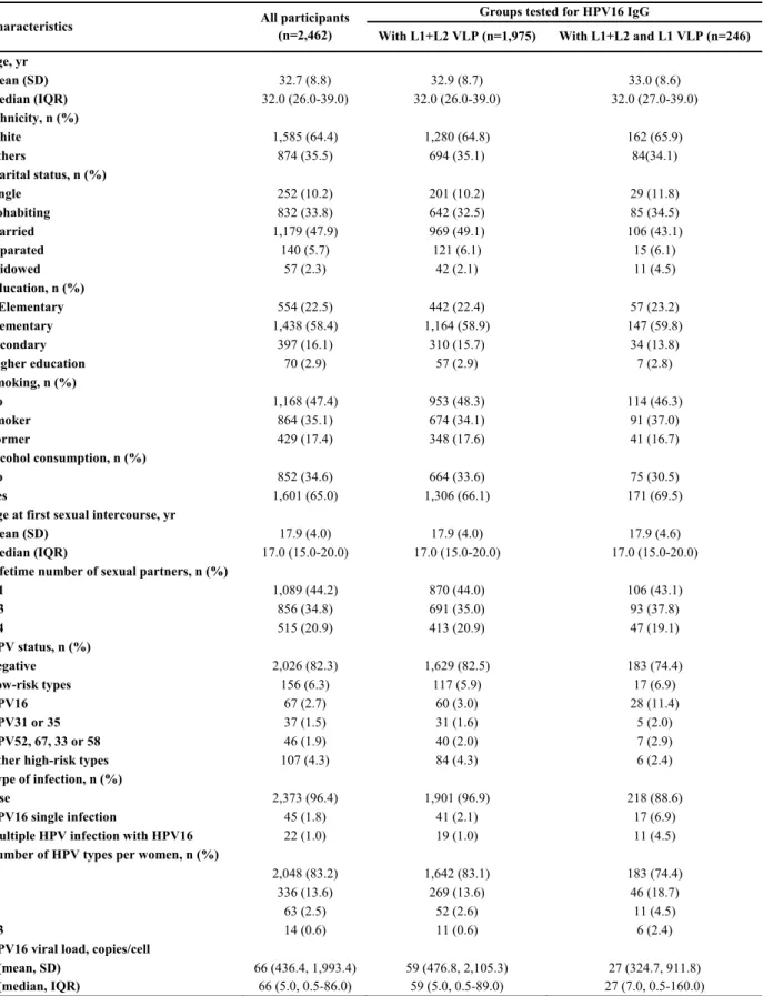
Table 1: Characteristics of the Ludwig-McGill cohort participants at baseline 54
Table 2: Linear regression between HPV16 seroreactivity and HPV status based on the phylogenetic relatedness to HPV16 at baseline
evaluated by three models of exposure 58
Table 3: Linear regression between HPV16 seroreactivity and HPV status at
baseline in the entire cohort 59
In the dissertation: Supplemental results
Table I: Linear regression between HPV16 seroreactivity and HPV status (single vs. multiple infection) at baseline in women tested by both
protocols and serum dilutions 70
Table II: Evaluation of age at enrollment as an effect modifier of the association between HPV16 seroreactivity and HPV DNA infection (HPV status by phylogenetic relatedness to HPV16) at baseline in
the entire cohort 71
Table III: Evaluation of age at enrollment as an effect modifier of the association between HPV16 seroreactivity and HPV DNA infection (single vs. multiple infection) at baseline in the entire cohort 72
Appendices:
Appendix I: Inclusion and exclusion criteria for the summary table of the
literature review i
Appendix II: Summary table of the literature review iii
List of figures
In the manuscript
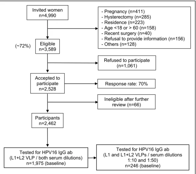
Figure I: Flowchart of the Ludwig-McGill cohort study participants 53
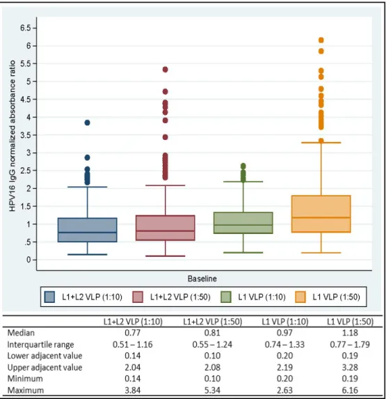
Figure II: Box-and-whiskers representation of untransformed HPV16 IgG normalized absorbance ratios (NAR) at baseline 55
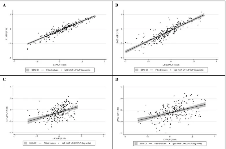
Figure III: Correlation between log10-transformed IgG normalized absorbance ratios (NAR) obtained by serum dilutions 1:10 and 1:50 at baseline 56
Figure IV: Receiver operating characteristic (ROC) curves of untransformed
IgG normalized absorbance ratios (NAR) 57
In the dissertation
Figure 1: Phylogenetic relatedness of alpha-9 HPV species 7
Figure 2: Human papillomavirus life cycle in the squamous epithelium 9
Figure 3: Natural history of HR-HPV infections and the likelihood of
progression according to disease severity 10
Figure 4: Bland-Altman plot of differences between log-10 transformed HPV16 IgG NAR obtained by two serum dilutions versus the mean
Figure 5: Correlation between log10-transformed HPV16 IgG NAR and
HPV16 viral load at baseline 73
Figure 6: Conceptual framework. The role of age on the acquisition of an
List of acronyms
A: Adenine
APC: Antigen presenting cells bp: Base pair
C: Cytosine
CERES: Comité d’éthique de la recherche en santé CI: Confidence interval
CIN II: Cervical intraepithelial neoplasia of grade II CIN III: Cervical intraepithelial neoplasia of grade III
cLIA: competitive multiplexed, Luminex based immunoassay cRIA: competitive radioimmunoassay
dATP: Deoxyadenosine triphosphate dCTP: Deoxycytidine triphosphate dGTP: Deoxyguanosine triphosphate DNA: Deoxyribonucleic acid
dNTP: Nucleoside triphosphate dTTP: Deoxythymidine triphosphate E1: HPV Early protein 1
E2: HPV Early protein 2 E4: HPV Early protein 4 E5: HPV Early protein 5 E6: HPV Early protein 6 E7: HPV Early protein 7
ELISA: Enzyme-linked immunosorbent assay G: Guanine
GST-L1: Gluthathione S-transferase-L1-flag-fusion protein HC2: Hibrid capture 2
HR-HPV: High oncogenic risk HPV type
HSIL: High grade squamous intraepithelial lesion HSV2: Herpes simplex virus type 2
IARC: International Agency for Research on Cancer IgG: Immunoglobulin G
Inj. C.: Injectable contraceptive IQR: Interquartile range
KCl: Potassium chloride L1: HPV Late capsid protein 1 L2: HPV Late capsid protein 2 LCR: Long control region Log: logarithm
LR-HPV: Low oncogenic risk HPV type
LSIL: Low grade squamous intraepithelial lesion LS-PCR: Low-stringency PCR
mg/mL: Milligram per millilitre MgCl2: Magnesium chloride
MHC: Main histocompatibility complex µL: microlitre
µM: micromole
MNCS: Milk and newborn calf serum mol/L: number of moles per litre NAR: Normalized absorbance ration ng: nanogram
oC: Degree Celsius OC: Oral contraceptive OD: Optical density OR: Odds ratio
PBS: Phosphate-buffered saline PCR: Polymerase chain reaction pmol: picomole
PRR: Prevalence rate ratio PV: Papillomaviruses r: Pearson’s correlation
R2: Coefficient of determination RCT: Randomized clinical trial
ROC: Receiver operating characteristic RR: Relative risk
SD: Standard deviation
STI: Sexually transmitted infection T: Timine
Taq: Thermus aquaticus Th1: T Helper cells type 1 Th2: T Helper cells type 2 Treg: Regulatory T cells
TRIS: Tris(hydroxymethyl)aminomethane TRIS-HCl: Tris-Hydrochloride
USA: United States of America VLP: Virus-like particle
List of abbreviations
etc.: Et cetera vs.: Versus
e.g.: From the Latin, Exempli gratia (For example) i.e.: From the Latin, Id est (That is)
Acknowledgements
First, I would like to thank Dr. Helen Trottier, my supervisor, for giving me the opportunity to join her team and for her unconditional support in all aspects of this work. She taught me epidemiology with joy, which I will always remember. I also thank Dr. Eduardo Franco, my co-supervisor, for his mentoring, believing in my talent and making me realize that it is never too late to start all over with passion. I would also like to acknowledge the ongoing support that I have received from previous supervisors: Drs. Luisa Lina Villa and François Coutlée. Their great references allowed me to receive numerous scholarships, including the Canadian Institutes of Health Research Master’s Award, thus obtaining the means to pursue my dreams.
Without the input of all Ludwig-McGill and Trottier’s team members, this work would not have been possible. Special thanks to João Manuel Grisi Candeias and Patrícia Thomann from the Ludwig-McGill team for the serological tests, Louise Laporte, Monica Zahreddine, Catherine Deshaies, Ndongo Sangare, Jean Claude Mutabazi, and Joseph Niyibizi from Dr. Trottier’s team for their valuable comments, technical support, and friendship.
Thanks to my dear friend Martin Chevrier for the English review and suggestions. Thanks to the professors, staff, and colleagues of the Public Health School for their valuable teaching. A special thank (in memoriam) to Dr. Jean Lambert who taught me linear regression with a lot of passion. All of them have made my return to school much easier and enjoyable.
I will be eternally grateful to the jury members of this memoir, Drs. Benoit Mâsse and Julie Bruneau, for their valuable comments and suggestions which made a significant difference in the quality of this work.
Thanks to my family and friends for their unconditional support. I did my best to remain present in their lives during the past two years. Last, but not least, I would reaffirm my love to Roger Marc Gagnon, my partner in life and my best friend. His brilliant mind certainly helped me completing this work in the best way possible. I am fully grateful for his love and support in this endeavor. Starting all over was a challenge, but we can now all consider it a
Introduction
Human papillomavirus (HPV) infection is a major public health concern globally. Approximately 10% of worldwide cancers are associated to viral infection and more than half of infection-related cancers in women are attributed to HPV (1).
Cervical cancer is a rare consequence of a common sexually transmitted HPV infection, most frequently with HPV16 (50%), the most significant genotype associated with the development of the disease (2). It is ranked as the fourth most common malignancy in women worldwide and the second most common cancer in women aged 15 to 44 years (3-5). In 2012, the world population of women aged ≥ 15 years who were at risk of developing cervical cancer was 2.7 million (6). About 528,000 new cases of cervical cancer were diagnosed in the same year and over 265,000 people died from it. The estimated cumulative incidence of cervical cancer worldwide is 14 cases per 100,000 women aged ≥ 15 years per year (4). The worldwide incidence increased by 0.6% annually between 1980 and 2010 (6). The estimated worldwide cumulative mortality of cervical cancer is 6.8 cases per 100,000 women aged ≥ 15 years per year (4). About 85% of the global burden occurs in developing regions, where the cumulative incidence and mortality estimations of cervical cancer are 15.7 and 8.3 cases per 100,000 women aged ≥ 15 years per year, respectively. While in more developed regions the statistics are 9.9 and 3.3 cases per 100,000 women aged ≥ 15 years per year, respectively (6, 7).
Most HPV infections are transient and are cleared within 1 or 2 years by the immune system (2, 8). Although not all infected women develop measurable HPV antibodies, about 60-70% seroconvert and retain their antibodies at low-levels in the serum (9-12). The duration
and cervical precancerous lesions are still unclear (9, 13-23). HPV16 DNA positive women tend to be more frequently seropositive than HPV DNA-negative women (13, 15, 24-28). Several studies have found a positive association between HPV16 seropositivity and HPV DNA positivity; however, some of them did not reach statistical significance (13, 15, 25, 27-33).
There is no gold-standard method for measuring antibodies to HPV infection (34, 35). Several serological assays measuring a wide range of anti-HPV16 antibodies with different properties are currently available for research purposes only (34, 36, 37). They measure humoral immune response of cumulative exposure to the virus (38). In the absence of efficient methods to harvest native antigens from tissue culture, researchers have used virus-like particles (VLP) to study HPV serology. They are composed by recombinantly expressed HPV capsid proteins which self assemble into VLPs lacking the viral genome (39). They can be composed by the major capsid protein only (L1) or L1 together with L2 protein, the minor capsid protein (40). L2 alone lacks the ability to form VLPs, but it can be incorporated when co-expressed with L1 (41).
The capsid proteins L1 and L2 are codified by the L1 and L2 genes, respectively (2). Sequencing analyzes of these genes have shown that L1 has the most conserved DNA sequence between different papillomaviruses. L2 DNA sequence is less conserved compared to L1 (42). There is no report in the literature evaluating which VLP type is better to detect HPV16 seroreactivity in enzyme-linked immunosorbent assay (ELISA) which is the most common method used for HPV seroepidemiological studies. Little is known if L1+L2 VLPs can be responsible for cross-reactivity between HPV types due to their degree of DNA
prozone effect, a type of interference resulting in false negatives or inaccurately low results which may be caused by a highly concentrated serum (34, 43).
In the present study, we compared two ELISA protocols (L1 VLP vs. L1+L2 VLP) with two serum dilutions (1:10 and 1:50) to measure HPV16 seroreactivity and investigated if HPV DNA positivity was associated with HPV16 seropositivity. We also assessed if the association was influenced by co-infection with other HPV types and viral load. Seroreactivity in our study was expressed in normalized absorbance ratio (NAR) to minimize the measurement errors due to intra- and inter-assay variability of ELISA assays (27, 30, 44-47). Although NAR is an arbitrary value and unitless, it is an internally standardized measure of seroreactivity (46).
This dissertation is composed of five main chapters. In the first chapter, we present an overview of the biology (viral structure, classification, life cycle, diagnosis and natural history), epidemiology of the papillomaviruses (risk factors of these infections based on DNA tests and questionnaires), and of the host immune response followed by a summary of the viral strategies to avoid it. We also present the viral-like particles and the main serological assays, followed by a review of the determinants of HPV16 seroreactivity (seroepidemiology). We conclude the first chapter presenting the relevance of this study. In chapter 2, we state our objectives, present the Ludwig-McGill cohort study, describe the participants of the study, the methods used to test our samples, and the statistical analyses. In this chapter, we emphasized that the methods used in this work are not currently available in the clinics and public health network. Next, we present the ethical considerations of this study and the author’s contributions to the Ludwig-McGill study. Chapter 3 presents the manuscript that will be submitted to “The Journal of Infectious Diseases”, containing the main results of this study.
The lab work was supervised by Dr. Luisa Lina Villa, and the statistical analysis by Drs. Helen Trottier and Eduardo Franco. João M.G. Candeias and Patrícia Thomann tested the HPV serology, and Andrea Trevisan tested the viral load and did the statistical analysis. In chapter 4, we present some supplemental results. In chapter 5, we discuss our findings in light of the literature, the limits and strengths of the study, and potential threats to internal and external validity. Finally, we present our conclusions.
Chapter 1. Literature review
1.1. Papillomaviruses
Papillomaviruses comprise a diverse group of viruses that are epitheliotropic, species-specific, and they can infect the skin and mucosa of animals and humans (2, 48). To date, they have been found in fish, reptiles, birds, and mammals (49-53). Considering that these viruses have coevolved with their hosts, they have been an evolutionary success for over 500 million years (54, 55).
1.1.1. Viral structure and classification
Human papillomaviruses belong to the family Papillomaviridae, a family of non-enveloped, small, and circular viruses with a double-stranded DNA genome of about 8,000 base pairs. The genome is divided into eight open reading frames (genes) — E1, E2, E4, E5, E6, E7, L1, and L2 — coding for ‘early’ (E) or ‘late’ (L) viral functions, and an untranslated long control region (LCR) (2). The structure of their capsid is composed of a virally encoded major coat protein, L1 and a minor coat protein, L2, which will be described afterwards. HPV infections are associated with certain anogenital and oropharyngeal cancers (2, 56). Links between HPV and cervical cancer were first suspected more than 40 years ago (57, 58).
HPV classification is based on the nucleotide sequence of the gene coding for the capsid protein L1 (48, 59). Types belonging to different genera share less than 60% similarity, different species within a genus have identity of DNA sequences between 60 and 70%, a novel genotype has less than 90% similarity to any other type, and identity of DNA sequences
between 98-99% defines a variant of type (48, 59). The family Papillomaviridae contains 49 genera (Papillomavirus α, β, γ, etc.), each of which is further divided into several species.
The Papillomavirus Episteme (PaVE) is a database of curated papillomavirus genomic sequences updated 4 times a year. The PaVE database was created with the objective to provide clinicians, epidemiologists, and bench scientists with a uniform data source (54). So far, about 350 types of papillomaviruses have been described of which more than 200 can infect humans (54, 60). About 40 human types exhibit tropism for the anogenital tract (1, 59). They are classified into two different groups according to their oncogenic risk. The first group is composed by low-risk types (LR-HPV), mainly represented by HPV6 and 11. They are found in 90% of genital warts and low-grade squamous intraepithelial lesions (LSIL) and rarely found in cancer. The second group is composed by high-risk types (HR-HPV), mainly represented by HPV16 and 18. They are associated with high-grade squamous intraepithelial lesions (HSIL) as well as carcinomas (2, 4, 61). HPV18 is most found in adenocarcinomas (4, 5). All HR-HPV types together account for up to 5% of all human cancers and are the necessary cause of 99.7% of cervical cancer, 90% of anogenital cancer, 40% of penile cancers, and 42–60 % of oropharyngeal carcinomas (5, 62, 63). HPV16 is found in about 50% of all cervical cancer cases, HPV18 in approximately 20%, HPV31, 33, 45, 52, and 58 in about 20%, and about 10% of all cases are caused by other HR-HPV types (4, 64, 65).
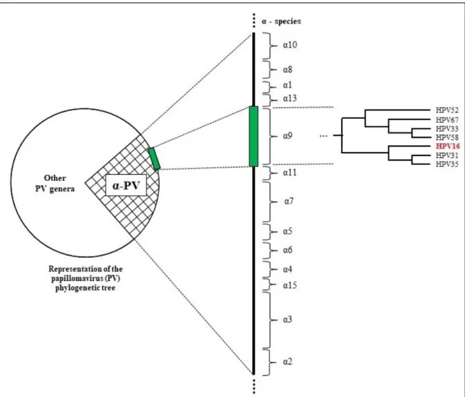
The number of new HPV types increases very quickly due to metagenomic sequencing, a high throughput technology for sequencing of biological samples (66). The genus alpha-papillomavirus contain 65 cutaneous and mucosal types as yet (60). Members of the alpha 9 and 7 species have been studied in more detail (67). HPV16 belongs to alpha 9 species
to HPV16, since they share a common immediate ancestor in the phylogenetic tree (Figure 1, page 7) (68). According to the International Agency for Research on Cancer (IARC), most of these viruses belong to the group of carcinogenic agents (group 1), except HPV67 which is considered probably carcinogenic (group 2B) (69).
Figure 1: Phylogenetic relatedness of alpha-9 HPV species. Inspired by Schiffman et al., 2011 (68).
1.1.2. HPV life cycle and diagnosis
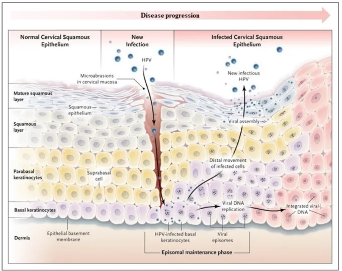
HPVs infect keratinocytes in the basal layer of the cervical epithelium at low copy numbers as a consequence of microlesions of skin or mucosa. During an infection, HPV genomes are found in the nucleus as episomes, circular extrachromosomal DNA (70). The infected cell divides and spreads laterally increasing the viral load. Some of these cells stops dividing and move into the suprabasal differentiating cell layers. Early viral genes are activated at this point to increase viral genomes to thousands.
Since HPV infection is asymptomatic, it is not possible to predict when it occurs and how soon after infection the presence of the virus can be detected in cervical cells. In clinics, the Pap test is used to look for abnormal cells in the cervix, while the HPV test looks for HPV DNA infection (71, 72). HPV test can find any of the HR-types of HPV that are commonly found in cervical cancer.
Progression to malignancy is frequently associated with loss or viral disruption in the E1/E2 regions and integration into the cellular DNA resulting in the loss of negative feedback control of viral oncogenes (E6 and E7) (73). The moment that integration occurs in the natural history of cervical HPV infections is a controversial issue (74-77). Expression of E6 and E7 oncoproteins is required to maintain the malignant growth of cervical cancer cells by inhibiting cellular tumour suppressors genes (2). The organization of the epithelium changes as the disease progresses (70). In the superficial layers of the epithelium, late viral genes are expressed, and L1 and L2 capsid proteins are formed to encapsidate the viral genomes. Infectious particles are released in the terminally differentiated outer epithelial layer (Figure 2, page 9) (2, 70, 78).
HPVs, especially alpha-species, are very successful infectious agents. They induce chronic infections with no serious sequelae, rarely kill the host and shed large amounts of infectious virus for transmission to other individuals (78).
Figure 2: Human papillomavirus life cycle in the squamous epithelium. Reproduced with permission from Kahn, 2009, Copyright Massachusetts Medical Society (70).
1.1.3. Natural history, prevalence and risk factors of HPV infections
HPV is a very common infection acquired via sexual activity (79). More than 80% of women will be infected in their lifetime (80). The incubation period of HPV infections may
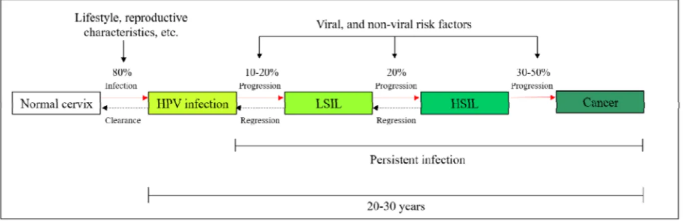
last from 3 weeks to more than 8 months (78). Genital warts may occur about 2 to 3 months after infection which end up regressing exponentially in 10-30% of patients within 3 months (78). Infection with HR-HPV types, such as HPV16 and 18 are usually transient and tend to be cleared in 12–18 months due to cell-mediated and humoral immune responses (8, 20, 64, 78, 81, 82). A small number of HPV infections persists, and the pathology may progress to LSIL (10–20%) and HSIL in some cases (20%). If not treated, advanced precancerous lesions may progress to cervical cancer (30-50%) (83-86). Therefore, cervical cancer is a rare consequence of a persistent HPV infection which can be harbored in a latent state for more than 20 years before progressing to cancer (Figure 3, page 10) (87, 88). Definition of HPV persistence varies significantly between studies due to several HPV detection methods available and several lengths of follow-up time (89-91). Consequently, comparison between studies can be challenging (92). In general, persistent infection means two or more HPV-DNA-positive tests, consecutively, in intervals of 4 to 6 months (93).
Figure 3: Natural history of HR-HPV infections and the likelihood of progression according to disease severity.
A decade ago, researchers published a meta-analysis based on 78 studies to investigate the age and genotype specific prevalence of HPV infections worldwide (94). Final analysis included 157,879 women with normal cytology. Overall worldwide HPV prevalence was 10.4% (95% CI: 10.2-10.7). Ten years ago, they observed a geographical variation of prevalence estimates by world region: in Africa HPV prevalence was 22.1% (95% CI: 20.9-23.4), Central America and Mexico 20.4% (95% CI: 19.3-21.4), Northern America 11.3% (95% CI: 10.6-12.1), Europe 8.1% (95% CI: 7.8-8.4), and Asia 8.0% (95% CI: 7.5-8.4) (94). Nowadays, the variance in prevalence among different regions of the world has diminished over time probably due to prevention programs (4). Recent data show that HPV prevalence in most regions mentioned above is around 4.0%, except in Asia, where no reduction was observed over the last decade (4). Less developed regions, such as Caribbean with 15.8% (95% CI: 12.2-20.2), South America 12.1% (95% CI: 11.6-12.7), and Eastern Europe 9.7% (95% CI: 9.1-10.4) have the highest HPV prevalence in women with normal cervical cytology worldwide (4).
HPV prevalence was high in women younger than 25 years of age, then decreased in older women in most of the world regions (94). In Africa, the Americas, and Europe, they observed a second peak of HPV prevalence in women aged 45 years or older. Unfortunately, most of age-prevalence data were shown graphically with no precise estimates in the text of the publication. They only provided detailed information on the HPV prevalence in women older than 45 years of age from the Americas. They showed that the overall estimate was higher in Central America (20.4%, 19.3–21.4) and South America (12.3%, 11.2–13.4) than in Northern America (11.3%, 10.6–12.1). HPV16, 18 or both were detected in 32% of the study participants (94, 95). The age-specific bimodal prevalence may be due to a cohort effect
(acquisition of new infections due to new sexual partners), the possibility of reactivation of latent infection or acquired immunity; however, more studies should be done to validate these hypothesis (16, 20, 93, 95, 96).
Overall HPV DNA prevalence grew with increasing severity of cervical disease (4, 94, 97). The more severe the lesion, the greater the probability of detecting the DNA of a HR-HPV type in cells from cervical smears. HR-HPV16 and 18 DNA prevalence according to severity of cervical disease in less developed regions of the world were: 4.4% (95% CI: 4.3-4.5) in normal cytology, 25% (95% CI: 24.1-25.9) in LSIL, 46.6% (95% CI: 45.8-47.4) in HSIL, and 69.5% (95% CI: 68.9-70.1) in cervical cancers (4).
There are some risk factors associated with the acquisition and persistence of HPV infections (98). Some of them can be measured by molecular biology techniques or questionnaires. Cumulative HPV exposure is associated with sexual behaviors, such as number of sexual partners, and concurrent relationships (96, 98, 99). Sexually transmitted infections, such as Chlamydia trachomatis, immunodeficiency virus, and bacterial vaginosis have also been found to be predictive of cervical HPV infection risk in epidemiological studies (96, 98, 100-102). Immunodeficiency appears to increase the host susceptibility to infection, since a higher prevalence of genital HPV is observed in immunosuppressed individuals, regardless of the cause of immunosuppression (79, 103).
There are other risk factors which are inconsistently associated to the acquisition of an HPV infection, particularly with respect to reproductive and genital health (98). Micro-abrasions caused during sexual intercourse in a dry and irritated genital tract due to tampon use could increase the susceptibility to HPV infection and decrease the rate of HR-HPV
is also a lack of consensus regarding smoking, use of oral contraceptive, condom use, and age at first intercourse (96, 99, 102, 106-108). Little is known about the association of frequent vaginal douching and HPV infections (109).
Researchers have also concentrated their efforts to understand the role of risk factors that favor persistence of HPV infection and mediate progression of precancerous lesions to cancer. Some authors have shown that older age and viral factors, such as genotype, molecular variants, and viral load are predictive of persistent HPV infection and progression to cervical cancer (79, 88, 110-115). Smoking, multiparity, long-term use of oral contraceptive, other sexually transmitted infections, and chronic inflammation also seem to increase the risk of persistence, and disease progression (96). Daily consumption of vegetables has also been associated with HPV clearance (92). Besides, it is very common to find co-infection with multiple HPV types in many epidemiological studies (15, 30, 116). However, the role of co-infection on the duration of the co-infection is not fully understood (101, 117).
1.1.4. An overview of the host immune response
This section is focused on the host immune response and the virus strategies to avoid it. Host immune response against pathogens can be divided in several basic phases that differ depending on the perspective. For the pathogen side, they must find a permissive host environment, successfully initiate infection of target cells and be able to replicate. For the host side, they have to initiate a series of events which includes initial recognition of the pathogen by sentinel host immune cells, establish a innate immune response and trigger an adaptive response to eliminate the pathogen (118). The innate and adaptive immune systems are often described separately; however, they usually act together (119).
1.1.4.1. Innate immunity
Innate immunity is the first line of defense from infection in a non-specific manner by detecting the pathogen and clearing most of microbial assaults (119, 120). It is rapid, does not require prior sensitisation, is not antigen-dependent and has no specific memory (120, 121). In our context, it is an epithelial barrier composed by cells (i.e., phagocytes, some antigen presenting cells, and the effector cells), several cellular antimicrobial products (e.g., cytokines and chemokines), and the complement cascade, a biochemical process that occurs in the blood to help cells of the immune system to eliminate invading pathogens (121).
Briefly, inflammation is the first sign of innate immune response which is triggered by cell injury or death. At this point, the actors of the innate immune system are recruited to solve the infection and kick-start the adaptive immune response if necessary (120).
1.1.4.2. Adaptive immunity
Adaptive immune response is specific and generally lethal to foreign antigens. Antibody-mediated humoral immune response clears free virus particles from body fluids preventing viral reinfection, while cell-mediated immune response kills infected cells and generate immune memory. Both systems are interconnected in some ways with the adaptive immunity becoming prominent several days after the onset of the innate immune response (119, 120).
Antigen-specific immune response is triggered when cells of the innate immune system are stimulated leading to the proliferation and differentiation of cells that compose the adaptive immune system. Lymphocytes T and B are born in the bonne marrow and are the
immune defenses depends on them (120). Next, we provide an overview of the humoral immune response.
1.1.4.2.1. Humoral immune response
Antibodies, also called Immunoglobulins (Ig), are Y-shaped glycoprotein molecules that are produced by plasma cells in response to an antigen. Since different antibodies recognize different antigens, antigen-binding sites are different for different antibodies which are the effector molecules of the humoral immune response (122). Five primary classes of antibodies exist based on the structure of their molecule. They are identified as IgG, IgM, IgA, IgD, and IgE, and are distributed and function differently in the body. IgG has four subclasses (IgG 1 to 4) and is the most frequent (75%) immunoglobulin in the serum. It is versatile because it can carry out all functions performed by all classes of immunoglobulins and provides long term protection (122).
Naïve lymphocytes B are activated when they first encounter an antigen. Only a few native antigens can directly activate B cells and generate plasma cells (120). Low levels of antibodies are produced after natural HPV infection. The response to a second round of infection is often faster than the primary infection because of the activation of memory B and T cells (78). A neutralizing antibody response highly type-specific to L1 is known to effectively prevent HPV infection (123). However, they are unable to kill established HPV-infected cells (62).
HPV vaccination was implemented in several countries in 2007. A systematic review and meta-analysis published at the “Lancet Infectious Diseases” in 2015 showed that HPV16 and 18 infections decreased between the pre-vaccination and post-vaccination periods by 68%
(RR: 0.32, 95% CI: 0.19 – 0.52) and anogenital warts decreased by 61% (RR: 0.39, 95% CI: 0.22 –0.71) in girls 13–19 years of age (124). Significant reductions in HPV 31, 33, and 45 infections were also observed in this age group of girls (RR: 0.72, 95% CI: 0.54 – 0.96) suggesting cross-protection. All these results were observed in high-income countries with female vaccination coverage of at least 50% (124). Vaccination can induce very high concentrations of neutralizing antibodies, at least 2 to 4 log units higher than in natural infections (78).
1.1.4.3. Viral strategies to avoid host immune response
The reason of the successful viral lifestyle is the ability of HR-HPV types to avoid host defence systems (78). The virus replication cycle itself is an immune evasion mechanism that helps the virus to evade the innate immune response and delay activation of adaptive immunity (78, 121).
The HPV life cycle depends on the keratinocyte differentiation program, production of viral particles is time-consuming, there is no cytolysis and no virally induced cell death; consequently, there is no inflammation. All key events occurs in a cell destined for desquamation away from the primary site of immune surveillance, the submucosa (121). During HPV life cycle, there is little or no release of pro-inflammatory cytokines as part of the innate immune response. Cytokines help to trigger the adaptive immune response and are important in the activation and migration of antigen-presenting cells (78). In addition, there is no viremia, and host dendritic cells are exposed to low levels of viral proteins during the natural history of HPV infection (78, 84, 120).
1.1.5. HPV seroepidemiology
In general, natural exposure to a virus results in a protective antibody response; however, seroconversion does not always occur following HPV infections. Only about half of infected women have detectable levels of anti-HPV antibodies in their bloodstream (10, 93). In addition, about half of seropositive women produce neutralizing antibodies (21). For women with incident HPV16 infections, the median time to seroconversion from DNA detection varies from 6-12 months (10, 125). The duration of natural immunity and whether it can protect against cervical precancerous lesions are still unclear (9). Serological assays may identify the individuals who had developed an immune response to previous exposure to HPV and may be protected against reinfection (18, 21, 126). However, some studies have shown that reinfection with the same type is possible suggesting no protection following a previous type-specific infection (20).
Although some researchers have concentrated their efforts to establish an international standard operating procedure for HPV serology, we still do not have a gold-standard method for measuring antibodies to HPV infection (34, 35). Consequently, we have no agreed definition of what level of response indicates effective seroreactivity making comparison between results obtained by different laboratories extremely difficult (34, 78). In addition, HPV serology has several limitations, such as low seroconversion after natural infection, antibody levels may decrease over time, and limited assay sensitivity (10, 13, 127). Due to this variety of technical and biological limitations, HPV serology has not been used in the clinics (45, 128).
available to measure antibody titers to HPV infection for research purposes. Finally, we present an overview of the most important findings in the literature about the determinants of HPV16 seroreactivity highlighting the association between HPV16 seroreactivity and HPV DNA positivity.
1.1.5.1. Virus-like particles (VLP): antigens for serological assays
The L1 and L2, major and minor viral capsid proteins, respectively, are assembled late in the HPV life cycle to compose the icosahedral capsid shell which has the function to protect the viral genome (129-132).
In the absence of efficient methods to harvest native antigens from tissue culture, serologic detection of HPV has used virus-like particles (VLP) (39, 45, 131, 133-135). They are non-infectious papillomavirus particles without the viral genome. VLPs display conformational and type-specific epitopes which are the part of an antigen molecule to which an antibody attaches itself. They are structurally similar to authentic virions, term used to designate viral particles outside living cells (39, 135). VLPs are produced in a variety of recombinant expression systems and are highly immunogenic inducing potent antibody responses due to their ability to activate both innate and adaptive immune responses (131, 133-139).
L1 protein can self-assemble to form empty VLPs that are the basis of the licensed HPV vaccines (41, 123, 131). L2 does not form VLPs, but it can be incorporated when co-expressed with L1 (40, 133). L1 has a highly conserved DNA sequence, and L2 is less-well conserved among different HPV types. Addition of L2 in the composition of VLPs can
possibly induce broader protection through cross-neutralizing antibodies, even across species (40, 62, 131).
1.1.5.2. Main serological assays
Serological assays confer an advantage over DNA methods because it is a single outcome that can represent infection from multiple anatomic sites (140, 141). They also can be used as an indicator of cumulative infection exposure to predict the risk of developing cancer and their precursor lesions, reinfection, reactivation, and clearance of infections. (14, 19, 30, 142, 143).
Several serological assays measuring a wide range of anti-HPV16 antibodies with different properties are currently available for research purposes (34, 36). The first assay developed for measuring HPV antibody titers was the athymic mouse xenograft system (144). Due to technical difficulties in testing a large number of sera using this protocol, several complementary assays have been developed (18). Each assay provides only a partial characterization of immune status. They differ quantitatively (i.e., throughput and detection range) and qualitatively (i.e., if they detect polyclonal antibodies which may be indicative of prior exposure or neutralizing antibodies which is indicative of immune protection). Because of that, comparison of seroprevalence across assays is not possible (18, 34, 121)
Several immunoassays have been developed during the last decades, such as the type-specific competitive radioimmunoassay (cRIA) and the pseudovirion-based neutralization assay. Both methods are labor intensive and only measure neutralizing antibodies (145, 146). The last generation of methods have used the Luminex technology to measure neutralizing or total IgG antibodies to VLP (competitive multiplexed, Luminex-based immunoassay, cLIA) or
to glutathione S-transferase-L1-flag-fusion proteins (GST-L1) (147-150). Luminex is a robust, sensitive, and high-throughput serological platform that can be used to measure antibodies to several HPV genotypes at the same time (151). Nevertheless, it is expensive and depends on monoclonal antibodies which specifically bind to only one epitope of the antigen to perform.
The most common serologic assay for HPV is the enzyme-linked immunosorbent immunoassay (ELISA) (152). It is type-specific, but it cannot differentiate between neutralizing and non-neutralizing antibodies. In fact, it measures antibodies to HPV VLPs that were secreted by different B cell clones within the body. So, ELISA measures a polyclonal response. Technically, it means that these antibodies can bind to different epitopes on the same antigen.
In ELISA protocols, antibody measurements have relied on determining VLP optical density (OD) values for serum samples and comparing them against negative and positive controls to detect HPV seroreactivity. However, OD values are prone to measurement errors due to intra- and inter-assay variability originated from daily variations in reagent batches and technical performance (e.g., pipetting, instrument readings, etc.) (46). Our team has proposed the use of normalized absorbance ratio (NAR) to circumvent these technical problems that can affect the validity of seroreactivity (27, 30, 44-47). NARs are calculated by dividing the mean blank-subtracted optical densities (OD) by the equivalent value of the control serum pool included in the same plate in triplicate. Although NAR is an arbitrary value and unitless, it is an internally standardized measure of seroreactivity (46).
Therefore, we are facing a unique opportunity to evaluate which VLP type can better capture the association between naturally acquired HPV16 seropositivity and HPV DNA
positivity using an optimized ELISA, and to investigate if L1+L2 VLPs can be responsible for cross-reactivity between HPV types.
1.1.5.3. Determinants of HPV16 seroreactivity
A review on HPV serology including 117 studies from several world regions has been published (153). Participants were women and men from several hours to over 90 years of age. Serological antibodies were detected with ELISA (78%), cLIA (12%), and other available methods (10%). HPV16 seropositivity was more prevalent in women than in men and peaked around ages 25-40 years in women. Some studies have reported that seroprevalence peaked twice in women. A possible explanation for the second peak at older ages (>50 years) is a reinfection or reactivation of a latent infection maybe by reduction of immune surveillance with increasing age followed by increasing viral load, and antibody induction (26). In young women from 9-26-year-old, HPV16 seroprevalence ranged from 0-31% in North America, 21-30% in Africa, 0-23% in Asia/Australia, 0-33% in Europe, and 13-43% in Central and South America (153).
To better understand the humoral immune response against HPV infections, several researchers from all over the world have identified the determinants of HPV16 seroreactivity. Sexual behavior seems to play an important role in the acquisition of HPV antibodies, particularly, the increased number of lifetime sexual partners (24, 27, 29, 30, 33, 38, 108, 154-161). The exact number of sexual partners that increase the likelihood of seroconversion varies from study to study and depends particularly on the presence of HPV DNA infection among partners. In the Ludwig-McGill cohort study the odds ratio of HPV16 seroreactivity at baseline for women who reported having had more than four lifetime sexual partners were
elevated >2.5-fold compared to women who reported 0-1 partner during their entire life (OR: 2.56, 95% CI: 1.97-3.53) in the analysis adjusted for age and HPV16 DNA positivity (30).
Other determinants of HPV16 seroreactivity were identified, such as smoking, marital status, seropositivity for HPV18, history of sexually transmitted disease other than HPV, hormonal contraceptive use, parity, frequency of sex, years since sexual debut, and high HPV16 viral load (24, 30, 108, 156-159). Age, age at first intercourse, stage of the disease, and cytologic diagnosis are still controversial determinants of HPV16 seroreactivity (24-30, 33, 38, 154, 156-158). However, all these factors might be related to HPV infection only. It is difficult to understand what is related to HPV DNA infection from what is related specifically to seroconversion. The role of potential confounders in the association between HPV16 seropositivity and HPV DNA positivity will be discussed in depth later.
Both cross-sectional and longitudinal study designs have reported the correlation between HPV16 DNA positivity and HPV16 seropositivity (13, 15, 25, 27-33). Based on inclusion and exclusion criteria which are shown in the appendix I, we prepared a summary table of the literature regarding this subject (Appendix II). In brief, all studies mentioned above have found a positive association between HPV16 seropositivity and HPV16 DNA positivity independently of any other factor (e.g., age, number of lifetime sexual partners, etc.). Three of them did not report statistically significant results for the association (29, 31, 32). Only one study presented the results adjusted for age and lifetime number of sexual partners (p=0.046) (25). HPV16 DNA positive women tended to be more frequently seropositive than HPV DNA-negative women (13, 15, 24-28). Only two studies found that HPV16 DNA positive women were less seropositive than HPV DNA-negative women which
peaks of HPV seroprevalence according to the age of the participants (26). The first peak is in young adult women (15–34 years) and the second in women older than 45 years old.
The partial analysis of the Finnish family HPV study conducted at Turku, Finland, and designed to evaluate dynamics of HPV infections within families used the Luminex technology with GST-L1 proteins as antigen to detect the antibody levels (150, 162). Authors reported no concordance between cervical DNA detection and co-existent seropositivity even in samples taken 12 months apart, but it showed that women who cleared their cervical HPV16 DNA infection had the highest HPV16 antibody levels, whereas those who acquired incident HPV16 DNA infections had the lowest antibody levels.
1.2. Relevance of the study
Cervical cancer is an important public health problem worldwide. There is no cervical cancer without an HPV infection. From a public health perspective, the government depends on seroconversion results obtained by standardized serological methods to decide the cost-effectiveness of vaccination. This study aims to increase the validity and precision of a serological instrument and; therefore, should contribute directly to the quality and reliability of the decisions from public health institutions.
From a clinical perspective, understanding why some women can produce antibodies after having naturally acquired an HPV infection and others not, particularly infection with HPV16, the most prevalent genotype, can help clinicians to drive personalised and more effective treatments.
Based on the assumption that both clinical and public health professionals depend on a highly performing serological instrument for decision-making, it is important to consider
every detail concerning the methodology used to measure HPV antibodies. An accurate instrument should be valid and precise, which is essential to compare epidemiological studies and ultimately use the findings for the benefit of the population. The lack of precision (whether there is or not dispersion in measurements) and validity (whether the estimation is near or not the true value) in measurements can produce random errors and bias. The epidemiological objective of this study is the analysis of the baseline data of a cohort of women tested by two ELISA protocols performed with two VLP types using two serum dilutions. We sought to identify which of them better capture the association between HPV16 seropositivity and HPV DNA positivity, that is which combination of conditions was the most accurate and precise to measure HPV seropositivity. Although comparisons between serological assays have been done, particularly to measure antibody responses after HPV vaccination, studies comparing L1 and L1+L2 VLP ELISA protocols to measure humoral immune response to naturally acquired HPV infection are lacking in the literature (34, 39, 163, 164).
This study provides data to increase the performance of serological methods with the potential to be used in clinics and public health decision-making in the future. A standardized method is crucial to validate the effect of the HPV vaccine in contrast to naturally acquired immunity. Our findings further our understanding of the natural history of HPV infections, provide us knowledge about the main determinant of HPV16 seroreactivity (HPV DNA positivity), and allow epidemiological researchers to design new epidemiological studies with the objective to answer remaining questions about this issue.
Chapter 2. Methodology
2.1. Objectives
The aims of this study were to compare two protocols (L1 only vs. L1+L2 VLPs) based on two serum dilutions (1:10 and 1:50) to measure HPV16 seroreactivity, to investigate whether HPV DNA positivity was associated with HPV16 seropositivity and to verify if the association was influenced by co-infection with other HPV types and viral load.
2.2. The Ludwig-McGill Cohort Study
The Ludwig-McGill Cohort Study is a large longitudinal investigation of the natural history of HPV infection and cervical neoplasia which was carried out at Ludwig Institute for Cancer Research, Sao Paulo Branch, Brazil, in collaboration with McGill University, Montreal, Canada. Its design and methods have been described in detail in previous work (165). The objectives of this prospective cohort study were : (1) study the epidemiology of persistent cervical HPV infection in asymptomatic women, (2) investigate whether persistent HPV infection increases cervical precancerous lesions, (3) search for determinants of persistent HPV infection, (4) search for molecular variants of HPV that may be associated with an increased risk of lesions, (5) investigate whether viral load is correlated with persistent infections and with lesion risk, (6) study the antibody response to HPV as a predictor of persistence and lesion progression, and (7) evaluate the involvement of patients’ genetics in mediating HPV persistence and lesion severity. Study participants are described below.
2.3. Methods
2.3.1. Study participants
Out of 3,589 women eligible to be enrolled in the Ludwig-McGill cohort study, 2,528 accepted to participate in the study which resulted in a response rate of over 70%. After further review restricted to eligibility, the cohort included 2,462 participants. The study population of this work is summarized in Figure I, page 53. They belong to a subset of Brazilian women attending a comprehensive maternal and child health program catering to low-income families in the city of Sao Paulo, Brazil, from 1993 to 1997. Participants were followed up on average for 6 years with some women who were followed for up to 10 years at scheduled returns every 4 months in the first year and once every 6 months thereafter.
In brief, two nurses were employed and trained specifically for the study. They recruited participants randomly from the daily lists of outpatients in the family medicine, gynecology, and family planning clinics at the Municipal Hospital Maternidade Escola Dr. Mario de Moraes Altenfelder Silva, popularly known as Maternidade Escola Vila Nova Cachoeirinha, Sao Paulo, Brazil. The inclusion criteria were: (1) being 18–60 years old, (2) being permanent residents of Sao Paulo, (3) had no intention to become pregnant over the next year, (4) had an intact uterus without referral for hysterectomy, (5) had no treatment for cervical disease within 6 months before enrolment, and (6) reported no use of vaginal medication in the 2 days prior to enrolment. Eligible participants answered baseline and follow-up questionnaires administered by the nurses to collect information on sociodemographic, lifestyle, and sexual, reproductive, and contraceptive characteristics.
detailed. The codebook of the baseline questionnaire is in the appendix III. Patient’s biological samples were collected at baseline and each scheduled visit. Cervical cell specimens were collected for Pap cytology and HPV DNA analyses and blood samples for HPV serology. Cervicographies were performed once within the first year for each participant at one of the first four visits as well as at 24 and 48 months.
Women recruited for the study were compensated with meal tickets which had a cash value honored by almost all shopping facilities, including groceries. To encourage compliance with follow-up visits, the value of the first meal ticket started at 5$ and increased by 5$ at each subsequent visit to a maximum of 20$ for every visit afterward. Meal ticket values were converted in US dollars to facilitate understanding.
2.3.2. Cervical specimens
An Accelon biosampler (Medscand Inc., Hollywood, FL, USA) was used to collect a sample of ecto- and endocervical cells. After preparation of the pap smear on a glass slide for cytology, remaining exfoliated cells were preserved in Tris-EDTA buffer (pH 7.4) at most 5 days at 4ºC and were then frozen. Samples were sent to the Ludwig Institute for Cancer Research in Sao Paulo for storage and testing. Pap smears were shipped to Montreal, where they were re-read by one of the Canadian collaborators. Cytopathology reports were based on the Bethesda system for cytologic diagnoses (166).
2.3.3. HPV detection and genotyping
Standard techniques were used to extract and purify DNA from cervical cells. In brief, samples were digested with 100µg/ml proteinase K for 3-18h at 55ºC, and the DNA purified by spin-column chromatography. Specimens were tested for the presence of HPV DNA by a
previously described PCR protocol amplifying a highly conserved 450 base pairs (bp) segment of the L1 viral gene flanked by MY09/11 or PGMY09/11 primers (167, 168). Genotyping of the amplified products was performed by hybridization with individual oligonucleotide probes labelled with P32 and specific for 27 HPV genital types whose nucleotide sequences for probes within the MY09/11 fragment have been published elsewhere (169). To verify the specificity of the hybridizations, we included more than 30 type-specific positive controls in all membranes.
Amplified products hybridizing to the generic probe, but not to any of the type-specific probes were further tested by restriction fragment length polymorphism analysis of the L1 fragment extending the range of identifiable HPV to more than 40 genital types (42). The informative enzymes for this analysis include BamHI, DdeI, HaeIII, HinfI, PstI, RsaI, and Sau3aI. (170). The genotypes tested included high oncogenic risk (HR-) HPV types 16, 18, 31, 33, 35, 39, 45, 51, 52, 56, 58, 59, 66, 68, 73, and 82, and low oncogenic risk (LR-) HPV types 6, 11, 26, 32, 34, 40, 42, 44, 53, 54, 57, 61, 62, 64, 67, 69, 70, 71, 72, 81, 83, 84, 89, and CP6108, plus other unknown types (20, 171). Testing for host DNA was performed using GH20 and PCO4 primers, which amplify a 268 bp region of human β-globin gene. Specimens were tested blindly with respect to all other participant-specific information and care was taken to avoid contamination in all procedures. Only samples that tested positive at least for β-globin were considered adequate and included in the analysis.
2.3.4. HPV serology
the enzyme-linked immunosorbent assay (ELISA). Recombinant HPV16 VLPs, composed by L1 only and L1 along with L2, were prepared in baculovirus (45). They were kindly provided by Dr. I. Frazer, University of Queensland, Australia and Dr. J. Schiller, National Institute of Health, United States, respectively. The ELISA protocol was performed as previously described (30, 46). Briefly, polystyrene microtiter plates were coated with 50 μL of a solution containing 2 mg of HPV16 VLP per 100 mL of PBS (phosphate-buffered saline) and incubated for 1.5 hours at 37°C. Plates were washed three times with calcium- and magnesium-free PBS and were then incubated with serum samples diluted 1:10 or 1:50 in PBS containing 0.5% skim milk and 0.1% newborn calf serum (PBS-MNCS) for 2.5 hours at 37°C. Following repeated washings, plates were incubated with 50 μL of a previously standardized dilution of peroxidase-labeled anti-IgG conjugate for 1 hour at room temperature. Following an additional washing cycle, a chromogen substrate mixture (0.1 mg/mL O-phenylenediamine and 0.003% hydrogen peroxide diluted in 0.15 mol/L PBS; pH 6.0) was added to the wells. Absorbances were read at 490 nm in a colorimetric plate reader after 45 minutes. Replicate blank wells with PBS-MNCS instead of diluted serum samples and a control human serum pool were included in all plates. The latter was included to control the inter- and intra-assay variation in reactivity that is inherent to immunoenzymatic techniques. A single batch of this serum pool was prepared in advance and used throughout the study. It was prepared from dozens of blood banks and normal clinical laboratory specimens from female adult donors at the AC Camargo Hospital in Sao Paulo. Specimens were then aliquoted and kept frozen at - 20°C. Absorbances were corrected for the fluctuation in seroreactivity of the serum pool as previously described (46). Seroreactivity was expressed as normalized absorbance ratio (NAR) by dividing the mean blank-subtracted optical density (OD) by the equivalent value of
the control serum pool included in the same plate in triplicate (46). Sample size analyzed for HPV16 IgG antibodies seropositivity in this study is described in detail in the item 2.3.6, entitled statistical analysis and illustrated in the Figure I, page 53.
2.3.5. Viral load
Cervical specimens found to be positive with the main PCR protocol (MY09/11) were retested by a quantitative PCR to measure viral burden known as low-stringency PCR (LS-PCR) (172). Briefly, a consensus primer pair (GP5/GP6) targeting the L1 gene of a broad spectrum of HPV was employed under low-stringency conditions to coamplify the specific HPV DNA fragment (140 bp) along with DNA sequences from the human genome present in the starting PCR mixture (173). A 192 bp DNA product homologous to a small region of the human chromosome X was selected to serve as internal control for the reaction. DNA extracted from two cervical carcinoma cell lines with known quantities of HPV copies (HeLa, 20–40 copies/cell of HPV18 and Caski, 400–600 copies/cell of HPV16) were used as viral load controls (174). Standards were prepared with a reference HPV16 plasmid kindly provided by Dr. E.M. de Villiers, Deutsches Krebsforschungszentrum, Heidelberg, Germany. They consisted of mixtures containing varying amounts of the reference HPV16 plasmid (corresponding to 0, 4, 20, 100, 500, and 2,500 viral copies/cell) added to a constant background of DNA extracted from human breast tissue which were tested in all reactions.
LS-PCR components in final volume of 20 μl were: 10 mM Tris-HCl, pH 8.3, 50 mM KCl, 3.5 mM MgCl2, 0.1 units of Taq DNA polymerase (Invitrogen, Grand Island, NY, USA), 10 ng of the template DNA, 200 μM of each dNTPs (dATP, dTTP, dCTP, and dGTP), and 10