Université de Montréal
2’//33
//7LDevelopment of a Binary Positive and Negative
Protein Fragment Complementation Assay using Yeast Cytosine
Deaminase
par
Po Hien Ear
Département de biochimie Faculté de médecine
Mémoire présenté à la faculté des études supérieures En vue de l’obtention du grade de
Maître ès science (M.Sc.) En biochimie
Avril 2005
© Po Hien Ear, 2005 Université de Montréal Faculté des études supérieures
q
r
J J_J
Université
de Montréal
Direction des bibliothèques
AVIS
L’auteur a autorisé l’Université de Montréal à reproduire et diffuser, en totalité ou en partie, pat quelque moyen que ce soit et sur quelque support que ce soit, et exclusivement à des fins non lucratives d’enseignement et de recherche, des copies de ce mémoire ou de cette thèse.
L’auteur et les coauteurs le cas échéant conservent la propriété du droit d’auteur et des droits moraux qui protègent ce document. Ni la thèse ou le mémoire, ni des extraits substantiels de ce document, ne doivent être imprimés ou autrement reproduits sans l’autorisation de l’auteur.
Afin de se conformer à la Loi canadienne sur la protection des renseignements personnels, quelques formulaires secondaires, coordonnées ou signatures intégrées au texte ont pu être enlevés de ce document. Bien que cela ait pu affecter la pagination, il n’y a aucun contenu manquant. NOTICE
The author of this thesis or dissertation has granted a nonexclusive license allowing Université de Montréal to reproduce and publish the document, in part or in whole, and in any format, solely for noncommercial educational and research purposes.
The authot and co-authors if applicable retain copyright ownership and moral rights in this document. Neither the whole thesis or dissertation, nor substantial extracts from it, may be printed or otherwise reproduced without the author’s permission.
In compliance with the Canadian Privacy Act some supporting forms, contact
information or signatures may have been removed from the document. While this may affect the document page count, it does not represent any loss of content from the document.
Ce mémoire intitulé:
Development of a Binary Positive and Negative
Protein Fragment Complementation Assay using Yeast Cytosine Deaminase
Présentée par:
Po Hien Ear
A été évalué par un jury composé des personnes suivantes:
Dr. Pascal Chartrand président-rapporteur
Dr. Stephen Michnick directeur de recherche
Dr. Muriel Aubry membre du jury
RESUME
Les essais de sélection facilitent les études en génétique, en biologie moléculaire et en biologie cellulaire. Notre laboratoire a développé plusieurs essais de complémentation de fragments protéiques (PCAs) basés sur des protéines rapportrices pour la survie, la fluorescence, les changements colorimétriques et la luminescence. Le PCA consiste à séparer le gène de la protéine rapportrice en deux et de les fusionner à deux gènes codant pour des protéines d’intérêt. Les gènes de fusion sont exprimés dans une cellule hôte et lorsque les deux protéines d’intérêt intéragissent ensemble, les fragments de la protéine rapportrice se rapprochent et se replient afin de retrouver sa conformation native de la protéine rapportrice. Les PCAs nous ont permis d’étudier les interactions protéine-protéine lors d’une pertubation dans une voie métabolique, de cribler des librairies de gènes pour identifier nouvelles interactions entre les protéines, et d’évoluer deux protéines afin d’obtenir une meilleure interaction réciproque.
Le présent travail consistait à développer un nouvel essai de complémentation de fragments protéiques avec la cytosine déaminase de Saccharomyces cerevisiae (yCD) pour une sélection binaire positive et négative. La yCD, une enzyme impliquée dans la voie de recyclage des pyrimidines, a été choisie car elle peut être utilisée comme protéine rapportrice pour le développement d’un essai de survie cellulaire ou de mort cellulaire avec du 5-fluorocytosine (5-fC). Le 5-FC est converti en 5-fluorouracil, un composé toxique pour les cellules, par la yCD. Pour le développement du PCA yCD, nous avons fragmenté la yCD en deux, à sept positions différentes, et fusionné chaque fragment à la fermeture éclair à leucines de GCN4 (Zip). Nous avons utilisé le PCA yCD et identifié deux sites de coupure permettant la régénération de l’activité cytosine déaminase. D’autre part, nous avons criblé les sept variants de fragments Y [Fi] contre les sept variants de fragments 2 [F2] et avons identifié une reconstitution optimale de l’activité avec la combinaison Zip [F1]yCD cut4 et Zip-[F2]yCD cuti. Cette combinaison contient une répétition d’un fragment de la yCD localisée au niveau de la deuxième hélice cc Pour démontrer que ce PCA peut être utilisé chez les cellules de mammifères, nous avons placé le PCA yCD sous le contrôle des promoteurs des gènes de l’antigène carcino-embryonnaire (CEA) et de la telomerase transcriptase inverse humaine (hTERT). Nous avons établi des lignées stables de cellules HEK 293 avec ces constructions. Les lignées stables ont été testées avec 5-FC et nous avons observé une diminution de croissance cellulaire due aux effets du PCA yCD
et du 5-FC. Une optimisation du PCA yCD sera requise pour son utilisation dans les cellules de mammifères.
MOTS CLÉs:
Selection positive et negative, essai de complémentation de fragments protéiques (PCA), essai de survie, essai de mort, cytosine déaminase de Saccharornyces cerevisiae (yCD), recyclage des pyrimidines, 5-fluorocytosine (5-fC), 5-fluorouracil (5-FU).
ABSTRACT:
Selection assays are fundamental for molecular biology and celi biology. Our laboratory has developed various protein-fragment complementation assays (PCAs) based on reporter proteins that when reconstituted provide for survival, are fluorescent, or can convert substrates to fluorescent, colored or luminescent products. PCA consists of rationally dissecting a reporter gene into two fragments and fusing these separated fragments to two genes of interest. The fusion genes are expressed in host ceils and when the two proteins of interest interact with each other, the two fragments are brought into proximity and refold to generate the reporter protein. PCAs have been used to study protein-protein interactions, screen for unknown protein interacting partners, and engineer optimal binding protein partners.
In this work, we developed a novel Binary Positive and Negative selection PCA, which can increase the specificity of a selection system and be used for multiple applications. This novel PCA utilizes the SaccÏzaromyces cerevisiae cytosine deaminase (yCD), an enzyme involved in the pyrimidine salvage pathway, which allows selection for celi survival, or celi death in the presence of 5-fluorocytosine (5-FC). 5-FC is a relatively non-toxic prodrug that can be converted to a cytotoxic compound, 5-fluorouracil by yCD. For the development of the yCD PCA, we fragmented yCD at seven different cut sites and fused each fragment to the GCN4 leucine zipper (Zip) domain, which can form a homodimer and bring the yCD fragments into close proximity. We tested the efficiency of yCD PCA using the celi death assay and demonstrated yCD PCA activity for two cut sites. In addition, we screened for yCD PCA activity by shuffling the seven variants of yCD fragment 1 [F1] against the seven variants of fragment 2 [F2]. We found an increased yCD PCA activity for the Zip-[Fi]yCD cut4 and Zip-[F2]yCD cuti, which contains two fragments that overlap in the region of the a2 helix of yCD. The presence of these overÏapping a2 helices in the yCD PCA could contribute to the stabilization of the full length enzyme in a monomeric form resulting in an enhanced yCD PCA activity. We developed and demonstrated both positive and negative selection assays in yeast using a yCD PCA containing the overlapping a2 helix fragment combination that gave the best activity. We then demonstrated a binary yCD PCA designed to specifically kill a
population of mammalian ceils. In this assay the two complementary Zip-fragment fusions were expressed under the control of two promoters demonstrated to be highly active in the celi une of interest. Specifically, we cloned the Zip-[F1IyCD cut4 and Zip-[F2JyCD cuti recombinant genes under the control of the promoters of carcinoembryonic antigen (CEA) and human telomerase reverse transcriptase (hTERT) respectively, and generated stable HEK 293 cell lines containing these two constructs. Using the yCD PCA and 5-FC death selection assay, we demonstrated inhibition of celi growth of the resulting stable celi unes compared to control ceil lines. Improvements in this death assay will be possible through
ongoing directed evolution efforts and optimization of selection conditions. Further a
survival assay for mammaiian ceils will be developed. We envisage many applications of
both the yeast and mammalian yCD PCAs including protein interaction dissection and optimization, small molecule inhibitor screening, lineage switching in development and cancer gene therapy efforts.
KEY WORDS:
Positive negative selection, survival assay, death assay, protein-fragment complementation assay (PCA), protein-protein interaction, yeast cytosine deaminase (yCD), pyrimidine salvage pathway, 5-fluorocytosine (5-FC), 5-fluorouracil (5-FU).
TABLE 0F CONTENT
RESUME iii
ABSTRACT y
TABLE 0F CONTENT vii
LIST 0F TABLES viii
LIST 0F FIGURES ix
CHAPTER 1: INTRODUCTION 1
1.1 Selection Assays 2
1.2 Protein fragment Complementation Assays 6
1.3 Applications of the Binary Positive and Negative PCA using the Death PCA 7
1.4 Candidates for Death and Survival-Death PCA 9
1.5 Yeast Cytosine Deaminase li
a) yCD in pyrimidine salvage pathway and survival selection assay 11
b) yCD Death Selection Assay 13
1.6 Selection of a cellular system for the development of the Binary Positive and
Negative PCA 15
1.7 Approaches for establishing the Binary Positive and Negative yCD PCA 15
a) Molecular Characteristics of yCD 17
b) Fragmentation of a Reporter Protein 17
e) Optimization of yCD PCA activity by fragments shuffling 17
1.8 Specific aims 1$
CHAPTER 2: MATERIALS AND METHODS 19
CHAPTER3: RESULTS 32
3.1 Development of yCD PCA in Yeast 33
3.2 Optimizing yCD PCA 41
3.3 Application of yCD PCA in cancer gene therapy 46
CHAPTER4: DISCUSSION 55
4.1 Binary Positive and Negative yCD PCA 56
4.2 yCD PCA Activity 56
4.3 Overlapping Fragment yCD PCA 57
4.4 Enhanced yCD PCA activity in Yeast 59
4.5 yCD PCA in Mammalian Ceil Lines 60
4.6 Conclusions 62
4.7 Perspectives 62
REFERENCES 64
ACKNOWLEDGEMENT 71
LTST 0F TABLES
Table 1: List of Death and Survival-Death PCA Candidates and Characteristics 10 Table II. Primers for generating yCD fragments at 7 cut sites and yCD full-length
enzyme 23
Table III. Primers for amplifying tissue specific promoters from genomic DNA of LoVo
colon cancer celi une 27
Table IV. List of Stable Ceil-lines generated or in the progress of being generated for this
study 51
LIST 0F FIGURES
Figure 1. Protein fragment Complementation Assay (PCA) 4
Figure 2. Binary Positive and Negative PCA 5
Figure 3. Pyrimidine Salvage Pathway and Survival Selection Strategy in S. cerevisiae.12
Figure 4. Pyrimidine Salvage Pathway and 5-Fluorocytosine Death Assay in S.
cerevisiae 14
Figure 5. yCD topology 16
Figure 6. yCD PCA construct scheme 21
Figure 7. yCD cut sites for PCA development 34
Figure 8. Recombinant genes generated from yCD or yCD fragments fused to GCN4
zipper sequence 36
Figure 9. yCD PCA expression detection normalized to PGK expression 37
Figure 10. yCD PCA activity as measured by sensitivity to 5-FC in liquid medium 39
Figure 11. yCD PCA and 5-FC sensitivity on solid media (using fragments ofcut
site 4) 40
Figure 12. Fragment swapping matrix of yCD-PCA activity in liquid medium 43
Figure 13. Comparison of yCD PCA activity using 5-FC sensitivity assay on solid
media 44
Figure 14. yCD PCA activity using the survival assay on solid selection media 47
Figure 15. Comparison of tissue specific promoters activity in various celi unes using a
transcriptional reporter assay 49
Figure 16. yCD PCA activity in HEK 293 mammalian ceils stable transfected with
plasmids under CMV and tissue specific promoters expressing 54
Figure 17. Structure of yCD 58
1.1 Selection Assays
Development of selection assays bas facilitated studies in genetics, molecular biology and biochemistry. The first molecular biology selection assay was adapted from a naturally occurring resistance vector carrying a penicillinase gene, which allowed StaphyÏococcus aureus to grow in media containing erythromycin (Lindbergi, Novick R, 1973). In 1977, Bolivar et al. developed other cloning vectors, which were more stable and have other
antibiotic resistance markers for survival selection assays. Nowadays, in addition to
survival selection assays established by antibiotic resistance markers, other positive selection assays have been developed based on auxotrophic markers (Barnes 1979; Chevallier, Bloch et al. 1980; Grimm, Kohh et al. 1988). Negative selection systems have also been developed based on strategies that cause ceil growth inhibition (Grimm, Kohli et al. 198$; Yagi, Nada et al. 1993). Consequently, applications of selection assays have shifted from simple phenotypic markers to sophisticated systems, which allow for simultaneous selection of multiple characteristics as discussed in the following two examples.
In some cases, combinations of multiple reporter proteins are required in order to establish a complex selection strategy. This is seen in the strategy used for generating targeted gene replacement (Capecchi 1994). The strategy consists of using two selection markers [the neomycin resistance gene (neo) and the Herpes Simplex Virus type I thymidine kinase gene (HSV1-TK)] as positive and negative selection reporter proteins. First, ceils are selected for integrating the neor gene into their genome by a positive selection assay using the antibiotic neomycin (G41$), which kilis cells that do not carry the
neot gene. Second, celis that integrated the disruption cassette with HSV1-TK gene at
random integration sites are killed using a prodrug, ganciclovir. After the dual selection events, surviving ceils represent those that harbored a targeted gene replacement.
In other cases, a single reporter protein can be used for multiple selection assays. For example, orotidine-5 ‘-monophosphate decarboxylase (OMPdecase), a reporter protein encoded by the URA3 gene, can be used in either a positive or a negative selection assay.
The positive selection can be established in a URA3 deletion (uraY) strain of
Saccharomyces cerevisiae (S. cerevisiae), a uracil auxotroph, by complementing for the
gene deletion with a plasmid canying the URA3 gene as a selection marker for growth in minimal medium lacking uracil (Gerbaud, Fournier et al. 1979). In contrast, the negative
selection assay consists of killing yeast celis carrying the URA3 gene with 5-fluoroorotic
acid (5-FOA). The combination of URA3 gene and 5-FOA have been widely used in
plasmid shuffling assays and specific targeted gene replacement (Boeke, Trueheart et al. 1987).
Development of multiple selection systems is both laborious and time consuming. For this reason, combining multiple selection strategies using an individual selection marker protein could increase the efficiency and provide for multiple applications from one selection system. This idea inspired us to develop a Binary Positive and Negative Selection strategy using Protein fragment Compiementation Assay (PCA). As described beiow, PCA involves reconstitution of a protein, often an enzyme, from complementary fragments and this reconstitution is driven by the interaction of proteins to which the individual fragments are fused (Figure 1). Thus, for example, it is possible to imagine a selection system for creating stable celi unes for two genes simultaneously in which plasmids harboring the genes also contain one of two individual PCA fragment-interacting protein fusions. Expression of both PCA partners in cells that have incorporated the two plasmids would then resuit in reconstitution of the PCA reporter protein activity and signaling incorporation of the two genes of interest. Equally, each of the PCA pairs could be expressed under the control of two different promoters that are uniquely active in a specific cell type, therefore allowing binary lineage selection. Finally, if the PCA reporter protein could allow for positive survival selection under one set of conditions but negative, death selection under another set, one would have a binary and switchable PCA (Figure 2). A binary positive and negative PCA could increase specificity for selecting a clonai population of cells or increase specificity for killing a particular population of celis. In addition, a binary positive and negative PCA could be used to screen for molecular characteristics of interacting protein partners such as optimal binding or identification of binding motifs.
In the following sections, I will describe: a) why PCA is a good method for the
establishment of this Binary Positive and Negative selection strategy; b) some specific applications of the Binary Positive and Negative PCA; c) selection of an appropriate
Figure 1. Protein fragment Complementation Assay (PCA). Fragment 1 [F1] of the reporter enzyme is fused to protein A and fragment 2 [F2] of the reporter protein is fused to
protein B. When protein A and protein B interact with each other, [Fi] and [F2] are
brought together and fold to reconstitute the reporter protein. A biological signal will be
detected in the form of ceil survival, fluorescence, luminescence, etc. depending on the nature of the assay.
No interaction
between A and B
No biological signal
-A ami B interact
with eacli other
Biological signal: Survival
fluorescence
Figure 2. Binary Positive and Negative PCA. When protein A and protein B interact with
each other, the reporter fragments [Fi] and [F2] are brought together, fold to reconstitute
the reporter protein and display an outcome of the selection assay as: A) Positive Selection PCA, where ce!! growth is observed or B) Negative Se!ection PCA, where celi death is observed.
A) Positive Selection PCA
B) Negative $election PCA
colonies
growth
reporter protein that can both regulate a metabolic pathway required for celi survival or inhibit celi growth under different conditions; d) determination of a cellular system for the selection assay; and e) approaches used for the development of the Binary Positive and Negative PCA.
1.2 Protein fragment Complementation Assays
Our iaboratory has deveioped several Protein fragment Complementation Assays
(PCAs) for studying dynamic protein-protein interactions in the context of living ceils.
PCA consists of rationaliy dissecting a reporter protein into two fragments and generate recombinant proteins with these separated fragments and two proteins of interest (Figure 1). Once the recombinant genes encoding for the recombinant proteins are constructed,
they are introduced into ceils where the fusion proteins are expressed. When the two
proteins of interest interact with each other, the cognate fragments are brought into proximity and refold to generate the reporter protein. PCAs that have been developed to the present are based on reporter proteins that generate signais in the form of survival, fluorescence or color. The first few PCAs estabiished were designed for survivai selection. These inciude murine Dihydrofolate Reductase Protein fragment Complementation Assay
(mDHFR PCA) (Pelletier, Campbeil-Vaiois et ai. 1998), aminoglycoside
phosphotransferase PCA, hygromycin B phosphotransferase PCA, and giycinamide
ribonucieotide transformyiase PCA (Michnick, Remy et ai. 2000). Fluorescent and
colorimetric PCAs have aiso been developed using mDHFR (Remy and Michnick 1999) and beta-iactamase (Galameau, Primeau et al. 2002) as reporter proteins.
Some of the advantages of using PCA (Michnick 2001) are the foilowing: First, PCA allows genes to be expressed in the relevant ceiluiar context and recombinant proteins can
appropriateiy undergo post-translational modifications. Second, PCA ailows the direct
detection of molecular interactions rather than via secondary events, such as transcriptional activation (Fieids and Song 1989). Third, PCA allows interactions to occur in bioiogically
relevant compartments of celis (Remy and Michnick 2001). Finally, one of the most interesting features of PCA is that both fragments of the reporter protein do flot
spontaneously interact and fold. Fragment interaction/folding is induced only by the
interaction of the interacting protein partners resulting in a clear “ail or none, binary resuit” (Pelletier, Campbell-Valois et al. 1998).
In addition to the above-mentioned properties of PCA, it can also be regulated from the level of gene transcription (Zhang, Ma et al. 2004). Therefore, PCA can be regulated and
controlled at two levels: first, at the level of gene expression and second, by protein
protein interactions. When one protein fused to a reporter fragment is expressed, no PCA signal can be detected. When both proteins, fused to respective reporter fragments, are expressed but no interaction occurs between them, no PCA signal can be detected
(Pelletier, Campbell-Valois et al. 1998). Only when both proteins, fused to respective
reporter fragments, are expressed and both proteins interact with each other, will a PCA signal be observed. The benefits of having two levels of regulatory points of PCA is the reason why we chose to develop the Binary Positive and Negative Selection strategy using
PCA. Binary Positive and Negative PCA could be used for applications in positive
selection assays (similar to the survival selection assay of mDHFR PCA) or negative selection assays (Figure 2). Negative selection PCA could also be refened to as death PCA since it consists of killing ceils carrying interacting protein partners fused to fragments of the reporter protein. Applications of the death PCA will be further covered in this project since no previous death PCA lias been developed.
1.3 Applications of the Binary Positive and Negative PCA using the Death PCA Taking advantage of the multiples regulatoiy points of the Binary Positive and Negative PCA, various selection assays can be established. Here, we focus on some of the possible applications of the death PCA:
First, death PCA could be used to increase specificity of killing cancer cells. Many studies suggest that a toxic gene can be expressed under the control of a promoter of an over expressed gene in cancer ceils in order to specifically kill the tumor celis (Nyati,
Sreekumar et al. 2002; Song, Kim et aI. 2003). However, a promoter that is highly expressed in cancer celis could also be expressed in other tissues of an organism. For example, although the carcinoembryonic antigen (CEA) promoter is highly active in colon cancer ceils, it is also highly expressed in the gastrointestinal ceils of the colon (Eades Perner, van der Putten et al. 1994; Chan and Stanners 2004). Hurnan telornerase reverse transcriptase (hTERT) promoter is another tissue specific promoter that is highly expressed in tumor celis (Song, Kim et al. 2003). In normal ceils, hTERT is expressed at very low level with the exception that it can also be expressed in leukocytes (Counter, Gupta et al. 1995) and stem ceils. We hypothesize that using death PCA and two promoters of highly expressed genes in cancer ceils wilI further increase the specificity for targeting tumor celis. This strategy consists of increasing the specificity of the toxic effect of suicide gene therapy to act in tumor ceils that highly express both tumor marker genes and flot in normal ceils that express only one of the tumor marker genes.
Second, a death PCA could be used for screening for bioactive molecules. Survival selection strategies such as the yeast two-hybrids assay have successfully been
demonstrated as an in vivo inhibitor screening method (Licitra and Liu 1996; Kato
Stankiewicz, Hakimi et al. 2002). However rnany survival selection strategies require a replica-plating step in order to recover clones. Replica-plating is time consurning and often leads to false-positives. A death selection tool would eliminate this step. Thus hundreds of thousands of molecules could be screened and only molecules with bioactive properties will stand out since they will disrupt the death selection mechanism and cells could grow and forrn colonies.
Finally, death PCA can be used to select for non-interacting mutants of an interacting protein pair. For example, protein A and protein B interact with each other. However, we would like to identify mutant forms of protein A that do not interact with protein B. In this case, we could use mutagenesis and death PCA to screen for mutant forms of protein A that do not bind to protein B. We could generate a library of protein A mutants and use death
PCA to screen for specific mutants. Since only non-interacting proteins fused to death
PCA fragments allow ceil growth, only ceils expressing mutant forms of protein A that do not bind to protein B will be able to grow and form colonies. Plasmids carrying mutant
forms of protein A can be isolated from the surviving colonies and sequencing of the mutant forms of protein A can be done in order to identify mutations that change the properties of protein A.
1.4 Candïdates for Death and Survival-Death PCA
Many proteins qualify to be a candidate for celi death selection assay or both survival
and death selection assay. However, small and monomeric proteins with well-known
structures are ideal candidates since they facilitate the process of developing a PCA. Unfortunately, flot many “death” reporter proteins satisfy these conditions. Therefore we included small dimeric proteins in our list of candidate proteins for the development of the death PCA (Table I). There is always the possibility to engineer a small dimeric protein to become a functional monomeric protein such as seen in the case of GFP (Campbell, Tour et al. 2002).
In general, death reporter proteins can be categorized into two classes according to how they activate the celi death pathways. The first class consists of cytotoxic proteins that
inhibit ceil division by directly interacting with the cellular machinery. For example,
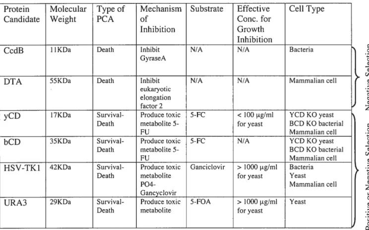
bacterial toxin CcdB inhibits DNA gyrase (Bemard and Couturier 1992) and promotes cell death by causing DNA lesions such as double-stranded breaks in DNA (Loris, Dao-Thi et al. 1999). Diphtheria toxin A is another toxic protein that causes celi death by covalently attaching ADP-ribose to the N-1 of the imidazole ring of diphthamide in eukaryotic elongation factor 2 (EF-2) (Oppenheimer and Bodley 1981). The second class consists of proteins that indirectly inhibit ceil division by activating a non-toxic compound to a toxic compound. The latter are also known as cytotoxic pro-drug converting enzymes. Herpes Simplex Virus-1 thymidine kinase (HSV1-TK), orotidine-5’-monophosphate decarboxylase (OMPdecase) and cytosine deaminase (CD) are some examples. Only proteins from this subset qualify for both survival and death assay and are therefore candidates for the development of the Binary Positive and Negative PCA. Among the candidates listed in this category, yeast cytosine deaminase (yCD) is an interesting candidate since the prodrug 5-fluorocytosine is widely available, inexpensive, and a small amount is required to cause
Table I: List of Death and Survival-Death PCA Candidates and Characteristics.
CcdB: Gene encoding for a toxin that inhibits bacterial DNA gyrase. DTA: Diphtheria toxin A.
yCD: Yeast cytosine deaminase. bCD: Bacterium cytosine deaminase.
HSV1-TK: Herpes Simplex Virus-I thymidine kinase.
URA3: Gene encoding for orotidine-5‘-monophosphate decarboxylase.
C -à Q Q Q C/D Q > Q
z
C -à Q Q Q Q > -à ce Qz
C Q > -à cdD CProtein Molecular Type of Mechanism Substrate Effective Ceil Type
Candidate Weight PCA of Conc. for
Inhibition Growth
Inhibition
CcdB 11KDa Death Inhibit NIA N/A Bacteria GyraseA
DTA 55KDa Death Inhibit N/A N/A Mammalian ceil eukaryotic
elongation factor 2
yCD 17KDa Survival- Produce toxic 5-FC < 100 tg/m1 YCD KO yeast
Death metabolite 5- for yeast BCD KO bacterial
FU Mammalian ceil
bCD 35KDa Survival- Produce toxic 5-FC N/A YCD KO yeast Death metabolite5- BCD KO bacterial
FU Mammalian ceil
HSV-TK1 42KDa Survival- Produce toxic Ganciclovir > 1000 ig/m1 Bacteria
Death metabolite for yeast Yeast
P04- Mammalian ceil Gancyclovir
URA3 29KDa Survival- Produce toxic 5-FOA > 1000 tg/m1 Yeast
Death metabolite for yeast
cytotoxicity in yeast and mammalian celis. HSVI-TK is also an interesting candidate for applications in mammalian celis since only 0.1 tM of the nucleoside analog, ganciclovir, is required to induce ceil death (Black, Kokoris et al. 2001). However, more than 1000 tg/m1 is required to temporarily inhibit celi growth in yeast (Wera, Degreve et al. 1999). Since our goal is to establish an assay that could be used in both yeast and mammalian celi systems, we chose to develop this survival and death PCA using yCD.
1.5 Yeast Cytosine Deaminase
Cytosine deaminase (CD) is an enzyme involved in the pyrimidine salvage pathway (Kurtz, Exinger et al. 1999) and was initially discovered in yeast and Escherichia cou (E. cou) in 1925 by Hahn and Schafer (Hahn and Schafer 1925). CD is not present in higher eukaryotic celis such as plant and mammalian celis (Nishiyama, Kawarnura et al. 1985). The yeast cytosine deaminase (yCD), has been used as a reporter protein for both positive and negative selection as discussed below (Wei and Huber 1996; Gallego, Sirand-Pugnet et al. 1999; Xiaohui Wang, Viret et al. 2001).
a) yCD in pyrimidine salvage pathway and survival selection assay
In S. cerevisiae, yCD is encoded by the fCYJ gene. The presence of this enzyme
allows yeast to use environmental cytosine as a source of pyrimidine for the nucleotide pooi (Figure 3) in the same way as bacteria (Grenson 1969). Cytosine enters into the celi using a purine-cytosine transporter encoded by the FCY2 gene. In the cytosol, cytosine is deaminated to uracil by yCD (Erbs, Exinger et al. 1997). Once cytosine is converted to uracil, uracil can be phosphoribosylated to uridine 5’-monophosphate (UMP) by uracil phosphoribosyltransferase (encoded by the FUR] gene) (Kern, de Montigny et al. 1990). UMP can be further phosphorylated to become uridine 5’-triphosphate (UTP) by uridine kinase (encoded by the URK] gene). UTP can then be converted by thymidylate synthase (encoded by the CDC2J gene) to thymidine monophosphate (TMP). Thus, yeast possesses a pyrimidine sauvage pathway that allows it to recycle exogenous cytosine for the ribose and deoxyribose nucleotide pool.
Figure 3. Pyrimidine Salvage Pathway and Survival Selection Strategy in S. cerevisiae.
When one of the key enzymes (encoded by URA2 or URA3 gene) of the pyrimidine de novo
pathway is disrupted, the ceil cannot produce uridine triphosphate (UTP). In order for the celi to survive, it must use the pyrimidine salvage pathway to import extracellular cytosine, deaminate cytosine to uracil by yCD (encoded by FCYJ gene), and modify uracil to UTP.
Proteins involved in this process are shown by their gene name in italics: aspartate
transcarbamylase (URA2), dihydrooratase (URA4), dihydrooratate dehydrogenase (URA I),
orotate phosphoribosyltransferase (URA5 and URAJO), orotidine-5 ‘-phosphate
decarboxylase (URA3), uridine kinase (URK1), purine-cytosine transporter (FCY2), yeast cytosine deaminase (FCY]), uracil phosphoribosyltransferase (FUR]).
De Novo Pathway
$aïvage Pathway
(INHIBITED)
Extracellular environrnent Cytosine FCY2
L-aspartate L-glutarnine Cytosolic environrnent
I
phosphate URA2 Cytosine
Carbamoyl-L-aspartate F120 H20 URA4 FCVI Dihydroorotate NH3 acccptor lIRAi PRPP Uracil reduced acceptor Orotate lIRA 10 pyrophosphate fURI PRPP lIRAS pyrophosphate Orotidine-5 ‘-phosphate UMP C02 URA3 ATP UMP URKI ADP URKI
UTP
$ URVIVAL
In addition to relying on the pyrimidine salvage pathway, S. cerevisiae can synthesize ribose and deoxyribose nucleotides using the de novo pyrimidine synthesis pathway. However, when the de novo pyrimidine synthesis pathway is inhibited, celis become uraci!
auxotrophs and thus require the essential nucleoside uracil for surviva!. Under these
conditions, the pyrimidine salvage pathway becomes the main source for generating uracil. Therefore, yCD cari become an important regu!atory enzyme that controls ce!! growth. This allows for the possibility of establishing a survival se!ection assay. Such a positive selection assay lias been established in yeast (Erbs, Exinger et al. 1997) and in mammalian cel!s (Wei and Huber 1996). In yeast, OMPdecase is one of the key enzymes in uracil synthesis and pyrimidine de novo synthesis (Figure 3). Disruption of the URA3 gene will force the ce!! to use yCD to convert environmental cytosine to uracil for ce!! surviva!. In mamma!ian ceils, gene knockouts in the pyrimidine de novo synthesis pathway also force
celis to utilize the pyrimidine salvage pathway for survival. However, pyrimidine de novo
synthesis can a!so be blocked by an inhibitor known as N-(phosphonacetyl)-L-aspartate
(PALA). PALA inhibits aspartate carbamyl transferase (CADases), an enzyme in the
carbamoyl-phosphate synthetase complex (Swyryd, Seaver et al. 1974). Mamma!ian celis treated with PALA wil! undergo apoptosis (Wei and Huber 1996) unless an exogenous cytosine deaminase enzyme (yCD) is introduced into the celi and cytosine is added as a supplement to the culture medium.
b) yCD Death Se!ection Assay
In addition to cata!yzing the deamination of cytosine, yCD can also deaminate 5-methylcytosine and 5-fluorocytosine (5-FC) (Wang, Sable et a!. 1950). 5-FC was initially synthesized in 1957 in prospective as an antitumor agent but instead was found to have uti!ity as an anti-fungal agent (Grunberg, Titsworth et al. 1963). yCD can deaminate 5-FC, a relative!y non-toxic compound, to 5-Fluorouraci! (5-FU), a toxic compound (Figure 3 in brackets). 5-FU can be ribosylated to 5-fluorouridine monophosphate (5-fUMP) by uracil phosphoribosy!transferase (FUR] gene). 5-FUMP is further phosphory!ated by uridine
kinase (URK] gene) to become 5-fluorouridine triphosphate (5-fUTP). 5-FUTP inhibits
ceil growth by incorporating into RNA (Fang, Hoskins et al. 2004; Lum, Armour et al. 2004) and directÏy inhibiting thymidy!ate synthase (CDC2] gene), an enzyme that controls
Figure 4. Pyrimidine Salvage Pathway and 5-Fluorocytosine Death Assay in S. cerevisiae.
Enzymes involved in this process are shown by their gene name in itaÏics. Environmental
cytosine is transported into the celi by the purine-cytosine transporter (ECY2). Yeast
cytosine deaminase (FCY1) deaminates cytosine (C) to generate uracil (U). Uracil phosphoribosyltransferase (FUR]) phosphoribosylates uracil to uridine monophosphate
(UMP). UMP is phosphorylated by uridine kinase (URKJ) to uridine diphosphate and
uridine triphosphate (UTP). UTP can incorporate into RNA and serve as a substrate for
thymidylate synthase (CDC2]) for the de novo synthesis of thymidine monophosphate
(TMP). 5-Fluorocytosine (5-FC) can also be processed by enzymes of the pyrimidine
salvage pathway and allow the establishment of a death assay. 5-FC and its derivatives are shown in brackets. FUTP is the toxic compound that causes inhibition of ceil growth.
5-FUTP can incorporate into RNA and inhibit thymidylate synthase.
C
FCY2 Fxtrace1Iu1ar environnient
I-FCJ Cytosolic environrnent H20 FCYI NH3 PRPP U 15-[UI 15-FUTPJ FURI RNA pyrophosphate URKI
Z
15-FUMPI UTP CDC2JATP ADP 15-FUIPI
TMP
*
DNA synthesis (Parker and Cheng 1990; Longley, Harkin et al. 2003). In S. cerevisiae, the effects of yCD and 5-FC resulted in morphological changes preventing daughter ceils to separate from the mother celis (Zhang, Zhang et al. 2002). Hence, yCD and 5-FC can be used as a selection tool for targeting ceil death. This negative seÏection lias been suggested for a plasmid shuffling assay by Erb et al. in 1997 and is currently being explored for applications in gene therapy.
1.6 Selection of a cellular system for the development of the Binary Positive and Negative PCA
The selection of the cellular system for establishing a Binary Positive and Negative PCA is dependent on the selection of the reporter protein. The objective is to select a system that is amenable to monitoring the signal of the PCA. Wild-type strain of S. cerevisiae lias a gene that encodes for cytosine deaminase (yCD). However, many yCD knockout strains of S. cerevisiae have been reported in small scale (Erbs, Exinger et al.
1997) and large scale (Giaever, Chu et al. 2002) studies. The latter large scale gene
disruption project utilized S. cerevisiae strain BY4741 (MATa ura3AO Ïeu2AO his3Al
metSAO) and BY4742 (MATa ttra3AO leu2AO his3A]
lys2AO)
for their gene disruptionproject. Since the URA3 gene is disrupted in botli BY4741 and BY4742 strains, the
pyrimidine de novo synthesis pathway can be blocked by removing cytosine from the
culture medium. yCD knockout ceils cannot grow under these conditions. yCD becomes the regulatory enzyme for the pyrimidine salvage pathway and can regulate ceil survival. These yCD knockout strains of BY4741 and BY4742 can be used for the development of our yCD PCA since botli tlie survival and deatli selection assays can be tested in tliese strains.
1.7 Approaches for establishing the Binary Positive and Negative yCD PCA
Tlie approach for developing yCD PCA consisted of carefully analyzing the structure of tlie protein and selecting specific sites for fragmentation. Furtliermore, in order to increase the possibility of finding tlie best yCD fragments that can generate highest yCD PCA activity, a fragment sliuffling experiment can be done.
Figure 5. yCD topology. yCD monomer is composed of six alpha (a) helices and five
beta
(I)
strands. The first a-lieux (ai) is found at the N-terminal of the protein followedby a f3-hairpin, (32, I3), a second a-helix (a2), a Rossmon fold (33-a3-(34-a4-f35), and
two short helices (a5-a6). The five [3-strands form a f3-sheet sandwiched between a
helices (al and a5) on one side and a-helices (a2, a3, a4, and a6) on the other side.
C-terminal
a) Molecular Characteristics of yCD
The structure of yCD has recently been solved (Ireton, Black et al. 2003; Ko, Lin et al.
2003). yCD differs significantly from bacterial cytosine deaminase (bCD) in terms of
quaternary structure, primary amino acid sequence, molecular mass, and relative substrate
specificities and affinities. yCD is a homodimer where the 17.5 KDa monomers are
arranged in a 2-fold symmetry axis. The dimer is formed from the association of the two monomers in a head-to-tail orientation. Each monomer forms a compact domain composed
of six alpha ta) helices and five beta (3) strands (Figure 5). The five -strands form a
f3-sheet sandwiched between a-helices (al and a5) on one side and a-helices (a2, a3, a4,
and a6) on the other side. There is one active site present per subunit. The active site
involves amino acid histidine-62, glutamate-64, cysteine-91, and cysteine-94. Each active site can bind a tetrahedral catalytic zinc ion, which helps to coordinate the substrate and participate in an acid/base catalysis mechanisrn. Amino acids histidine-62, cysteine-91 and cysteine-94 are also involved in the coordination of the zinc molecule.
b) Fragmentation of a Reporter Protein
The fragmentation sites were chosen in regions of the enzyme flot involved in secondary structure elements such as aipha-helices or beta-strands of the protein. Cutting reporter proteins in loop regions connecting one secondary structure element to another is favored. In the case that the reporter protein is an enzyme, we select for loop regions not involved in the active site of the enzyme. Cutting within secondary structure elements could interfere with the refolding of the reporter protein. Thus, the idea is to fragment the reporter protein in such a way that it could easily refold to its functional structure resembling the native protein.
c) Optimization of yCD PCA Activity by Fragments Shuffling
It lias previously been shown that two fragments of an enzyme containing overlapping amino acid residues could rearrange to generate a functional enzyme (Taniuchi and Anfinsen 1971; Ostermeier, Nixon et aÏ. 1999). In addition, it lias also been known that
elimination of some residues in a peptide fragment could increase or decrease the activity of an engineered protein (Ostermeier, Nixon et al. 1999). By shuffling yCD fragment 1 versus yCD fragment 2 of the different cut sites, different PCA combinations can be screened in order to increase the activity of yCD PCA.
1.8 Specific aims
The goal of this project is to develop a Binary Positive and Negative PCA using yeast cytosine deaminase (yCD PCA). In brief, this study consisted of: 1) fragmenting yCD to generate the PCA; 2) testing yCD fragments for yCD PCA activity using the death and survival assay as well as improving yCD PCA activity; and 3) demonstrating a possible application of the Death PCA in a mammalian celi system.
CHAPTER 2: MATERIALS AND METHODS
2.1 Yeast genomic DNA, yCD knockout yeast strains, and substrates
S. cerevisiae strain BY4743 (diploid iira3AO leu2AO his3A] met5AO lvs2AO) was used for isolation of genomic DNA containing two wild-type copies of the yCD gene. S. cerevisiae 3Y4741 yCD knockout and BY4742 yCD knockout, generated as part cf the yeast gene knockout collection (Giaever, Chu et al. 2002), were used for yCD PCA since their genomic copy of yCD have been disrupted. Ail yeast strains were gifts from Dr. Howard Bussey. YPD (yeast extract, peptone, and dextrose) and synthetic defined medium with amino acids complementation (SDC) were prepared as described by Guthrie et al.
(Guthrie C, Fink GR 1991). YPRO62w knockout strains were propagated in medium
containing 200 1g/ml cf Geneticin® (G41$) purchased from Invitrogen (Burlington,
Ontario). 5-fC and cytosine [purchased from Sigma (Oakville, Ontario)] were diluted in water to a concentration of 10 mg/ml and kept as stock solution.
2.2 yCD PCA development in yeast
Yeast expression vectors p4i3Gall and p4l5Gall (Mumberg, Muller et al. 1995), used for yCD PCA development, were gifts from Dr. Howard Bussey. First, GCN4 leucine zipper and one third of the iinker sequence coding for amine acids GGGGS was amplified by polymerase chain reaction (PCR) with pfu polymerase, purchased from Fermentas (Burlington, ON), from the plasmid pcDNA3.1-Zip-[Fl.2] mDHFR (Remy and Michnick
1999) using the following primers: 5’-cgc tctaga ggg
ATGAACACTGAAGCCGCCAGGCG-3’ and 5’-ccg ctcgag cta tccgga gccaccgccacc
GCGTTCGCCAACTAATTTC-3’. GCN4 leucine zipper was cloned into the multiple cloning sites cf p4l3Gall and p4l5Gall vectors at the XbaI and XhoI restriction sites. A unique BspEi site was added as part cf the linker sequence for cloning DNA fragements downstream cf the GCN4 leucine zipper sequence (Figure 6). The vectors carrying these GCN4 leucine zipper sequence are named p4l3Gall-Zip and p4l5Gall-Zip.
Figure 6. yCD PCA construction scheme. First, the gene of interest is cloned into the vector, for example GCN4 cou-cou leucine zipper (Zip) sequence cloned into the multiple cloning sites with a flexible linker and BspEI of p4l3Gall or p4l5Gallvectors using the
XbaI and XhoI sites. Next, yCD or yCD fragments are cloned downstream of the Zip
sequence using BspEI and XhoI sites.
Stepi: Insert Zip and 5 arnino acids(Saa) sequence with BspEl restriction site in multiple cloning sites of vector using XbaI and Xhol sites.
Step2: insert 10 amino acids sequence and yCD fragment using BspEI and XhoJ sites.
Xbal BspEI X ho! p4.l3 Gali or p4i5GaIl
I
BspEI XhoI p413 Gall/Zip or p4l5Gall/ZipyCD gene and yCD fragments were amplified from S. cerevisiae strain 3Y4743 genomic DNA using pfu polymerase (Fermentas, Burlington, Ontario) with the remaïning linker sequence codïng for amino acïds GGGGSGGGGS. Conditions used for PCR were the following: 95°C (5 mm), 25 cycles of [95°C (1 mm), 55°C (1 mm), 72°C (1 mm)], 72°C (3 mm). The primers used for amplifying yCD and yCD fragments are listed in Table II. These PCR products were cloned into p4l3Gall and p4l5Gallvectors downstream of GCN4 leucine zipper sequence using BspEI and XhoI restriction sites and T4 DNA ligase purchased from Fermentas (Burlington, Ontario).
2.3 Yeast Ceil Transformation
Competent yeast celis were prepared and stored at —80°C (Knop, Siegers et al. 1999). In brief, MATa, MATa, and diploid yCD knockout yeast colonies were inoculated for 6 hours in 5 ml of YPD medium with 200 ig/ml G418. These pre-cultures were used to start a 100 ml culture for overnight incubation. The next day, when the culture reached
0D600 0.5 to 0.7, cells were harvest by centrifugation at 1500 rpm for 5 min at room
temperature (RT). The pellets were washed with 5 ml of sterile water, centrifuged for 5 min at 1500 rpm, and washed with 5 ml of Sorbitol Buffer (1 M sorbitol, 1 mM EDTA, 10 mM Tris pH 8.0, 100 mM LiOAc). Pellets were resuspended in 720 tl of Sorbitol Buffer and 80 tl of Salmon Sperm DNA at 10 mg/ml stock solution purchased from Sigma (OakviÏle, Ontario).
Yeast cell transformationwas performed by mixing 100 ng of plasmid DNA with 25
il of frozen competent yeast celis in a 1.5 ml microtube. 150 d of PLATE solution (40% PEG 4000, 100 mM Lithium Acetate, 10 mM Tris pH 7.5 and 0.4 mM EDTA) was added.
The sample vas mixed and allowed to incubate for 30 min at RT. 20 pi of
Dimethylsulfoxide (DMSO), purchased from Fisher Scientific (Fairlawn, NJ), was added, and incubated at 42 °C for a 20 min heat shock. Cells were pefleted by centrifugation at 2000 rpm for 3 min. The supernatant was discarded and the pellet was resuspended in 200
tl of PLATE solution. 20 il of the sample was plated on synthetic defined medium with
amino acid complementation for selection (6-well plate) and allowed to incubate at 30 °C for 3 days.
D
Table
II.
Primers
for
generating
yCD
fragments
at
7
cut
sites
and
yCD
full-length
enzyme.
Nucleotide
sequences
in
uppercase
correspond
to
sequences
that
directly
anneal
on
yCD
gene,
nucleotide
sequences
in
lowercase
from
the
sense
primers
column
correspond
to
flexible
linker
sequences,
and
underligned
nucleotide
sequences
correspond
to
restriction
sites.
5-ggc tee gga ggt gga ggt tct gga ggt ggc gga tct ATGGTGACAGGGGGAATGGC-3’ 5-ggc tee gga ggt gga ggt tel gga ggt ggc gga tct ATGGTGACAGGGGGAATGGC-3 5’-ggc tcc gga ggt gga ggt tct gga ggt ggc gga tct ATGGTGACAGGGGGAATGGC-3 5’-ggc tcc gga ggt gga ggt tct gga ggt ggc gga tct ATGGTGACAGGGGGAATGGC-3’ 5’-ggc tcc ga ggt gga ggt tet gga ggt ggc gga tct ATGGTGACAGGGGGAATGGC-3 5-ggc tcc gga ggt gga ggt tct gga ggt ggc gga tct ATGGTGACAGGGGGAATGGC-3 5’-ggc tcc gga ggt gga ggt tct gga ggt ggc gga tct ATGGTGACAGGGGGAATGGC-3 5’-ggc tee gga ggt gga ggt tct gga ggt ggc gga tct GGATCCGCCACACTACAT-3 5-ggc çç ggt gga ggt tct gga ggt TFAGAGGGCAAAGTGTACAAAG-3 5-ggc tee ga ggt gga ggt tct gga ggt ggc gga tct AAAGTGTACAAAGATACCAC-3’ 5-ggc tee ga gt gga ggt tel gga ggt ggc gga tel GTGTACAAAGATACCACT-3 5-ggc tee gga ggt gga ggt tet gga ggt ggc gga tct GATACCACFFTGTATACG-3’ 5-ggc tee gga ggt gga ggt tet gga ggt ggc gga tct GTFAATFFCAAAAGTAAGGGC-3’ 5-gge tee gga ggt gga ggt tet gga ggt gge gga let GACGATGAGAGGTGTAAA-3’ 5-gge tee gga ggt gga ggt let gga ggt gge gga tet ATGGTGACAGGGGGAATGGC-3 Anti-sense Primers 5’-eeg eec ecg etegag eta CTTTFGAAATCTCATGTT-3 5’-eeg eec ceg etegag eta TCTCCCACAGTHTCCAAAGTGGAG-3 5’-eeg ccc ceg etegag eta GCCCTCTAATCTCCCACAG-3’ 5-ccg ccc eeg etc eac eta TTTGCCCTCTAATCTCCC-3’ 5-ccg ccc eeg ctegag eta TFfGTACACTFTGCCCTC-3 5-ceg eec eeg ctcgag cta GTFCTCACCGACAACACA-3 5-eeg eec eeg etcgag eta AACAACAACAACCTCGTG-3 5-eeg ccc ceg etegag cta CTCACCAATATCTFCAAACC-3 5-eeg eec eeg etegag eta CTCACCAATATCTfCAAACC-3 5-ccg ccc eeg eteeae eta CTCACCAATATCTfCAAACC-3 5’-ccg ccc ecg ctcag eta CTCACCAATATCTTCAAACC-3 5-ceg eec ccg etegag cIa CTCACCAATATCTFCAAACC-3’ 5-ecg ccc ecg etegag eta CFCACCAATATCTfCAAACC-3’ 5-eeg ccc ecg ctegag eta CTCACCAATATCFTCAAACC-3 5-eeg ccc eeg etegag eta TCTCCCACAGTITTCCAAAGTGGAG-3 Gene NameSense Primers FIyCDeutl FI yCDeut2 FI yCDeut3 FI yCDeut4 fi yCDeut5 Fi yCDeut6 FI yCDeut7 F2yCDeut 1f2yCDeut2 F2yCDcut3 F2yCDcut4 F2yCDcut5 F2yCDeut6 F2yCDcut7 C-ter
2.4 Protein Expression in Yeast
Colonies were inoculated in 1 ml of synthetic defined medium (6.7 g/L of yeast nitrogen base without ammonium sulphate and amino acids [purchased from BioshopJ), 1 g/L monosodium glutamic acid, 2 g/L amino acid drop-out lacking methionine, lysine, histidine, or leucine, 200 ig/ml G418, and 2% raffinose (SC -met -lys -his or—leu +G418). The cultures were grown overnight and then induced with 2% galactose for 24 hrs. Celis were collected and prepared for protein extraction using Lyticase method (Sambrook J. 1989). The cell pellet was washed with Phosphate-Buffered Saline (PBS). The pellet was resuspended in Stabilizing Buffer A [1 M sorbitol, 10 mM MgC12, 2 mM dithiothreitol (DTT), 50 mM potassium phosphate pH 7.8, 100 igIml, and phenylmethylsulfonyl fluoride (PMSF)] and incubated at 30 °C for 10 min. The sample was pelleted, resuspended in Stabilizing Buffer 3 (1 M sorbitol, 10 mM MgC12, 2 mM DTT, 25 mM potassium phosphate pH 7.8, 25 mM sodium succinate pH 5.5, 100 tg/ml PMSF), and incubated at 30 °C for 2 min. Then 10 d of Lyticase vas added to the sample and incubate at 30 °C for 30 min. Protoplasts were collected by centrifugation at 12 000 rpm for 10 min at 4 °C, resuspended in 100 tl No-salt lysis buffer (50 mM HEPES pH 7.0, 1% NP-40, 1 tg/ml aprotinin, and 100 ig/m1 PMSF), and incubated for 30 min on ice. Protein concentration was determined using Bio-Rad DC Protein Assay [purchased from Bio-Rad (Hercules, CA)] since 1 % NP-40 was used in the lysis buffer.
for western blot analysis, 30 tg of protein extracts were loaded per well onto a
12.5% sodium dodecyl sulfate polyacryÏamide gel electrophoresis (SDS-PAGE). After electrophoresis, proteins from the acrylamide gels were transferred onto PVDF membranes. The membranes were blocked with 5% milk in Tris-NaCÏ Buffer (50 mM Tris pH 7.5, 150 mM NaC1, 0.2% Tween) overnight. Membranes were probed at RT for one hour with anti yCD purchased from Biogenesis (Poole, England) at a 1/1000 dilution and with anti-sheep HRP purchased from Upstate ceil signaling solutions (Lake Placid, NY) at a 1/5000 dilution. Membranes were washed 3 times 10 min with Tris-NaC1 Buffer after each antibody incubation. Membranes were revealed with Western Lightning® Chemiluminescence Reagent Plus purchased from PerkinElmer (Boston, MA). Membranes were stripped with stripping solution purchased from Promega (Madison, WI) and re
blocked with 5% milk in Tris-NaC1 Buffer for 30 min at RT. Membranes were probed at RT for one hour with anti-yeast 3-phosphoglycerate kinase (anti-PGK) purchased from Molecular Probes (Eugene, Oregon) at a 1/1000 dilution, washed 3 times for 10 min with Tris-NaCI Buffer, probed with anti-mouse HRPO purchased from BD Biosciences (San
Diego, CA) at a 1/5000 dilution, and revealed with Western Lightning®
Chemiluminescence Reagent Plus.
2.5 5-FC Death Assay on Liquid and Solid Medium
Yeast colonies were inoculated ovemight in 1 ml of SDC-met-lys-his-leu + G418 +
2% raffinose. The next day, 20 d cf the culture was transferred to 1 ml of SDC-met-Iys
his-leu + G418 + 2 % raffinose and 2 % galactose for a 6 hrs induction at 30°C.
For 5-FC death assay in liquid medium, either 500 or 5000 celis were transferred te a 96-well plate with selection medium containing 0, 100 or 1000 tg/ml of 5-FC. Amount
cf ceils were determined based on optical density measurement at 600 nm (OD 600nm). OD
600nm cf 1 equals to 106 cells per ml. Samples were incubated at 30°C with shaking for 1 to 2 days. After the incubation time, 200 tl samples were transferred to a 96-well plate for
measuring OD 600nm with Spetramax 190 from Molecular Devices (Sunnyvale, CA). The
percentage of relative growth inhibition vas calculated as follows: {(OD ioonm non-treated
celis —OD 600nm treated cells) / (OD 6nm non-treated ceils)]
* 100.
For 5-FC death assay on solid medium, around 5000 ceils were transferred to 1 ml
of SDC-met-lys-his-leu + G418 + 2% raffinose and 2% galactose (with and without 5-FC)
for an 18 hrs pre-incubation at 30°C shaking. After the pre-incubation period, 10 d of the
samples were plated on solid selection medium: SDC-met-Iys-his-leu + 2% Agar +G418 +
2% raffinose and 2% galactose (with and without 5-FC). Plates were incubated at 30 °C for either 2, 3, or 6 days.
2.6 Survival Assay on Solid Medium
Yeast colonies were inoculated ovemight in 1 ml of SDC-met-lys-his-leu + G418 +
2% raffinose. The next day, 20 tl cf the culture vas transferred to 1 ml of SDC-met-Ïys
his-leu + G41$ + 2% raffinose and 2% galactose fora 6hrs induction at 30°C. 10 d ofthe
samples were plated solid selection medium: SDC-met-lys-his-!eu + 2% agar + G418 + 2%
raffinose and 2% galactose (with and without cytosine). Plates were incubated at 30 °C for either 2, 3, or 6 days.
2.7 Mammalian Celi Lïnes and Reagents
Mammalian HEK293 was a gift from Dr. Guy Boileau’s laboratory. Mammalian colon cancer ce!! unes CaCo-2 and LoVo were generously provided by Dr. C!ifford Stanners’ laboratory. Mammalian 6, 12, 96-we!! culture plates are treated for ce!! cu!ture
and are purchased from Corning COStar (Acton, MA). Du!becco’s Modified Eag!e
Medium (DMEM), Minimum Essential Medium a!pha-Medium (a-MEM), Du!becco’s Phosphate-Buffered Sa!ine (PBS) and Trypsin-EDTA are from Invitrogen (Burlington,
Ontario). Fetal Bovine Serum (FBS) is provided by Wisent (Saint-Jean-Baptiste de
Rouville, Quebec). Transfecting reagent Fugene 6 is from Roche Diagnosics (Basel,
Switzer!and). ZeocinlM vas purchased from Invitrogen (Bur!ington, ON)
2.8 Construction of Mammalian Celi Promoter Characterization Vectors Primers used to amplify tissue specific promoters are !isted in Table III.
Carcinoembryonic antigen promoter
(
CEAp from -407bp to —43bp), CEA enhancer (CEAefrom -6.lkb to —4kb), CC6 promoter (CC6p from -281bp to —2bp), and CC6 enhancer (CC6e from-1.24kb to —0.585kb) were amp!ified with pfu po!ymerase (Fermentas) from LoVo cell !ine genomic DNA using the fol!owing PCR condition: 95°C (5min), 25 cyc!es
of [95°C (imin), 62°C (lmin), 72°C (imin)], 72°C (3min). Human telomerase reverse
transcriptase prornoter (hTERTp) was amp!ified with pfu polymerase (Fermentas) from LoVo ceil line genomic DNA using the following PCR conditions: 95°C (Smin), 25 cycles
of [95°C (lmin), 60°C (30sec), 72°C (30sec)], 72°C (3min). The PCR reaction was
supplemented with 250 ig of bovine serum a!bumin and 0.1 M of betaine both purchased
from Sigma (Oakvil!e, ON). CEAp, CC6p and hTERTp were c!oned into pGL3prom,
Sense Primers
5’-cccggg ctcgag CCCGGGACCCTGCTGGGYFTC-3’ 5’- cccggg acgcgt GGTFACATTACAAAGTGGAAT-3’ 5 ‘-cccggg ctcgag CTFGCFFCTCAGAGCATCTTC-3’ 5 ‘-cccggg acgcgt CACAGCAATAAACACAATGAT-3’ 5’-atatat ctcgag AGTGGATTCGCGGGCACAGA-3’
Anti-sense Primers
5 ‘-cccggg aagctt GAGTI’CCAGGAACGÏT]TGTC-3’ 5 ‘-gggggg ctcgag CGGCTCACTGCAACCTCTGCCTC-3 5’-cccggg aagctt GGTCTCTGCTGTCTFCTCTGT-3’ 5 ‘-gggggg ctcgag TFATGTGACTCTAATTTCCTG-3’ 5 ‘-atatat aagctt AGGGCTFCCCACGTGCGCAG-3’ CEAp
CEAe CC6p CC6e hTERTp
Table III. Primers for amplifying tissue specific promoters from genomic DNA of LoVo colon
cancer celi une.
Promoter Name
purchased from Promega (Madison, WI) vector by removing the original SV40promoter
with XhoI and Hindili. The new vectors are subsequently named pGL3!CEAp,
pGL3/CC6p and pGL3/hTERTp. CEAe and CC6e were cloned upstream of CEAp and CC6p using Miul and XhoI sites generating plasmids: pGL3/CEAep and pGL3/CC6ep.
In order to generate the negative control vector, the original pGL3prom vector was digested with XhoI and Hindiil to remove the SV4O promoter. The linearized vector was treated with Mung Bean Nuclease [purchased from NEB (Beverley, MA)] to remove the 5’nucleotide extensions and Iigated with T4 DNA Ligase [purchased from Fermentas
(Burlington, Ontario)]. This new promoterless vector was named pGL3noProm.
pGL3/CEAp, pGL3/hTERTp, and pGL3noProm vectors were confirmed with sequencing.
2.9 Mammalian celi Promoter assay vector
2 X10⁵ HEK 293 and CaCo2 celis were seeded in 12-well plates purchased from Corning
(Acton, MA). The following day, celis were co-transfected respectively with 1 tg of
pGL3/CEAp, pGL3/CEAep, pGL3/CC6p, pGL3/CC6ep, pGL3/hTERTp, pGL3prom, and pGL3noProm plasmid DNA and 100 ng of pRL-CMV [purchased from Promega (Madison, WI)] using Fugene (Roche) transfecting reagent. pRL-CMV is a plasmid that carnes the RenilÏa luciferase gene under the Cytomegolovirus (CMV) promoter. 24 hours after the transfection, ceils from each well of the 12-well plate were trypsinized and re-plated in
96-well clear-bottom white plate. 48 hours after the transfection, celis from the same
transfection were assayed for firefly luciferase and Renitla luciferase activity. Firefly
luciferase activity was assayed by using Promega Bright-GloT’ Luciferase Assay kit, purchased from Promega (Madison, WI), for quantification of firefly luciferase activity. In brief, 100 pi of the substrate was directly added to celis in the 96-well plate and celis were
lysed. Luminescence signal was acquired for 30 sec using PACKARD FUSION
MULTIDETECTION PLATE READER purchased from PerkinElmer (Woodbridge,
Ontario). Activity of Renilla luciferase was quantified by the addition of 1 !IM of
Coelenterazine [purchased from Biotium, (Hayward, CA)] and luminescence signal was acquired for 30 sec. Resuits of the firefly luciferase activity were normalized to resuits of
the RenilÏa luciferase activity. Each plasmid was tested in three to four different
transfection experiments. Data from the promoter assay varied relative to differences in transfection efficiency from each transfection. Therefore, resuits of only one transfection experiment are represented since the overali trend of the promoter activity assay is the same.
2.10 Mammalian Expression Vectors Construction
Mammalian expression vectors pcDNA3. 1 neo and pcDNA3. 1 zeo were purchased
from Invitrogen. These expression vectors were chosen for their antibiotic resistance
genes, which were used to generate stable cell unes. Zip-yCD, and Zip-[Fl]yCDcut4 were amplified from yeast expression vectors (p4l3Gall series) and sub-cloned into pcDNA3. lneo using Hindili and XbaI. Zip-yCD and Zip-[f2]yCDcut4 were amplified from yeast expression vectors (p4l5Gall) and sub-cloned into pcDNA3.lzeo using Hindili
and XbaI. This generates the following vectors: pcDNA3.lneo/Zip-yCD,
pcDNA3. 1 neo/Zip-[Fl]yCD cut 4, pcDNA3.1 zeo/Zip-yCD, and pcDNA3. 1 zeo/Zip
[F2]yCD cut4. The GCN4 leucine sequence vas replaced by the library optimized
WinZipAl (WinA) or WinZipB 1 (Win B) leucine zipper (Pelletier, Arndt et al. 1999). WinA has the following amino acid sequence: STTVAQLEEKVKTLRAQNYELKS
RVQRLREQVAQLAS. WinB lias the following amino acid sequence:
STSVDELQAEVDQL QDENYALKTKVAQLRKKVEKLSE. WinA was generated by
overlapping oligonucleotide fragments and PCR using oligos: f-WinA:
5’-GGGAAGCFFACCATGTCCACCACCGTGGCCCAGCTGGAGGAAAAGGTGAA AACCCTGAGAGCCCAGAACTACGAGCTGAAGTCC-3’ and r-WinA: 5 ‘-GCCCAG AACTACGAGCTGAAGTCCAGAGTGCAGAGGCTGAGAGAACAAGTCGCCC AGCTGGCCTCCGGTGGCGGTGGCTCCGGA TAG -3’. WinB was generated by overlapping oligonucleotide fragments and PCR using oligos: f-WinB: 5’-GGGAAGCTTACCATGTCCACCTCCGTGGACGAGCTGCAGGCCG
AGGTGGACCAGCTGCAGGACGAGAACTACGCCCTGAAGACC-3’ and r-WinB: 5’-GACGAGAACTACGCCCTGAAGACCAAGGTGGCTCAGCTGAGAAAGAAGGTGG AGAAGCTGTCCGAAGGTGGCGGTGGCTCCGGATAG-3’. PCR was performed at 95°C (5 mm), [95°C (1 mm), 55°C (1 mm), 72°C (1 mm)] for 25 cycles, 72°C (3 mm) with Pfx AccuPrime from Invitrogen (Burlington, Ontario). Giving rise to the following set of
vectors: pcDNA3.lneo/WinA-yCD, pcDNA3.lneo/ WinA-[F1]yCD cut 4, pcDNA3.lzeo/
WinB -yCD, and pcDNA3.lzeo/WinB-{F2IyCD cut4. Ail of the above vectors were
confirmed by sequencing and have the original CMV promoters of the pcDNA3.lneo and pcDNA3. lzeo.
In order to obtain tissue specific promoters, CEAp, and hTERTp were digested from pGL3/CEAp and pGL3/hTERTp with MluI and Hindili restriction sites and sub-cloned into ail previously constructed pcDNA3. lneo or pcDNA3. lzeo based vectors by removing the CMV promoter with MluI and Hindili sites. This generated the following plasmids:
pcDNA3. lneo/CEAp/WinA-yCD, pcDNA3. 1 neo/CEAp/WinA-[F1IyCD cut 4,
pcDNA3. 1 zeo/hTERTp/WinB -yCD, and pcDNA3. 1 zeo/ hTERTp/WinB -[F21 yCD cut4.
[F2]yCD cuti was sub-cloned downstream of promoter specific vectors using BspEI and XhoI to generate pcDNA3.lzeo/WinB-[F2]yCD cuti, and pcDNA3.lzeo/hTERTp /WinB-[F2]yCD cuti.
2.11 Generating Stable Transfectants
2.5 X iO ceils were seeded in 12-wells plates 24 hrs prior to transfection. Cells were transfected with 1 g of plasmid DNA. The following combinations of plasmids were used: a) CEAp /WinA-[F1]yCD cut4 and hTERTp/WinB-[f2]yCD cut4, b) CMVp /WinA [F1]yCD cut4 and CMVp/WinB-[F2]yCD cut4, c) hTERTp/WinA-yCD, d) CMVp/WinA
yCD, and e) pcDNA3.lneo and pcDNA3.izeo mock vectors.) pcDNA3.lneo and
pcDNA3.lzeo mock vectors. 48 hrs after transfection, celis were grown in medium
containing 400 ig/m1 of G418 and 400 pg/m1 of ZeocinTM in order to select for stable ceil lines. Selection medium was changed every 2 days. After 7 to 21 days, stable celi unes were trypsinized and transferred to 5 cm Petri dishes. Subsequently cells were transferred to 10cm Petri dishes and grown in medium containing 200 pg/m1 of G418 and 200 tg!mI ofZeocinhM. These stable celi unes were allowed to reach approximately 70% confluency and frozen for later use.
2.12 5-FC Assay in Mammalian Ceils and MIT Assay
1 X10⁴ celis were seeded in 96-well plate with culture medium containing 200 ig/ml of G418 and 200 tg/m1 of ZeocinTM. After 24 hrs of incubation, the medium was changed for medium containing 0, 100 or 1000 ig/ml 5-FC and 200 tg/m1 of G418 and
200 tgIml of ZeocinTM. Media were changed once every two days. After six days of
treatment with 5-FC, celi viability was assayed by measuring the activity of dehydrogenase enzymes presence in metabolically active ceils using the Colorimetric (MTT) assay for celi survival and proliferation kit purchased from Chemicon (Temecula, CA). Briefly, 10 pi of 3-(4,5-dimethylthiazol-2y1)-2,5-diphenyl tetrasodium bromide (MTT) in PBS, was added to each well of the 96-well plate and incubated for 3 hrs at 37°C. After the incubation time, 100 pi of solubilizing solution was added to each well in order to lyse ceils and solubilize the product. Conversion of the yellow MTT substrate to a blue product was measure at 570
nm and 630 nm (Molecular Device Spectramax 190). The quantity of blue product
produced is calculated by substracting the optical density (OD) value at 570 nm minus the
OD at 630 nm. The percentage of relative growth inhibition is calculated with the
following formula:
= [(OD57Onm6yJnm untreated - OD57Oim6OOnm 5-FC treated)/ °D57Onm6OOnrnuntreated]
* 100
CHAPTER3: RESULTS
3.1 Development of yCD PCA in Yeast
We have chosen to develop a Positive/Negative Selection PCA in yeast for mainly two reasons. First, it is both time- and cost-efficient to use yeast for the developmental stage of our yCD PCA since no sophisticated equipment or material are required. A simple vector system (p4l3Gall and p4l5Gall) with auxotrophic markers (Mumberg, Muller et al. 1994) and basic transformation protocol (Knop, Siegers et al. 1999) allowed us to study the effect of our fusion gene in yeast celis. It was not necessary to establish stable cell unes as would have been for mammalian ceils in order to maintain stable expression of the
recombinant genes. In addition, we took advantage of the haploid/diploid life cycle of
yeast in order to introduce one fragment of our yCD PCA in MATa mating type and the other fragment in MATa mating type and then mated the MATa and MATa to generate diploid celis carrying the two fusion genes. The second reason vas that we hoped also to establish a negative selection PCA in yeast for future application in screening projects. In
vivo negative selection systems have been established to screen for bioactive molecules in
bacteria and in yeast based on the concept of a reverse two hybrid assay (Licitra and Liu 1996; Horswill, Savinov et al. 2004). The yCD PCA could be used for similar applications and provide a more direct method of signal output since yCD PCA does not involve the transcriptional regulatory system of a reporter gene (Michnick 2004).
3.1.1 Design and Construction of yCD PCA
Topological analysis of yCD structure revealed several loop regions of the protein
that could serve as sites for fragmentation of the protein. Only one large loop region,
composed of 10 amino acid residues, located between a2-helix and 133-strand (Figure 7), was identified from the yCD structure (Protein Data Bank file: ÏUAQ). We considered this loop region as a good eut site for yCD fragmentation since it dissects the active site of the enzyme, glutamine-64 (E-64), from the zinc ion-binding signature motif (C-91-X-X-C-94)
of yCD which coordinates the substrate for the deamination process. Hence, yCD
fragments generated from cuts in this region were predicted to be unlikely to possess full
enzymatic activity alone. We therefore chose four eut sites in this region (Figure 7). In
addition, we have included other loop regions for yCD fragmentation in this study. Other
eut sites were located in the 132-a2, f34-a4, and (35-a5 loop regions. In brief, the seven eut
Figure 7. yCD cut sites for PCA development. Ail cut sites are in ioop regions of the protein. Cut site 1 is between K-56 and G-57. Cut site 2 is between R-73 and L-74. Cut site 3 is between G-76 and K-77. Cut site 4 is between K-77 and V-78. Cut site 5 is between K-80 and D-$1. Cut site 6 is between N-111 and Vii2. Cut site 7 is between V
132 and D-133.
C-terminal Cuti
Legend for Cut Sites: Cuti: K-56/G-57 Cut2: R-73/L-74 Cut3: G-761K-77 Cut4: K-77/V-78 Cut5: K-80/D-81 Cut6: N-111/V-112 Cut7: V-132/D-133 Cut7 N-terminal Cut2 Cut3 Cut4 Cut5
sites were: cut site 1 (between amino acid 56-57), cut site 2 (between amino acid 73-74),
cut site 3 (between amino acid 76-77), cut site 4 (between amino acid 77-78), cut site 5
(between amino acid 80-81), cut site 6 (between amino acid 111-112), and cut site 7 (between amino acid 132-133). This enzyme is very small and almost every a-helix and 3-strand interacts with the substrate. Fragmentation of yCD between amino acid residues that serve to make contact with the substrate and hold it in place represents a potential ctit site for yCD PCA since this could prevent the enzyme from catalyzing the dearnination
reaction. We avoided cutting in the middle of secondary structure of a-helices and
f3-strands since this could affect important structural elements for refolding of the enzyme.
Constructions of fusion genes, used to determine yCD PCA activity, were done in p4l3Gall or p4l5Gall vectors and are listed in Figure 8. The fusions genes were cloned downstream of the Gali promoter which gives a high level of expression in yeast when yeast celis are grown in medium with galactose as an inducer and no glucose (Ronicke, Graulich et al. 1997). fragments of yCD or full length yCD were fused at the C-terminus
of the GCN4 leucine zipper. A linker region composed of 15 or 12 amino acids
[(GGGGS)3 or (GGGGS) 2GGI was inserted between the zipper and yCD fragments or
yCD. All fusion genes presented in this work were generated with the same strategy.
3.1.2 fusion Protein Expression
Expression of the fusion proteins was verified by Western blot using polyclonal anti-yCD antibody (Biogenesis). Detection of yeast 3-phosphoglycerate kinase (PGK), a ubiquitously expressed protein in yeast, was used as a loading control to indicate that a
similar amount of protein had been loaded in each well of the acrylamide gel. In
comparison to the mock samples (ceÏÏs transformed with the mock p4Ï3GalÏ or p4Ï5GalÏ vectors) most yCD-fusion proteins were detected. Zip-[F2]yCDcut7 was expressed at a very low level (figure 9) since it could only be detected on the same western blot at a longer exposure (data not shown). Unfortunately, the polyclonal anti-yCD antibody non specifically detected other yeast cellular proteins (20 Kda and 24 Kda in size) in addition to yCD. The expression of Zip-[fl]yCD cut6, Zip-[FljyCD cut7, and Zip-[F2jyCD cutl (Figure 9) fusion proteins could not be confirmed by western blot analysis using the
Figure 8. Recombinant genes generated from yCD or yCD fragments fused to GCN4 zipper sequence. A) yCD Fragment 1 [F1] and yCD full-length enzyme, generated from the different cut sites, were fused to GCN4 zipper forming sequences and cloned into p4l3Gal 1 plasmid. B) yCD Fragment 2 [F2] and yCD full-length enzyme, generated from the different eut sites, were fused to GCN4 Zipper forming sequences and eloned into p4l5Gal 1 plasmid.