Université de Montréal
Dissecting Cell Cycle Protein Complexes using the
Optimized Yeast Cytosine Deaminase Protein-
fragment Complementation Assay
“You too can play with an edge”
1par Po Hien Ear
Département de Biochimie Faculté de médecine
Thèse présentée à la Faculté des études supérieures en vue de l’obtention du grade de Ph.D.
en Biochimie Novembre, 2011
© Po Hien Ear, 2011
Université de Montréal Faculté des études supérieures
Cette thèse intitulée :
Dissecting Cell Cycle Protein Complexes using the
Optimized Yeast Cytosine Deaminase Protein-
fragment Complementation Assay
“You too can play with an edge”
1
présentée par : Po Hien Ear
a été évaluée par un jury composé des personnes suivantes :
Pascal Chartrand, président-‐rapporteur Stephen W. Michnick, directeur de recherche
Daniel Zenklusen, membre du jury Sachdev Sidhu, examinateur externe Paul S. Maddox, représentant du doyen de la FES
Résumé
Les protéines sont les produits finaux de la machinerie génétique. Elles jouent des rôles essentiels dans la définition de la structure, de l'intégrité et de la dynamique de la cellule afin de promouvoir les diverses transformations chimiques requises dans le métabolisme et dans la transmission des signaux biochimique. Nous savons que la doctrine centrale de la biologie moléculaire: un gène = un ARN messager = une protéine, est une simplification grossière du système biologique. En effet, plusieurs ARN messagers peuvent provenir d’un seul gène grâce à l’épissage alternatif. De plus, une protéine peut adopter plusieurs fonctions au courant de sa vie selon son état de modification post-‐traductionelle, sa conformation et son interaction avec d’autres protéines. La formation de complexes protéiques peut, en elle-‐même, être déterminée par l’état de modifications des protéines influencées par le contexte génétique, les compartiments subcellulaires, les conditions environmentales ou être intrinsèque à la croissance et la division cellulaire. Les complexes protéiques impliqués dans la régulation du cycle cellulaire sont particulièrement difficiles à disséquer car ils ne se forment qu’au cours de phases spécifiques du cycle cellulaire, ils sont fortement régulés par les modifications post-‐ traductionnelles et peuvent se produire dans tous les compartiments subcellulaires. À ce jour, aucune méthode générale n’a été développée pour permettre une dissection fine de ces complexes macromoléculaires. L'objectif de cette thèse est d'établir et de démontrer une nouvelle stratégie pour disséquer les complexes protéines formés lors du cycle cellulaire de la levure Saccharomyces cerevisiae (S.
cerevisiae).
Dans cette thèse, je décris le développement et l'optimisation d'une stratégie simple de sélection basée sur un essai de complémentation de fragments protéiques en utilisant la cytosine déaminase de la levure comme sonde (PCA OyCD). En outre, je
décris une série d'études de validation du PCA OyCD afin de l’utiliser pour disséquer les mécanismes d'activation des facteurs de transcription et des interactions protéine-‐protéines (IPPs) entre les régulateurs du cycle cellulaire. Une caractéristique clé du PCA OyCD est qu'il peut être utilisé pour détecter à la fois la formation et la dissociation des IPPs et émettre un signal détectable (la croissance des cellules) pour les deux types de sélections.
J'ai appliqué le PCA OyCD pour disséquer les interactions entre SBF et MBF, deux facteurs de transcription clés régulant la transition de la phase G1 à la phase S. SBF et MBF sont deux facteurs de transcription hétérodimériques composés de deux sous-‐unités : une protéine qui peut lier directement l’ADN (Swi4 ou Mbp1, respectivement) et une protéine commune contenant un domain d’activation de la transcription appelée Swi6. J'ai appliqué le PCA OyCD afin de générer un mutant de Swi6 qui restreint ses activités transcriptionnelles à SBF, abolissant l’activité MBF. Nous avons isolé des souches portant des mutations dans le domaine C-‐terminal de Swi6, préalablement identifié comme responsable dans la formation de l’interaction avec Swi4 et Mbp1, et également important pour les activités de SBF et MBF. Nos résultats appuient un modèle où Swi6 subit un changement conformationnel lors de la liaison à Swi4 ou Mbp1. De plus, ce mutant de Swi6 a été utilisé pour disséquer le mécanisme de régulation de l’entrée de la cellule dans un nouveau cycle de division cellulaire appelé « START ». Nous avons constaté que le répresseur de SBF et MBF nommé Whi5 se lie directement au domaine C-‐terminal de Swi6.
Finalement, j'ai appliqué le PCA OyCD afin de disséquer les complexes protéiques de la kinase cycline-‐dépendante de la levure nommé Cdk1. Cdk1 est la kinase essentielle qui régule la progression du cycle cellulaire et peut phosphoryler un grand nombre de substrats différents en s'associant à l'une des neuf protéines cycline régulatrice (Cln1-‐3, Clb1-‐6). Je décris une stratégie à haut débit, voir à une
associer la cycline appropriée(s) requise(s) à l’observation d’une interaction en utilisant le PCA OyCD et des souches délétées pour chacune des cyclines. Mes résultats nous permettent d’identifier la phase(s) du cycle cellulaire où Cdk1 peut phosphoryler un substrat particulier et la fonction potentielle ou connue de Cdk1 pendant cette phase. Par exemple, nous avons identifié que l’interaction entre Cdk1 et la γ-‐tubuline (Tub4) est dépendante de Clb3. Ce résultat est conforme au rôle de Tub4 dans la nucléation et la croissance des faisceaux mitotiques émanant des centromères. Cette stratégie peut également être appliquée à l’étude d'autres IPPs qui sont contrôlées par des sous-‐unités régulatrices.
Mots-clés : Saccharomyces cerevisiae, sélection positive et négative, essai de
complémentation des fragments protéiques, cytosine désaminase de levure, 5-‐ fluorocytosine, cycle cellulaire, facteurs de transcription, Swi6, activités transcriptionnelles, mécanisme de régulation, phase G1/S du cycle cellulaire, kinase dépendante des cyclines (CDK), cycline, complexes entre substrats et protéines, interactions protéine-‐protéines.
Abstract
Proteins are the end-‐products of gene interpretative machinery. Proteins serve essential roles in defining the structure, integrity and dynamics of the cell and mediate most chemical transformations needed for everything from metabolic catalysis to signal transduction. We know that the central dogma of molecular biology, one gene = one mRNA = one protein is a gross simplification and that a protein may do different things depending on the form in which its mRNA was spliced, how and where it is post-‐translationally modified, what conformational state it may be in or finally, which other proteins it may interact with. Formation of protein complexes may, themselves, be governed by the states in which proteins are expressed in specific cells, cellular compartments or under specific conditions or dynamic phases such has growth or division. Protein complexes involved in mitotic cell cycle regulation are particularly challenging to dissect since they could only form during specific phases of the cell cycle, are highly regulated by post-‐translational modifications and can be found in any subcellular compartments. To date, no general methods have been developed to allow fine dissection of these protein complexes. The goal of this thesis was to establish and demonstrate a novel strategy for dissecting protein complexes regulating the budding yeast Saccharomyces
cerevisiae (S. cerevisiae) mitotic cell cycle.
In this thesis, I describe my development and optimization of a simple survival-‐ selection Protein-‐fragment Complementation Assay using the prodrug-‐converting enzyme, yeast cytosine deaminase as reporter (OyCD PCA). I further describe, in a series of proof of principle studies, applications of the OyCD PCA to dissect the mechanism of transcriptional activation by key mitotic transcription factors and to dissect protein-‐protein interactions (PPIs) among regulators of the mitotic cell cycle.
A key feature of the OyCD PCA is that it can be used to detect both formation and disruption of PPIs by virtue of having positive readouts for both assays.
I applied the OyCD PCA in a strategy to dissect interactions between the key transcription factors of the G1/S phase: SBF and MBF. These two heterodimeric transcription factors are composed of, respectively, two distinct DNA-‐binding subunits named Swi4 and Mbp1 and a common transcription activation subunit called Swi6. I took advantage of the dual selection by OyCD PCA to engineer a specific mutant of Swi6 in order to demonstrate the rewiring of a transcriptional network. We isolated Swi6 with mutations found in its C-‐terminal domain previously identified for binding Swi4 and Mbp1 and important for SBF and MBF activities. Our results support a model where Swi6 undergoes a conformational change upon binding to Swi4 or Mbp1. In addition, this Swi6 mutant was used to dissect the regulatory mechanism that governs the entry of S. cerevisiae to a new round of cell division also known as START. We found that the SBF and MBF repressor Whi5 directly binds to the C-‐terminal domain of Swi6.
Finally, I applied the OyCD PCA to dissect the yeast cyclin dependent kinase Cdk1-‐protein complexes. Cdk1 is the essential kinase that regulates cell cycle progression and can phosphorylate a large number of different substrates by teaming up with one of nine cyclin regulatory proteins (Cln1-‐3, Clb1-‐6). I describe a strategy to identify interaction partners of Cdk1 that can easily be scaled up for a genome-‐wide screen and associate the complexes with the appropriate cyclin(s) required for mediating the interaction using the OyCD PCA and deletion of the cyclin genes. My results allow us to postulate which phase(s) of the mitotic cell cycle Cdk1 may phosphorylate proteins and what function potential or known substrates of Cdk1 may take on during that phase(s). For example, we identified the interaction between Cdk1 and the γ-‐tubulin (Tub4) to be dependent upon Clb3, consistent with its role in mediating nucleation and growth of mitotic microtubule bundles on the
spindle pole body. This strategy can also be applied to study other PPIs that are contingent upon accessory subunits.
Key words:
Saccharomyces cerevisiae, positive and negative selection, Protein-‐fragment
Complementation Assay, yeast cytosine deaminase, 5-‐fluorocytosine, cell cycle, transcription factors, Swi6, rewiring transcriptional activities, mechanism of regulation, G1/S phase, cyclin dependent kinase Cdk1, cyclin, substrates and protein
Table of Content
Résumé………..…...….iii
Abstract……….…....vi
List of tables………..xii
List of figures……….………..xiii
List of abbreviations………..…...…xv
Chapter 1: Introduction……….……….1
1.1 Protein complexes governing cellular activity……….……1
1.2 Protein-‐protein interaction networks (PINs) and the understanding of protein functions……….………...…………2
1.3 Reagents for dissecting protein complexes………3
1.3.1 Fishing for binding mutants by random mutagenesis………...…4
1.4 A dual selection assay to engineering proteins with specific interaction: A matter of life and death ………5
1.5 Existing positive and negative selection assays for screening interaction specific mutant(s)………..……6
1.5.1 Reverse yeast two-‐hybrid assays ………7
1.5.2 Split-‐ubiquitin assay using Ura3 as reporter……….…………9
1.6 Towards a Protein-‐fragment Complementation Assay for a life and death selection………..10
1.6.1 Yeast cytosine deaminase as a reporter for life and death PCA…….……….12
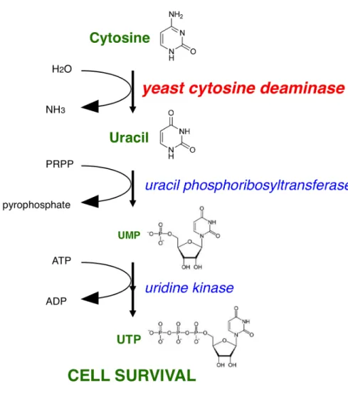
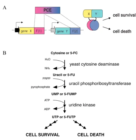
1.6.2 The role of yCD in the pyrimidine salvage pathway and utilization in a survival selection assay………..……….…..13
1.6.3 yCD Death Selection Assay………..15
1.6.4 Molecular Characteristics of yCD………16
1.6.5 Stability of yCD………...……18
1.6.7 Optimization of yCD PCA activity by fragment shuffling and error-‐prone PCR
mutagenesis………...…19
1.7 Application of OyCD PCA to study protein complexes that govern the cell cycle………....19
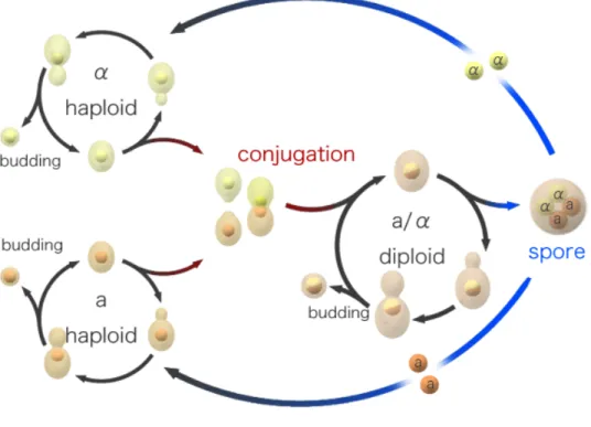
1.8 The S. cerevisiae sexual and mitotic cell cycles………20
1.9 Yeast CDKs involved in cell cycle regulation……….…22
1.9.1 Cdk1………...23
1.9.2 Regulators of Cdk1………24
1.9.3 Cyclins and the cell cycle………...………26
1.9.4 Cdk1 substrates………..28
1.9.5 Pho85………....29
1.9.6 START and G1 to S-‐phase cell cycle transcription factors: SBF and MBF…...…30
1.9.7 Activation and inactivation of SBF and MBF……….32
1.9.8 Conservation of cell cycle regulation between yeast and metazoa……….33
1.9.9 Remodeling of the cytoskeleton during the cell cycle and the morphogenesis checkpoint………..34
1.10 Objectives of this thesis………..34
Chapter 2: A generalized life or death selection strategy to detect formation or disruption of protein-protein interactions ………..…..37
Abstract………...……..38
Introduction………39
Results……….…...43
Discussion………...….64
Material and methods………...…66
Reference………..73
Abstract………...79
Introduction……….…80
Results………..83
Discussion………..…98
Material and methods………..101
Reference……….107
Chapter 4: Systematic Screens to Identify and Dissect Cdk1-cyclin Complexes in Vivo……….……….109
Abstract………...…..111
Introduction………112
Results………...115
Discussion………...128
Material and methods………..…132
Reference………...137
Chapter 5: General Discussion……….….141
5.1 Insights into yCD structure and function………141
5.2 Development of other biomolecular death assays ………...142
5.3 Dissecting cell cycle protein complexes using OyCD PCA……….143
5.3.1 Swi6 dimerization……….144
5.3.2 Swi6 and Whi5 interaction………..144
5.3.3 Dissecting cyclin-‐Cdk1 complexes……….…………...145
5.4 Perspective………146
5.4.1 False positive rate of OyCD PCA in the Swi6 screen……….146
5.4.2 Engineering specific binders against proteins and post-‐translationally modified sequences.. ……….147
5.4.3 Beyond the survival and death selection assays of OyCD PCA………148
List of tables
Chapter 1
Table 1. Different classes of yeast and human cyclins.………..27
Chapter 2
Table 1. PCA activity of the different yCD fragment combinations………..46 Table 2. Mutations identified in yCD fragments from the error-‐prone PCR library screen..………52 Table 3. Mutations identified by combining mutations found in yCD fragments from the error-‐prone PCR library screen. .………53 Table 4. List of primers……….67
Chapter 4
Table 1. List of genes encoding proteins that interact with Cdk1 tested by OyCD PCA……….122
List of figures
Chapter 1
Figure 1. Limitation of the yeast two-‐hybrid (Y2H) assays………8
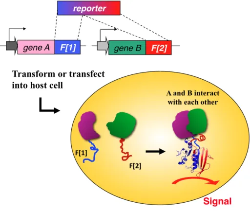
Figure 2. Protein-fragment Complementation Assays (PCA)………...11
Figure 3. Pyrimidine Salvage Pathway in S. cerevisiae………...14
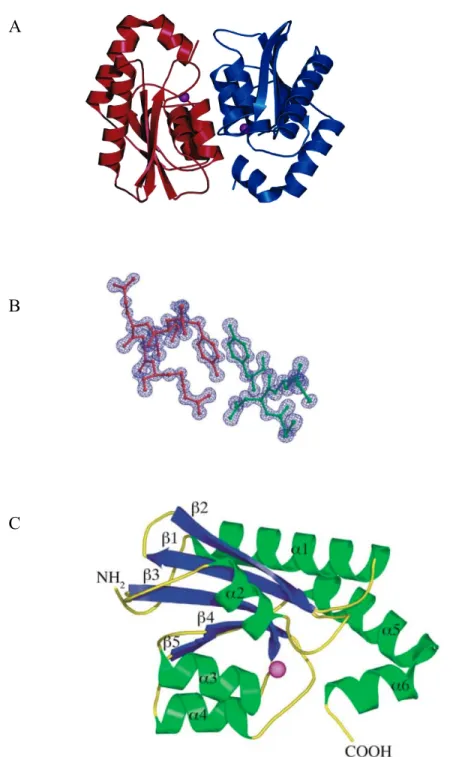
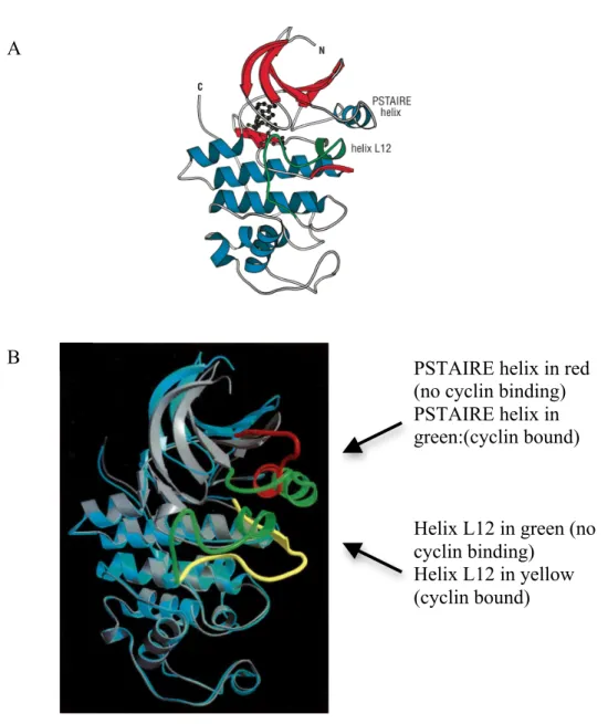
Figure 4. Structural characteristic of the yeast cytosine deaminase………...17
Figure 5. Sexual life cycle of S. cerevisiae………...…………...21
Figure 6. Structural feature of Cdk2………..………...…………...25
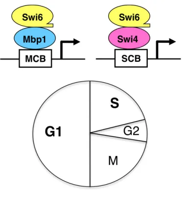
Figure 7. MBF and SBF transcription factors………..31
Chapter 2
Figure 1. A positive and negative selection PCA based on yCD.………...41
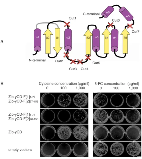
Figure 2. Development of the yCD PCA………...45
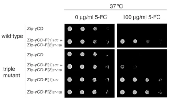
Figure 3. Activity of yCD PCA with the thermostabilizing triple mutations at 37 oC..47
Figure 4. Optimizing yCD PCA activity...………...48
Figure 5. Optimization of yCD PCA………...50
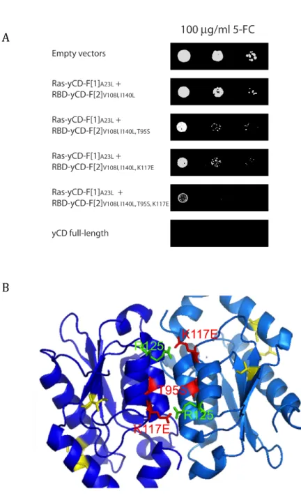
Figure 6. Mapping mutations identified in yCD fragments that increase yCD deamination of 5-‐FC to 5-‐FU onto the yCD structure..………...54
Figure 7. Effect of Y121A mutation on the optimized yCD PCA..………....55
Figure 8. OyCD PCA activity is mediated by protein-‐protein interaction……….……….57
Figure 9. Detecting interactions among transcription factor subunits..……….………....58
Figure 10. Dual selection properties of OyCD PCA……….……….………....61
Figure 11. Titration of FK506 concentration required for the disruption of FM1 OyCD PCA……….….……..62
Figure 12. Disruption of interaction between FM1 homodimer using FK506…….…...63
Chapter 3
Figure 1. MBF and SBF transcription factors………...84
Figure 2. Strategy for engineering a Swi6 mutant. ………...86
Figure 3. Selection process for re-‐engineering Swi6.………...87
Figure 4. Mutation in Swi6 before and after selection…………...88
Figure 5. Transcription assays for Swi6* mutants.…………...89
Figure 6. SBF and MBF reporter assays. .…………...90
Figure 7. Interaction between the Mbp1, Swi4 and Swi6 by pulldown assays...92
Figure 8. Interaction between the C-‐terminal domain of SBF and MBF determined by OyCD PCA……….………....…………...93
Figure 9. Expression of wild-type and 2m-‐Swi6 fused to OyCD-‐F[1]...94
Figure 10 Chromatin immunoprecipitation of MBF and SBF complexes using the affinity purified Swi6 antibody in wild-type (wt) SWI6 and 2M-SWI6 strains…...95
Figure 11. Interactions between Swi6 and Whi5.…...97
Figure 12 Model for allosteric regulation of Swi6………..…..…99
Chapter 4 Figure 1. Dissecting Cdk1 complexes using the OyCD PCA..……...………….…………..…116
Figure 2. Protein-‐protein interaction results between Cdk1 and the sixty-‐eight test proteins.………..………118
Figure 3. The OyCD PCA results in different cyclin deletion strains…………..…………123
Figure 4. Factors influencing the activity of OyCD PCA.…………..………..…126
List of abbreviations
S. cerevisiae: Saccharomyces cerevisiae Escherichia coli: E.coli
DNA: Deoxyribonucleic acid RNA: Ribonucleic acid
mRNA: messenger Ribonucleic acid ATP: Adenosine triphosphate C: Cytosine
U: Uracil
5-‐FC: 5-‐fluorocytosine 5-‐FU: 5-‐fluorouracil
yCD: Yeast cytosine deaminase
PCA: Protein-‐fragment Complementation Assay TF: Transcription factors
Cdk: Cyclin dependent kinase Cln or Clb: Cyclin
YPD: Yeast extract Peptone Dextrose SC: Synthetic complete
PCR: Polymerase chain reaction OD: Optical density
PAGE: Polyacrylamide gel electrophoresis Kb: Kilo bases
KDa: Kilo Dalton ml: milliliter μl: microliter mg: milligram μg: microgram mM: millimolar μM: micromolar
To Casandro and Leopaul, for making me understand the value of time.
“I don't know anything, but I do know that everything is interesting if you go into it deeply enough.” Richard Feynman
* Death PCA Pumpkin. Sculpted by Vincent Messier. Winner of the Université de Montréal, Biochemistry Department Pumpkin Competition 2011.
# Everyone can play with an edge. Built by members of the Michnick laboratory, 2011.
*
#
Acknowledgement
Supervisor:
This thesis would not be possible without the support from Stephen Michnick. No one is perfect in this world but people like Steve are rare and particularly admirable. One would think that someone of his status has accomplished everything already and yet, throughout all these years, I have witnessed how he is still constantly looking for ways to self exceed. Many thanks to Steve for giving me the freedom to explore my research interests (yes I have too many) and finding my own path. I have benefited tremendously from all these experiences and have finally found myself. I have also come to understand the notion of time and will tame my ambition accordingly. Also, thank you Steve for allowing me to understand the framework of an excellent article. I now see the parallel between the role of a scientist and an artist. A manuscript that we prepare is similar to a painting. The beauty of the final product depends on the composition, the color, the layering and the perspective. Although I haven’t accomplished such masterpiece, I am content to understand the recipe. I extremely appreciate this creative exercise and will apply it to all my future work. In summary, I am very proud to be an apprentice of such a talented master.
Members of the Michnick Lab:
I thank all members (past and present) of the Michnick lab not only for sharing their scientific expertise but also for their friendship and life experiences. You are my second family! Although I am not gone yet, I’m missing you all already. Thank you for all the nicknames that you have given me: The Energizer Bunny, La vieille croûte, Madame Po, The Shark without Teeth, THE MAN, The GAL, etc. Literally some
guys mean them in the utmost positive way and I am very honored!!! Vincent Messier and Mohan Malleshaiah, thanks for taking this roller coaster ride with me from the being till the end! I don’t know how I could have done this without both of you. Merci Vincent pour m’avoir écouté malgré la barrière et le mur que j’avais construis. Merci FX Valois-‐Campbell pour m’avoir guidé au début et Alexis Vallée-‐Bélisle pour être ma source d’inspiration! You rock! Merci Ingrid Remy pour m’avoir montrée c’est quoi l’efficacité. Thanks to Michael Booth, Leena Kaing, Diala Abd Rabbo and Jacqueline Kowarzyk for helping me with my projects. Sorry if I was a slave driver sometime. Mike and Jacqueline, without you the Cdk1-‐cyclin project would not have gotten so far. You are my two angels sent from heaven! Merci Emmanuel Levy pour ton SUPER script et tes parties sur la terrace! Merci Abedellali Kelil pour tes conseils! Thank you Luz Carillo, Amy Lerman, Emmanuelle Tchekanda, and Eugene Kanshin for helping me with the GAL project. Even though it is not included in this thesis, it was still 40% of my Ph.D. work. Hopefully, we will see the fruit of our hard work very soon. Also, thank you Luz for proof reading my introduction!!! Thanks Carol Mazurek for sharing all your detailed protocols, recipes, life experiences and stories! Merci Jean-‐François Paradis pour m’aider avec TOUT, incluant Casandro! Sans toi, je ne sais pas si je peux me rendre jusqu’à là. Louis-‐Philippe, merci pour tes mots de sagesse. Tu es mon “power supply”! And yes Arturo, I will call to check up on you so keep up the hard work. Durga, call me if the boys are out of line. I will come and help you duct-‐tape them to the tree. Edi Stefan, Livia Otte and Valérie Jacquier thank you for being my role models!
Université de Montréal:
Many thanks to the wonderful people at Université de Montréal. Special thanks to the members of my thesis committee (Norman Brisson and Muriel Aubry) for guidance, constructive criticisms and encouragement!!! Thanks to my pre-‐doc committee members: James Omichinski, Benoît Coulombe and Jean-‐Claude Labbé.
Thank you also to Pascal Chartrand, Daniel Zenklusen, and Sachdev Sidhu for accepting to evaluate this thesis.
I would also like to thank my friends and colleagues from the department who generously try to help me with crazy experiments requested by referees (especially referee #3). Jimmy Dikeakos and Paola Di Lello, you guys are the best! Even though my experiments didn’t work, I feel so fortunate to have friends in the field who care. Simon Drouin, merci pour ton encouragement et m’avoir montré comment faire les expériences de ChIP.
Un gros merci à tout le personel du Département de biochimie de l’Université de Montréal. Sylvie Beauchemin et Monique Vasseur, merci pour m’avoir calmé pendant les situations les plus stressantes! Vous êtes vraiment des anges! Merci également à Anne Lessard et toute l’équipe du Baluchon ainsi que le CPE Château des Neiges (Thérèsa, Krystèle, Sylvie, Ouiza, Nathalie, Marlène, Sophia, Josée et Pascal) pour s’occuper de Casandro et Leopaul. Sans vous, je ne pourrai compléter cette thèse!
Funding:
I would like to acknowledge the Falcuté des Études Supérieure de l’Université de Montréal for the Bourse d’Excellence scholarship. In addition, I would like to thank the university for the Bourse Olive-‐Beaudry-‐Leriche de la Faculté de Médecine and Merck-‐Frosst for scholarships that allowed me to attend international conferences. Many thanks to the FQRNT for the postdoctoral fellowship award! This serves as a big motivation for me to complete my thesis and continue in science!
Previous supervisors and their lab members:
laboratory and share their passion for science, you have allowed me to build a solid foundation in research and armed me with useful tools for my graduate studies. Maureen, thank you for taking the risk and encouraging Ally and I with our Science Fair projects. Without you, I would have probably been a chartered accountant by now. Not that there is anything wrong with being an accountant. It’s just not me and I don’t think I would have been very happy. Denis, thanks for all the stimulating discussions. It means a lot since I was just a summer student and you were the big boss. Your questions really got me thinking on the right path. Dave, being in your lab has opened many doors for my career. Thank you. Annette, thanks for the opportunity that you have given me. To all members of these labs, thank you for teaching me so much! Merci M. Dr. Alain Guimond pour m’avoir appris la biologie moléculaire. Merci Joanne Magoon et Christiane Cantin pour partager vos expériences. Anne Lenferink, you are my Iron Woman role model in science. Merci Jean-‐Pierre Falgueyret et Marc Ouellet pour m’avoir stimulé à penser d’une façon critique et travailler efficacement.
Friends and Family:
Thanks to my girl friends outside this university for keeping me “normal”. My Chane, Tori, Neang, Lee, Sara, Diane, Yao, Ally and Vivien you keep me rooted to the real world and this is really important. (Diane, thanks for your help with the Excel formula! It is still super helpful!!!)
Finally, I would like to thank my family (yes Neang, you are included here too) for their moral support and helping me take care of Casandro and Leopaul. Thank you donating your precious time! Thanks to Carlos for understanding my ambition and letting me have things my way for most of the time. Apologies to Cas and Leo for not being able to spend as much time with you as I should and to my parents for not choosing a “normal” career like everyone else in the family.
Introduction
1.1 Protein complexes governing cellular activity
The simplest entity of life is the cell, whether those that make up complex metazoa such as ourselves or unicellular organisms such as bacteria or fungi. Cells are mainly composed of water and four classes of macromolecules: proteins, nucleic acids, lipids and polysaccharides (Alberts, Bray et al. 1994). Interactions among these macromolecules mediate all of the molecular transformations between matter, energy and information transfers necessary to sustain life (Monod 1968).
Proteins play a very important role among the four classes of macromolecules. Not only do they participate in their own synthesis, they are also involved in the synthesis of the other three classes of macromolecules (nucleic acids, lipids and polysaccharides). How can proteins accomplish all these different functions? Often they interact with other proteins and form functional complexes for relaying information from the extracellular to the intracellular environment or regulate their enzymatic activity. Particularly important classes of complexes are those whose functions are regulated during the cell cycle. Since a cell is constantly growing, dividing or dying at a given time, the functions of these protein complexes are intrinsically coupled to the mitotic cell cycle. However, understanding the functions of protein complexes during the cell cycle remains challenging due to the lack of existing tools for dissecting diverse protein complexes. In this thesis, I will present the development of a novel tool that allowed us to systematically dissect the budding yeast, Saccharomyces cerevisiae (S. cerevisiae) G1-‐S cell cycle transcription factors (SBF and MBF) and cyclin-‐dependent protein kinase complexes in order to understand their different functions.
1.2 Protein-protein interaction networks (PINs) and the
understanding of protein functions
Studying protein-‐protein interactions (PPI) is a general strategy for understanding the function(s) of an unknown protein or discovering novel function(s) of a characterized protein. In recent years, massive genome sequencing projects has given rise to the identification of thousands of genes. Many of these genes still have unknown function(s). This has motivated the design of methods to systematically detect protein-‐protein interactions on a large scale in order to define potential functions of genes.
The genome of the unicellular organism S. cerevisiae was sequenced in 1996 (Goffeau, Barrell et al. 1996). It has 5798 consensus open reading frames (ORFs) that potentially represent the number of genes in its genome (Goffeau, Barrell et al. 1996). What are the functions of these genes? Several protein-‐protein interaction networks (PINs) have been generated using the yeast two-‐hybrids screens (Y2H) (Ito, Tashiro et al. 2000; Uetz, Giot et al. 2000; Ito, Chiba et al. 2001), tandem affinity purification followed by mass-‐spectrometry analysis (TAP-‐MSs) (Gavin, Bosche et al. 2002; Ho, Gruhler et al. 2002; Gavin, Aloy et al. 2006; Krogan, Cagney et al. 2006) and Protein-‐fragment Complementation Assays based on the murine dihydrofolate reductase as reporter enzyme (mDHFR PCA) (Tarassov, Messier et al. 2008) in order to better assign functions to all the yeast genes. The results of these studies yield protein interaction network (PIN) data that serve as a valuable treasure chest of information for discovering mechanisms that regulates protein complexes and for inferring the functions of uncharacterized proteins according to the “guilt-‐by association” concept (Oliver 2000). This means that the function of an unknown protein can be assigned to it by grouping it with its interacting partners. The established PINs allow us to identify many new components of protein complexes that govern basic cellular processes such as transcription, translation, signal
transduction and cell cycle regulation and their mechanism of regulation. Despite all these efforts, 866 ORFs remain uncharacterized.
Ultimately biochemists engaged in understanding some cellular process begin with the supposition that process is somehow mediated by individual or groups of complexes and thus the goal is to identify the component subunits that are necessary to a process and then dissect out the roles of the individual subunits (Alberts 1998).
1.3 Reagents for dissecting protein complexes
The functions of protein complexes can be modulated using small molecules or genetic perturbations. However, such small molecules for inhibiting protein activity or protein-‐protein interaction are rare. Thus, it is a major challenge to identify novel molecules specific to a particular protein (Arkin and Wells 2004). Genetic perturbations represent an attractive avenue for dissecting protein complexes. With the availability of its genomic sequences, a systematic single gene deletion of almost 5000 S. cerevisiae non-‐essential genes has been accomplished (Giaever, Chu et al. 2002). Many mutant strains carrying two deleted genes have been reported (Tong, Lesage et al. 2004; Costanzo, Baryshnikova et al. 2010) and efforts to obtain the entire array of double deletion strains are on going.
A finer level of protein complex dissection can be achieved by using truncation, deletion or point mutations of individual subunits (also know as a missense mutation). Like gene deletion, mutations in a protein can be used to dissect protein complexes and their functions. A C-‐terminal truncation variant of a protein can be obtained by simply introducing a nonsense mutation to the DNA sequence of the gene in order to generate a premature stop codon. Mutating the first ATG codon and introducing another ATG codon further on in the gene sequence can obtain an N-‐
identifying interacting domains (independently folding regions) or binding motifs (small linear sequences) of a protein. Point mutations can provide more fine-‐ detailed information about the chemical basis of an interaction or, in cases of sites that are post-‐translationally modified, study the effects of these modifications on binding. Yet, there is a drawback to using specific mutants to systematically dissect protein complexes since they cannot be easily predicted or identified from the primary amino acid sequence of the protein of interest.
1.3.1 Fishing for binding mutants by random mutagenesis
Aside from step by step approaches to identify mutants that will disrupt a binding interface, random approaches have been found to be effective. Libraries of gene point, truncation or internal deletion mutants can be generated. There are various ways to introduce mutation(s) into a gene for generating a library (Bonsor and Sundberg 2011). Many laboratories use mutagenesis generated by error-‐prone polymerase chain reaction (ePCR) to introduce mutations into the gene sequence due to its simplicity and low cost. This PCR method uses a variation in the amount of manganese (Mn) or magnesium (Mg) and an unequal amount of nucleotide concentration to force a low fidelity DNA polymerase from Thermus aquaticus (TAQ) to introduce mutation during a PCR reaction (Cadwell and Joyce 1992). Although this technique does not allow for an exhaustive coverage of the amino acid sequence, it has the potential to generate a mutant of desired characteristics when used with the appropriate binding selection strategy. Such methods have been used to disrupt complexes, for example, to increase the fluorescent properties of fluorescent proteins including green and red fluorescent protein (Campbell, Tour et al. 2002) and infrared fluorescent protein (Shu, Royant et al. 2009) .
Error-‐prone PCR can be scalable for high-‐throughput screening and has been the method of choice to generate libraries of mutants in genome-‐wide projects. For
example, error-‐prone PCR was used to generate libraries of temperature-‐sensitive mutant for over one thousand essential S. cerevisiae genes (Ben-‐Aroya, Coombes et al. 2008). Similar approaches could be envisioned for screening for disruption of protein-‐protein interactions using yeast two-‐hybrid or PCA screening methods.
1.4 A dual selection assay to engineering proteins with specific
interaction: A matter of life and death
In order to identify mutation(s) that affect a PIN, an ideal tool to screen for a missense or nonsense mutations should provide a positive output for detecting both the interaction itself and disruption of that interaction. Additional features of the tool would be that full-‐length proteins can be expressed in their native cellular compartments with appropriate post-‐translational modifications and since the readout is direct and independent of transcription, the strategy even can be applied to transcription factors. Finally, the tool should allow for easy recovery of the mutant of interest. In addition, this tool should be sensitive and allow detection of conformational changes between the two interacting proteins (Remy and Michnick 1999; Tarassov, Messier et al. 2008).
A simple solution to a dual selection system that can be used to re-‐engineer interactions between specific proteins or to dissect protein complexes is to use a reporter protein that is bifunctional. Ideally, such a reporter protein would provide simple dual readouts: for example, cell survival or cell death. A selection strategy based on a simple output such as cell growth is favored for library screening since these are inexpensive and require no sophisticated equipment for signal detection. An additional feature of such a tool is that it could also be used to identify contingent interactions. Specifically if an interaction between proteins X and Y is mediated by a third protein Z, then a detector of the interaction of X and Y would give a positive
There are a small number of known reporter proteins that can regulate both cell survival and cell death. They often fall into the category of prodrug-‐converting enzymes. These enzymes regulate cell survival by participating in pathways that lead to the synthesis of essential metabolites. However, in addition to modifying their natural substrates, they can also convert unnatural and benign compounds (so called prodrugs) to products that are toxic to cells. The most well known prodrug-‐ converting enzymes used in positive-‐negative clonal selection strategies include the herpes simplex virus thymidine kinases (HSV-‐TK1 and HSV-‐TK2), yeast cytosine deaminase (yCD), and orotidine 5-‐phosphate decarboxylase (Ura3 of S. cerevisiae) (Capecchi 1980; Nishiyama, Kawamura et al. 1985; Boeke, Trueheart et al. 1987).
Enzymes that regulate key metabolic pathways and have known inhibitors cannot act as a reporter for a dual PPI selection assay since blocking the activities of the enzymes will automatically inhibit cell survival. An example of a reporter from this category is the imidazoleglycerol-‐phosphate dehydratase of S. cerevisiae (His3). His3 can be used as an auxotrophic marker yet it can also be inhibited by a competitive inhibitor called 3-‐Amino-‐1,2,4-‐triazole (3-‐AT) (Brennan and Struhl 1980).
1.5 Existing positive and negative selection assays for screening
interaction specific mutant(s)
There are two S. cerevisiae based PPI assays that can be used for both survival and death selection assay. They are based on the gene product of URA3 as reporter protein. Ura3 is an enzyme of the pyrimidine de novo synthesis pathway that converts orotidine 5-‐phosphate to uridine monophosphate. In addition, Ura3 can also convert 5-‐Fluoroorotic acid (5-‐FOA) to 5-‐fluorouracil (5-‐FU) (Boeke, Trueheart et al. 1987), a compound that can further be modified to 5-‐fluorouridine-‐ triphosphate (5-‐FUTP) which can induce cell death (Fang, Hoskins et al. 2004).
1.5.1 Reverse yeast two-hybrid assays
The reverse yeast two-‐hybrid (rY2H) system is a yeast two-‐hybrid (Y2H) assay using Ura3 as a reporter protein and 5-‐FOA as prodrug for inducing cell death when two proteins of interest bind to each other (Leanna and Hannink 1996; Vidal, Brachmann et al. 1996). It is based on the reconstitution of the Gal4 transcription factor from its split DNA binding (DB) and transactivating (TA) domains fused to two test proteins (Fields and Song 1989). When the two test proteins interact, the transactivation domain of Gal4 is brought into proximity with its DNA binding domain resulting in the activation of the URA3 reporter gene in the nucleus. In the presence of the prodrug 5-‐FOA, cells expressing the URA3 gene will be sensitive and cannot grow. Equally, a rY2H with a ribosomal protein of the large (60S) ribosomal subunit (Cyh2) as reporter gene has been developed (Leanna and Hannink 1996). In the presence of cycloxehamide, cells that express the CYH2 gene do not grow. A disadvantage of Cyh2 over Ura3 is that this reporter protein can only be used for death selection.
Since the output of the rY2H system is cell death, a mutation or small molecular that disrupts the interaction between two test proteins will result in cell survival. This particular strategy facilitates the screening process of library of mutant proteins for non-‐binding mutant(s) or small molecules that can inhibit a specific PPI without the tedious replica-‐plating step. It has successfully been used to identify mutants for proteins such as the yeast MAPK scaffold protein Ste5 (Inouye, Dhillon et al. 1997).
A central limitation of Y2H assays is that since they work at the level of transcriptional machinery, they cannot be used in a trivial way to study protein complexes involved in transcription itself or perhaps other complexes involved in
Figure 1. Limitation of the yeast two-‐hybrid (Y2H) assays. The Y2H system is based on splitting Gal4 into its DNA binding (DB) and activation (AT) domains and fusing these to two proteins of interest (X and Y). The interaction between protein X and protein Y will activate the transcription of the reporter gene. (A) Y2H assay cannot be used to study the interaction between Gal4 and its interaction partner(s). (B) If protein X has a transcriptional activation (TA) domain fused to DB domain of Gal4, it will activate the transcription of the reporter gene. C) If protein X contains a DB domain, it could bind other genomic DNA sequence and result in a decreased of transcription of the reporter genes. D) If protein X binds to a transcriptional repressor, it can repress the transcriptional activity of Gal4.