Assessment of the Combination between Mass Spectrometry
and Computational Chemistry for the investigation of
Supramolecular Assemblies
Gaseous Flying Boxes as a Case Study
CARROY GLENN
Dissertation présentée en vue de l’obtention du grade académique de Docteur en SciencesAnnée Académique 2017-2018 Promoteurs : Prof. Pascal Gerbaux
Prof. Edwin De Pauw Prof. Jérôme Cornil
Acknowledgments
« Allo?
Hey Glenn, Pascal here, how are you?
Hey Pascal, I am fine, thank you, what about you?
I am OK, look, I might have a solution if you still want to do a PhD, do you want to hear it? »
This looks closely to the phone call I received from Pascal Gerbaux in April 2013. Back then, I was working in the pharmaceutical industry after two failures at the F.R.I.A contest where I tried to obtain a PhD financial support. If I am here today, if I know what I know and if, somehow, I am a little bit good at what I do; the credit first goes to Pascal. Boss, I would like to thank you for believing in me, even though I know I have not been easy to manage all the time. I am proud I have been able to witness and participate to the evolution of the S2MOs lab
along the years. Now the social rehabilitation begins ( ;-) ) but be sure I will never forget those almost 7 years in total spent on your (dark?) side. Thank you very, very much for everything.
« What?
I have a question about “subject A”, I don’t understand what’s happening.
Yes but did you think about “subject B”, “subject C”, have you check your references, internet, books, stars, tarot cards and read the coffee spots?
…. I said I don’t understand! »
When you start to work with Julien De Winter, you better be patient and learn how to not be easily offended but actually, if you think deeper about it, everything Julien is doing helps you to be better at what you do , to be smarter and a better scientist. When you get to know him, under his sarcasms and bad humors, there is someone helpful and gentle, which will always find a moment to help you at whatever you need him, a tattoo lover (long live Instagram), someone devoted to his work and a true kickass scientist. Now my time is over at the lab, I can say I admire that guy, but won’t tell him to much (kidding). Julien, thank you for all the questions and discussions we had, we had some disagreements, but you always responded and I will never forget that.
« You can be a Jedi [I] but you will not studi [I] »
Jerôme Cornil knows how to make good English tips for myself! Jérôme always impressed me
with how he was able to deal with so many different subjects and (experimental/ computational) technics. He always came with some insides and solutions when we had meetings for our different publications and issues. Master in jokes and spoonerisms (well, Pascal is also not bad at it), I will always remember those meeting, which were perfect mix of hard work and fun time. Thank you Jérôme for all your help and advices along those years.
« You can take more processors, they won’t see it. *don’t worry face* »
I worked with Vincent Lemaur from the very start of my master thesis and I feel so lucky I have been able to meet such a nice person. I think nobody will say it differently: Vincent will never say no to help anybody asking even if he is flooded with work and he truly rocks in sciences. Vincent, thank you so much for your patience (#toomanyblankspaceinthefile) and for have been my Master Yoda in computational chemistry for almost 6 years!
« Well, your thesis is over isn’t it? »
Right after the last congress I had the chance to spoke too, those were the first words I heard from Edwin de Pauw. Somehow, he got it right: I had two months to finish my PhD but he also looked confident looking at my work and that was the most important part. Mister De Pauw, we did not meet that much but I cannot thank you enough for saying “yes” to be co-supervisor of this thesis a few years ago. Thank you for your trust.
« Glenn, it is 3PM, bring the Minion! »
This is not a breaking news: you have to be a little crazy to become a scientist and my colleagues at the S2MOs make no exceptions. Thank you Marie, Thomas, Corentin, Emilie, Tiffani, Quentin, Romain, Manu, Sébastien D., Sébastien H., Fabrice, Géry and Eric for all those
great moments spent together whatever it was for talking about sciences, shopping, videos games, music, making jokes, etc. and of course thank you for your support.
In particular, I would like to thank Marie who has been a real support through the first 2 years of my PhD. I will never forget our crazy screaming sessions when we were both working for hours in front of our computers. Thank you for your encouragement and support. After Marie, Emilie came in my office and putting two geeks like us in the same room was maybe not the best idea lol. Emilie, thank you for all our chats on shopping and gaming, for your crazy stupid music and your ugly dogs on facebook (yes, they are ugly, believe it :-P) but also for having my back during this last year. Lastly, thank you Corentin for being so supportive and kind for almost three years.
« Dude, how are your shoeboxes going on? »
It is not always easy to make your non-scientist people understand what you do, so yes: I explain my host-guest complexes as shoeboxes since your sneakers will not fit a boots box and vice-versa. Of course, there is many people I would like to thank here but I will focus on some of them.
Steve and Romain have been my friends for respectively 25 and 13 years, even though they
are not chemists, they were always interested about my research and truly helped me going through it, thank you guys, you are rock stars, let’s have another 30 plus years of great friendship!
Another rock star is my dear Orel / Aurélie / Sally / Bébéééé / Bichetaud, we have been together for more than 3 years and I feel so lucky to have her in my life. I learn from her every day, self-made women, true champion and hard-work girl, she is all about passion and getting stuff done. Thank you for helping me being a better man without forgetting the crazy child in me, I am so proud of you and us. I love you “costaud”. Where is the gym?
Speaking off self-made women, here comes the best: my mum Lucia. There are not enough words to say how much I am proud of her and of the fighter she is. If I am the man I am today, her and my grandparents have all the credit for it. Thank you for supporting me in whatever are the crazy things I do and even if you do not understand them.
Finally yet importantly, there is the man of my life, Roger, my grandfather. If I was able to go to the University, it is mostly because of him and my grandmother Noëlle. Papy, you have always been an example for me and I cannot thank you enough for the trust you have in the man I have become. Thank you for having been there all the time, from reciting multiplications tables when I was six to the final stages of this PhD.
« Do or do not, there is no try »
Master Yoda.
I guess this is the best way to describe a PhD, whatever you start it or have to put an end to it. Thank you to the University of Mons (UMONS) and University of Liège (Liège Université) for their financial support.
I can just hope you will enjoy the next two hundred pages as much as I enjoyed writing them. It has been an incredible ride, time for something new.
Thank you all so much.
Abstract
Non-covalent interactions result from the associations of different molecules held together by intermolecular forces. Nowadays, supramolecular chemistry is in constant evolution and can probably be considered as one of the main research fields in modern chemistry. Numerous applications in drug delivery, water treatment, catalysis, etc. are already well controlled and more are expected soon.
A peculiar area in the domain of supramolecular assemblies is the host-guest chemistry in which a big chemical receptor, the host, can strongly bind within an inner cavity at least one small molecule, the guest. The structures and physicochemical properties of host-guest assembles are obviously studied directly in solution with common methods such as NMR and UV-vis spectroscopy. Nevertheless, with the development of soft ionization methods that can preserve non-covalent interactions from the solution to the gas phase, mass spectrometry appears to be an efficient analytical tool in the study of supramolecular assembles especially with the emergence of ion mobility in association with mass spectrometry. This thesis represents a contribution towards the applicability of MS methods to investigate supramolecular objects, considering that the data generated upon MS are related to gaseous ions and that any shortcut linking the gas phase structures to the condensed phase topologies is definitively not straightforward.
In this PhD, we report different studies carried out on complexes involving cucurbiturils as macrocycle hosts. Cucurbit[n]urils are macrocycles obtained by the condensation reaction of n glycoluril units (n=5 to 14) and have been extensively studied for the outstanding binding abilities with amino-guest molecules. The selection of cucurbiturils as model host has been motivated due to the rigid character of those macromolecules. For our studies, we used mass spectrometry, associating Electrospray ionization, collision-induced dissociation and ion mobility, with computational chemistry to advantageously study cucurbituril systems in association with different amino compounds. Basically, our studies demonstrated the applicability of MS-based methods to probe the gas phase structures of complex ions, provided computational chemistry is implemented in the workflow to optimize the structures of non-covalent ion candidate to further compare to the experimental data. In particular, the
relative energies of the inclusion/exclusion structures have to be determine for each association under study using theoretical calculations to confirm the experimental observations.
In the first part of this PhD, the influence of the experimental and instrumental conditions on the nature of the detected ions has been investigated with a special emphasis on the in-solution reaction time, saying the time spent by the host and the guest in in-solution prior to the ionization process, and on the size of the guest, which may affect the in / out ratio of the detected ionized associations. Similarly, the key-role played by the ion optics on the topology of the supramolecular ions transferred from the solution to the gas phase upon Electrospray Ionization has been highlighted.
Secondly, a mass spectrometric study has been performed on a click reaction using the inner cavity of a cucurbit[6]uril host as a catalyzer. According to our results using CID, ion mobility and energy resolved-CID experiments, we observed that the cycloaddition reaction within the CB[6] cavity is an extremely fast process and that the rate determining step in the overall cycloaddition process as measured in solution is the egression of the cycloadduct products from the cavity.
In the final section of this PhD thesis, we report a joint experimental and theoretical study carried on a bitopic cucurbituril receptor (ns-CB[10]). We first demonstrated upon MS measurements that the creation of the ternary complexes is under homotropic allosteric control. We also reported that CID experiments, with the control of the kinetic energy, must be also carefully used when trying to deduce relative binding affinities from CID energy thresholds.
Overall, in the present PhD research work, the advantages and drawbacks of mass spectrometry as an analytical tool for the study of supramolecular chemistry have been highlighted with a special emphasis on the conservation of the complex topology all along the path of the ions from the solution to the detector of the mass spectrometer.
Table of content
I.
Introduction:
1. Outlook ... 1
2. Supramolecular Chemistry ... 3
2.1 Non-covalent Interactions ... 5
2.1.1 Van Der Waals Interactions ... 6
2.1.2 - stacking ... 7
2.1.3 Hydrophobic interactions ... 8
2.1.4 Hydrogen bonds ... 10
2.1.5 Dative and ionic bonds ... 11
2.1.6 Exotic non-covalent bonds ... 12
2.2 Host-Guest Complexes ... 14
2.3 Investigation Methods for Non-Covalent Associations ... 17
2.3.1 UV-Visible Spectroscopy... 18
2.3.2 Nuclear Magnetic Resonance (NMR) Spectroscopy ... 20
2.3.3 Isothermal Titration Calorimetry ... 23
2.3.4 Extraction Method ... 26
3. Supramolecular Mass Spectrometry ... 28
3.1 Electrospray Ionization ... 29
3.2 Selected examples of the use of Electrospray in supramolecular chemistry... 32
3.2.1 Supramolecular associations ... 33
3.2.2 Complete virus ionization... 37
3.2.3 Gas phase reactivity of supramolecular assemblies ... 38
3.3 False positive detection ... 40
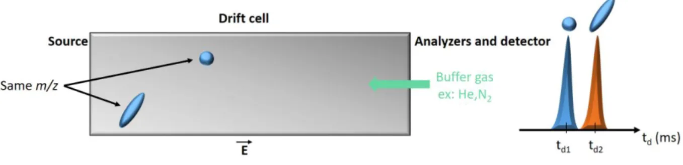
4. Ion Mobility Mass Spectrometry (IMMS) ... 41
4.1 Description of the Gas Phase Ion Motion ... 41
4.2 Ion Mobility Spectrometry ... 43
4.4 Experimental considerations for ion mobility experiments: arrival time distribution 47
4.5 Ion Mobility Mass Spectrometry: incorporation of Drift-Cell Units ... 50
4.5.1 Drift Tube Ion Mobility Measurements ... 50
4.5.2 Traveling- Wave Ion Mobility Measurements ... 52
5. The Cucurbituril Macrocycle Family ... 57
5.1 Synthesis ... 57
5.2 History ... 59
5.3 Structural and chemical properties ... 61
5.4 Host-guest chemistry ... 64
5.5 Guest complexation mechanism with cucurbiturils ... 66
5.6 High-affinity binding ... 68
5.7 Applications ... 71
5.7.1 CB[n] recognition in biological systems ... 71
5.7.2 Catalysis in the inner cucurbituril cavity ... 74
5.7.3 Photoswitching reaction operated using cucurbituril and product self-sorting release………. ... 76
References ... 80
II.
Aim of the thesis:
Aim of the thesis ... 92References ... 93
III.
Experimental part:
1. Mass Spectrometers ... 941.1 Waters Q-ToF 2 ... 94
1.1.1 Description ... 94
1.1.2 Experimental procedure ... 95
1.2 Waters Synapt G2-Si ... 96
1.2.1 Description ... 96
1.2.2 Experimental procedure ... 97
2. Overview of the Mass Spectrometer Acquisition Modes ... 100
3. Energy-Resolved Collision Induced Dissociation ... 101
4. Collision Cross Section Calibration Method for TWIMS Analysis ... 104
5. Nuclear Magnetic Resonance Measurements ... 107
6. Materials ... 108
7. Theoretical calculations ... 108
References ... 109
IV.
Results and discussion:
Chapter 1: Influence of Equilibration Time in Solution on the Inclusion/Exclusion Topology Ratio of Host-Guest Complexes Probed by Ion Mobility and Energy-Resolved Collision-Induced Dissociation. 1.1 Introduction ... 1111.2 Results and discussion ... 113
1.3 Conclusions ... 125
References ... 126
Chapter 2: Flying cages in Traveling Wave Ion Mobility: insidious effect of the instrumental parameters on the topology of the host-guest complexes. 2.1 Introduction ... 129
2.2 Results and discussion ... 131
2.2.1 Adamantylamine
CB[6] and diaminohexane@CB[6] as exclusion and inclusion model complexes ... 1322.2.2 Aniline
CB[6] or aniline@CB[6] as exclusion or inclusion complexes ... 1372.2.3 Halogeno-aniline
CB[6] or halogeno-aniline@CB[6] as exclusion or inclusion complexes ... 1452.2.4 PXDCB[6] or PXD@CB[6] as exclusion or inclusion complexes ... 149
2.2 Conclusions ... 151
Chapter 3: Probing the cucurbituril-catalysed 1,3-cycloaddition by mass spectrometry.
3.1 Introduction ... 156
3.2 Results and discussion ... 157
3.3 Conclusion ... 164
References ... 165
Chapter 4: Homotropic allosterism: in-depth structural analysis by mass spectrometry and computational chemistry of the gas-phase non covalent complexes associating a double-cavity cucurbit[n]uril-type host and size-selected protonated amino compounds. 4.1 Introduction ... 167
4.2 Results and discussion ... 170
4.2.1 ESI time-of-flight (TOF) measurements ... 170
4.2.2 Collision-induced dissociation experiments ... 175
4.2.3 Ion mobility spectroscopy experiments (IMS) ... 177
4.2.4 Structural analysis of the ternary complex ions (+2 / +3) by computational chemistry ... 180
4.2.5 Experimental and theoretical investigation of the size specificity of the complexation reaction. ... 185
4.3 Conclusions ... 187
References ... 189
Chapter 5: Energy-resolved Collision-induced Dissociation of Non-Covalent Ions: Charge- and Guest-dependence of the Decomplexation Reaction Efficiencies. 5.1 Introduction ... 192
5.2 Results and discussion ... 195
5.2.1 Experimental results: mass spectrometry study of the host/guest associations ……….195
5.2.2 Estimation of the dissociation barriers ... 203
5.2.3 CID reactions of the 2+ ternary complexes ... 212
5.2.4 CID reactions of the 3+ ternary complexes ... 214
References ... 217
V.
Conclusion & outlook:
Conclusion & Outlook ... 219VI.
Annexes:
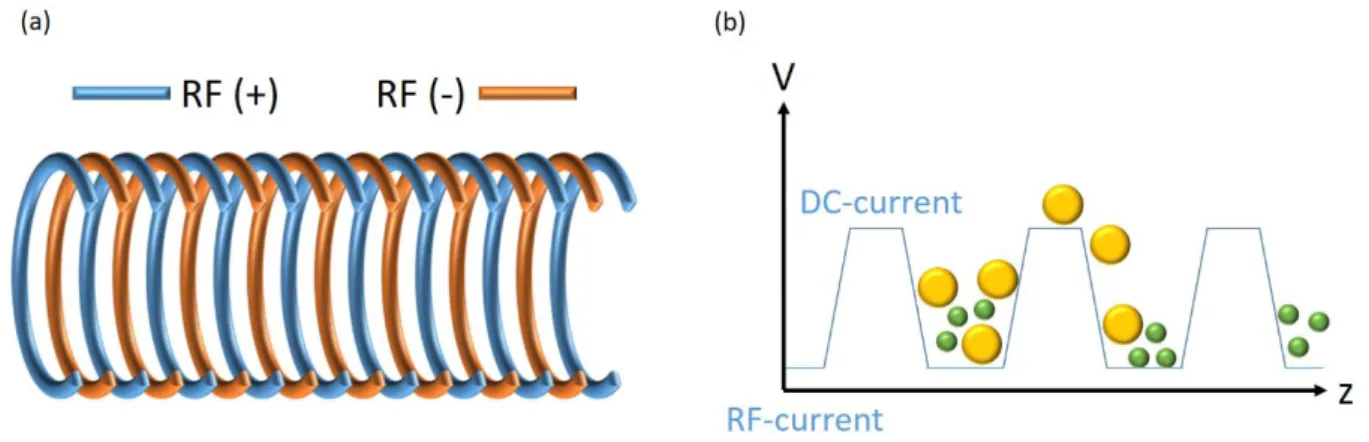
1. Mass Spectrometry: additional notes ... 2231.1 Tandem Mass Spectrometry ... 223
1.2 Triwave ... 224
2. Density Functional Theory ... 227
2.1 The Thomas-Fermi Model ... 228
2.2 The Hohenberg and Kohn Theorems ... 228
2.3 The Kohn-Sham Method ... 230
2.4 Exchange-correlation Energy Assessment methods ... 232
2.4.1 Local Density Approximation (LDA) ... 232
2.4.2 General Gradient Approximation (GGA) ... 233
2.4.3 Hybrid Functional... 233
2.5 The B97D Functional ... 234
2.5.1 Description of the B97D energetic terms ... 234
2.5.2 Description of the dispersion forces energetic term ... 236
References ... 237 Accomplishments
0
I. Introduction
Outlook
Supramolecular Chemistry
Supramolecular Mass Spectrometry
Ion Mobility Mass Spectrometry
The Cucurbituril Macrocycle Family
1
1. Outlook
In modern chemistry, mass spectrometry (MS) methods are increasingly used to study non-covalent complexes extracted from the solution phase to the rarefied gas phase. The development of new ionization source such as the electrospray allowed such interactions to be preserved through the phase transfer [1]. In the gas phase, ions do not interfere with each other chemically. Consequently, dynamic processes do not play a role anymore and the properties of the supramolecular objects can be monitored, with a special interest in the determination of the composition, i.e. stoichiometry of the association. New insights can be gained from the examination of the gas phase stability and reactivity. However, huge differences can be expected when compared to the solution phase properties so that the use of MS-based methods to study non-covalent associations must be handled with great care, especially when trying to deduce the solution properties from the MS data [1, 2].
Nowadays, MS-based methods appear more and more prone to provide fast and accurate information about supramolecular systems [1]. Mass spectrometry can go far beyond the analytical characterization of the complexes in terms of their exact mass, charge state, and stoichiometry. Indeed, mass spectrometry offers quite a large panel of methods that can provide structural information on the complexes. In particular, the “secondary structure” of the non-covalent associations, i.e. the relative position of the non-covalently bound subunits in the complex can be efficiently probed by collision-induced dissociation [3], associative ion/molecule reactions (H/D exchange) [4] and ion mobility [5] experiments. Relative binding energies of non-covalent associations in the gas phase can also be inferred by MS-based methods by performing ligand exchange reactions in the gas phase [6]. Critical energies of fragmentation for non-covalent complexes in the gas phase are also often estimated by performing energy-resolved CID experiments and determining the center-of-mass energy required for the decomposition / decomplexation reaction to occur [7]. However, such experiments do not afford any thermochemical data such as binding energies but threshold energies are estimated.
2 In addition, the use of computational chemistry in association with mass spectrometry permits a better understanding of the properties of the supramolecular association in the gas-phase. Indeed, secondary structures alongside with relative binding-guest energies can be determined through the evaluation of optimized geometries of the complex ions. Straightforward comparison with MS data, such as experimental collisional cross-sections, are also doable thanks to the evaluation of theoretical cross-sections calculated from optimized structures. Finally, absolute binding energies for gas-phase non-covalent complexes are nicely evaluated by using computational chemistry. [6,8]
Herein, a deep inside in the gas phase properties of the cucurbit[n]urils containers has been handled. Cucubit[n]urils are macrocyclic receptors constructed by the association of n glycoluril repeat units [9]. These pumpkin-shaped molecules present a hydrophobic inner cavity and two identical carbonyl portals, making them suitable for encapsulation of hydrophobic molecules or of the hydrophobic part of molecules in aqueous media. The main drawback of cucurbiturils is their low solubility in water requiring low pH or high ionic strength to ensure their dissolution upon carbonyl portal protonation or cationization [10]. On the other hand, the cucurbit[n]uril (CB[n]) family of molecular containers has attracted huge interest due to their outstanding recognition properties, to the exceptional strength of their interaction (ka up to 1017M-1) with various guests and to the numerous applications offered by
the encapsulation propensity of the cucurbituril family members [11]. Cucurbiturils are extensively demonstrated to form stable inclusion complexes with various protonated alkyl- and aryl(di)amines [9-11].
Lastly, the structural modifications of non-covalent complexes due to experimental and/or instrumental parameters have to be treated with a special emphasis. Indeed, in solution, (supra)molecules are characterized by certain conformations, which might not be preserved during the ionization/desolvation processes due to external stimuli (temperature, pH) or inherent voltages used within the mass spectrometer. Understanding the impact of those parameters is critical when attempting to consider MS as an additional tool for structural characterization of non-covalent objects. Then, one of the main objectives of the present PhD thesis is to assess how the gaseous structures can be envisaged as a probe of the condensed phase structure, despite the dramatic changes in environment undergone by the molecules upon Electrospray ionization and transfer within the high vacuum of the mass spectrometer.
3
2. Supramolecular Chemistry
Resolving around intermolecular interactions, supramolecular chemistry has constantly evolved over the last decades, to finally become one of the main research field in modern chemistry.
The basic concepts of this new research area have been formulated in the late nineteenth century. Back then, in 1891, A. Villiers discovered the first macrocycle compound: a cyclodextrin [12]. Later on, in 1893, Alfred Werner proposed a first definition of coordination chemistry [13] while Emil Fischer introduced the “lock and key” concept between enzyme and substrate in 1894 [14]. Nevertheless, more than 40 years were needed to see the apparition of the German Word for “Supramolecule” in the literature when Karl Wolf introduced the word “Übermolekül” (1937) to describe the interactions of coordinately saturated species like dimers of carboxylic acids [15]. Indeed, supramolecular chemistry has not been directly recognized as a research area in its own right for two reasons: (1) the scientists’ perception, which consisted to believe that the molecular properties were weakly influenced by their environment; and (2) the lack of available methods to explain the dynamic nature of the long range interactions relevant to supramolecular systems [16].
A definition of supramolecular chemistry has been given by Jean-Marie Lehn as followed: “chemistry of molecular assemblies and of the intermolecular bond” in 1979 [6]. Actually, in 1987, Lehn was awarded the Nobel Prize, alongside with Donald Cram and Charles Pedersen for their works on cryptands [18-20] (figure I.1).
4 From this moment, supramolecular chemistry has been in constant expansion as revealed by
figure I.2, which represents the number of referenced articles dealing with supramolecular
chemistry over the years.
Figure I.2: number of referenced articles when looking for “supramolecular chemistry” as key
word (website consulted in March 06, 2017) [22]. 1987: year of the Nobel Prize of Lehn, Cram and Pedersen.
Jean-Marie Lehn gave a more elegant definition of supramolecular chemistry in 1995,
presenting this new domain as the:
‘‘Chemistry beyond the molecule, bearing on the
organized entities of higher complexity that result from
the association of two or more chemical species held
together by intermolecular forces.” [23].
In conclusion, supramolecular chemistry is defined by the non-covalent interactions between molecules and their structural complementarity. Furthermore, the notion of “self-assembly” [24] has been introduced in order to describe the property of complementary molecules to spontaneously make associations in solution.
5 At this point, it is important to note that the reactions leading to the establishment of supramolecular interactions are reversible. Accordingly, the formation of supramolecular assemblies does not only depend on the complementarity between the partner structures but also on how the environment and external stimuli will influence the kinetics of the complexation or decomplexation reaction. Therefore, options exist to finely tune the release mechanism of trapped-guest [25].
The next section aims to cover some of the intermolecular interactions commonly responsible of the formation of supramolecular assembles.
Different types of intermolecular interactions exist and are characterized by different range of energy values. Those non-covalent bonds allow for the formation of supramolecular assemblies even though their energies are weak in comparison to ionic (>45 kcal/mol) or covalent bonds (≈90 kcal/mol for a single C-C bond). Actually, ionic bonds are defined as “strong interactions” created on the basis of a high electronegativity difference between the covalently bounded atoms [26]. In fact, one or several electron(s) are transferred from the less electronegative atom to the one with the higher electronegativity. Covalent bonds, also named molecular bonds, involve the sharing of one or more electron pairs between atoms for each atom attaining a stable electronic configuration.
Contrariwise, the non-covalent bonds, detailed here after, do not necessitate an electron transfer from one molecule to another [26]. Non-covalent bonds range from tens of kcal/mol for coordinative bonds to only a few kcal/mol for van der Waals interactions [16]. They can be divided into several different classes and the following sections only present a brief overview of the most common interactions found when studying supramolecular associations. Table I.1 summarizes the different non-covalent bonds presented in an ascending order of the interaction energy values.
6
Table I.1: summary of some of the most common supramolecular interactions and their
energy range [26].
Interaction type Energy (kcal.mol
-1) in
chloroform van der Waals interactions 0.1 – 1
- stacking 2 – 5 Hydrophobic interactions 1 – 10 Hydrogen bonds Weak: < 1 Moderate: 1 – 4 Strong: 5 – 10 Ionic bond >45
2.1.1 Van Der Waals Interactions
Van der Waals interactions (VDW) represent the weakest supramolecular interactions (0.1 - 1 kcal/mol). Those interactions arise from the permanent or induced dipoles within molecules [27]. VDW forces are a superposition of attractive and repulsive interactions, which evolves with the distance r between atoms, respectively in an r-6 and r-12 dependence according to the
Lennard-Jones potential [28].
Actually, this type of non-covalent bond gathers three interactions presented in figure I.3: (A) Keesom Forces, (B) Debye forces and (C) London forces, each of them depending on the kind of dipole involved in the interaction between the partners [28].
7
Figure I.3: representation of the van der Waals interactions: (A) Keesom forces, (B) Debye
forces and (C) London forces (adapted from reference [29].
2.1.2
-
stacking-systems (such as benzene) may weakly interact with other Π-systems through Π-stacking interactions. Nevertheless, those interactions are quite complex since two systems will not interact in a perfect face-to-face manner [30]. In that case, all partial charges on the facing atoms would would repulse each other. Actually, two different orientations exist (figure I.4): (A) face-to-face and (B) edge-to-face (3-5 kcal.mol-1). This last orientation is stabilized by a
favorable attraction between one benzene Π-cloud and the positive hydrogen atoms of the other.
8
2.1.3 Hydrophobic interactions
Hydrophobic interactions, or hydrophobic effect, result from the association of non-polar molecules in a polar solvent. The goal of this clustering is to minimize the energetically unfavorable surface between polar/protic and nonpolar/aprotic molecules [16].
Such interactions are generally demonstrated by the formation of micelles in water, resulting from the association of the hydrophobic parts of amphiphilic molecules in order to no longer interact with the polar solvent (figure I.5) [31]. In that way, only the polar parts of the molecules will be exposed to the polar solvent.
Figure I.5: general representation of the hydrophobic effect in water.
In the case of supramolecular complexes, a more complete definition has been proposed by
Franck Biedermann et al. in 2014 [32], which describes the contribution of the hydrophobic
effect as a driving force for the guest encapsulation process.
This definition involves the liberation of multiple water molecules, called “high energy water molecules” from the host cavity when one guest molecule will make its way through the macrocycle to finally form an inclusion complex. This high-energy water release will contribute to an enhancement of the entropy while the rearrangement of both the solvent molecules (old and new water molecules together) and the host-guest association generate a gain in enthalpy. An example of this revisited version of the hydrophobic effect is represented in
figure I.6 through the formation of a ternary compound (2 guests for one host) of
9
Figure I.6: formation of a ternary complex of CB[8] in aqueous solution. The first binding
(Ka,1) increases the energetic frustration of the residual cavity water. Therefore, the second
binding is energetically favored (adapted from reference [33]).
In this example: (a) the CB[8] host and the auxiliary guest (AG) are surrounded by solvent molecules. The solvent molecules (water) are represented in light blue while the solvent molecules filling the host cavity are represented in red. Due to the ingression of AG inside the cavity of CB[8] (b), a part of the red “high energy” water molecules contained in the host cavity will be released and therefore participate to the driving force of the ingression process of AG. Following the same principle, the release of the last “high energy” water molecules contained in the binary complex CB[8]●AG (c) will contribute to the driving force of the ingression process of the analyte (A) (d). The formation of the first binary complex (b) results in a strong exothermic reaction (ΔH up to -60 kJ.mol-1) for the complexation of aromatic guest, in
particular if that guest is large enough to displace all cavity water molecules for the CB[8] host (AG= naphthalene for example) [32-33].
10
2.1.4 Hydrogen bonds
Hydrogen bonds are one of the most important non-covalent bonds when studying supramolecular associations [34]. In addition to their important role in biochemistry (protein folding, DNA structure, etc.), they were also greatly employed in the design of artificial supramolecules.
Hydrogen bonds (figure I.7) arise from the interaction between an electron deficient acceptor (atom H) and a highly electronegative donor atom (oxygen, nitrogen, etc.).
Figure I.7: representation of hydrogen bonds in water [35].
Two reasons explain the pivotal role of hydrogen bonds in host-guest chemistry. The first one is that many host-guest complexes have been studied in non-competitive solvents enhancing the strength of the hydrogen bonds. The second reason is related to a very important intrinsic property of the hydrogen bond: its directionality [16]. Indeed, it allows the chemist to control / modulate the geometry of the complexes and even to design specific host for a peculiar guest.
Hydrogen bonds (H-bonds) are characterized by two parameters: - The distance between the donor and acceptor atoms. - The angle between H-bonds (α on figure I.7).
Accordingly, one should distinguish strong H-bonds from moderate and weak H-bonds. Strong H-bonds yield to binding energies of 5 up to 10 kcal.mol-1 for an angle between 175 – 180° and
a heteroatom distance that varies from 1.5 to 2.5Å [36]. Moderate bonds possess binding energies of 1 to 4 kcal.mol-1 , for a distance between 2.5 and 3.2Å and an H-bond angle of 130
11 distance up to 4Å and angles values between 90 and 150°. In addition, this classification expressed the more covalent and inflexible nature of strong H-bonds, while moderate and weak ones are more flexible and display a more electrostatic character [16]. Lastly, hydrogen bonding between neutral molecules should always be distinguished from charged hydrogen bonds. Indeed, H bonding interactions involving ions are significantly stronger. For example, the strongest H-bond known involves F-H ---- F- and presents a binding energy of 38 kcal.mol -1 [16].
2.1.5 Dative and ionic bonds
Dative and ionic bond-types are both high-energy non-covalent interactions (up to 90 kcal.mol-1) characterized by a higher covalent nature than other supramolecular interactions.
Ion-ion interactions (ionic bond) result from the high electronegativity (En) difference between the atoms and implies the transfer of one electron from the less electronegative atom to the higher electronegative atom. Accordingly, an ionic bond corresponds to an electrostatic attraction between two oppositely charged ions
[37]. As an example, the NaCl compound (figure I.8) can be cited where Na is positively charged (Na+, cation, En=1) and Cl
is negatively charged (Cl-, anion, En=2.8). The distance between
the two opposite charges will become a geometric factor for supramolecular aggregates even though no particular direction is privileged for ionic interaction [16] as shown on the adjacent picture of a NaCl crystal [38].
12
2.1.6 Exotic non-covalent bonds
Beside the most famous supramolecular interactions cited above, some interactions are more peculiar. Some of them are briefly presented in the next sections.
2.1.6.1 Multipole forces
Multipole forces result from short contact interactions of intrinsic dipoles together. As an example, the crystal structure of alloxan (pyrimidine-2,4,5,6-tetraone)[39] (figure I.9) features a grid of short, almost orthogonal intermolecular C=O···C=O contacts with distances of about 2.8 Å and C=O···C angles in the range between 155° and 163°. Regarding the chemical structure of alloxan, one would expect molecules to form a hydrogen-bonded structure, given the existence of two acidic NH units per molecule. In fact, the perpendicular nature of the C=O···C=O interaction is best described as electrostatic, where the partial positive charge residing on the C atom of the intrinsically polar carbonyl unit represents an electrostatic center of attraction for the partially negatively charged O atom of an adjacent C=O fragment [40]. Such electrostatic interactions between intrinsically dipolar units can be highly directional and may be the cause of very short contact distances, bearing some structural resemblance to interactions between nucleophiles and carbonyl groups (see sections I.2.1.1 and I.2.1.4).
Figure I.9: Crystal structure of alloxan (CSD-code: ALOXAN) featuring orthogonal
13 Beside their important role in small molecules crystals, multipoles forces deeply influence the secondary structure in protein-ligand interactions [41]. For example, the C-F···C=O short orthogonal interaction profile can be found in the complex of the potent serine protease inhibitor ZK-807834 bound to the active site of its biological target, factor Xa (figure I.10) [41].
Figure I.10: X-ray crystal structure showing a short, orthogonal C-F···C=O contact between
the serine protease inhibitor ZK-80784 and factor Xa as well as another intramolecular C-F···C=O close contact of the inhibitor [41]. Color code: inhibitor skeletons: green; C: gray; O:
red; N: blue; S: yellow; F: cyan.
In this complex, one fluorine atom of the central pyridine of the inhibitor core is close contact (d(F···C) = 2.90 Å, angle (F-C-O) = 91°, angle (C-F-C) = 153°) with the side chain amide carbonyl unit of Gln 192. At the same time, the fluorine atom enters into a second intramolecular close contact with almost ideal sheared parallel geometry (d(F···C) = 3.0 Å, angle (F-C-O) = 88°, angle (C-F-C) = 90)) with a nearby carboxyl unit present in the inhibitor.
In fact, the contributions of orthogonal dipolar interactions (saying multipole interactions) to the secondary structure of proteins and to molecular recognition in biochemical systems have been recognized and an increasing number of cases are now identified.
14 2.1.6.2 Halogen bonding
Halogen bonding results from the attractive non-covalent interaction between the electrophilic site of a bound halogen atom and a Lewis base [42]. Recently, Diederich et al reported the detection of a halogen bonded capsule in the solid state, in solution and in the gas phase (figure I.11) [43].
Figure I.11: Halogen-bonded supramolecular capsules. 1···2 (R = n-undecyl) studied in
solution and in the gas phase. 1C6···2C6(R = n-hexyl) studied in the solid state [43].
In this example, the halogen bonded donor 1 and acceptor (Lewis base) 2 are constructed from resorcin[4]arene cavitands. The halogeno-bounded capsule 1···2 has been observed in solution and in the gas phase and is characterized by a substantial association constant of Ka= 5370 M-1 (ΔG0 = -4.85 kcal mol-1).
In supramolecular chemistry, host-guest chemistry defines the association of two or more molecules or ions held together by non-covalent bonds. In general, the “host” molecule possesses a central cavity, which can include at least one smaller molecule called “guest”. Consequently, the guest molecule will be isolated from the bulk solvent located outside of the container host. Therefore, it is important to design suitable host depending on which substrates will bind the host central cavity.
15 Fabricating synthetic containers, in order to highly control the guest-binding process and change its reactivity, has been a great challenge for the last forty years [44, 45]. A special regard has been dedicated to the development of molecules characterized by high selectivity interactions such as cryptands, crown ethers, cyclodextrines and more recently cucurbiturils and calixarenes (figure I.12). Those compounds are designed to interact with smaller “guest molecules” through non-covalent interactions.
Figure I.12: representation of different molecular hosts: (A) Crown ether, (B) Calixarene, (C)
Cyclodextrine and (D) Cucurbituril.
The interest in the synthesis of macro-containers has been highlighted in 1971 with the pioneering work of Breslow on cyclodextrins as enzyme mimics [46]. Back then, Breslow catalyzed the para-chlorination of anisole trapped inside α-cyclodextrin and introduced the representation of an inclusion complex as shown in figure I.13 a. The full structure of anisole is presented on the figure while the cyclodextrin host is schematically represented by a barrel shape. This simple representation resumes the principle of host-guest chemistry on its own where the “small” anisole molecule is fully encapsulated inside the “big” host cavity.
Figure I.13: (a) schematic drawing of the anisole-α cyclodextrin complex (adapted from
16 From that point, specific host synthesis has constantly progressed with the design of hundreds of new molecules such as calixarenes and resorcinarenes [48], carcerands [49] and many more.
According to a recent review [50], the most representative non-covalent bonds in host-guest chemistry are hydrogen-bonded associations and metal coordination, which both offer huge interior spaces. For example, figure I.14 presents a metalo-supramolecular compound constituted of 72 components (M24L48) [51], 48 identical ligands organized around 24
palladium metal ions (Pd2+) which has been revealed using mass spectrometry. Closely related,
the hydrogen bonded hexamers of resorcinarene (60 H-bonds) and pyrogallarene presents an inner cavity volume of about 1200 Å3 [52].
Figure I.14: structure of the metallo-supramolecular compound M24L48 [51].
All previous sections aimed to present some different types of interactions and phenomena related to supramolecular chemistry. Nevertheless, it is now important to stress that supramolecular associations do not only depend on individual non-covalent bonds but also on the structural complementarity, the charge distribution and the surrounding environment of the molecules involved into the binding event [16]. Consequently, selective binding is a combination of steric fit, matching of the charge distribution and spatial arrangement that results in maximizing the attractive forces between host and guest while minimizing repulsive forces. In other words, binding events depends on molecular complementarity between the host and guest compounds as much as it depends on the environment.
This necessity in structure complementarity has been extensively demonstrated according to Fischer’s Lock-and-key principle in the interaction of enzymes with a substrate [53]. As presented in scheme I.1, an enzyme will only bind to a substrate that will correctly fit its active
17 site geometry, saying a substrate that will exhibit the complementary structure of the enzyme active site, while other less adapted substrates will not interact with the enzyme. This simple representation reveals the specificity of an enzyme or host molecule for a peculiar substrate or guest molecule.
Scheme I.1: representation of the Lock-and-Key principle of Fischer [53].
Alongside with the synthesis of more specific synthetic hosts, high-performance characterization methods have also been developed. The next section will cover some of the most used methods to describe supramolecular structures.
In order to characterize supramolecular interactions, different methods are available. The next sections will only cover a few of those methods by a brief presentation of the information they can offer concerning supramolecular complexes. Nevertheless, several books and reviews are dealing with those characterization methods, providing many details and examples to demonstrate how powerful these tools can be for the study of supramolecular associations. Among all these works, some have been of great interest in the redaction of this PhD and are cited in the reference section [54-56].
18
2.3.1 UV-Visible Spectroscopy
Ultraviolet-visible spectroscopy, also called absorption spectroscopy, is a spectroscopic method involving the absorption of photons in the UV and visible wavelength domain (100-700 nm, an even near infrared up to 1400 nm) by molecules, ions or complexes. The absorption of photons will induce electronic transitions within the sample. Absorption spectroscopy is complementary to fluorescence spectroscopy, in that fluorescence deals with transitions from the excited state to the ground state, while absorption measures transitions from the ground state to the excited states [57].
In the field of host-guest chemistry, UV-visible spectroscopy can be used either as a qualitative as well as quantitative characterization method. In the first case, UV-visible spectroscopy can be used to highlight the complexation of a guest molecule by a supramolecular container by observing the variation of absorption when progressively adding one of the two compounds [58]. As an example, figure I.15 presents the UV-visible absorption spectrum of Novocain as a function of the β-cyclodextrine concentration [59]. Here, the complex formation is highlighted by the increment of the Novocain absorption intensity in addition to a shift towards higher wavelength (over 289 nm) when adding more cyclodextrine host to the guest solution. The inclusion of Novocain into the cyclodextrine cavity explains the wavelength shift since the inclusion provides a low-polarity environment to the guest chromophore.
Figure I.15: (a) chemical structures of the host β-cyclodextrine and Novocain guest. (b)
evolution of the UV-visible absorption spectra of Novocain (5.10-5M) upon addition of
19 For the quantitative part, UV-visible spectroscopy can be used to determine the in-solution binding constant of a supramolecular association alongside with the complex stoichiometry and its thermodynamic parameters (enthalpy, entropy and Gibbs free energy) [58, 60].
Figure I.16 presents different data obtained using spectrophotometric method for the
determination of caffeic acid self-complexation (figure I.16 a) [61]. Here, KdB is the equilibrium
dimerization constant (figure I.16 b), B1 and B2 are monomers and dimers of caffeic acid, respectively. KdB can be obtained from a numerical analysis of the experimental concentration
dependence of the molar absorptivity of caffeic acid, here in acetonitrile and water solutions,
figure I.16 c. The value of the dimerization constant has been computed by nonlinear
regression based on the Lavenberg-Marquardt algorithm by Origin software and equals 2.95 x10³ M-1 in water. The authors showed that heating the aqueous solution of caffeic acid deeply
modifies the absorption spectra of the molecules [61]. As temperature increases, the absorption intensity increases which reflects a dissociation of the molecular associated forms in the solutions. Figure I.16 d shows the graph of ln KdB as a function of 1/T, which is linear.
The magnitude of the enthalpy of compound under study can be estimated from the slope according to van't Hoff equation (figure I.16 e): 𝑑𝑙𝑛(𝐾𝑑𝐵)
𝑑(1
𝑇)
= −𝛥𝐻
𝑅 where, ∆H° is the molar
enthalpy change, R= 8.31 J.mol-1.K-1, the universal gas constant and T the temperature in
Kelvin. The entropy is derived from Gibbs free energy and enthalpy. The Gibbs free energy can be expressed as ∆G=-RTln KdB. The calculated values of Gibbs free energy, enthalpy, and
entropy of caffeic acid self-association are -34.06, -63.20 k J.mol-1 and -88.84 k J.mol-1
20
Figure I.16: (a) structure of the caffeic acid, (b) dimerization reaction of caffeic acid, (c) the
mole fraction of monomer and dimer versus total concentration of caffeic acid in water solution studied by UV-Visible spectroscopy, (d) Van’t Hoff plots for self-association of caffeic
acid at concentration C=5.83x10⁻⁵ M, (e) calculation of the thermodynamic data (adapted
from reference [61]).
2.3.2 Nuclear Magnetic Resonance (NMR) Spectroscopy
Nuclear Magnetic Resonance spectroscopy (NMR) exploits the magnetic properties of certain atomic nuclei. This experimental method relies on the nuclear magnetic resonance phenomenon that occurs with some nuclei when they are placed in a magnetic field [62]. Using NMR, detailed information about structures and chemical environment of organic molecules can be found starting from either solution or solid samples. Therefore, NMR analyses are mostly used to identify substances due to the unique character of the NMR signature. As in the case with UV-visible spectroscopy, the formation of a host-guest complex can also be highlighted using NMR spectroscopy. Figure I.17 presents the proton-NMR spectrum (1H
21 NMR) recorded for the complexation of the host chromene (alone in 3) with aminocaprylic acid as the guest (alone in 1) [63]. In this study, Fedorova et al. performed 1H NMR titrations
upon successive addition of ω-aminocaprylic acid to a solution of chromene in acetonitrile. Spectral changes were observed until the molar ratio of 1: 1 is reached (figure I.17 b2); further addition of the amino acid does not change the resonance frequencies.
Figure I.17: (a) proposed spatial arrangement of the complex involving a chromene
derivative (2,2 -diphenyl - 7,8,10,11,13,14,16,17,19,20 - decahydro -
2H-[1,4,7,10,13,16]hexaoxacyclooctadeca[2,3-g]chromene) and the acid guest (b) 1H NMR
spectra (aliphatic region) of the amino acid (1), the 1:1 complex (2) and chromene (3).
For the chromene host, the most pronounced changes in proton resonances can be observed in the proximity of the methylene protons of the crown ether unit (H7-20 and 8-19) whereas the resonances associated to the chromene moiety barely change. On the other hand, the characteristic upfield shift of the guest ω-aminocaprylic acid resonances (up to 0.22 ppm) and splitting of H-3’’ and H-7’’ multiplet signals of the guest indicate the formation of an inclusion complex.
Here again, NMR spectroscopy can be used either as a qualitative and quantitative characterization method. Indeed, in the latter case, NMR can be used to determine the in-solution binding constant of a supramolecular association alongside with the complex stoichiometry and its thermodynamic parameters [64]. The methodology employed will be the same as that using UV-Visible spectroscopy: the host or guest concentration will be
22 modified in order to study the modification in chemical shifts upon the formation of the complex. From those data, different algorithm methods will be used in order to finally obtain the thermodynamic parameters of the complex formation [65].
Furthermore, besides common experiments, the NMR method is also used to measure the molecular diffusion of non-covalent assemblies; this method is referred to “diffusion NMR” [66]. Molecular diffusion is the random translational motion of particles due to their initial thermal energy and is related to the size of the diffusing compound [67].
Briefly, a typical experiment to measure molecular diffusion, actually to measure the self-diffusion coefficient of a compound, consists in acquiring a set of spectra employing different values of the field gradient strength, or the length of the gradient pulse, while the other parameters of the NMR experiment are held constant. Then, by plotting the intensity of the echo (or response) versus one of those two parameters, it is possible to obtain the self-diffusion coefficient from the decay of the echo intensity [68]. As an example, the encapsulation of benzene inside a tetraureacalix[4]arene dimer (figure I.18 a) has been proven using diffusion NMR method [69]. When the calixarene dimer is prepared in a mixture of C6H6 and C6D6, a new peak at 4.4 ppm is observed in the 1H NMR spectrum (figure I.18 b).
In fact, the middle part of the figure (figure I.18 b) depicts the signal decay (echo) as a function of the diffusion gradient strength (named G). The diffusion coefficients extracted therefrom for the peak associated to the complex (4.4 ppm) along with that of the peak of bulk benzene (7.17 ppm) and one representative peak of the dimer (at 1.95 ppm) are represented in figure
I.18 c. The complex (at 4.4 ppm), is found to have a much lower diffusion coefficient than
‘‘free’’ benzene (at 7.17 ppm). Indeed, the peak at 4.4 ppm is found to have the same diffusion coefficient as the peaks representing the dimer (D= 0.34.10-5 cm2s-1). Herein, the use of
diffusion NMR demonstrates how easy it is to reach the conclusion that the entire capsule diffuses as a single entity since both the ternary complex (1 guest benzene for 1 calixarene dimer) and the empty dimer capsules are characterized by the same diffusion coefficient.
23
Figure I.18: (a) structure of the tetraureacalix[4]arene and formation of the ternary complex,
(b) 500 MHz 1 H NMR spectra of diffusion experiment in a 80 : 20 (v/v) benzene–benzene-d6 solution showing the signal intensity decay as a function of the pulsed gradient strength (G).
For clarity only the signal of 1 at 1.95 ppm and the signals attributed to “free” and encapsulated benzene at 7.15 and 4.4 ppm, respectively are shown, the extracted diffusion
coefficients are presented in (c) (adapted figure from reference [69]).
2.3.3 Isothermal Titration Calorimetry
Any physical or chemical reaction induces variation of enthalpy when the reaction occurs. Isothermal titration calorimetry (ITC) is a direct method allowing measurement of the heat energy change (saying change in enthalpy) of a reaction at constant temperature [70]. Since ITC analysis is carried out at a given temperature, it allows the direct measurement of the reaction binding constant (Ka), reaction stoichiometry (n), enthalpy change (ΔH) and entropy
change (ΔS, more details below). In other words, ITC experiments provide the complete thermodynamic profile of a molecular association [70].
24 The in-solution formation of non-covalent complexes results from multiple steps. Each of these steps is associated to a change in the enthalpy due to the making and breaking of non-covalent bonds (such as the interaction between solvent and host prior to the complexation). Therefore, the calorimetric experiment does not just include measurement of the heat associated with the formation of a complex, but also the heat associated with other potential accompanying events such as protonation/deprotonation, solvent rearrangement and conformational changes in the complexes [71].
Figure I.19 a depicts the instrumental set-up of a power compensation calorimeter [72]. Both
cells are filled: the reference cell with pure solvent while the sample cell contains a solution of one of the two host-guest partner (generally the host). The addition of the guest solution by a computer-driving syringe induces a heat effect that is counter-regulated by the solvent cell to maintain ΔT at zero.
As an example, figure I.19 b, illustrates the ITC data output, which consists in a number of heat pulses that decreases (from left to right) with the progressive saturation of the host by incremental addition of the guest compound (presented in figure I.19 c) [73]. The upper part of figure I.19 b presents downward directed pulses indicating the diminution of the feedback current necessary to keep a zero temperature difference with respect to the reference cell due to the exothermic character of the association between the phosphate guest with the cryptand host. The integration of the heat pulses, when plotted versus the nominal molar ratio of the injected compound over the one in the cell, yields a titration curve that exhibits a characteristic sigmoidal shape (bottom part of figure I.19 b). Data treatment of the sigmoidal curve allows for the determination of the molar enthalpy ΔH0 from the extrapolated step
height of the curve. From the same curve, the stoichiometry n of the binding process can be determined from the position of the inflection point along the molar ratio axis (X-axis) and the binding constant Ka is derived from the slope at the inflection point of the sigmoid [72].
25
Figure I.19: (a) instrumental set-up of a power compensation calorimeter. (b) ITC titration of
a cryptand by phosphate guest (pH=7), structures presented in (c) (adapted from reference [73]).
ITC is a versatile, label sensitive and destruction-free, quite rapid (up to three hours) instrumental method that necessitates less than micromolar amounts of compounds to define thermodynamic parameters of reversible molecular associations [72]. Actually, the reliability and accuracy of ITC are unsurpassed. Therefore, ITC is considered as the gold standard in the characterization of intermolecular interactions in solution [72]. Nevertheless, one has to realize that calorimetric spectroscopy is equilibrium-dependent, which is in contrast with some facets of supramolecular chemistry. For example, molecular recognition exists because of non-equilibrium conditions and relies more on kinetic selectivity rather than on thermodynamic phenomena and therefore cannot be described by calorimetry spectroscopy [74].
26
2.3.4 Extraction Method
Supramolecular chemistry covers different research areas such as molecular recognition, binding and phase transport of chemical compounds that led to several advances in analytical techniques, catalysis, environmental processes etc [44, 75]. Therefore, understanding the binding properties of a host in an organic phase over chemical species diffusing in aqueous solution has been of great interest over the years.
The so-called liquid-liquid extraction enables to assess several supramolecular properties such as binding strength, selectivity, speciation and of course phase transfer associated to supramolecular receptors toward selected cations, anions, zwitterion, salts in two-phase systems (aqueous versus organic phases) [76].
The principle of solvent (liquid-liquid) extraction is presented in figure I.20. The first step corresponds to the extraction process (a) and starts with two solutions, aqueous (aq) and organic (org), which are immiscible. The aqueous phase contains the compound of interest (solute) and the organic phase the complexing agent, also called extractant. The phase transfer of the solute is driven by the complex formation between the solute and the extractant and is normally accomplished by mixing the two phases intensively (middle of
figure I.20 a). After phase separation, the solute recovery and the extractant regeneration
occur from the organic phase (right of figure I.20 a) in a second step by means of a back extraction (schematized in figure I.20 b) [44, 75].
Figure I.20: representation of an extraction process divided in two parts: (a) the extraction,
27
Figure I.21 presents an example of the relative selectivity abilities of two different
guanidinium compounds (Guadi A and Guadi B) towards different anions (a) [77]. Guadi B extracts the strongly hydrophilic SO42- ion with higher efficiency than Br- or HPO42-. On the
other hand, Guadi A, the monotopic guanidinium derivative, does not present a comparable behavior yielding to an optimum structural arrangement from Guadi B with the sulfate anion (b). In fact, this latter result has been confirmed with molecular modeling calculations of the structure of the corresponding 1:1 complex [77] in which the anion is bound by the cooperative action of a salt bridge and at least two strong hydrogen bonds (figure I.21 b).
Figure I.21: (a) extraction of Br-, I-, HPO
42- and SO42- by guanidinium compounds Guadi A and
Guadi B. (b) structures of Guadi A and Guadi B complexed respectively with Cl- and SO 42- in
CHCl3 (adapted from reference [77]).
In addition to those extraction experiments, isothermal titration calorimetry measurements (ITC, presented in section I.2.3.3) of sulfate binding by the above receptors in methanol attested the dominating role of solvation during the complex formation [78]. This last example constitutes a good demonstration of how the complementarities of several different characterization methods can be useful in the study of supramolecular assemblies.
The different methods presented in this last section aim to characterize non-covalent associations in solution. Accordingly, the influence of the solvent might sometimes alter the nature or the conformation of the assemblies as well as drive the complexation process. Nevertheless, being able to study supramolecular assemblies without any influence of their environment is dramatically needed in order to define intrinsic properties of those associations. Therefore, mass spectrometry has become a great additional tool to the characterization method armada already available in the field of supramolecular chemistry.
28
3. Supramolecular Mass Spectrometry
Mass Spectrometry (MS) is an experimental method operating in high vacuum (up to 10 -9mbar), allowing the detection of gas phase ions of several origins. Since the vaporized
compounds are no longer in interaction with their environment, the molecular intrinsic
properties (elemental composition, stoichiometry, secondary structures of non-covalent associations, etc.) can be approached using mass spectrometry. Indeed, once the molecules
have been desolvated and ionized; the gas phase ions do not possess any solvation layer and are isolated in the gas phase. Therefore, isolated compounds of interest can be studied by mass spectrometry without any influence of their environment [79].
In the late nineteenth-century, the prequels of mass spectrometry have been discovered when Eugen Goldstein highlighted the influence of a magnetic field over a particles ray emitted from a gas discharge tube [80]. In 1897, Joseph John Thompson measured the mass-to-charge ratio of electrons and obtained the Nobel Prize in physics (1906) for his work on discharges in gases and the discovery of electrons [81]. Later on, in 1913, Thompson has been able to separate particles of different mass-to-charge ratios (isotopes 20Ne and 22Ne)[82].
Since that moment, mass spectrometry has considerably evolved alongside with its use in modern research as a performant analytical tool (figure I.22).
Figure I.22: number of referenced articles when looking for “mass spectrometry” as key word
(website consulted in March 07, 2017) [83]. 0 5000 10000 15000 20000 25000 30000 35000 1927 1930 1933 1936 1939 1942 1945 1948 1951 1954 1957 1960 1963 1966 1969 1972 1975 1978 1981 1984 1987 1990 1993 1996 1999 2002 2005 2008 2011 2014
29 Over the last decades, Supramolecular Mass Spectrometry has been constantly used to help answering many questions about characterization (stoichiometry, elemental composition, etc.), structural aspects (connectivities, relative positions, etc.) and reactivity of supramolecular associations in solution and in the gas phase [81, 83].
Mass spectrometry actually offers a large set of methods available to characterize non-covalent associations (see section III), providing the three “S” outlined by McLafferty [84] to this new research field: specificity, sensitivity and speed.
Concerning the research field of supramolecular chemistry, mass spectrometry has been able to adapt to this new research area thanks, in particular, to the development of soft ionization methods such as electrospray (ESI) [85, 86]. Over the last decades, the technical advances in mass spectrometry have allowed for the investigation of many types of different molecular containers in the gas phase [87].
Supramolecular associations are characterized by weak energy interactions between host and guest molecules. Accordingly, those intermolecular bonds have to be preserved when studying those assemblies in mass spectrometry, in other words when those compounds will be transferred from the solution to the gas phase. Therefore, “hard” ionization methods, such as the Electronic Ionization (EI), cannot be employed when studying non-covalent complexes. Indeed, those methods are accompanied by a high fragmentation ratio, which will probably result in the separation of the host-guest association through the ionization process.
Initially developed for the study of proteins (which cannot either be analyzed using EI method), Electrospray ionization (ESI) has been proposed by John Fenn in 1984 [85]. Electrospray is a soft ionization method, which permits the preservation of non-covalent interactions from a solution to the gas phase of a mass-spectrometer. Fenn, alongside with
Koichi Tanaka (figure I.23), has been awarded a Nobel Prize in chemistry “for their
development of soft desorption ionization methods for mass spectrometric analyses of biological macromolecules" in 2002 [88].
30
Figure I.23: Nobel prizes in Chemistry, laureates of 2002 [88].
The Electrospray is a soft, continuous and atmospheric operable ionization source, which allows the analysis of high molecular mass compounds such as proteins, polymers and supramolecular compounds through their corresponding mono or polycharged ions.
Electrospray is triggered by the application of an electric field at atmospheric pressure on a polar sample solution heading through a capillary at a low flow rate (5 µl/min) (figure I.24) [85]. In addition, the capillary is submitted to a desolvation gas flow (N2) which contributes to
the sample evaporation. The application of the electric field causes an accumulation of charges at the liquid surface located at the end of the capillary leading to the formation of the Taylor Cone. The he accumulated ions will be cations if the potential applied on the capillary is positive; oppositedly, anions will be accumulated if the applied potential is negative. Accordingly, this potential leads to a separation of the charges in the solution. Once the amount of charges in the Taylor Cone overcomes the liquid surface tension, the cone explodes in a spray of highly charged droplets (electrospray) containing analytes and solvent molecules and cations (Na+, K+, …) [89].
Those droplets are stable as long as the liquid surface tension balances the coulombic repulsion. Nevertheless, with the temperature in the source (80-120°C), the solvent evaporates and the size of the droplets decreases down to a point where the coulombic repulsion is no more balanced. This limit, called the limit of Rayleigh, leads to the explosion of the droplet in a new spray of smaller charged droplets (Coulomb explosion). Finally, after several droplet explosions, the ions enter the mass spectrometer (around 1 mbar) [89, 90].
![Figure I.2: number of referenced articles when looking for “supramolecular chemistry” as key word (website consulted in March 06, 2017) [22]](https://thumb-eu.123doks.com/thumbv2/123doknet/5852741.142183/17.892.123.765.222.545/figure-referenced-articles-looking-supramolecular-chemistry-website-consulted.webp)


![Figure I.22: number of referenced articles when looking for “mass spectrometry” as key word (website consulted in March 07, 2017) [83]](https://thumb-eu.123doks.com/thumbv2/123doknet/5852741.142183/41.892.128.757.734.1054/figure-number-referenced-articles-looking-spectrometry-website-consulted.webp)

![Figure I.44: number of referenced articles when looking for “cucurbituril” as key word [148]](https://thumb-eu.123doks.com/thumbv2/123doknet/5852741.142183/72.892.107.793.561.882/figure-number-referenced-articles-looking-cucurbituril-key-word.webp)
