HAL Id: hal-01017616
https://hal.archives-ouvertes.fr/hal-01017616
Submitted on 2 Jul 2014HAL is a multi-disciplinary open access archive for the deposit and dissemination of sci-entific research documents, whether they are pub-lished or not. The documents may come from teaching and research institutions in France or abroad, or from public or private research centers.
L’archive ouverte pluridisciplinaire HAL, est destinée au dépôt et à la diffusion de documents scientifiques de niveau recherche, publiés ou non, émanant des établissements d’enseignement et de recherche français ou étrangers, des laboratoires publics ou privés.
Conformational analysis of some pyridinium phenolates
and synthetic precursors based on X-Ray and IR
characterisations
Hélène Chaumeil, Markus Neuburger, Patrice Jacques, Théophile Tschamber,
Vincent Diemer, Christiane Carré
To cite this version:
Hélène Chaumeil, Markus Neuburger, Patrice Jacques, Théophile Tschamber, Vincent Diemer, et al.. Conformational analysis of some pyridinium phenolates and synthetic precursors based on X-Ray and IR characterisations. Tetrahedron, Elsevier, 2014, 70 (19), pp.3116-3122. �10.1016/j.tet.2014.03.066�. �hal-01017616�
1
Conformational analysis of some pyridinium phenolates and
synthetic precursors based on X-Ray and IR characterisations
Hélène Chaumeila*), Markus Neuburgerb), Patrice Jacquesc), Théophile Tschamberd), VincentDiemera),Christiane Carrée)
a)
Laboratoire de Chimie Organique et Bioorganique, Université de Haute Alsace, Ecole Nationale Supérieure de Chimie de Mulhouse, Institut Jean-Baptiste Donnet, 3 bis rue Alfred Werner, 68093 Mulhouse Cedex, France.
b)
Laboratory of Chemical Crystallography, Department of Chemistry, University of Basel, Spitalstraβe 51, 4056 Basel, Switzerland.
c)
Laboratoire de Photochimie et d’Ingénierie Macromoléculaires, Université de Haute Alsace, Ecole Nationale Supérieure de Chimie de Mulhouse, Institut Jean-Baptiste Donnet, 3 bis rue Alfred Werner, 68093 Mulhouse Cedex, France.
d)
Laboratoire de Chimie Moléculaire, CNRS, UMR 7509, Université de Strasbourg, Ecole Européenne de Chimie, Polymères et Matériaux, 25 rue Becquerel, 67087 Strasbourg, France.
e)
CNRS, Laboratoire Foton (UMR 6082), ENSSAT, 6 rue de Kerampont, CS 80518, 22305 Lannion Cedex, France.
Abstract:
This manuscript reports X-Ray and IR characterizations of representative pyridinium phenolates, model compounds for nonlinear optics. These analyses reveal the close dependence existing between molecular structure and the contribution of quinone and zwitterionic limiting forms. The bond length alternation (BLA) values, the well-known parameter correlated to hyperpolarisability β, are also discussed and compared with literature data.
1- Introduction
Π-conjugated derivatives that possess an electron donor group at one end and an electron attracting group at another end, usually called push-pull compounds have attracted much attention because of their interesting optical properties, as well as the opportunity they give for a fundamental understanding of the interaction between these compounds and light. Among push-pull molecules, the biphenyl derivatives present a special enticement due to the ability of phenyl rings to rotate around intercyclic bond. The variation of twist angle induces a modulation of the charge transfer between the two aromatic parts of the molecule in the particular case of biphenyls with a zwitterionic character, that is, for molecules with a ground state dominated by a charge separated resonance form.1-10 Their interaryl twist angle is readily
tuned by introducing sterically hindered substituents at ortho-ortho’ positions of the intercyclic bond.
A particular attention has been paid to pyridinium phenolates as model structure of twisted intramolecular charge transfer molecules, that is, in other word TICTOID,7-15 their ground
state geometry being represented as a linear combination of zwitterionic and quinoïd resonance structures (Scheme 1).16-17
In the present manuscript, the solid-state structures elucidated by X-ray diffraction analysis of representative pyridinium phenolates and some of their synthetic precursors, as well as IR analysis, are reported. Furthermore, we study the dependence existing between molecular structure and contribution of the two limiting forms related to the two pyridinium phenolates series.
3 Θ N O R R4 R5 R2 R3 R1 R1 N O R R4 R4 R2 R3 R1 R1 Θ Zwitterion quinone TICTOID chromophores
Scheme 1: Limiting forms (zwitterion and quinone).
2- Discussion
We have earlier reported the synthesis of two pyridinium phenolate series 1a-f and 2a-e (Scheme 2) sterically crowded by alkyl substituents at ortho positions of the interaryl bond.
18-19
The two synthetic pathways used are quite similar. However, in the particular case of 1a-f, the last two steps were the N-alkylation of pyridinyl phenols 3a-f and the cautious deprotonation of 4a-f. N O R4 R5 N OH R1 R1 R3 R2 N OH R1 R1 R3 R2 I N O R1 R1 R3 R2 1a-f 3a-f 4a-f a R1 = R2 = R3 = H b R1 = H, R2 = H, R3 = Me c R1 = H, R2 = R3 = Me d R1 = H, R2 = R3 = Et e R1 = H, R2 = R3 = iPr f R1 = R2 = R3 = Me 2a-e 2a R4 = R5 = H 2b R4 = H, R5 = Me 2c R4 = R5 = Me 2d R4 = R5 = Et 2e R4 = R5 = iPr
The low solubilities of 1a-f have prevented their non-linear optical (NLO) characterizations.20
However, the introduction of bulky tert-butyl groups at ortho position of the phenolate function reduces the formation of aggregates and significantly enhanced the solubilities of 2a-e. Experimental measurements of NLO properties of 2a-d with twist angle achieving 50° have confirmed the predicted enhancement in quadratic response with the raise of twist angle.10
It is worth recalling here that many theoretical studies, as well as experimental measurements concerning the optical properties of various pyridinium phenolates, have been conducted since the early 90’s. In particular, sustained efforts have been exerted to design chromophores with ever-stronger NLO responses.3, 6, 8, 11-13, 21-25 Recently, the synthesis of 5 and 6 crowed by
four methyl groups at ortho-ortho’ positions of the intercyclic bond was achieved (Scheme 3).11-13 Unfortunately, the too low solubility of 5 makes its NLO characterization impossible.
6 possesses a strong electron accepting dinitrile function and exhibits hyperpolarizabilities that are an order of magnitude above the best performing conventional chromophores to date. Exaltation of NLO response is attributed to the interplay of three configurations, which include, beside the neutral and the zwitterionic forms, a biradical one. Unfortunately, the large dipole moments of these derivatives5 and 6 lead to high aggregation propensities. Their biradical character compromises their chemical stability and as a consequence seriously limits their use in integrated optical devices.
I N O N OH N OH N CN CN I 5 6 7 8 Na
5
2.1- X-ray results
The X-ray structures of compounds 5 and 6 as well as those of their synthetic intermediates have been published.11-13 Thus, the comparison of the structure of some of our intermediates
and pyridinium phenolates with those of Marks’s group is of great interest in order to evaluate the structural changes induced by the torsion and the different functionalities anchored on the molecule backbones.
Line compounds B1 B2 B3 B4 B5 B6 B7 C-O Θ Θa) BLAb) BLAi) 1 3e 1.335 1.379 1.390 1.490 1.397 1.390 1.387 1.364 81.5 2 3f 1.332 1.385 1.385 1.496 1.403 1.4015 1.393 1.379 76 3 7 1.331 1.391 1.401 1.493 1.401 1.390 1.393 1.358 85.7 4 4a 1.445 1.364 1.396 1.472 1.395 1.388 1.386 1.363 23.9 5 4c 1.342 1.365 1.399 1.481 1.404 1.390 1.385 1.363 55 6 4d 1.335 1.381 1.390 1.490 1.405 1.397 1.386 1.374 75.4 7 8 1.338 1.383 1.396 1.497 1.400 1.392 1.391 1.364 86.1 8 1a 1.350 1.369 1.409 1.457 1.411 1.377 1.421 1.262 5.7 0.039 0.013 9 1ac) 1.385 1.366 1.447 1.379 1.449 1.448 1.467 1.243 0.057 10 1ad) 1.375 1.361 1.446 1.411 1.443 1.362 1.464 1.244 11 1ae) 1.375 1.359 1.445 1.408 1.444 1.359 1.466 1.234 0 0.093 0.076 12 5e) 1.368 1.372 1.446 1.442 1.450 1.371 1.454 1.246 56.91 0.079 0.055 13 5f) 1.345 1.383 1.402 1.489 1.406 1.387 1.411 1.312 86.9 0.021 -0.013 14 6g) 1.339 1.3445 1.377 1.380 1.406 1.404 1.501 1.492 1.406 1.405 1.388 1.3825 1.401 1.401 0.019 0.016 15 2a 1.353 1.356 1.424 1.429 1.419 1.365 1.462 1.262 11.0 0.073 0.052 16 2ah) 1.410 (1.380) 1.257 (1.244) 12 (0) 17 2b 1.352 1.363 1.408 1.446 1.402 1.370 1.457 1.267 28.9 0.054 0.029 18 2bh) 1.423 (1.384) 1.261 (1.245) 35 (23) B1 B2 B3 B4 N O R R4 R4 R2 R3 R1 R1 B5 B6 B7
7
a)
Calculated angles in solution (in gas phase).
b) BLA = [(B3-B2)+(B5-B6)+(B7-B6)]/3 (reference25). c) Calculated values.3 d) Calculated values.27 e) Calculated values.25 f)
5 crystallized with NaI. Experimental values.11-13
g)
Experimental values. There are two independent molecules of 6 in the unit cell.11-13
h)
Calculated values in CH3CN10 (or in gas phase). i)
BLA = [(B3-B2)+(B5-B4)+(B7-B6)]/3 (this work).
Table 1: Average of bond lengths (Å), twist angle (°) from X-Ray analysis (present work) or literature values (experimental or theoretical) and bond length alternation values (BLA) of twisted pyridinium phenolates studied. All theoretical values and their corresponding BLA are in italics.
As expected, the X-ray structures of 3e and 3f clearly indicate a classic aromatic framework (Table 1). Their ORTEP ( Oak Ridge Thermal Ellipsoid Plot) representations are shown in Table 2. All aromatic bonds are of similar lengths and the distance of intercyclic bond (around 1.49 Å) matches with this of biphenyl (1.515 Å).26 The two aryl moieties are twisted
out of plane. The angle values are, respectively equal to 81.5° and 76°. All these structural features, except for dihedral angle, are very close to those of 7, precursor of 5 (Table 1, entry 3).
Table 2: ORTEP diagram of compounds 3e,f, 4a-d and 2a-b.
The architecture of biaryliodide salts 4a, 4c and 4d also respect an aromatic biaryl framework. Once again, the same structural data can be drawn from the X-ray structure of Marks’s 8 intermediate (Table 1, entries 4-7).
The X-ray analysis of the two series of pyridinium phenolates must be considered independently. Compounds 1a, 5 and 6 crystallize in a monoclinic crystal system: 1a with two molecules of water, 6 with molecules of water and acetone, 5 as complexes with NaI units. The strong affinity of Na+ with the phenolate oxygen atom of 5 leads to a head to head arrangement of two molecules linked by a Na+ cation. The crystal packing of this
3e 4a 1a 2a
3f 4c 2b
9 chromophores consists in a centosymmetric dimer of two dimeric complexes linked between two neighbouring pyridinium phenolates from each complexes arrayed in an antiparallel fashion.12 This last feature together with the presence of a hydrogen bond between water and
charged oxygen atom of 1a or the charged carbon of malonodinitrile moietyof 6 promotes the zwitterionic forms, as shown in our solvatochromism studies dealing with compounds 2a-e.17
On the other hand, 2a and 2b crystallize in orthorhombic crystal system without any co-crystallized molecule or cation.
All experimental intercyclic bonds and C-O distances of compounds 1a, 2a, and 2b (Table 1, entries 8, 15, 17) are slightly shorter than those of pyridinyl phenols and pyridinyl phenol salts (Table 1, entries 1-7). However, it should be especially noted that these bonds are shorter than typical simple intercyclic biphenyl (1.47826) and C-O bonds, and longer than C=O quinone double bonds (1.222Å26). It is probably a result of the contribution of quinone
limiting form to ground state. However, these bond lengths increase as the twist angle increases displaying the lessening of the conjugation. As a result, 5 respectively 6, whose positive and negative charges are properly localized on pyridinium nitrogen and either on phenolate or on the charged carbon of malonodinitrile moiety, exhibit significantly less quinoïdal character than 1a respectively 2a.
Calculated aromatic bond lengths of 1a are alternately shorter or longer3, 25, 27 than those
derived from X-ray analysis (Table 1, entries 8-11). Besides, the intercyclic and C-O calculated bond lengths are all underestimated, indicating the too large contribution of the quinone limiting form in calculations. Such an underestimation of calculated bond lengths is depicted for compounds 5 and 2a-b (entries 12-13 and 15-18).25, 10 Nevertheless, twist angles
existing between the two aromatic rings of 2a-b match well with those obtained by semi-empirical measurement (entries 15-18).10
2.2- Discussion about bond length alternation (BLA)
The fact, that quadratic hyperpolarisability β has been correlated to the bond length alternation (BLA) parameter,28-31 prompted us to investigate it for representative molecules
reported in Table 1. BLA is defined as the average length differences between single and double bonds in polyene chains. Thus, in the case of D-π-A molecules, BLA is positive for neutral form, zero for the cyanine limit structure and negative for the zwitterionic contributor. It should be noted that some authors adopt the opposite convention.32 Interestingly, it was
recently shown that the sign of BLA could be inversed as the length of polyene chain increased.32
Our main objective is to determine to what extend the torsion angle could modify the structure. However, we first should pay attention to the fact that the presently studied heterocyclic betaines are not typical donor-acceptor π conjugated molecules. Their charge transfer (CT) electronic transition arises owing to the interaction of the different π orbitals present in the D, A moieties through the σ bonds of the biphenyl core (Scheme 4). Moreover, the above mentioned classification of BLA is - strictly speaking - only valid for planar molecules. However, we contend that the BLA values reported inTable1 can be predictive of thestructures studied here. More importantly, these BLA values considered in a comparative approach can give valuable information about both the dependence of the structure on the torsion angle and the change induced by solvatation, compared to the X-ray data corresponding more or less to non polar solvent. In addition, it should be mentioned that the BLA calculation is not straightforward. The formula used here (BLA = [(B3-B2)+(B5-B4)+(B7-B6)]/3), where bonds are numbered starting from nitrogen-carbon bond B1 to carbon-oxygen bond B8, B4 being the C-C intercyclic bond) (Table 1), seems us the most appropriate and differs considerably from the one reported by Liu.25 Finally, some compounds
11 Fortunately, these substituents correspond to low Hammett constants and are not taken into account when calculating the BLA, as recently performed in our paper on solvatochromism17.
With these restrictions in mind, the following discussion reveals interesting features.
D-π−σ−π−Α D-π−σ−π−Α D-π−σ−π−Α polyene form cyanine form polymethine form (neutral) (zwitterionic)
δ δ
Scheme 4: Ground state of pyridinium phenolate molecule viewed as a combination of two valence-bond (VB) forms. (Note the presence of a σ bond in the molecules studied here).
From our X-ray analysis, the model compound 1a has a BLA value of 0.013 (entry 8) corresponding to a moderate quinone type structure, confirmed by its short C=O length of 1.262 Å. Interestingly, the strongly twisted compound 5 reported by Ratner et al.11-13 has a
negative value of BLA (-0.013) (entry 13). These features clearly point out that the torsion modifies significantly the structure towards a zwitterionic one. This is also confirmed by the consequent elongation of the C=O bond, this length being now of 1.312 Å.
Now, comparing 2a and 2b, we observe that the increasing torsion (from 11° to 28.9°) decreases the BLA (from 0.052 to 0.029) concomitantly to a very small increase of carbonyl bond length (from 1.262 to 1.267 Å) (entries 15 and 17). Consequently, 2b has stilla quinone type structure. Unfortunately, it was not possible to get good quality crystals of 2c, 2d and 2e. Further, inspection of Table1 reveals that calculated bond lengths can drastically be different,
depending on the level of quantum chemistry approach. This remark underlines the failure of quantum chemistry to describe properly these structures (entries 9, 10, 11, 12, 16, 18). Anyway, the formula for BLA used by Liu et al.25 seems inappropriate by considering the
very small decrease of BLA (from 0.093 to 0.079), when going from 1a to 5, and the fact that 5 has a too high positive value for such a twisted molecule.
Finally, the present investigation puts in light the fact that the low value of BLA for 1a (0.013), indicative of a very moderate polar structure, is in contradiction with the strong blue shift solvatochromism of its UV absorption, recently reported.17 Indeed, the 1a
solvatochromism relies on a high decrease of the factor µg(µe-µg) (µg and µe being
respectively the dipole moments of the ground and Franck Condon states).10 This implies a
high value of µg and a consequent decrease of µg upon excitation, apparently in contradiction
with a cyanine like structure having a small dipole moment. But, we propose to level off this contradiction by admitting that solvatation is strong enough to induce structural changes towards a zwitterionic form. Indeed, such a structure undergoes an intense blue solvatochromism, as exemplified by the empirical parameter ET(30).
2.3- IR results
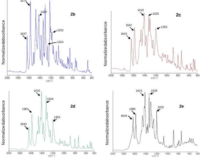
The IR spectra of compounds 2b-e recorded in solid-state (KBr pellet) are shown in Figure 1. All derivatives exhibit sharp intense bands in the 1000-1700 cm-1 region. Among these transitions, many of them can be easily assigned knowing that the IR spectrum of 2a has been theoretically and experimentally thoroughly studied in the past.6,8 The band around 1200 cm-1
corresponds to the methyl-N stretch while this around 1325 cm-1 is ascribed to the intercyclic C-C stretches between pyridinium and phenoxide rings. The transition around 1580 cm-1 mainly corresponds to the quinoïdal double bond stretch, combined with a slight contribution of pyridinium stretch and ring carbon-carbon stretching vibrations. The band around 1640 cm
-1
mainly corresponds to a pyridinium stretch. This interpretation differs from this of Ratner, but is confirmed by literature6,8 and by comparison between IR spectra of pyridinyl phenols and pyridinium salts, precursors of 2a-e. 18-19 Actually, the band around 1600-1640 systematically lacks for all the spectra of pyridinylphenols but exists in all the spectra of pyridinium salts. It must be added that the CH3 bending of tert-butyl groups is commonly
13 observed as a doublet around 1385-1395 cm-1 and 1370 cm-1 while the degenerated bending give raise to a strong band at around 1400 cm-1.33 Bands are actually observed at 1402 and
1361 cm-1 in the FTIR spectrum of 2,4-di-tert-butylphenol 33 and at 1390 and 1370 cm-1 in the
case of 3,3’,5,5’-tetra-tert-butyldiphenylquinone.34-35 In contrast, the spectrum of 2a exhibit
only a single sharp band at 1393 cm-1.19 Two bands are again observed for 2b-e (around 1414-1420 cm-1 and 1373-1378 cm-1). This explains why all spectra are normalized in absorbance taking the transition at 1410-1420 cm-1 as a reference. As usual, the frequency region 1350-1430 cm-1 remains not further interpreted.
Figure 1: IR spectra of compounds 2b-e as KBr pellet. (All spectra were normalized in absorbance taking the transition at 1410-1420 cm-1 as reference).
N o rm a liz e d a b s o rb a n c e N o rm a liz e d a b s o rb a n c e N o rm a li z e d a b s o rb a n c e N o rm a li z e d a b s o rb a n c e 1637 1575 1420 1202 1324 2b 1643 1587 1410 1326 1203 2c 1643 1587 1410 1326 1203 2d 2e 1634 1586 1415 1339 1202
The transition located around 1325 cm-1 (intercyclic C-C stretching) can be expected to be sensitive to the twist angle variations. Unfortunately, band superposition and likely covering with C-O stretching vibration make undetectable any change in bond intensity. The C=O stretching mode (ν(C=O)) around 1580 cm-1 has a different behaviour despite the weak contribution of pyridinium stretch. A decrease of the intensity of this transition from 2b to 2d is expected, arguing for the lowering contribution of the quinoïdal limiting form as the twist angle raises. Such a decrease is observed going from 2b to 2c. The torsion angle for 2c and 2b being very close (tables 3), the intensity decrease of ν(C=O) from 2c to 2d is logically extremely weak. At the same time, as it might be expected and in agreement with literature,36
an increase of the wavenumber with the torsion. This well-known hypsochromic effect is easily explained by the increase in bond strength as the conjugation vanishes. Actually, in the present work, with the torsion and the concomitant decrease in conjugation, the wavenumber increases from 1575 cm-1 for 2b to 1587 for 2c or2d. The interpretation of 2e spectrum is trickier, since iso-propyl and tert-butyl groups give similar characteristic doublets in the symmetric CH3 bending region. However, the band located at 1586 cm-1 would be less
intense than this at 1587 cm-1 in the case of 2d, once the iso-propyl contribution deduced. Attempt to transpose this study to compounds 1a-e is so far not feasible. Any increase in twist
angle affects all stretching and bending mode. As a consequence, no method of standardisation can be obviously proposed.
2a 2b 2c 2d 2e
δ(tert-butyl) /ν(C=O) 0.40 0.68 0.72 0.80
Θ 12 35 45 48 54
Table 3: Bands absorption ratio’s of δ(tert-butyl) to ν(C=O) and twist angles obtained from numerical simulations.10
15 3- Conclusion
All considered synthetic intermediates correspond to aromatic structures. In contrast, the shift and intensity variations of IR bands, being in agreement with BLA variations, confirmfirstly, that quinoïde and zwitterionic limiting forms contribute to the ground state structure of pyridinium phenolates; secondly, that the zwitterrionic form becomes predominant, as the twist angle increases. Finally exploring potential applications for nonlinear optics, it should be kept in mind that the solvent or, generally speaking, the medium can modified drastically the conformation of these molecules.
4- Experimental part
The synthesis of studied compounds was previously described.18-19
All X-Ray structures were determined from single crystals obtained by slow evaporation of acetonitrile solutions. The suitable samples were analysed on a Nonius Kappa CCD diffractometer at 173K using graphite-monochromated Mo Kα-radiation with λ = 0.71073 Å. The Nonius suite was used for data collection and integration. The structure was solved by direct methods using the program SIR92.37 Least-squares refinement against F was carried out
all non-hydrogen atoms using the program CRYSTALS.38 Chebychev polynomial weights
were used to complete the refinements. Plots were produced using CAMERON.39 Space
group, lattice parameters and other relevant information are listed in Table 4. Crystallographic data, excluding structure factors, for the structures in this paper were deposited with the Cambridge Crystallographic Data Center. Crystallographic Data Center. CCDC 768158 to 768162 contain the supplementary crystallographic data for, respectively 3f, 4c, 1a, 2a and 2b, CCDC 983983 to 983985 the supplementary crystallographic data for 3e, 4d and 4a. Copies of the data can be obtained, free of charge, on application to the CCDC, 12 Union
IR spectra were recorded on a Perkin-Elmer spectrum 65 FTIR spectrometer. 2b: IR (KBr): ν = 1637, 1575, 1476, 1428, 1420, 1381, 1344, 1323, 1303, 1213, 1202, 1180 cm-1. 2c : IR (KBr): ν = 1643, 1587, 1466, 1425, 1410, 1373, 1326, 1304, 1203 cm-1. 2d : IR (KBr): ν = 1643, 1587, 1487, 1466, 1425, 1410, 1373, 1326, 1304, 1251, 1203 cm-1. 2e : IR (KBr): ν = 1634, 1586, 1469, 1428, 1415, 1379, 1367, 1257, 1201 cm-1. Compounds 3e 3f 4a 4c 4d Chemical formula C15H19N1O 2 C15H17N1O 1 C12H14IN O2 C14H16INO C16H22I1N1 O2 Formula weight 245.32 227.31 331.15 341.19 387.26
Crystal appearance Colourless
plate Colourless plate Colourless plate Colourless plate Colourless plate Size mm3 0.12·0.28·0. 28 0.13·0.28·0. 32 0.04·0.10·0 .20 0.12·0.20·0.2 9 0.10·0.16·0. 21
Crystal system Trigonal Monoclinic Orthorhom
bic
Monoclinic Orthorhomb
ic
Space group R -3c P 21/c P 21 21 21 P 21/a P c a b
Unit cell dimensions Å a =
16.3055(5), b = 16.3055(5) c = 27.6692(10) a = 8.5707(4) b = 9.6582(4) c = 15.7429(7) a = 7.13960(10 ), b = 10.2823(2) c = 17.1010(3) a = 8.13180(10), b = 15.9024(2) c = 11.0125(2) a = 9.50760(10), b = 13.8415(2) c = 25.4627(4) Cell angles α = 90 β = 90 γ = 120 α = 90 β = 100.206(2) γ = 90 α = 90 β = 90 γ = 90 α = 90 β = 102.9067(8) γ = 90 α = 90 β = 90 γ = 90 Volume Å3 6370.8(4) 1282.54(10) 1255.41(4) 1388.10(4) 3350.88(8) Density (calculated) 1.151 1.177 1.752 1.633 1.535 F(000) 2376 488 648 672 1552 Θmax 27.030 30.052 27.857 27.851 29.984 Minimal/maximal Transmission integration 0.98/0.99 0.98/0.99 0.78/0.90 0.63/0.76 0.74/0.83 µ 0.076 0.073 2.537 2.292 1.913 Total number of reflection 42363 13891 11348 12373 27364 Independent reflections (merging r) 1641 (0.089) 3732 (0.029) 2995 (0.082) 3309 (0.042) 4882 (0.079) Data/restraints/paramet ers 949/0/99 2058/0/154 2006/5/155 2445/0/154 2623/0/181 Goodness of fit on F2 0.9865 1.0045 0.8873 0.9961 0.9759 Reflection threshold expression
I>1.00u(I) I>2.0u(I) I>3.00u(I) I>3.00u(I) I>3.00u(I)
R indice (observed data)
0.801 0.0487 0.0272 0.0241 0.0293
wR indice (all data) 0.1189 0.0766 0.0250 0.0282 0.394
Minimal/maximal residual electron density
17
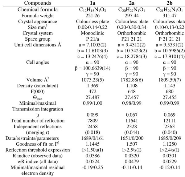
Compounds 1a 2a 2b
Chemical formula C12H15N1O3 C20H27N1O1 C21H29N1O1
Formula weight 221.26 297.44 311.47
Crystal appearance Colourless plate Colourless plate Colourless plate
Size mm3 0.02·0.14·0.22 0.20·0.30·0.34 0.10·0.13·0.22
Crystal system Monoclinic Orthorhombic Orthorhombic
Space group P 21/a P21 21 21 P 21 21 21
Unit cell dimensions Å a = 7.1003(2)
b = 11.6103(3) c = 13.2476(4) a = 9.4312(2) b = 10.3423(2) c = 18.2784(3) a = 9.5331(2) b = 10.5986(2) c = 17.9101(4) Cell angles α = 90 β = 100.6639(14) γ = 90 α = 90 β = 90 γ = 90 α = 90 β = 90 γ = 90 Volume Å3 1073.23(5) 1782.88(6) 1809.59(7) Density (calculated) 1.369 1.108 1.143 F(000) 472 648 680 Θmax 27.487 27.457 27.455 Minimal/maximal Transmission integration 0.99/1.00 0.98/0.99 0.99/0.99 µ 0.099 0.067 0.069
Total number of reflection 7809 11641 12111
Independent reflections (merging r) 2458 (0.018) 2328 (0.044) 2363 (0.040) Data/restraints/parameters 1689/0/161 1651/0/200 1685/0/209 Goodness of fit on F2 1.1445 1.507 1.1250
Reflection threshold expression I>1.50u(I) I>2.5\s(I) I>2.4\s(I)
R indice (observed data) 0.0386 0.0320 0.0301
wR indice (all data) 0.0524 0.0479 0.0529
Minimal/maximal residual electron density
-0.19/0.25 -0.11/0.14 -0.12/0.14
Table 4: Crystal data and structure refinement for studied compounds.
Acknowledgments
The authors are indebted to the Région Alsace and the Programme Interreg III A Rhenaphotonics for their financial supports. We would like to gratefully acknowledge Dr. Albert Defoin for his valuable help in guiding this research work and for numerous fruitful comments and Prof. S. Walter for valuable comments during the preparation of the manuscript.
References
1. Albert, I. D. L.; Marks, T. J.; Ratner, M. A. J. Am. Chem. Soc. 1997, 119, 3155-3156. 2. Albert, I. D. L.; Marks, T. J.; Ratner, M. A. J. Am. Chem. Soc. 1998, 120,
11174-11181.
3. Pati, S. K.; Marks, T. J.; Ratner, M. A. J. Am. Chem. Soc. 2001, 123, 7287-7291. 4. Kang, H.; Facchetti, A.; Zhu, P.; Jiang, H.; Yang, Y.; Cariati, E; Righetto, S.; Ugo, R.;
Zuccaccia, C.; Macchioni, A.; Stern, C. L.; Liu, Z.; Ho, S.-T.; Marks, T. J. Angew.
Chem. Int. Ed. 2005, 44, 1-5.
5. Brown, E. C.; Marks, T. J.; Ratner, M. A. J. Phys. Chem. B 2008, 112, 44-50.
6. Fort, A.; Boeglin, A.; Mager, L.; Amyot, C.; Combellas, C.; Thiébault, A.; Rodriguez, V. Synth. Met. 2001, 124, 209-211.
7. Zalesny, R.; Bartkowiak, W.; Stryrcz, C.; Leszczynski, J. J. Phys. Chem. A 2002, 106, 4032-4037.
8. Boeglin, A.; Fort, A.; Mager, L.; Combellas, C.; Thiébault, A.; Rodriguez, V. Chem.
Phys. 2002, 282, 353-360.
9. Niewodniezanski, W.; Bartkowiak, W. J. Mol. Model. 2007, 13, 793-800.
10.Boeglin, A.; Barsella, A.; Fort, A.; Mançois, F.; Rodriguez, V.; Diemer, V.; Chaumeil, H.; Defoin, A.; Jacques, P.; Carré, C. Chem. Phys. Lett. 2007, 442, 298-301.
11.Kang, H.; Facchetti, A.; Stern, C. L.; Rheingold, A. L.; Kassel, W. S.; Marks, T. J.
Org. Lett. 2005, 7, 3721-3724.
12.Kang, H.; Facchetti, A.; Jiang, H.; Cariati, E.; Righetto, S.; Ugo, R.; Zuccaccia, C.; Macchioni, A.; Stern, C. L. Liu, Z.; Ho, S.-T.; Brown, E. C.; Ratner, M. A.; Marks, T. J. J. Am. Chem. Soc. 2007, 129, 3267-3586.
13.Marks, T. J.; Kang, H. US patent, US2006/0237368 A1.
14.Vonlanthen, D.; Mishchenko, A.; Elbing, M.; Neuburger, M.; Wandlowski, T.; Major, M. Angew. Chem. Int. Ed. 2009, 48, 8886-8890.
15.Duvanel, G.; Grilj, J.; Chaumeil, H.; Jacques, P.; Vauthey, E. Photochem. Photobiol.
Sci. 2010, 9, 908-915.
16.Chaumeil, H.; Jacques, P.; Diemer, V.; Le Nouën, D.; Carré, C. J. Mol. Struc. 2011,
1002, 70-75.
17.Jacques, P.; Graff, B.; Diemer, V.; Ay, E.; Chaumeil, H.; Carré, C.; Malval, J.-P.
Chem. Phys. Lett. 2012, 531, 242-246.
18.Diemer, V.; Chaumeil, H.; Defoin, A.; Fort, A.; Boeglin, A.; Carré, C. Eur. J. Org.
Chem. 2006, 2727-2738.
19.Diemer, V.; Chaumeil, H.; Defoin, A.; Fort, A.; Boeglin, A.; Carré, C. Eur. J. Org.
Chem. 2008, 1767-1776.
20.Diemer, V.; Chaumeil, H.; Defoin, A.; Boeglin, A.; Barsella, A.; Fort, A.; Carré, C.;
Proc. Spie-Org. Optoelectron. Photonics II 2006, 6192, 559-565.
21.Barzoukas, M.; Fort, A.; Boy, P.; Combellas, C.; Thiébault, A. Nonlinear Optics 1994,
19 22.Barzoukas, M.; Fort, A.; Boy, P.; Combellas, C.; Thiébault, A. Chemical Physics
1994, 185, 65-74.
23.Runser, C.; Fort, A.; Barzoukas, M.; Combellas, C.; Suba, C.; Thiébault, A.; Graff, R.; Kintzinger, J.-P. Chem. Phys. 1995, 193, 309-319.
24.Rotzler, J.; Vonlanthen, D.; Barsella, A.; Boeglin, A.; Fort, A.; Mayor, M. Eur. J. Org.
Chem. 2009, 1096-1110.
25.Liu, L.; Xue, Y.; Wang, X.; Chu, X.; Yang, M. Int. J. Quantum Chem. 2012, 112, 1086-1096.
26.Allen, F. H.; Kennard, O.; Watson, D. G.; Brammer, L.; Orpen, G.; Taylor, R. J.
Chem. Soc. Perkin Trans. II, 1987, S1-S19.
27.Fabian, J.; Rosquete, G. A.; Montero-Cabrera, L. A. J. Mol. Struct. THEOCHEM 1999, 469, 163-176.
28.Marder, S. R.; Perry, J. W.; Tiemann, B. G.; Gorman, C. B.; Gilmour, S.; Biddle, S. L.; Bourhill, G. J. Am. Chem. Soc., 1993, 115, 2524-2526.
29.Marder, S. R.; Gorman, C. B.; Tiemann, B. G.; Cheng, L.-T. J. Am. Chem. Soc., 1993,
115, 3006-3007.
30.Marder, S. R.; Perry, J. W. Adv. Mater. 1993, 5, 804-815.
31.Gorman, C. B.; Marder, S. R. Proc. Natl. Acad. Sci. 1993, 90, 11297-11301. 32.Pinheiro, J. M. F.; de Melo, C. P. J. Phys. Chem. A 2011, 115, 7994-8002.
33.Kalaichelvan, S.; Sundaraganesan, N.; Dereli, O.; Sayin, U. Spectrochimica Acta Part
A 2012, 85, 198-209.
34.Pastor, S. D. J. Org. Chem. 1984, 49, 5260-5262. 35. http//sdbs.db.aist.go.jp
36.Souchay, P.; Tatibouët, F.; Barchewitz, P. J. Phys. Radium 1954, 15, 533-535.
37.Altomare, A.; Cascarano, G.; Giacovazzo, G.; Guagliardi, A.; Burla, M. C.; Polidori, G.; Camalli, M. J. Appl. Crystallogr. 1994, 27, 435-436.
38.Betteridge, P. W.; Carruthers, J. R.; Cooper, R. I.; Prout, K.; Watkin, D. J. J. Appl.
Crystallogr. 2003, 36, 1487.
39.Watkin, D.J.; Prout, C. K.; Pearse, L. J. CAMERON; Chemical Crystallography Laboratory: Oxford, UK, 1996.