Published in : Journal of Fish Biology (1999), vol. 55, n°4, pp. 795-808 DOI: 10.1006/jfbi.1999.1037
Status : Postprint (Author’s version)
Early development of the chondrocranium in
Chrysichthys auratus
Vandewalle, Pierre1, Chikou, Antoine2, Lalèyé, Philippe2, Parmentier, Eric1, Huriaux, Françoise3, Focant, Bruno3
1Université de Liège, Laboratoire de Morphologie fonctionnelle et évolutive, 22 quai Van Beneden, B-4020 Liège, Belgium
2Universite Nationale du Benin, Faculté des Sciences Agronomiques, Laboratoire d'Hydrobiologie, B.P.526, Cotonou, Bénin
3Université de Liège, Laboratoire de Biologie cellulaire et tissulaire, 20 rue de Pitteurs, B-4020, Liège, Belgium
KEYWORDS: Siluriformes; Chrysichthys auratus; ontogeny; chondrocranium. ABSTRACT
The inception and development of the cartilaginous cephalis skeleton of Chrysichthys auratus is described from hatching to about 18 days post-hatching. At hatching, no skeletal structure is present. Not until day 3 do clearly delimited cranial primordia become apparent. As in many siluriforms, the neurocranium is platybasic from the start, the suspensorium constitutes, with Meckel's cartilage and the hyoid bar, a single cartilaginous element, and the junction between the front and rear of the neurocranium is complete on day 4. By day 8 the quadrato-mandibular joint has formed and the tectum posterius has appeared. Cartilage reduction first a ects the trabecular bars, then, markedly, the visceral arches. By day 18 the braincase floor has almost disappeared.
INTRODUCTION
Nearly all developmental studies of fishes show that the chondrocranial elements are the first to appear (de Beer, 1937; Hubendick, 1942; Daget, 1964; Elman & Balon, 1980; Vandewalle et al., 1992, 1997). The chondrocranium thus first gives the head its shape. The shape of the head is subject to constraints related to the lifestyle of the fish. In siluriforms, the skull is platybasic with a large hypophyseal fenestra and often small eyes, whereas most teleosts display a trophibasic skull (Daget, 1964). In catfish the skull is also generally flattened dorso-ventrally, thus increasing stability on the ground (Alexander, 1965). Small eyes imply small ocular muscles and thus rudimentary or absent myodomes (Daget, 1964; Alexander, 1965). This chondrocranium corresponds more with a benthic life and/or nocturnal activities (Adriaens & Verraes, 1997). Reduction of the eyes is doubtless compensated for by the presence of mobile barbels with a tactile, sensory and gustatory function and a well-developed Weberian apparatus (Alexander, 1965; Chardon, 1968; Gosline, 1975;
Published in : Journal of Fish Biology (1999), vol. 55, n°4, pp. 795-808 DOI: 10.1006/jfbi.1999.1037
Status : Postprint (Author’s version)
This paper deals with the postembryonic development of Chrysichthys auratus (Geo roy Saint-Hilaire) [Claroteidae sensu Mo (1991) and Teugels (1996)]. It is a sequel to a previous study of the osteocranium of this fish (Vandewalle et al., 1995). This catfish possesses large, almost lateral eyes and a relatively elevated skull. The nomenclature used to describe the developing skeletal structures is based principally on the works of
de Beer (1937) and Daget (1964).
Figure. 1. Chrysichthys auratus: lateral views of skeletal organization at 12 h (a) (cells only)
and 2 days (b) (cartilaginous formations in dotted regions). BCA, Commissura basicapsularis; BOT, lamina basiotica; CBR, ceratobranchial; HB, hyoid bar; HSY, hyosymplectic; IH, interhyal; MCA, Meckelian cartilage; OPR, opercular process; OTCAP, otic capsule; PC, parachordal cartilage; PLOC, pila occipitalis; PQ, pars quadrata; RPR, retroarticular process; TR, trabecula.
MATERIALS AND METHODS
Chrysichthys auratus fry were obtained by semi-natural fertilization carried out at the
Faculty of Agronomical Sciences in Cotonou (Benin). The fry were related at a temperature of 27) C. Batches of 25 fry were sampled at hatching (one day post-fertilization) and 12 h and 1, 2, 3, 4, 6, 8, 10, 12, 14, 16, and 18 days post-hatching. The fry were fixed in a CaCO3-buffered 10% formalin solution, alcian-blue stained,
trypsin cleared, and finally stored in glycerin (Taylor & Van Dijk, 1985). The cleared specimens were studied with a Wild M5 stereoscopic microscope.
RESULTS
AT HATCHINGNo cartilaginous cephalic structures are present. 12H POST-HATCHING [Fig. 1(a)]
No cartilaginous structures are stained but chondrocytes are organized so as to form parachordal bars, a commisura basicapsularis anterior, a lamina basiotica, partial trabecular bars, hyosymplectics, a pars quadrata, hyoid bars, and Meckel's cartilages. DAY 1
Chondrocytes delimit complete trabecular bars. DAY 2 [Fig. 1(b)]
Published in : Journal of Fish Biology (1999), vol. 55, n°4, pp. 795-808 DOI: 10.1006/jfbi.1999.1037
Status : Postprint (Author’s version)
Meckelian cartilages are alcian blue stained. Chondrocytes constitute the first anlagen of the otic capsule wall and pila occipitalis, and prefigure an opercular joint process at the level of the hyosymplectics and a retroarticular process at the level of Meckel's cartilages.
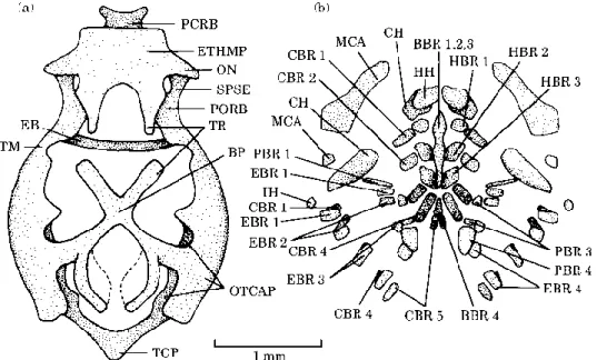
Figure 2. Chrysichthys auratus: dorsal (a) and lateral (b) views of the chondrocranium at 3
days. BCA, Commissura basicapsularis; CBR, ceratobranchial; CPR, coronoid process; EB, epiphysial bridge; ETHMP, ethmoid plate; HB, hyoid bar; HSY, hyosymplectic; HY, fenestra hypophysea; IH, interhyal; MCA, Meckelian cartilage; OPR, opercular process; OTCAP, otic capsule; PC, parachordal cartilage; PCRB, lamina precerebralis; RPR, retroarticular process; TM, taenia marginalis; TR, trabecula.
DAY 3 (Fig. 2)
All structures are stained in blue. In front, the trabecular bars fused in an ethmoid plate; the taeniae marginales, epiphyseal bridge, and lamina precerebralis are forming. The joint between the hyosymplectics and braincase is conspicuous. The hyosymplectics, Meckel's cartilages, and the hyoid bars constitute a single cartilaginous part. Meckel's cartilages each bear a nascent dorsal coronoid process.
DAY 4 (Fig. 3)
The lateral walls of the otic capsules have formed. The laminae basioticae are fused along the median line to form an acrochordal cartilage. This cartilage, the trabecular bars, and the ethmoid plate thus close the hypophyseal fenestra.
The taeniae marginales are connected to the ethmoid plate by the commissurae sphenoseptales and the lamina precerebralis.
The laminae orbitonasales have appeared and they link the taeniae marginales to the trabeculae: these structures delimit the olfactory foramina. The epiphyseal bridges has formed, separating a prepineal fontanella from the rest of the dorsal cranial opening. Independent partes palatinae are present and each hyosymplectic now bears a nascent pterygoid process. The branchial basket includes a basibranchial, Five pairs of ceratobranchials, and four pairs of epibranchials.
Published in : Journal of Fish Biology (1999), vol. 55, n°4, pp. 795-808 DOI: 10.1006/jfbi.1999.1037
Status : Postprint (Author’s version)
days. AC, Acrochordal cartilage; BCA, Commissura basicapsularis; CBR, ceratobranchial; CPR, coronoid process; EB, epiphysial bridge; EBR, epibranchial; ETHMP, ethmoid plate; HB, hyoid bar; HSY, hyosymplectic; HY, fenestra hypophysea; IH, interhyal; MCA, Meckelian cartilage; OL, olfactory foramen; ON, lamina orbitonasalis; OPR, opercular process; OTCAP, otic capsule; PC, parachordal cartilage; PCRB, lamina precerebralis; PP, pars palatina; PTPR, pterygoid process; RPR, retroarticular process; SPSE, commissura sphenoseptalis; TM, taenia marginalis; TR, trabecula.
DAY 6 (Fig. 4)
The pyterygoid processes have lengthened. Meckel's cartilages and the left and right hyoid bars are clearly separated and the joints between the mandible and the partes quadratae seem to be forming. A pair of distinct hypohyals has appeared at the front of the hyoid bars, this marking the end of the ceratohyals behind them. The fourth pharyngobranchial are present, the third epibranchials bear an uncinate process, and the fourth epibranchials have broadened.
DAY 8 [Fig. 5(a), (b),(c)]
The parachordals have fused and constitute, with the acrochordal cartilage, the basal plate. The tectum posterius closes the rear of the neurocranium dorsally. The trabecular bars display a fissure for the internal carotid artery. The quadrato-mandibular joints are conspicuous and the interhyals have separated from the ceratohyals. A posterior basibranchial, the hypobranchials of the first, second, and third arches, and the first and third pharyngobranchials have appeared. The first pharyngobranchials are slender.
DAY 10 [Fig. 5(d)]
The laminae preorbitales are present. The trabecular bars are divided in two parts. DAY 14 (Fig. 6)
The lamina precerebralis is dorsally expanded.
Most cartilaginous components of the splanchnocranium are undergoing reduction except for the palatines, interhyals, hypohyals, basibranchials, hypobranchials, and first and third pharyngobranchials. The central part of the hyosymplectics and partes quadratae are no longer stained by alcian blue. The pterygoid processes are reduced
Published in : Journal of Fish Biology (1999), vol. 55, n°4, pp. 795-808 DOI: 10.1006/jfbi.1999.1037
Status : Postprint (Author’s version)
at their centre. The mandible remains cartilaginous in the middle of these two branches and where it articulates with the partes quadratae.
Figure 4. Chrysichthys auratus: dorsal (a) and lateral (b) views of the chondrocranium and
ventral view (c) of the visceral arches at 6 days. AC, Acrochordal cartilage; BBR, basibranchial; BCA, Commissura basicapsularis; CBR, ceratobranchial; CH, ceratohyal; CPR, coronoid process; EB, epiphysial bridge; EBR, epibranchial; ETHMP, ethmoid plate; FACI, fissura arteriae carotis internae; HB, hyoid bar; HH, hypohyal; HSY, hyosymplectic; HY, fenestra hypophysea; IH, interhyal; MCA, Meckelian cartilage; ON, lamina orbitonasalis; OTCAP, otic capsule; PBR, pharyngobranchial; PC, parachordal cartilage; PP, pars palatina; PQ, pars quadrata; PTPR, pterygoid process; SPSE, commissura sphenoseptalis; TM, taenia marginalis; TR, trabecula.
DAY 18 (Fig. 7)
The floor of the neurocranium and the tectum posterius are considerably reduced and the main components of the splanchnocranium (except for Meckel's cartilages) are clearly reduced at their extremities.
DISCUSSION
INCEPTION AND DEVELOPMENT OF THE CARTILAGINOUS STRUCTURES
As in Clarias gariepinus (Burchell) (Siluriformes, Clariidae) (formerly Clarias lazera, see Teugels, 1986) and many teleosts, chondrification of the neurocranium begins, in
C. auratus, at the same time as that of the splanchnocranium (de Beer, 1937; Adriaens & Verraes, 1977; Vandewalle et al., 1992). Yet in Heterobranchus longifilis Valenciennes (Clariidae, Siluriformes), the primordia of Meckel's cartilage and of the hyoid arch appear first (Vandewalle et al., 1997), while in Heteropneustes fossilis (Bloch) (Siluriformes, Heteropneustidae), the neurocranium is the first skeletal element
Published in : Journal of Fish Biology (1999), vol. 55, n°4, pp. 795-808 DOI: 10.1006/jfbi.1999.1037
Status : Postprint (Author’s version)
to appear (Srinivasachar, 1959).
Figure 5. Chrysichthys auratus: dorsal view of the visceral arches (a), dorsal (b) and lateral (c) views of the chondrocranium at 8 days and lateral view of the chondroneurocranium at 10 days (d). BBR, Basibranchial; BP, basal plate; CBR, ceratobranchial; CH, ceratohyal; CPR, coronoid process; EB, epiphysial bridge; EBR, epibranchial; ETHMP, ethmoid plate; FACI, fissura arteriae carotis internae; HB, hyoid bar; HBR, hypobranchial; HH, hypohyal; HSY, hyosymplectic; IH, interhyal; MCA, Meckelian cartilage; OL, olfactory foramen; ON, lamina orbitonasalis; OTCAP, otic capsule; PBR, pharyngobranchial; PCRB, lamina precerebralis; PORB, lamina preorbitalis; PP, pars palatina; PQ, pars quadrata; PTPR, pterygoid process; SPSE, commissura sphenoseptalis; TCP, tectum posterius; TM, taenia marginalis; TR, trabecula.
Neurocranium
In C. auratus and C. gariepinus, no cephalic structure is observed at hatching. This contrasts with H. fossilis and Barbus barbus (L.) (Cypriniformes, Cyprinidae) (Srinivasachar, 1959; Vandewalle et al., 1992), where several elements are present at this time. In the latter species, this precocity is only apparent, since hatching occurs 4 days post-fertilization. (Vandewalle et al., 1992).
In C. auratus as in other teleosts, the floor of the neurocranium appears first. The parachordals are fused from the start with the trabecular bars, these being well separated with a distance between them, bounding laterally a wide hypophyseal space. The neorocranium is thus platybasic at the outset of development, as in most catfish (Adriaens & Verraes, 1997; Vandewalle et al., 1997). In B. barbus the parachordals and trabecular bars are separate when they appear (Vandewalle et al., 1992).
Figure 6. Chrysichthys auratus: dorsal view of the chondroneurocranium (a), lateral view of the
suspensorium and mandible (b), and dorsal view of the hyobranchial apparatus (c) at 14 days. BBR, basibranchial; BP, basal plate; CBR, ceratobranchial; CH, ceratohyal; CPR, coronoid process; EB, epiphysial bridge; EBR, epibranchial; ETHMP, ethmoid plate; HBR, hypobranchial; HH, hypohyal; HSY, hyosymplectic; IH, interhyal; LETHM, lateral ethmoid cartilage; MCA,
Published in : Journal of Fish Biology (1999), vol. 55, n°4, pp. 795-808 DOI: 10.1006/jfbi.1999.1037
Status : Postprint (Author’s version)
Meckelian cartilage; ON, lamina orbitonasalis; OPR, opercular process; OTCAP, otic capsule; PBR, pharyngobranchial; PCRB, lamina precerebralis; PP, pars palatina; PQ, pars quadrata; PTPR, pterygoid process; SPSE, commissura sphenoseptalis; TCP, tectum posterius; TM, taenia marginalis; TR, trabecula.
In C. auratus, the trabecular bars fuse rapidly in an ethmoid plate, forming anteriorly the first junction between the left and right halves of the neurocranium, as in Salmo sp. (Salmoniformes, Salmonidae), B. barbus, and Merluccius capensis (Castelnau) (Gadiformes, Merluciidae) (de Beer, 1937; Badenhorst, 1989a; Vandewalle et al., 1992). In H. fossilis, C. gariepinus, and H. longifilis, on the contrary, the hypophyseal fenestra is closed first posteriorly by the acrochordal cartilage (Srinivasachar, 1959;
Vandewalle et al., 1997; Adriaens & Verraes, 1997). This is a major difference in the construction of the neurocranium, and one for which it is hard to find a functional justification. The reason of this difference is probably embedded in phylogeny. Maybe the early posterior closure of the hypophyseal fenestra is a shared derived character for clariids and heteropneustids?
Shortly after the first elements of the floor of the neurocranium appear, the braincase walls develop with the inception of the commissurae basicapsulares anteriores and otic capsules. This appears as a general feature of skeletal development in catfish. In the cyprinid B. barbus, the commissurae basicapsulares are present before the parachordals and trabecular bars join (Vandewalle et al., 1992).
From day 4, in C. auratus, the dorsal arch of the orbits, composed of the taeniae marginales, grows forward, forms the epiphyseal bridge, and joins on day 6 with the ethmoid plate by means of the commissurae sphenoseptales; concomitantly the acrochordal cartilage forms and closes the hypophyseal fenestra posteriorly. At this stage the hypophyseal fenestra is already filled partially by the parasphenoid bone (Vandewalle et al., 1995). In other siluriforms the development of the dorsal arch is similar to that of C. auratus, but the ethmoid plate only appears when the taenia marginalis is sufficiently developed to join the front and rear of the neurocranium (Bamford, 1948; Srinivasachar, 1959; Surlemont & Vandewalle, 1991; Adriaens & Verraes, 1997; Vandewalle et al., 1997). This state of development, i.e. a braincase
Published in : Journal of Fish Biology (1999), vol. 55, n°4, pp. 795-808 DOI: 10.1006/jfbi.1999.1037
Status : Postprint (Author’s version)
floor connected dorsally (teaniae marginales and commissurae sphenoseptales) and ventrally (trabecular bars) to the ethmoid region and a hypophyseal fenestra occupied by the parasphenoid, before closing of the braincase roof, appears common not only in catfish (Bamford, 1948; Srinivasachar, 1959; Weisel, 1967; Vandewalle et al., 1991,
1997; Adriaens & Verraes, 1997) but also to many other teleosts (Elman & Balon, 1980; Vandewalle et al., 1992, 1997; Watson & Walker, 1992; Cubbage & Mabee, 1996; Mabee & Trendler, 1996).
Figure 7. Chrysichthys auratus: ventral view of the chondroneurocranium (a) and dorsal view of
the visceral arches (b) at 18 days. BBR, Basibranchial; BP, basal plate; CBR, ceratobranchial; CH, ceratohyal; EB, epiphysial bridge; EBR, epibranchial; ETHMP, ethmoid plate; HBR, hypobranchial; HH, hypohyal; IH, interhyal; MCA, Meckelian cartilage; OTCAP, otic capsule; PBR, pharyngobranchial; PCRB, lamina precerebralis; PORB, lamina preorbitalis; SPSE, commissura sphenoseptalis; TCP, tectum posterius; TM, taenia marginalis; TR, trabecula.
This level of neurocranial development is compatible with the transition from endogenous to exogenous feeding: the floor of the neurocranium isolates the developing nervous system from the buccal cavity, subject to the physical particularities of the food (Verraes, 1974; Vandewalle et al., 1997; Adriaens & Verraes, 1998; Wagemans et al., 1998; Gluckmann et al., 1999).
In nearly all fish where the chronology of development has been well studied, the next element to appear is the tectum posterius, linking at last the posterior lateral walls of the braincase. Although no observation confirms this view, backward extension of the tectum posterius might correspond with fusion with the first supraneural, as in C. gariepinus (Adriaens & Verraes, 1997).
At this stage of development, the ratio of the height of the neurocranium to its length shows that the neurocranium of catfish is not particularly depressed as compared with that of other teleosts (Kindred, 1919; de Beer, 1937; Bamford, 1948; Srinivasachar, 1959; Elman & Balon, 1980; Badenhorst, 1989a; Watson & Walker, 1992; Vandewalle
et al., 1992, 1997; Adriaens & Verraes, 1997; Wagemans et al., 1998; Gluckmann et
al., 1999), and that it is much broader (except for Ictalurus nebulosus (Lesueur), formerly Ameiurus nebulosus) (Kindred, 1919; Srinivasachar, 1959; Surlemont & Vandewalle, 1991; Adriaens & Verraes, 1997; Vandewalle et al., 1997).
Published in : Journal of Fish Biology (1999), vol. 55, n°4, pp. 795-808 DOI: 10.1006/jfbi.1999.1037
Status : Postprint (Author’s version)
Splanchnocranium
Like that of the neurocranium, the first primordium of the splanchnocranium consists only of chondrocytes. No separation between these cell masses is discernible. The splanchnocranial cell mass represents the hyosymplectic, the pars quadrata, Meckel's cartilage, and the hyoid bar. Not until day 3 is chondrification complete. As in several other siluriforms (Surlemont et al., 1989; Arratia, 1990; Surlemont & Vandewalle, 1991;
Adriaens & Verraes, 1994, 1997; Vandewalle et al., 1997), Meckel's cartilage, the hyoid bar, the interhyal, the hyosymplectic, and the pars quadrata constitute a single cartilaginous part. In Arius felis (L.) (formerly Galeichthys felis), Meckel's cartilages are not linked to the rest of the suspensorium (Bamford, 1948). In I. nebulosus and H.
fossilis, only part of the suspensorium, consisting of the hyosymplectic, the pars
quadrata, and the interhyal, constitutes a single cartilaginous part, the mandible and hyoid bar being separate from it (Kindred, 1919; Srinivasachar, 1959). In most other teleosts, all elements of the suspensorium are separate (Norman, 1926; de Beer,
1937; Bertmar, 1959; Langille & Hall, 1987; Badenhorst, 1989b; Vandewalle et al., 1992; Watson & Walker, 1992; Wagenmans et al., 1998; Gluckmann et al., 1999). At least partial fusion of the suspensorium, i.e. of the hyosymplectic with the pars quadrata, may be a synapomorphy among siluriforms. Another is the appearance on day 4, in C. auratus, of a pars palatina independent of the rest of the suspensorium (Arratia & Schultze, 1990). The pars palatina, distinct from the rest of the suspensorium, is related to the original movements of the maxillary in catfish (Gosline, 1975; Ghiot et al., 1984).
As in I. nebulosus, C. gariepinus, and H. longifilis (Kindred, 1919; Adriaens & Verraes, 1997; Vandewalle et al., 1997), the hyosymplectic develops an opercular process at an early stage and the mandible displays very early a nascent retroarticular process, then a coronoid process. These particularities are not common to all siluriforms (Srinivasachar, 1959). Formation of the opercular process corresponds with the appearance of the opercular bone (Vandewalle et al., 1995). In teleosts, the coronoid process is one of the insertion sites of the adductor muscles of the mandible (Winterbottom, 1974). In C. gariepinus, adductor 2 of the mandible is present very early in development and movements of the lower jaw are observable before the appearance of the quadrato- mandibular joint: this implies cartilage bending (Surlemont
et al., 1989). The early presence of a coronoid process in C. auratus suggests that a mandibular musculature exists and hence that buccal movements with cartilage bending are possible.
Splitting of the splanchnocranium in C. auratus begins as in C. gariepinus and H. longifilis (Surlemont & Vandewalle, 1991; Adriaens & Verraes, 1997; Vandewalle et al., 1997) with the appearance of the quadrato-mandibular joint, followed at a late stage by the joint between the interhyal and hyoid bar. The interhyal becomes totally independent in C. auratus by day 14; this is not the case in H. longifilis or C. gariepinus (Adriaens & Verraes, 1994; Vandewalle et al., 1997). In the latter species, the interhyal disappears by day 100 (Adriaens & Verraes, 1994).
The first four ceratobranchials appear together in C. auratus, whereas in C. gariepinus, the first ceratobranchial appears first, then the next three (Surlemont et al., 1989;
Surlemont & Vandewalle, 1991; Adriaens & Verraes, 1997), and in H. longifilis, the first three appear before the fourth (Vandewalle et al., 1997). These variations may simply
Published in : Journal of Fish Biology (1999), vol. 55, n°4, pp. 795-808 DOI: 10.1006/jfbi.1999.1037
Status : Postprint (Author’s version)
reflect the difference in observation times for the three species. The fifth ceratobranchial, however, always appears last, concomitantly with the epibranchials and posterior pharyngobranchial in all three species. Late formation of the fifth ceratobranchial and of the pharyngobranchial seems frequent in teleosts; it is concomitant with the appearance of the first toothed dermal ossifications which they bear (Badenhorst, 1989b; Vandewalle et al., 1992, 1995; Cabbage & Mabee, 1996;
Doi et al., 1997; Gluckmann et al., 1998; Wagemans et al., 1998). The pharyngeal teeth are present just before the disappearance of the yolk sac (Vandewalle et al., 1995). At hatching, respiratory exchanges occur by means of the skin and the highly vascularized yolk sac. The size of this sac diminishes gradually. In parallel with this, the first four branchial arches appear and probably increasingly ensure respiration until total resorption of the yolk sac. The fifth ceratobranchials and the pharyngobranchials never have a respiratory function and in this regard, their formation can be delayed. However, since they constitute the pharyngeal jaws, they must be functional at the time of the swift transition from endogenous to exogenous feeding.
CARTILAGE REDUCTION
In teleosts, cartilage reduction corresponds with formation of the bony parts. In C.
auratus, reduction begins when all the elements of the chondrocranium are in place. It
affects first the trabecular bars, which become pinched to form the fissure of the carotid artery before separating from the braincase. This reduction of the trabecular bars occurs, in catfish, after formation of the tectum posterius (Adriaens & Verraes, 1997;
Vandewalle et al., 1997), while in other teleosts it occurs first (Badenhorst, 1989b;
Vandewalle et al., 1992, 1997; Wagemans et al., 1998; Gluckmann et al., 1999). It may be that the very wide neurocranium of catfish requires posterior consolidation before it splits. It is surprising to note that in Oryzias latipes Jordan & Snyder (Atherinomorpha, Adrianichthyidae), the trabecular bars are never, at any time, linked to the base of the braincase (Langille & Hall, 1987).
The next reductions of the braincase, in cases where these have been observed, occur long after reduction of the trabeculae and affect the epiphyseal bridge (Vandewalle et
al., 1997) or the taeniae marginales (Wagemans et al., 1998). In C. auratus, however, the braincase floor is reduced to a kind of cross: according to Vandewalle et al. (1995), there remains between the prootics, the parasphenoid, and the exoccipitals a non-ossified area with a very similar shape.
In other teleosts, cartilage reduction first affects the splanchnocranium: in H. longifilis, the hyoid bars and ceratobranchials regress before the trabecular bars (Vandewalle et
al., 1997), while in Scophthalmus maximus (L.) (Pleuronectiformes, Scophthalmidae) Meckel's cartilages are the first to regress (Wagemans et al., 1998). In C. auratus, all of the visceral arches would seem to regress simultaneously, allowance being made for the time elapsed between the two successive stages observed. In B. barbus, reduction of the splanchnocranium first affects the fifth ceratobranchials. The only constant feature of splanchnocranial reduction in teleosts seems to be that it first affects the hyoid bars and branchial elements at their centre, where these elements ossify first (Langille & Hall, 1987; Vandewalle et al., 1992, 1995, 1997; Tilney & Hecht, 1993; Kohno et al., 1996; Mabee & Trendler, 1996; Adriaens & Verraes, 1998;
Wagemans et al., 1998; Gluckmann et al., 1999).
Published in : Journal of Fish Biology (1999), vol. 55, n°4, pp. 795-808 DOI: 10.1006/jfbi.1999.1037
Status : Postprint (Author’s version)
hyosymplectic-pars quadrata seems to preclude the presence, even transient, of a symplectic (Vandewalle et al., 1995, 1997; Adriaens & Verraes, 1997). A rudimentary symplectic does seem to exist in Diplomystes camposensis Arratia, 1987 (Siluriformes, Doplimystidae) (Arratia, 1992).
CONCLUSIONS
It is hard to consider that there exists a general plan of chondrocranium construction in teleosts or even siluriforms, where closing of the hypophyseal fenestra is variable. Yet as noted by Adriaens & Verraes (1997), catfish do share some particularities as regards the development of the cartilaginous skull. The neurocranium is platybasic with a wide hypophyseal fenestra bordered posteriorly by the acrochordal cartilage and not by the notochord; the epiphyseal bridge is well developed; the lamina precerebralis constitutes an internasal septum part; the lamina orbito-nasalis closes the front of the orbit; the otic capsule is almost complete, lacking only the tectum synoticum. The splanchno- cranium is characterized by the following features: early fusion of several elements which will separate in the course of development, a pars palatina isolated from the rest of the suspensorium, the fact that the left and right Meckelian cartilages and hyoid bars are joined at first, and that the fifth ceratobranchials and the pharyngobranchials are the last elements of the hyobranchial system to appear.
At the time the yolk sac disappears, all teleosts whose development is well known display a well-developed branchial respiratory system. Moreover, their bucco-pharyngeal apparatus is in place and their buccal cavity is separated from the braincase. All this makes it possible for the fry to survive.
The chondrocranium of all teleosts undergoes, after its construction, a reduction period corresponding with the appearance of the enchondral ossification (Weisel, 1967;
Vandewalle et al., 1992, 1997; Adriaens & Verraes, 1998; Wagemans et al., 1998;
Gluckmann et al., 1999). In C. auratus, these cartilage reductions do seem to follow an original way, and confirm the lack of a general reduction sequence in siluriforms (Srinivasachar, 1958; Vandewalle et al., 1997; Adriaens & Verraes, 1998).
It could be that once aquatic respiration and exogenous feeding are operational, the constraints imposed by the need to survive and to occupy the environment become widely divergent for different species, thus justifying a species-specific sequence of conversion from a cartilaginous to a bony skeleton (Osse, 1990; Vandewalle et al., 1992, 1995; Adriaens & Verraes, 1998).
This work was supported by grants of the 'Fonds national de la Recherche scientifique', 'Agence générale de Cooperation au Développement' and 'Communauté française' of Belgium. The authors thank K. Broman for linguistic assistance. B.F. is Research Associate of 'Fonds national de la Recherche scientifique de Belgique'.
References
Adriaens, D. & Verraes, W. (1994). On the functional significance of the loss of the interhyal during ontogeny in Clarias gariepinus Burchell, 1822 (Teleostei: Siluroidei). Belgium Journal of Zoology 124, 139-155.
Adriaens, D. & Verraes, W. (1997). The ontogeny of the chondrocranium in Clarias gariepinus: trends in siluroids. Journal of Fish Biology 50, 1221-1257.
Published in : Journal of Fish Biology (1999), vol. 55, n°4, pp. 795-808 DOI: 10.1006/jfbi.1999.1037
Status : Postprint (Author’s version)
Adriaens, D. & Verraes, W. (1998). Ontogeny of the osteocranium in the african catfish, Clarias gariepinus Burchell (1922) (Siluriformes: Clariidae): ossification sequence as a response to functional demand. Journal of Morphology 235, 183-237.
Alexander, R. McN. (1965). Structure and function in catfish. Journal of Zoology 148, 88-152.
Arratia, G. (1990). Development and diversity of the suspensorium of trichomycterids and comparison with loricarioids (Teleostei: Siluriformes). Journal of Morphology 205, 193-218.
Arratia, G. (1992). Development and variation of the suspensorium of primitive catfishes (Teleostei: Ostariophysi). Bonner Zoologische Monographien 32, 1-148.
Arratia, G. & Schultze, H. P. (1990). The urohyal development and homology within osteichthyous. Journal of Morphology 203, 247-282.
Badenhorst, A. (1989a). Development of the chondrocranium of the shallow-water cape hake Merluccius capensis (Cost.). Part 1: neurocranium. South African Journal of Zoology 24, 33-48.
Badenhorst, A. (1989b). Development of the chondrocranium of the shallow-water cape hake Merluccius capensis (Cost.). Part 2: viscerocranium. South African Journal of Zoology 24, 49-57.
Bamford, T. W. (1948). Cranial development of Galeichthys felis. Proceedings of Zoological Society (London) 118, 364-391.
Chardon, M. (1968). Anatomie compareée de l'appareil de Weber et des structures connexes chez les Siluriformes. Annales du Musée royal de l'Afrique centrale, Sciences zoologiques 169, 1-227.
Cubbage, C. C. & Mabee, P. M. (1996). Development of the cranium and paired ®ns in the zebrafish Danio rerio (Ostariophysi, Cyprinidae). Journal of Morphology 229, 121-160.
Daget, J. (1964). Le crane des teleosteens. Mémoire du Museum National d’Histoire Naturelle, Série A, Zoologie 31, 167±340.
de Beer, G. R. (1937). The Development of the Vertebrate Skull. Oxford: Clarendon Press. Doi, M., Ohno, A., Kohno, H., Taki, Y. & Singhagraiwan, T. (1997). Development of feeding ability in red snapper Lutjanus argentimaculatus early larvae. Fisheries Science 63, 845-853.
Elman, J. F. & Balon, E. K. (1980). Early ontogeny of white sucker Catostomus commersonii, with steps of saltatory development. Environmental Biology of Fishes 5, 191-224.
Ghiot, F. & Bouchez, N. (1980). The central rod of the barbels of a South American catfish, Pimelodus clarias. Copeia 1980, 908-909.
Ghiot, F., Vandewalle, P. & Chardon, M. (1984). Comparaison anatomique et fonctionnelle des muscles et des ligaments en rapport avec les barbillons de deux familles apparentées de poissons Siluriformes Bagroidei. Annales de la Société Royale Zoologique de Belgique 114, 261-272.
Gluckmann, I., Huriaux, F., Focant, B. & Vandewalle, P. (1999). Postembryonic development of the cephalic skeleton in Dicentrarchus labrax (Pisces, Perciformes, Serranidea). Bulletin of Marine Science, in press.
Gosline, W. A. (1975). The palatine-maxillary mechanism in catfishes with comments on the evolution and zoogeography of modern siluroids. Occasional Papers of the Californian Academy of Sciences 120, 1-31.
Published in : Journal of Fish Biology (1999), vol. 55, n°4, pp. 795-808 DOI: 10.1006/jfbi.1999.1037
Status : Postprint (Author’s version)
rutilus. Arkiv für Zoologi 34A, 1-35.
Kohno, H., Ordonio-Aguilar, R., Ohno, A. & Taki, Y. (1996). Morphological aspects and improvement in feeding ability in early stage larvae of milkfish, Chanos chanos. Ichthyological Research 43, 133-140.
Kindred, J. E. (1919). Development of skull in Ameirus nebulosus. Illinois Biological Monographs 5, 7-121.
Langille, R. M. & Hall, B. K. (1987). Development of the head skeleton of the japanese medaka, Oryzias latipes (Teleostei). Journal of Morphology 193, 135-158.
Mabee, P. M. & Trendler, T. A. (1996). Development of the cranium and paired fins in Betta splendens (Teleostei: Percomorpha): intraspecific variation and interspecific comparisons. Journal of Morphology 227, 249-287.
Mo, T. (1991). Anatomy, relationships and systematic of Bagridae (Teleostei: Siluroidei) with a hypothesis of siluroid phylogeny. Theses Zoologicae 17, 1-216.
Osse, J. W. M. (1990). Form changes in fish larvae in relation to changing demands of function. Netherlands Journal of Zoology 40, 362-385.
Srinivasachar, H. R. (1958). Development of the skull in catfishes: part V. Development of the skull in Heteropneustes fossilis (Bloch). Proceedings of the Academy of Natural Sciences of India 24B, 165-190.
Srinivasachar, H. R. (1959). Development of the skull in catfishes: part III. The development of the chondrocranium in Heteropneustes fossilis (Bloch) (Heteropneustidae) and Clarias batrachus (Linn.) (Clariidae). Morphologisches Jahrbuch 101, 373-405.
Surlemont, C. & Vandewalle, P. (1991). Développement postembryonnaire du squelette et de la musculature de la tête de Clarias gariepinus (Pisces, Siluriformes) depuis l'eclosion jusqu'à 6-8 mm. Canadian Journal of Zoology 69, 1094-1103.
Surlemont, C., Chardon, M. & Vandewalle, P. (1989). Skeleton, muscles and movements of the head of a 5-2 mm fry of Clarias gariepinus (Burchell) (Pisces Siluriformes). In Progress in Zoology 35 (Splechtna, H. & Hilgers, H., eds), Trends in Vertebrate Morphology, pp. 459-462. Stuttgart: Gustav Fisher Verlag.
Taylor, W. R. & Van Dyke, G. C. (1985). Revised procedures for staining and cleaning small fishes and other vertebrates for bone and cartilage study. Cybium 9, 107-121.
Teugels, G. (1986). A systematic revision of the african species of the genus Clarias (Pisces; Clariidae). Annales du Musée Royal de l'Afrique Centrale, Sciences Zoologiques 247, 1-199.
Teugels, G. (1996). Taxonomy, phylogeny and biogeography of catfishes (Ostariophysi, Siluroidea): an overview. Aquatic Living Resources 9, 9-34.
Tilney, R. L. & Hecht, T. (1993). Early ontogeny of Galeichthys feliceps from south east coast of South Africa. Journal of Fish Biology 43, 183-212.
Vandewalle, P., Focant, B., Huriaux, F. & Chardon, M. (1992). Early development of the cephalic skeleton of Barbus barbus (Teleostei, Cyprinidae). Journal of Fish Biology 41, 43-62.
Vandewalle, P., Laleye, P. & Focant, B. (1995). Early development of cephalic bony elements in Chrysichthys auratus (Pisces, Siluriformes, Bagriidae). Belgium Journal of Zoology 125, 329-347.
Vandewalle, P., Gluckmann, I., Baras, E., Huriaux, F. & Focant, B. (1997). Postembryonic development of the cephalic region in Heterobranchus longifilis. Journal of Fish Biology 50, 227-253.
Published in : Journal of Fish Biology (1999), vol. 55, n°4, pp. 795-808 DOI: 10.1006/jfbi.1999.1037
Status : Postprint (Author’s version)
Verraes, W. (1974). Discussion on some functional morphological relations between some parts of the chondrocranium and the osteocranium in the skull base and the skull roof, and some soft head parts during the postembryonic development of Salmo gairdneri Richardson, 1836 (Teleostei: Salmonidae). Forma et Function 7, 281-292.
Wagemans, F., Focant, B. & Vandewalle, P. (1998). Early development of the cephalic skeleton in the turbot. Journal of Fish Biology 52, 166-204.
Watson, W. & Walker, H. J., Jr (1992). Larval development of sargo (Anisotremus davidsonii) and salema (Xenistius californiensis) (Pisces, Haemulidae) from the Southern California bight. Bulletin of Marine Science 51, 360-406.
Weisel, G. F. (1967). Early ossification in the skeleton of the sucker (Catostomus macrocheilus) and the guppy (Poecilia reticulata). Journal of Morphology 121, 1-18.
Winterbottom, R. (1974). A descriptive synonymy of the striated muscles of the Teleostei. Proceedings of the Academy of Natural History (Philadelphia) 125, 225-317.