MINISTRE DE L ENSEIGNEMENT SUPÉRIEUR ET DE LA RECHERCHE SCIENTIFIQUE
UNIVERSITÉ FERHAT ABBAS DE SÉTIF
FACULTÉ DES SCIENCES ET SCIENCES DE L INGENIEUR THESE DE DOCTORAT ES SCIENCE
Département d Optique et de Mécanique de Précision
Par
Ihaddadène Nabila
THEME
Contribution à l étude de la fissuration par fatigue des
ciments osseux Biomatériaux appliqués aux prothèses de
hanche
---Contribution to the study of fatigue cracked surfaces of
bone cements Biomaterial used in hip prosthesis
Membres du jury:
Président: Prof. Zegadi Rabah Université de Sétif Rapporteur : Prof. Bouzid Said Université de Sétif Examinateur : Dr. Mazouz Hamoudi Université de Batna Examinateur : Dr. Marouche Abdallah Université de M sila Examinateur : Dr. Smata Lakhdar Université de Sétif Membres invités : Dr. Bouzid Abderazek Université de BBA
First of all (On Sky scale), Thanks be to God for my life. You have made my life more bountiful. May your name be exalted, honored, and glorified.
On Earth scale, this research work would not have been possible without the help of many people who I would like to thank lots.
I especially want to thank my advisor, Prof. Said Bouzid, for his entire confidence made in me. I was delighted to interact with Prof. Luca Cristofolini by attending Rizzoli Orthopaedic Institutes and having him as my co-advisor. He was abundantly helpful and offered invaluable assistance, support and guidance. Thanks Prof. Luca for all.
I can t forget Paolo Erani who was always accessible in Rizzoli Laboratory and willing to help me with my research. Thanks Paolo.
Prof. Rabah Zegadi, Prof. Abdellah Marouche, Dr. Lakhdar Smata, Dr. Hamoudi Mazouz and Dr. Abderazek Bouzid deserve special thanks as my thesis committee members.
I would like to convey thanks to Prof. Slimane Barhoumi (Rector of f M sila s University) as my research was supported in part by the University of M Sila.
Furthermore, Dr Rachid Ahmed Ouamer has always been a constant source of encouragement during my doctoral study. He offers advice and suggestions whenever I need them. Thanks Dr Rachid.
My deepest gratitude goes to my family for their unflagging love, and support throughout my life; this work is simply impossible without them. I cannot ask for more from my sister, Razika, as she is simply perfect. I have no suitable word that can fully describe what she does for me. I remember her constant support when I encountered difficulties and I remember, most of all, her delicious dishes. Special thanks to my aunt Mrs Nouara Zaamoum.
I would also like to thank Prof. L H. Yahia who opened my mind to work on hip replacements. For all people who I haven t cited and helped and inspired me during my doctoral study thanks a lot.
Nabila Tanmirt inwen terni
Contents
INTRODUCTION
I Physico-Chemical properties of PMMA bone cement
I.1 History of acrylic bone cements 1
I.2 Chemico-physical properties of acrylic bone cements 2
I.2.1 Chemistry of the acrylic cement 2
I.2.1.1 Acrylic bone cement components 2
I.2.1.2 Polymerization process 4
I.2.1.2.1 Polymerization reaction 6
I.2.2 Polymerization heat 6
I.2.2.1 Effect of PMMA bead size 8
I.2.2.2 Effect of initiator to activator ratio 8
I.2.2.3 Effect of radiopacifiers 9
I.2.2.4 Effect of liquid to powder ratio 9
I.2.3 Residual monomer 9
I.2.4 Volumetric Shrinkage 10
I.2.5 Viscosity 10
I.2.6 Porosity 12
I.2.6.1 Cement mixing methods 13
I.2.7 Molecular weight 15
I.2.8 Glass transition temperature 16
Summary 18 II Mechanical properties of bone cements II.1 Introduction 20
II.2 Quasi-static proprieties 21
II.2.1 Tensile properties 21
II.2.2 Compressive properties 22 II.2.3 Flexural properties 23
II.2.2.4 Shear properties 25
II.3 Dynamic proprieties 27
II.3.1 Fracture Toughness 27
II.3.2 Fatigue properties 33
II.3.2.1 Fatigue resistance 33
II.3.2.3 Fatigue Crack Growth (FCP) 38 II.3.3 Creep 40
Summary 43 III Materials and methods III.1 Experimental Groups 44
III.2 Preparation of the cement specimens 46
III.3 Fatigue crack propagation tests 46
III.4 Roughness measurement 48
III.4.1 Roughness parameters 50
III.4.1.1 Ra Roughness 50
III.4.1.2 Rt Roughness 50
III.6 Count of cleaved pre-cured beads 52
III.6 Scanning Electron Microscopy 53
III.7 Statistical analysis 54
Summary 55
IV Results and discussion
IV.1 Crack growth rate 56
IV.2 Roughness measurement 57
IV.3 Count of cleaved pre-cured beads 66
IV.4 Fractography 72
IV.5 Limitations 78
CONCLUSION
Annex 1 Medical definitions Annex 2 Engineer definitions
Annex 3 Results Statistical analysis References
INTRODUCTION
Acrylic bone cements have been widely used in total hip replacements since 1960s as a fixation agent between the implant and the host bone. Hip Arthroplasty, is the definitive, surgical treatment for the severe pain and loss of function caused by hip arthritis. In this surgical procedure, the damaged arthritic cartilage and bone in hip joint are surgically removed and replaced by components of metal and plastic that provide bearing surfaces for the new artificial hip joint.
PMMA bone cement is prepared in the operating room at the time of surgery mixing the tow components; polymethylmethacrylate (PMMA) powder and methyl methacrylate (MMA) liquid monomer at an approximate ratio of two to three parts powder to one part monomer, according to the manufacturer instructions. Typically, the powder component consists of PMMA (and or copolymers), radiopacifier such as Barium sulphate (BaSO4) or Zirconium
dioxide (ZrO2) and di-benzoyl peroxide (BPO) as an initiator. The liquid component contains
the monomer MMA, dimethyl-p-toluidine (DMPT) as an accelerator and the inhibitor (Hydroquinone). The mixed cement is initially a viscous fluid that can be poured into bone cavity prepared to receive a prosthetic device. After curing, PMMA must assure a rigid fixation of the prosthetic components. Curing process is the result of the polymerisation of methyl methacrylate (MMA) monomer, in the presence of pre-polymerised PMMA, initiated by benzoyl peroxide (BPO) and activated at room temperature by the presence of a tertiary amine the most classical being N,N dimethyl-p-toluidine (DMPT).
A number of new formulations of acrylic bone cements have been developed with components that differ slightly or markedly from those in the current generation of commercial formulations. Thus alternative monomers, polymers, inhibitors, accelerators and radiopacifiers have been proposed in order to reduce toxicity and improve handling and mechanical properties. The incorporation of antibiotics in polymethylmethacrylate (PMMA) bone cements for the treatment and prevention of infection in orthopaedics has become clinical practice during the last two decades.
The average person takes about 1000000 steps in one year and thus, in hip joint, about 106 fatigue cycles (repeated loads) per year are applied to the bone cement. After long term use, it does fail under cyclic loading. Fatigue bone cement is a critical failure mechanism leading to revision surgery. This mechanism includes crack initiation and subsequent crack propagation under cyclic loading. However the exact mechanism of cement failure is not well defined. Acrylic cements used in total hip replacement contain small pockets of trapped air or monomer and exhibit regions of incomplete mixing between polymerized methyl-methacrylate (MMA) monomer and the PMMA powder, in addition to voids created by radiopacifier particles (such as BaSO4). These porous regions can become highly stressed due
to the stress concentrating effects of the voids and may act as sites for the initiation of small cracks. The formation of these microcracks may take place during the cure of the acrylic
cement (by thermal stressing and residual stressing due to the shrinkage of the cement) or when the prosthesis becomes loaded in uses. These flaws (micro-voids or small cracks), under in vivo loading first extend slowly by coalescence of voids and microcrack propagation (stable crack) and then later there is fast (unstable) growth culminating in the fracture of the cement layer. Pore size and pore size distribution were found to affect the crack initiation and fatigue resistance. Porosity can be affected by the mixing method. PMMA bone cements have been traditionally mixed by hand in the operating theatre. Improved mixing techniques have been developed but they have not yielded consistently superior cements.
Although the fatigue crack propagation has been widely studied in the past to asses the effect of the environment and the effect of different additives (antibiotics, radiopacifiers, reinforcing agents, etc.) added to the basic formulation, crack surfaces remain qualitatively inspected. It has been reported previously, that the fractured surface appearance showed two different morphologies in relation to the crack propagation rate; rough fatigue surface and smooth catastrophic fracture. In the present study, untitled:
Contribution to the study of fatigue cracked surfaces of bone cements
Biomaterial used in hip prosthesis
Two quantitative inspections of the fractured surfaces were elaborated based on roughness measurement and amount of cleaved pre cured beads. In this investigation three different morphologies were observed in the fractured surfaces; slow fatigue propagation which is relatively smooth, rough fast cyclic propagation and smooth sudden fracture.
This study focuses on four chapters. Two chapters are discussed in the theoretical part; two others in the experimental section. The first one deals with the physico-chemical properties of PMMA bone cement, followed by mechanical properties of acrylic cements in Chapter Two. Chapter three describes the material and methods used in the experiments, the last one reports the results found and discusses them. My thesis begins with an introduction and ends with a conclusion.
Finally, I have added some medical and chemical definitions in the two first annexes for engineers and non engineers users respectively, to facilitate the comprehension of the terms that appear confused. The terms written in bold are explained in the two annexes.
PHYSICO-CHEMECAL
PROPERTIES OF
PMMA BONE
CEMENT
This Chapter gives answers to the following questions: What is PMMA bone cement?
Why we use it? (Domains of concern) How it can be obtained?
What are PMMA s negative effects? What are PMMA s physical properties?
I.1 History of acrylic bone cements
Acrylic bone cements are the most frequently used materials for the fixation of a total joint prosthesis,(1,2,3,4,5) although other means of prosthetic component fixation are preferred in certain cases such as bony in-growth or press fit. These polymeric materials composed principally of poly methyl methacrylate (PMMA) have been widely known since 1930.
Originally, PMMA better known as Plexiglas or Perspex, was utilized above all for producing safety glass, but in the thirties, both an English company called ICI() and a German researcher, Otto Röhm, proposed the use of PMMA in dentures (dental prostheses),(2,6) their product, however, had technical difficulties (vacuum preparation and a high polymerization temperature, above 100°C).
The date of 1940 marked the beginning of the modern era of PMMA with the discovery of its cold polymerization (which occurs at room temperature) by Schnebel(6)(1940) and the two German companies Degussa and Kulzer(2)(1943). This process, using tertiary aromatic amines DMPT as a co-initiator is still valid to this day. During the forties, PMMA was used for implantation in the human body(6) (reconstruction of the skull 1941, filling the defects of the skeleton 1943, hip implant 1949 ..). Since the end of the World War II, PMMA bone cements were developed independently in several countries.(2)
After understanding the potential of PMMA, Kiaer (1951), Haboush (1953) and Wiltse (1957) proposed it as cement in hip prosthesis surgery.(2,6) This marked the fifties; Indeed, Sir John Charnley gave rise to the modern history of bone cement by using it for the first time in 1958(2,7) to fix cephalic prosthesis on the femur. He called the material used bone cement on acrylic basis .
The positive results regarding the biocompatibility of PMMA implants at an early date (Henrichsen et al.1953; Wiltse et al.1957; Lehmann and Jenny 1961; Hullinger 1962)(2) led to clinical interest in this material. Rather, since its availability was guaranteed, acrylic bone cements were used in various medical fields such as dentistry, cranioplasties, plastic surgery, orthopedic surgery, etc.
In 1969, professor Buchholz made a first tentative to add some antibiotics to the cement, to decrease the incidence of infection, this date marks the beginning of the cements with antibiotics.(7) Thus, the first antibiotic-loaded bone cement marketed was Refobacin-Palacos R (addition of Gentamicyn sulfate in Palacos R) by Merck and Kulzer.(2,7)
A uniform and reproducible testing basis for acrylic bone cements was developed, first, in the US (ASTM F451) in 1978. A short time later, based on the American standards, the protocol ISO 5833/1 (1979) was developed.(2) Today, all bone cements must fulfill the requirements of either ISO 5833/2 (1992) or ASTM F451 standards.(2,3)
Acrylic bone cement, so-called two-component systems is commonly prepared by mixing a solid part made of a pre-polymerized poly methyl methacrylate (PMMA), benzoyl peroxide (BPO) as an initiator, and radio-pacifiers, with a liquid part containing methyl methacrylate (MMA), N, N-dimethyl-p-toluidine as an accelerator and inhibitor. Each cement manufacturer recommends a specific monomer to polymer ratio, expressed as grams per milliliter.(8) Methods of cement mixing that are extrinsic factors may affect the properties of the final product.
Since PMMA bone cement s conception, efforts at improving its properties have been made by changing either the solid or the liquid part (incorporation of various ceramics into the powder such as hydroxyapatite, calcium phosphates and bioglass. Co-monomers, cross-linking agents, and new activators have been added to the liquid monomer).(3,8) Currently, many types of cement are available on the market for orthopedic interventions, with a wide variety of physical, chemical and mechanical properties.
In orthopedic surgery, PMMA based cement is routinely used to fill cavities in bone, reinforce weakened bone and act as a fixation agent for surgically implanted joint replacement prosthesis (our domain of interest). In 2000 s, about one million implants are fixed with bone cement world wide.(9)
I.2 Chemico-physical properties of acrylic bone cements
I.2.1 Chemistry of the acrylic cement
I.2.1.1 Acrylic bone cement components
Most commercially available cements consist of two separate components (Figure I.1): a powder containing spherical pearls of pre-polymerized PMMA (and or copolymers), with an average diameter of some tens of microns (µm)(2,4) and a liquid made principally of monomer methyl methacrylate MMA. The di-benzoyl peroxide (BPO) initiator is incorporated into the powder and the chemical activator, mostly N, N-dimethyl-p-toluidine (DMPT) is added to the liquid in order to encourage the polymer and monomer to polymerize at room temperature (cold curing cement). The BPO can be either contained in the polymer balls or added to the
polymer as a powder.(2) To prevent spontaneous polymerization of the liquid during storage,
a small quantity of retardant hydroquinone (HQ) is added as a stabilizer or an inhibitor. In the commercial bone cements, radiopacity is accomplished by the addition of an inorganic compound, like barium sulphate (BaSO4) or zirconium dioxide (ZrO2) to the powder, in a
proportion ranging between 8% to 13% for the barium sulphate and 9-15% for the zirconium dioxide. The ZrO2 particles are larger than those of BaSO4 with a medium size of around
10µm, as shown in Figure I.2. Bone cements with zirconium dioxide have a higher opacity than those containing barium sulphate. Colorant (chlorophyll or methylene blue) is added to both the liquid and the powder, to make the cement more easily visible in the operating room especially during revision procedures. The powder component in antibiotic loaded bone cement, additionally, contains an antibiotic (such as gentamicyn) or a combination of antibiotics (such as gentamicyn and clindamycin).
The actual production of the cement dough is performed by nurses in the operating room at the time of surgery, mixing the two components of the commercial package conveniently pre-dosed by the manufacturer at an approximate ratio of two to three parts powder to one part
monomer.(2)
(a) Powder (b) Liquid
Barium sulfate(BaSO4) Zirconium dioxide (ZrO2)
Figure I.2 SEM of inorganic fillers.(10)
In Table I-1, are listed the most commercial acrylic bone cement formulations, in popular use.
Table I-1 Name and main composition of most widespread PMMA based bone cements.(4)
Name Source Powder Composition Liquid Composition
Simplex P
Howmedica, Inc. (Rutherford, NJ, USA).
Shannon Industrial Estate Co.(Clare, Ireland). Copolymer (PMMA/Styrene) 75% PMMA 15% BPO n.a BaSO4 10% MMA 97.4% DMPT 2.6% Hydroquinone75ppm Zimmer
Regular(5) Zimmer, Inc. (Warsaw, IN, USA).
PMMA 89.25% BPO 1.19% BaSO4 10.00% MMA 97.27% DMPT 2.73% Hydroquinone75ppm Zimmer LVC (Low viscosity)
Zimmer, Inc. (Warsaw, IN, USA).
Copolymer (PMMA/Styrene)89.25% BPO 0.75% BaSO4 10% MMA 97.27% DMPT 2.73% Hydroquinone75ppm Zimmer
Osteobond Zimmer, Inc. (Warsaw, IN, USA).
Copolymer (PMMA/Styrene) 87.5% BPO 1.2-2.5% BaSO4 10% MMA 99.25% NNDT 0.75% Hydroquinone75ppm Palacos R
Manufacturer: Haereus, Kulzer Gmbh, Germany.
Distributed by:
-Smith & Nephew Orthopedics, Inc (Memphis, TN, USA) -Schering-Plough (Welwyn, UK). -Merck (Darmstad, Germany).
Copolymer (PMMA/PMA) 84% BPO -ZrO2 15% Chloropyll 200ppm MMA 98% DMPT 0.02% Chloropyll 267ppm
CMW-1 Wright Medical Technology (Arlington, TN, USA). PMMA 88.85% BPO 2.05% BaSO4 9.10% MMA 98.2% DMPT 0.82% Hydroquinone20ppm Ethanol 0.945% Ascrobic acid 0.02% CMW-3 Johnson and Johnson/ Depuy PMMA 88.00% MMA 96.54%
(Warsaw, IN, USA). BPO 2.00% BaSO4 10% DMPT 2.49% Hydroquinone20ppm Ethanol 0.945% Ascrobic acid 0.02% Cemex RX
Tecres SpA (Sommacampagna, VR, Italy). PMMA 88.27% BPO 2.73% BaSO4 9% MMA 99.1% NNDT 0.9% Hydroquinone75ppm Cemex Extra Low viscosity
Tecres SpA (Sommacampagna, VR, Italy). PMMA 85.00% BPO 3.00% BaSO4 12.00% MMA 98.20% DMPT 1.80% Hydroquinone75ppm Cemex Isoplastic
Tecres SpA (Sommacampagna, VR, Italy). PMMA 84.3% BPO 2.7% BaSO4 13% MMA 9.1% NNDT 0.9% Hydroquinone75ppm Cemex System
Tecres SpA (Sommacampagna, VR, Italy). PMMA 85.00% BPO 3.00% BaSO4 12.00% MMA 98.20% NNDT 1.80% Hydroquinone75ppm Cemex Fluoride LV
Tecres SpA (Sommacampagna, VR, Italy). PMMA 85.00% BPO 3.00% BaSO4 6.00% NaF 6.00% MMA 98.20% DMPT 1.80% Hydroquinone75ppm
The main differences between these twelve formulations are in their molecular weights and the relative amounts of homopolymer and copolymer, with other differences being in the quantity of other components (Radiopacifying agents, Chlorophyll ). Altering the concentrations of the individual reagents affects setting time, polymerization temperature and the material properties of the solidified product.
According to Lewis,(5) the most widespread acrylic cements used in the United States are: Simplex P (Howmedica, Inc; Rutherford, NJ USA), Zimmer Regular and Zimmer low viscosity cement (LVC, Zimmer, Inc; Warsaw, IN), Palacos R (Smith & Nephew Orthopedics, Memphis, TN) and CMW-1 and CMW-3 (Wright Medical Technology, Arlington, TN). In the Norwegian Arthroplasty Register,(11) Palacos w/gentamicyn is the most used cement (66%) in both primary and revision Total Hip Arthroplasty (THA). Currently, in Emilia Romania (Italy) there are four commercial formulations in popular use for THA: Surgical Simplex P, Cemex, Amplicem3 and Palacos R.(12) In Algeria I haven t unfortunately, succeed to gather any information.
All Cemex cements deviate most distinctly from the original 2:1 ratio.(2)
I.2.1.2 Polymerization process
At the moment of mixing, the solid particles of polymer are swollen by monomer and, due to the effect of the system (Activator/ Initiator), polymerization starts inside and around the swollen particles, transforming the MMA multi-molecular liquid system into a solid PMMA macro-molecular one in a very aggressive exothermic reaction.
The polymerization process of acrylic bone cements can be divided into four basic steps(2) as shown in Figure I.3: the mixing phase, the waiting phase, the working phase, and the hardening phase.
I. Mixing phase
During the mixing phase, the liquid is added to the powder or inversely, forming a homogenous mass after thorough mixing. Some cements can be easily mixed, others can be
homogenized only with great difficulty. The cement should be mixed homogeneously in order to minimize the number of pores (see Sect. I.2.6). Moreover, the more powerfully and longer the dough is mixed, the more porous it will be.(2)
II. Waiting phase
During this period, the cements accomplish a suitable viscosity for delivery of bone cement. It remains sticky dough.
III. Working phase
The working phase is the time during which the cement can be manipulated with ease. Rather, the surgeon can easily apply cement to the femur, during this time period. The dough must not be sticky, and its viscosity should be suitable for application (see Sect. I.2.5).
IV. Hardening phase
The hardening phase indicates the moment at which the cement is completely hardened. Hardening is influenced by the cement temperature, the operating room temperature as well as the body temperature.
The surgical preparation of PMMA bone cement can be divided into three time periods: mixing time (includes mixing and waiting phases), working time and hardening time. These time periods which present the handling properties of the material, depend strongly up on the environmental temperature(2,4,7) as well as on the mixing system.(2,13) According to ISO 5833 standard, the manufacturer is obliged to present the handling properties of his material in form of a graph(2) as outlined in Figure I.4 for Palacos high viscosity cement.
Figure I.3 Schematic graph of the polymerization process of PMMA-based bone
cements.(2)
I. Mixing Phase
II. Waiting Phase
IV. Hardening Phase III. Working Phase
Mixing time: 3 to 5 mn at 20°C Working time: 7 to 10 mn at 20°C Hardening time: 10 to 15 mn at 20°C ♦ Wetting
♦ Cement relatively liquid (Low viscosity)
♦ Swelling + polymerization
♦ Increase of viscosity
♦ Polymer chains, less movable
♦ Sticky dough
♦ Chain propagation
♦ Reduced movability
♦ Increase of viscosity
♦ Heat generation
♦ Chain growth finished
♦ No movability
♦ Cement hardened
Figure I.4 Handling properties of Palacos HV cement.(7)
Breush et al. 1999(2) reported that the mixing time of bone cement is standardized in only two thirds of all cases, whereas, the working time is standardized in approximately 88% of all cases. The hardening phase of the cement under operating conditions can significantly differ from the statements in the manufacturer s instructions conducted in laboratory under defined conditions of temperature, humidity, etc.(2)
I.2.1.2.1 Polymerization reaction
Polymerization begins by the addition mechanism in which DMPT cleaves BPO initiator
at room temperature, forming a phenyl radical as shown in Figure I.5.
The activated initiator attacks the double bond of the MMA monomer forming a
monomer radical that can attack the double bond of the next monomer by the adding
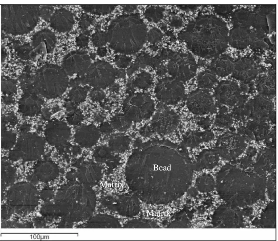
mechanism shown in Figure I.6, propagating, thus, the polymer chain. The growing polymer chains encapsulate the PMMA beads within a solid matrix. It is assumed that the smallest beads of the PMMA phase (<20µm) undergo complete dissolution in the presence of MMA
monomer increasing the viscosity of the curing mass. In contrast, the surface of the bigger
particles are partially dissolved and incorporated in the cured bone cement maintaining the spherical shape. When tow reactive radical chain ends meet, they react to form non-reactive completed polymer chains, within 10-15 mn after the start of mixing at 20°C.
Negative effects of PMMA bone cement have been attributed to its high temperature reached during polymerization,(2,4,5,7,13) monomer release to the surrounding tissues,(2,4,5,7,13)
and volumetric shrinkage.(2,4,5,7)
I.2.2 Polymerization heat
The conversion of MMA to PMMA is an exothermic reaction, releasing an energy of 52 kJ per mole of MMA.(2,4,7,13,14) This important energy that gives rise to the increase of the local temperature (peak temperature) is caused only by the monomer MMA.
I II III
I. Mixing time II. Working time III. Hardening time
0 1 2 3 4 5 6 7 8 9 10 11 12 13 14 15 16 17 18 19 20 min
Figure I.5 Formation of phenyl radical by reaction of DMPT with the BPO initiator.(14)
The temperature peak depends on the MMA-PMMA ratio,(15) the composition of the liquid and solid components,(16) the concentration of BPO and DMPT, the average size of the PMMA,(4,14,17,18) etc.
For a long time, the maximum temperature reached during the hardening phase of PMMA cements was known to cause tissue necrosis (50-60°C) and hinder the efficacy of joint prostheses.(2,4,7,14,19) In real conditions (in vivo), there are several factors that contribute to dissipate the heat produced, including the thin layer of cement (3-5 mm), the blood circulation, heat dissipation in the vital tissue connected with the cement, and heat dissipation of the system attained via the prosthesis.(4,20,21)
A temperature/time diagram (Figure I.7) shows best the progress of the polymerization temperature
Figure I.7 Diagram temperature/time of bone cement.(4)
The temperature increases only slowly during the mixing phase. It starts to rise rather quickly with increasing viscosity of the cement dough. During the hardening phase, the temperature rises explosively and soon reaches its maximum (gel effect). After this, a speedy decrease in temperature is registered due to the polymerization behavior.
The effect of each component on the kinetic control of the polymerization process of bone cement is detailed as follows:
I.2.2.1 Effect of PMMA bead size
Park and Lakes(22) considered that the properties of bone cement could be affected by the powder size, shape and distribution. Indeed, the characteristics of the curing process of acrylic cements, the peak temperature and setting time are better when using PMMA particles of relatively large diameter and wide size distribution, lower peak polymerization temperature and longer setting time.(17,18)
It has been found that there is a linear relationship between the average size of PMMA beads and the peak temperature for acrylic systems, prepared in the same experimental conditions.(4,17)
I.2.2.2 Effect of initiator to activator ratio
Classical kinetic parameters were strongly related to BPO and DMPT concentrations. Moreover, the rate of radical formation is dependent on the concentrations of the activator and initiator. Pascual et al.(17) reported with use of the more conventional liquid/powder cement mixture that, the peak temperature increases by increasing the concentration of both components. Inversely, this impact is unclear when using two liquid solutions.(23) Increasing
Setting Time Peak Temperature Time Temperature Dough Time Working Time
the concentration of both BPO and DMPT will speed up the overall polymerization process, decreasing setting time.(17,23)
I.2.2.3 Effect of radiopacifiers
In the cured cement, radiopaque particles are found in the interbead matrix filling the spaces between the relatively large pre-polymerized beads. A number of studies(17,24,25) reported that radiopacifiers such as BaSO4 or ZrO2 had no noticeable influence on the curing
properties of bone cement.
I.2.2.4 Effect of liquid to powder ratio
At high L/P ratios, an abundance of monomers react exothermically, increasing the peak
polymerization temperature. Moreover, the relative concentration of initiator (a component
of powder) is decreased, so the monomers are activated slowly, increasing setting time.(26,27)
I.2.3 Residual monomer
Generally, polymerization of MMA is never completed since the mobility of the
monomer is greatly decreased (increase of viscosity) at high conversion rates, thus residual monomer remains in the final product. The proportion of non converted residual monomer
remaining in the hardened cement is in the range of 2-6%(2,7)(Figure I.8). Within 2 to 3 weeks, this rate falls to 0.5 %(28),due to a slowly progressing, continuous polymerization. The main part of the eluted monomer quickly passes into blood stream and disappears just as quickly. A more reduced share of the residual monomer is either speedily exhaled or metabolized(29) in the Kreb s cycle (Figure I.9).
Investigations of Schlag et al.1976 show that MMA cannot be the cause of loosening or respiration and circulation reactions.(2)
Figure I.9 Metabolization of MMA.(7)
I.2.4 Volumetric Shrinkage
During the polymerization process, only a few large polymer molecules develop from the multitude of monomer ones, this determines a volumetric shrinkage (Material contract). Indeed, the monomer molecules are separated by an intermolecular distance of 4 A°, whereas after the reaction, due to the formation of covalent bonds, they will be separated by atomic bond distances of 1.9A°.(4)
Pure MMA shows a shrinkage of approximately 21-22% as shown in Figure I.10 during
polymerization,(2,7) which is intolerable for an application as cement for joint prosthesis
fixing. However, mixing monomer and pre-polymerized polymer particles reduces considerably the volumetric shrinkage, theorical contract, in this case varies from 6-7%.(7) Besides, the presence of porosity (air bubbles) reduces furtherly the amount of volumetric shrinkage making it as much as 2-4% of the initial volume (Figure I.10). Vacuum mixing (to avoid pores) leads to a slightly higher shrinkage (Davies et al.1990).(2)
Volumetric shrinkage of the cement is not disadvantageous since, its contraction at the cement bone interface will favor the building of new bone. Moreover, the shrinkage of the material will be compensated, in vivo, by its dilatation due to a slightly absorbent characteristic.
I.2.5 Viscosity
Viscosity is a physical parameter, which characterizes a material s resistance to flow. Materials with a high viscosity do not flow readily; materials with a low viscosity are more fluid.
The viscosity of the acrylic bone cement used in THA is an important material property for determination of the proper handling characteristics. In the operating theater, the cement viscosity must be low enough (not too low) to make it very easy to force the dough through the delivery system (syringe) and cause it to flow and penetrate into the interstices of the spongious bone, under any given pressure, in a very short time.(2,4,5,7)
However, cement with too low viscosity cannot withstand the bleeding pressure in the
femur.(2,7) Blood is included in the cement, resulting in gaps and laminate formation.(2,4,7)
These inclusions with a high fracture risk affect the mechanical stability of the implant.(30) 1.step Ester Hydrolysis Methacrylic acid+Methanol Methyl methacrylate 2.step Addition of water
Hydroxy butyric acid Methacrylic acid
3.step Degradation in the
Krebs cycle
Carbon dioxide + water Hydroxy butyric acid
Figure I.10 Volumetric shrinkage of PMMA based cement.(7)
High viscosity cement may introduce cement interface (cement-bone and cement-implant) gaps. This phenomenon due to a poor cement penetration into the bone tissue and/or poor cement flow properties.(4)
Dynamic viscosity, η, of bone cement decreases with an increase in shear rate, and increases with an increase in time as the polymerization reaction proceeds(4,5) (pseudo plastic behavior of bone cement).
The ideal cement would display a low and practical unchanged viscosity with time during the working period, followed by a very short time to full polymerization(31) (increase of viscosity).
Cement viscosity is increased by the addition of fibers, greater molecular weight of the
polymer, solubility of the polymer in the monomer, variation in the powder composition or
bead size distribution, and the temperature of the cement components.(4) Pre-chilling the cement components reduces the viscosity of some cement formulations.(4,7)
Acrylic cements on the market are divided according to their viscosity into low, medium, and high viscosity cements. In Table I-2 are summarized the handling characteristics of these three cement categories.
In vitro determination of viscosity is usually accomplished with the use of a capillary extrusion or rotational (plate and cone) rheometer.(5)
Volumetric shrinkage of pure MMA
100 ml Liquid MMA -79 ml Solid PMMA
100 ml dough -93-94 ml cured
cement PMMA Theoretical Volumetric
shrinkage of PMMA bone cement
-94-95 ml cured cement 100 ml dough
Volumetric shrinkage of vacuum mixed bone cement
Volumetric shrinkage of hand mixed bone cement
Table I-2 Handling characteristics of low, medium and, high viscosity cements.
Cement Type Handling properties
Low viscosity(LV) Medium viscosity
(MV) High viscosity(HV)
Mixing phase
-Long lasting liquid phase. -Low viscosity
wetting phase. -The dough Remains
sticky for 3 mn.
-Low viscosity wetting phase.
-The dough is no longer sticky after 3 mn
at the latest.
-Short wetting phase. -Quickly lose of their
stickiness.
Working phase
-Quickly increase of viscosity. The dough becomes
warm fast. Short working phase
1-2 min
-Slow and continuous invariance increase of
viscosity
-Unchanged viscosity -Slow increase toward the end of this
phase.
-Long working phase. Hardening phase Time period 1-2 mn Time period 1.5-2.5 mn Time period 1.5-2 mn
I.2.6 Porosity
The structure of bone cement is porous on both the macroscopic and microscopic scales (see Figure I.11). Thus, there are two types of pores in fully polymerized cement: macropores (pore diameter>1mm) and micro pores (pore diameter≈0.1-1mm).(32)
Figure I.11 Porosity of bone cement.(33)
powder polymer constituents,(2,4,34,35,36) incorporation of air during wetting and mixing of the components,(1,2,4,17,34,35,36) entrapment of residual monomer during
polymerization(2,4,17,34,35,36) (evaporation of the volatile monomer), entrapment of air during
the dough transfer,(4,34,35,36) and introduction of blood or tissue into the cement during implantation.(1,2)
Porosity of the cement can be influenced by the mixing method (see Sect. I.2.6.1). Relative to hand mixing, centrifugation or vacuum mixing are methods of pore reduction. The pre-cooling of the monomer, polymer and mixing vessels lead to a significant reduction of the number and the volume of pores.(2) Another source of micro-porosity in PMMA bone cement is the inclusion of radiopacifiers,(37) BaSO4 or ZrO2. These particles do not adhere to newly
polymerized PMMA matrix, but they are contained in small cavities that behaves as small pores (0.1-30µm), occupying up to about 6% of the cement volume.(38) Pressurization of the implanted doughy cement (delivery system) has also been proposed as means to reduce porosity.(4,5)
The presence of pores in the fully polymerized material may have a detrimental and a beneficial effect on the stability of the implant, and hence, on the artificial joint life. Pores would act as stress risers and crack nucleation sites for micro cracks, leading to early cement failure. However they may also act as crack decelerators.(39,40) Thereby, prolonging the life of the implant. The current consensus is that every effort should be made to substantially reduce the number and size of both macro and micro-pores in order to improve the mechanical properties of the acrylic cements.
Apparent density measurements,(35) use of a high powered optical or metallurgical microscope with incident light,(41) comparison of radiographs of the specimen with that of a standard metal plate containing holes of 1 to 10 mm in diameter, and use of a computerized image analysis system(42,43) are the usual methods for the in vitro determination of porosity.
I.2.6.1 Cement mixing methods
Mixing method has an important role in determining the mechanical properties of acrylic cements. There are four (4) cement-mixing methods: manual or hand mixing, centrifugation, vacuum mixing and, combined mechanical mixing.
In manual mixing, the powder component is added to the liquid in a polymeric bowel (see
Figure below) and manually stirred with a polypropylene spatula at 1 Hz or 2 Hz (not grater than 2 Hz in order not to beat air into the mixture)(2,44) for a period of time between 45 and 120 s.
In centrifugation mixing, the hand mixed mass is immediately poured into a syringe that is
then promptly placed in a centrifuge at 2300-4000 rpm for a period of time between 30 and 180 s.(42) Centrifugation is a method of pore reduction compared to hand mixing. The extent of such reduction depends on the storage temperature of the liquid monomer prior to mixing.(5)
For vacuum mixing, a number of proprietary (Simplex Enhancement Mixer, Stryker High Vacuum System, MITAB, Optivac, Stryker MixevacII, Sterivac, Mitvac, Cemvac Merck, Bonelock and Cemex Systems) and experimental chambers(35,36) have been used. Each chamber or proprietary has its own steps to follow. Depending on the chamber (or proprietary) used, vacuum is applied and the cement components are mixed at reduced pressure to obtain the dough. Compared to hand mixing, considerable reduction in porosity (micro-porosity) was observed for vacuum mixing.
A number of combined mixing devices have been used: hand mixing in stainless bowel on vibrating plate (50 vibrations/s)(46), a motor coupled to eccentric unit that creates the action of vibration (motion in two directions), in which a cement holder is fixed, the motor is typically run at 500 rpm for 120 s during the mixing of the constituents,(47) and a proprietary machine called the Universal Mixing Machine that simultaneously mixes and centrifuges the cement constituents for typically 12 s.(48)
Other emergent techniques for mixing exist also, based on a combination between ultrasonic cleaner and vibration after hand mixing,(49) pressurization under vacuum mixing.(50)
One new combined mixing method proposed by Lewis consists of the application of a passive vacuum to the cement constituents followed by their simultaneous mechanical mixing and centrifugation. This technique leads to cement with excellent physical and mechanical properties.
The usual procedure after mixing is to inject immediately the cement dough into a cartridge or tube. The dough is usually pressurized with a cement gun into the bone cavity.(4,5)
Cemex System is instead the unique device being a mixing and delivering system at a time: powder and liquid are pre-dosed and contained in a syringe like container(4) (see Figure below).
I.2.7 Molecular weight
When powder polymer and liquid monomer are mixed, macromolecules that have a variable number of linked monomers will be created. Therefore, different molecular weights co exist in the solidified product.(2,51) It is assumed that monomer dissolves the smaller particles of the powder (<20µm) forming thus, the interbead matrix and partially dissolves the surface of the bigger particles (pre-polymerized beads) which are thus completely incorporated in the cured cement.(52)
Gel permeation chromatography (GPC) is used to determine the molecular weight (Mw) of the two polymer phases (polymer powder and cured cement).(26,33,52)
The typical distribution of the molecular weights of the various macromolecules that form a polymeric product is similar to the shape of a ball, whose width is tied to the distribution of the molecular weight (see Figure I.14)
The values of the average molecular weights can be defined as follows:
1-The average numerical molecular weight (Mn), expresses the average according to the number of the macromolecules present. This parameter is of a highly interest in the study of reaction mechanisms.
∑
∑
= i i i n N M N M (I-1) Where,Mi is the mass of the nTh macromolecule present and Ni is the nTh macromolecule present.
2-The average ponderal molecular weight (Mw) expresses the average according to the mass of the macromolecules present, which is connected to the mechanical characteristics and processability of the material.
∑
∑
= i i 2 i i w M N M N M (I-2)Figure I.14 The typical distribution of the molecular weights of bone cements.(39)
Bone cement like any implantable biomaterial must be sterilized before its introduction into the body. It s well known that ethylene oxide gas (EtO) or gamma irradiation is used to
sterilize the powder constituents, whereas the liquid monomer typically is sterilized by membrane filtration.(33)
The structure of polymers seems to be affected by sterilization techniques.(53) When irradiated, PMMA undergoes chain scission leading to a reduction in its molecular weight.(33,53,54,55) Large decreases in molecular weight of the PMMA powder were observed with increasing gamma irradiation dose.(33) However, fumigation with ethylene oxide has no influence on the molecular weight of the cement.(33,54,56) Additionally, the molecular weight of the PMMA can further decease over time during in vivo use.(57)
The molecular weight influences the swelling properties, the mechanical properties and the working phases of the different cements.
I.2.8 Glass transition temperature
On heating, PMMA bone cements change their state from glass-like and brittle to elastic at glass transition temperature (Tg), the material starts to soften and the Brownian motions cease.(2) These micro-movements influence the thermal expansion coefficient, the bending modulus and mechanical and electric absorption of the material (Material s parameters).(2)
Recently, Tg is used as characteristic of bone cements (Thanner et al 1995).(2) Several methods can be used to determine the glass transition temperature: torsional fatigue testing, shear modulus determination and the dilatometric method DSC (see Figure I.15)
Figure I.15 Determination of the glass transition temperature using the dilatometric method.(2)
Glass transition temperature depends on the molecular weight, water content and the molecular structure of the monomer used (hydrophilic/hydrophobic behaviour).
Experiments reveal(2) that there is no change in temperature when bone cements (samples) are stored in dry environment, a continuous decrease in temperature can be registered when stored in water at 37°C. However, there are no further changes, when the samples are water-saturated.
Bone cements are always in a humid environment at 37°C after implantation and become saturated with water after only a few weeks, thus, cements with Tg s of 40-50°C (dry specimens) could lead to disastrous clinical results.(2)
According to Kühn,(2) Cemex cement and simples P present a relatively high Tgs in dry environment and remain above body temperature (~70°C) after 8 weeks of storage in water at 37°C.
Another transition temperature reported by various authors is beta transition. This last is related to the rotation of the bonds in the cement molecule.(58,59)
Summary
This chapter allows us to retain the following points:
• Polymethylmethacrylate (PMMA) widely known since 1930 was used for the first time, in 1958 as a fixation agent of a femoral implant, by Sir John Charnley, who named it bone cement on acrylic basis.
• In reality, acrylic bone cements were used in various medical fields such as dentistry, cranioplasties, plastic surgery, orthopaedic surgery, etc.
• Acrylic bone cement, so-called two-components system is commonly prepared by mixing a solid part made of a pre-polymerized poly methyl methacrylate (PMMA), benzoyl peroxide (BPO) as an initiator, and radio-pacifiers, with a liquid part containing methyl methacrylate (MMA), N, N-dimethyl-p-toluidine as an accelerator and inhibitor. Each cement manufacturer recommends a specific monomer to polymer ratio, expressed as grams per milliliter.
• Bone cement is formed from an exothermic reaction of benzoyl peroxide initiator present in Polymethylmethacrylate (PMMA) powder and N,N-dimethyl-p-toluidine in methylmethacrylate monomer liquid transforming after few minutes (10 to 15 mn), the MMA multi-molecular liquid system into a solid PMMA macro-molecular one.
• Negative effects of PMMA bone cement have been attributed to its high temperature reached during polymerization, monomer release to the surrounding tissues, and volumetric shrinkage.
• For a long time, the maximum temperature reached during the hardening phase of PMMA cements was known to cause tissue necrosis and hinder the efficacy of joint prostheses. In real conditions (In vivo), there are several factors that contribute to dissipate the heat produced, including the thin layer of cement (3-5 mm), the blood circulation, heat dissipation in the vital tissue connected with the cement, and heat dissipation of the system attained via the prosthesis.
• The proportion of non converted residual monomer remaining in the hardened cement is in the range of 2-6%. Within 2 to 3 weeks, this rate falls to 0.5 %, due to a slowly progressing, continuous polymerization. The main part of the eluted monomer quickly passes into blood stream and disappears just as quickly.
• Volumetric shrinkage (Material contract of approximately 2-4 %) of the cement is not disadvantageous since, its contraction at the cement bone interface will favor the building of new bone. Moreover, the shrinkage of the material will be compensated, in vivo, by its dilatation due to a slightly absorbent characteristic.
• Viscosity is a physical parameter, which characterizes a material s resistance to flow. Acrylic cements on the market are divided according to their viscosity into low, medium, and high viscosity cements. In the operating theater, the cement viscosity must be low enough (not too low) to make it very easy to force the dough through the delivery system (syringe) and cause it to flow and penetrate into the interstices of the spongious bone, under any given pressure, in a very short time. Therefore, cement viscosity is an important material property in determining the proper handling characteristics.
• The structure of bone cement is porous on both the macroscopic and microscopic scales. In general, pores are referred to as micropores or as macropores, depending on whether their maximum dimension is smaller or larger than 1 mm. Pores in the cement arise from different causes. These include the evaporation of the liquid monomer during curing, entrapment of air during mixing and mixture application, inclusion of radiopacifiers, interoperative bleeding, bone preparation, and cement implantation technique. There are four
(4) cement-mixing methods: manual or hand mixing, centrifugation, vacuum mixing and, combined mechanical mixing. Relative to hand mixing, centrifugation, or vacuum are methods of pore reduction.
• As with any implantable biomaterial, acrylic cement must be sterilized before its introduction into the body. Sterilization methods have been shown to affect molecular weight. Large decreases in molecular weight of the PMMA powder were observed with increasing gamma irradiation dose. However, fumigation with ethylene oxide has no influence on the molecular weight of the cement. Additionally, the molecular weight of the PMMA can further decease over time during in vivo use.
• On heating, PMMA bone cements change their state from glass-like and brittle to elastic at glass transition temperature (Tg). Glass transition temperature depends on the molecular weight, water content and the molecular structure of the monomer used (hydrophilic/hydrophobic behaviour).
MECHANICAL
PROPERTIES OF
ACRYLIC BONE
CEMENTS
It is believed that mechanical failure of the bone cement layer is the cause of aseptic loosening. For this reason it is necessary to know the mechanical properties of bone cements.
II.1 Introduction
Bone cements, as aforementioned by many authors, are used for the fixation of artificial joints. When used in total hip Arthroplasty, PMMA fill the free space between the metallic component and the predetermined bone site. This technique allows a stable fixation of the prosthesis to the bone.(2,60,61) The interaction between the cement and the adjacent bone tissue and between the cement and the prosthesis is only mechanical.(2,62,63) The main task of PMMA is to transfer complex, varying physiological loads from the prosthesis to the bone.(1,2,5,33,64)
The in vivo integrity and performance of bone cement are necessary for longevity of the prosthetic system.(2,60) Indeed, if the external stress factors are superior to the ability of the cement to transfer the force, a break will result. PMMA fragments thus formed could trigger phenomena of peri-prosthetic osteolysis and lead to the mobilisation and even failure of the prosthesis.(60,65) However, the state of stress in the cement mantle is influenced by many factors, tied on the thickness of the cement layer, structure of the cement (distribution and dimension of the pores),(1) the shape of the metallic implant and materials that constitute it, and the bond conditions of the prosthesis.
It is believed that mechanical failure of the bone cement layer at any or all of the weak link zones (the implant-cement interface, the cement mantle, and the cement-bone interface) is the cause of aseptic loosening.(66,67) For this reason it is necessary to test the mechanical properties of bone cements under standard conditions. Numerous studies have focused on improving the static and dynamic properties of bone cement in order to maximise its resistance to applied stress.
The American Society for Testing and Materials (ASTM-F451) and the International Standards Organization (ISO 5833) have fixed minimum values for the critical properties (Table II-1), these standards are limited as they cover some mechanical properties in compression and bending.(2,3)
Table II-1 ISO Critical values for mechanical properties of bone cements.(2,65)
Compression strength (MPa) Bending strength (MPa) Elastic or Bending modulus (MPa)
>70 >50 >1800
Compressive strength was the first mechanical criterion implemented by American society
for testing and Materials standards (ASTM) in the USA in 1978.(2) All surgical grades PMMA must fulfil these minimum requirements.
Several data on the mechanical properties of bone cements can be found in literature. However, there is no consensus on any unique value for any of the mechanical properties due to the different kind of cements and many trial techniques and conditions that have been used.(1)
Mechanical property determinations can be performed at different times after polymerization, or specimens can be stored for various time periods (some as long as two years)(1) in air, de-ionized water, distilled water or physiological solutions (Isotonic saline, or serum) at room temperature or at elevated physiological body temperature.(2,5) However, complex storage conditions and long storage times are prohibitive for most studies.(1) ASTM and ISO standards recommend storage in air or distilled water at 37°C for periods of 1 to 7 days for most mechanical property testing of Acrylic bone cement.(2)
mechanical properties.(1,59) Temperature and loading rate at the time of testing, also affect the measured mechanical properties.(1)
PMMA bone cement fails during in vivo use at stress levels far below the critical values seen in their static tests (Table II-1). In reality, the cement mantle is subjected to cyclical loads (repeated loads) and the fatigue phenomena appear to be the cause of mechanical failure of the PMMA mantle.(1,5,60,62,68,69) Failure of the cement most commonly occurs by the growth of fatigue cracks until a critical size when unstable propagation of the crack occurs and cement fractures.
Mechanical fatigue testing can be conducted as tensile, compressive or bending tests.(2) Unfortunately, mechanical fatigue testing on bone cement is not currently regulated by any international standard.(65)
II.2 Quasi-static proprieties
II.2.1 Tensile properties
In clinical service the hip implant is subjected to static or quasi-static tensile forces during daily activities.(5,65) In vitro determination of the quasi-static tensile properties involves the use of a dog-bone specimen, as exhibited in Figure II.1, at a cross-head displacement speed ranging from 5-30.5 mm/min.(49,70) The same experiments were also carried out with a cross head speed of 1 mm/min according to ISO 527 standard.(10)
Figure II.1 A dog bone specimen used in tensile tests.
The values of the ultimate tensile (UTS), tensile modulus of elasticity (Et) and tensile
strain at the fracture point (emaxt) are determined from the applied load versus elongation
curve that is fully linear, since cement is brittle or quasi-brittle material.(5) Davis et al.(72) in their study, found no significant difference in tensile strengths of three types of bone cements; Simplex P, LVC and Zimmer Regular bone cement.
A number of studies(10,14,15,44,71) have shown that the addition of Barium Sulphate (BaSO4)
decreases significantly tensile strength, which, however, is not observed when Zirconium dioxide (ZrO2) is added. These inorganic fillers interrupt the polymerizing matrix and produce
pores that act as fracture initiation sites, in the case of BaSO4, diminishing cement tensile
strength. The cauliflower like morphology of ZrO2 particles allowed for mechanical
anchorage, which could avoid the decrease in the tensile strength.(10) Pascual et al.(27) considered that the quasi-static tensile properties couldn t be affected by the powder size distribution, however, the tensile strength and the tensile strain of the test specimens were higher for formulations with lower DMPT content.
All commercially available cement requires mixing powder and liquid. The mixing methods have a large impact on the tensile properties, in fact, vacuum or centrifugation
mixing lead to a mean of 44% increase in UTS relative to hand mixing. Typical values of maximum tensile stress are between 24 MPa and 49 MPa.(5)
II.2.2 Compressive properties
The hip implant is subjected to static or quasi-static direct compressive forces, during certain daily activities, for example, in the one legged stance.(5,73) Additionally, in hip
Arthroplasties, the cement mantle may act as a compressive wedge between the femoral
stem and the bone tube.(74,75) Therefore, compression tests should be justified. In vitro determination of compressive properties has been carried out according to the ASTM-F451 or ISO 5833 standards for acrylic bone cements.(2,5,10,65,71) Compression trials, are performed on cylindrical samples of 6 mm in diameter and 12 mm in height (see Figure below) at a cross-head speed of 20(2,5,10,65) or 25.4(5,73) mm/min.
Figure II.2 Compressive test specimen.
The ultimate compressive strength (UCS) and compressive modulus (Ec) should be
estimated from the stress-deformation curve shown in Figure II.3. The slope of the first part of the curve characterises the stiffness of the material (E-modulus) and the maximum of the curve (yield point) defines the stress and strain beyond which the material will deform plastically. The ultimate compressive strain (emaxc) is obtained as the strain at the UCS
point. The 2% offset method is used to obtain the ultimate compressive strength and the ultimate compressive strain (emaxc).
Figure II.3 Compressive strain
For a given mixing method, the formulation exerts a profound effect on the quasi-static
compressive properties (see Figure II.4).
Figure II.4 Compressive strength of plain bone cements, according to ISO 5833
standards.(2)
The addition of inorganic fillers to PMMA bone cement has been shown to reduce quasi-static compressive strength,(15,77) because they could act as flaw sources.(44) Compressive
strength and elastic moduli seem to be poorly affected by the size of PMMA particles.(17) Lautenschlager et al.(78) showed that Simplex P loaded with Gentamicyn led the compressive strength to drop below 70 MPa. The same result was observed for Simplex P loaded with a mixture of Meropenem and Vancomicyn.(65) Some authors (65,79,80) reported that compressive strength of PMMA bone cements decreased with increasing dosages of antibiotic.
The compressive strength of PMMA bone cement has shown a slight increase in the first three months after mixing and fairly constant values after that.(44,81) The test parameters are of decisive importance, in fact, increases in strain rates result in quasi-static property increases(59) (modulus of elasticity and ultimate strength) due to the viscoelastic nature of PMMA.
Concerning mixing methods, Trieu et al.(48) reported that for Simplex P, there was no statistically significant difference between (UCS) and (Ec) of hand mixing (HM) and
simultaneous mechanical mixing and centrifugation (MEC) specimens. These findings are in contrast, with the results obtained by Lewis(73) for CMW-3 Cement. In addition, Trieu et al.
(48)
found in their study that (UCS) and (Ec) were significantly lower for (MEC) specimens
when compared to vacuum mixing, however, these differences were not significant in Lewis investigation.(73)
II.2.3 Flexural properties
of the artificial joint, a combination of shear, tension and compression stresses are found and therefore the study of bending tests are justified.(5)
In vitro determination of flexural properties of bone cements (flexural strength and modulus) has been performed in accordance with the ASTM-790(82) or ISO 5833 standards. The flat samples used in the ISO bending strength tests (see Figure below) have to be immersed for 50±2h in water or Ringer solution at 37±1°C(2,65) to simulate physiological conditions. According to ISO 5833, bending trials are performed at a cross head speed of 5 mm/min.
Figure II.5 Bending trials on a flat sample using a four-point test bench.(65)
The flexural strength value is obtained from the follow relation:
[
]
2 3 bh Fa MPa Fs = (II-1) WhereF [N] is the applied force;
b [mm] is the average width measured for the sample; h [mm] is the average thickness measured for the sample;
a [mm] is the distance between the two force application points on the sample on the
testing device.
The modulus of elasticity known as the flexural modulus (Eb) is a measure of the stiffness
of a material. The higher the modulus, the less the material is deformed.(2) The bending elastic modulus is also calculated, using the follow formulation:
[
]
3 4 fbh a F MPa Eb = ∆ ⋅ (II-2) Wheref is the difference between the arrow values corresponding to the loads 15 N and 50 N;
N
F =35
∆ is the interval of variation of the load applied between the two points of evaluation of the arrow f.
variables: cement formulation, mixing method, and curing and aging conditions.(1,2,5) The values of bending strength and elastic modulus of the most common commercial bone cements (according to ISO standards) are given in Table II-2.
Table II-2 ISO test values of flexural properties of the most commercial bone cements.(65)
Cement Types Bending Strength (MPa) Elastic Modulus(MPa) Palacos R CMW-3 Cemex System Cemex Rx Cemex Isoplastic Simplex P 66±2 59±4 58±4 61±4 59±9 77±3 2912±315 2698±239 3223±360 2956±196 3029±179 2679±39
It has been shown(15,83) that the addition of BaSO4 to PMMA bone cement causes a
reduction in its flexural strength. Numerous studies on the effect of adding antibiotics to bone cements assert that loaded cements show a reduction in their flexural properties.(65,78,84,85) Askew et al.(85) demonstrated in their investigation that mixing the cement under vacuum up to 700 mmHg increased the flexural properties of the cement by reducing its porosity. However, these properties show a positive but a weak strain rate dependency.(86)
Short Storage times, less than 60 days, in water in the same manner, probably do not affect the mechanical properties of acrylic bone cement since the absorption of water by PMMA cement is very slow as it is hydrophobic.(2,87) According to Kühn,(2) the bending strength was found to be not affected by the environment storage (dry for 16h or wet for 50h), whereas, the flexural modulus depend on.
Finally, various studies in literature,(5) have been performed on the flexural properties of acrylic cement using different specimen sizes ranging from 5×6×50 mm to 10×10×110 mm, support spans (25.4 or 50.0 mm), loading rates ranging from 0.033 to 7.6 mm/s, and loading types (3-point or 4-point).
II.2.2.4 Shear properties
During certain normal daily activities, the three weak link zones are subjected to shear forces.(5,88,89,90) Thus shear properties of the cement are relevant to the life of the implant. Lewis in his art review(5) cited only two reports(47,91) of the in vitro determination of the ultimate shear strength (USS) of bone cements, while, numerous papers of shear properties of bone cement interface and cement stem interface exist in literature.(88,89,90,92)
In one study(91) the acrylic specimens were rectangular in cross section, measuring 55×51×6,5 mm; in the other study(47) they were circular measuring 9 mm in diameter and 24 mm in length. Two different loading rates were used in these studies (47,91) 1.3 and 0.18 mm/min.
The load versus displacement response for the bone cement experienced a linear response followed by yielding and an exponential type decay to complete failure.
The values of USS are affected by cement formulation, mixing method, and curing and aging conditions as shown in table II-3. The addition of BaSO4to PMMA bone cement has
been shown to reduce its quasi static shear strength.(83,93) This latter was found to decrease in loaded bone cements.(78)
Shear strength of PMMA bone cement shows a positive but weak strain rate de
Table II-3 USS data for most commercial bone cements cited by Lewis.(5)
Shear Strength
(Mpa) From Formulation Mixing Details
Curing, Aging, And Test Environment Conditions Double Shear Test D732 Reference
Manual; per manufacturer s instructions Cure time of 24-48h 42.7 - Linden Mechanical mixer(vibration/shaker);500 rpm for 120s Cure time of 24-48h 48.4 Linden Manual; 1 storke/s Aged in 37°C water for at least 24h; Tested in37°C water - 37.0 Kindt-Larsen et al. Simplex P CC stored at 5°C prior to vacuum mixing; 20 kPa(absolute) Aged in 37°C water for at least 24h; Tested in37°C water - 69.0 Kindt-Larsen et al. Manual; per manufacturer s
instructions Cure time of 24-48h 48.7 - Linden Mechanical mixer(vibration/shaker);500 rpm for 120s Cure time of 24-48h 50.2 - Linden CC stored at 5°C prior to manual mixing; 1storke/s
Aged in 37°C water for at least 24h; Tested in37°C water - 33.0 Kindt-Larsen et al. Zimmer LVC CC stored at 5°C prior to vacuum mixing; 20 kPa(absolute) Aged in 37°C water for at least 24h; Tested in37°C water - 62.0 Kindt-Larsen et al. CC stored at 5°C prior to manual mixing; 1storke/s
Aged in 37°C water for at least 24h; Tested in37°C water 33.0 Kindt-Larsen et al. Palacos R CC stored at 5°C prior to vacuum mixing; 20 kPa(absolute) Aged in 37°C water for at least 24h; Tested in37°C water - 50.0 Kindt-Larsen et al. CC stored at 5°C prior to manual mixing; 1storke/s
Aged in 37°C water for at least 24h; Tested in37°C water - 32.0 Kindt-Larsen et al. CMWTM-1 CC stored at 5°C prior to vacuum mixing; 20 kPa(absolute) Aged in 37°C water for at least 24h; Tested in37°C water - 63.0 Kindt-Larsen et al. CC: Cement components
II.3 Dynamic proprieties
II.3.1 Fracture Toughness
During normal daily activities, a cemented femoral implant is stressed cyclically by physiological loads of up to five times body weight.(1,5,94,95) Additionally, bone cement used in
hip implants contains discontinuities such as voids, flaws or other stress concentrations,
resulting from porosity and irregular PMMA bone interface.(37,96) This combination of features is well known to make the cement vulnerable to fracture, thus threatening the life of the prosthesis. Indeed, numerous studies have reported that failure of the femoral arthroplastic components is mediated by extensive fractures in the cement mantle.(44,97,98) Initiation of such cracking is generally thought to begin with debonding of the bone cement from the metal prosthesis(66) and /or from voids associated with porosity either in the cement mantle(99) or at the biomaterial interfaces.(100) Under applied physiological loads, catastrophic failure of PMMA bone cement will inevitably result in failure of the entire cemented implant which is complicated by a complex biological response of the bone (PMMA debris realised from micro-cracking can trigger a localized immune response that leads to bone resorption(101)) leading to aseptic loosening.(63,64,66,101,103)
Accordingly, the fracture property of the fully polymerized cement is very important. Fracture mechanics approach has been widely adopted as the appropriate way of characterizing mechanical behaviour of the cement mantle.(59) The fracture mechanics analysis(1) takes into consideration three variables, namely applied stress, flaw size and a material property related to the resistance of the material to the propagation of the flaw. There are two approaches to the identification of this material property: the energy approach and the
stress intensity approach.
According to the energy approach, the crack extension occurs when the energy available for crack growth is adequate to overcome the resistance of the material to the creation of new surface. For an infinite plate (see Figure II.6) made of a linear elastic material with a through the thickness crack of length 2a and subjected to a tensile stressσ, the energy release rate, G, which is the rate of change of potential energy with increasing crack area is defined as follows: E a G 2× × = (II-3) Where
σ is the applied stress; a is half the crack length; E is young s modulus.
As the applied stress or the crack length increase, G, increases, until it reaches a critical value, Gc, when unstable crack occurs.
The stress intensity approach is based on analysis of the stress concentration that occurs at the tip of a crack. The analysis depends on the mode of loading and the existence of plane
strain conditions. For a linear elastic material presenting a sufficient thickness B for plane strain conditions, the stress concentration at the tip of the crack in Figure II.6 can be
described by a Mode I (tensile load applied perpendicular to the plane of the crack and its direction of propagation), plane strain, stress intensity factor given by the follow formulation:
a
KI =σ π× (II-4)
Where:
σ is the applied stress; a is half the crack length;
I
K is the Mode I stress intensity factor.
Figure II.6 Infinite plate with a through-the-thickness crack.(1)
Similarly, as the applied stress or the crack length increase, KI, increases, and when it reaches
a critical value, KIC, unstable (catastrophic) crack propagation occurs. This critical value of
the Mode I, plane strain, stress intensity factor is called the fracture toughness, a measure of material s ability to resist catastrophic crack propagation. The material s resistance to the combined effects of stress and flaws (known as fracture toughness) depends only on the geometry of the crack,(1,63,95) not the geometry of the object hosting the crack.
The assumptions(1) in linear elastic fracture mechanics (LEFM) are that the material be linearly elastic and the material fractures in a brittle manner, that is, without plastic deformation prior to crack propagation. Really, real materials, such as PMMA bone cement, rarely behave in such a restricted manner. In order to resolve this problem certain criteria have been established to allow the description of the behaviour of real materials in terms of linear elastic fracture mechanics. The zone of plastic deformation at the crack tip (plastic zone, damage or craze zone) must be very small compared to the thickness and the crack length of the material structure. The applicability of linear elastic fracture mechanics (LEFM) to the study of crack propagation and fracture of PMMA bone cement has been clearly established.(59,63,64,95,103)
Under clinical conditions, the cement is loaded in Mode I(5,96) and linear elastic conditions exist, so, the fracture toughness was obtained in vitro as the critical value of the Mode I stress intensity factor (KIC) for various cements.
Despite the absence of a standard specific to acrylic resin, two procedures based on the recommendation of two American standards have been used to determine the KIC value of
bone cement.(10,58,59,63,64,103,104,105) The two existent specifications are: the ASTM-E399-90 2a
B
σ σ