Science Arts & Métiers (SAM)
is an open access repository that collects the work of Arts et Métiers Institute of
Technology researchers and makes it freely available over the web where possible.
This is an author-deposited version published in: https://sam.ensam.eu
Handle ID: .http://hdl.handle.net/10985/15735
To cite this version :
Nolwenn FOUGERON, Hélène PILLET, Wafa SKALLI, Pierre-Yves ROHAN - Subject specific hexahedral Finite Element mesh generation of the pelvis from bi-Planar X-ray images - In: Société de biomécanique, France, 2017-10-27 - Computer Methods in Biomechanics and Biomedical Engineering - 2017
Any correspondence concerning this service should be sent to the repository
Subject Specific hexahedral Finite Element Mesh
Generation of the Pelvis from Bi-Planar X-ray Images
N.Fougeron
a*, A. Macron
a,b, H. Pillet
a, W. Skalli
aand P-Y. Rohan
aa Institut de Biomécanique Humaine Georges Charpark, Arts et Metiers ParisTech, 151 bd de l'Hôpital, 75013. Paris, France
bCEA, LIST, Interactiv Robotics Laboratory, F-91191 Gif-sur-Yvette, France
Keywords: FEM ; Hexahedral mesh ; Subject specific ; Pelvis; Bi-planar X-rays
1. Introduction
Finite Element Modelling is becoming an ever more important tool in the field of biomedical engineering for many applications including the investigation of the mechanisms responsible for the onset of pathologies, for surgical planing and for the evalution of the impact of medical devices on patient outcome.
Finite Element models are generally built from patient medical image data, such as computed tomography (CT) or magnetic resonance imaging (MRI) (Linder-Ganz et al. 2008). However, image segmentation and finite element mesh design are long and tedious processes that represent major bottlenecks for the fast generation of patient-specific Finite Element Meshes. Many attempts have been made in the literature to circumvent this problem and today several fast and robust methods have been developed for automatically generating tetrahedral meshes of arbitrary geometries. However, for a wide range of applications, hexahedral-based meshes are preferred: First, to achieve the same solution, accuracy for a given analysis requires far more tetrahedral elements than hexahedral elements, and this leads to higher computational costs (both time and memory). Second, it is well known that for incompressible and/or nearly incompressible materials, 4-noded tetrahedra with linear shape functions tend to lock and become overly stiff, generally producing acceptable displacement results but relatively inaccurate results for stresses.
Recent developments in 3D reconstruction techniques from calibrated bi-planar X-ray imaging provide a promising alternative tool for patient-specific 3D modelling of the spine, lower limb, pelvis (Mitton et al. 2006) and external envelope.
The aim of this study is to develop a methodology for the automatic hexahedral subject specific Finite Element mesh design of the pelvis based on 3D reconstruction from bi planar X-rays. The model obtained should preserve acceptable mesh quality to allow for mechanical simulations.
2. Methods
2.1 Generic Mesh design
A generic 3D-reconstruction model of the pelvis (anatomical atlas) was used to design the generic Finite Element mesh. Half of the surface mesh of the generic 3D reconstruction (sacrum and one ilium wing) was patched using NURBS surfaces in Geomagic Studio
version 12 software (Geomagic U.S. Corp., Research
Triangle Park, NC, USA). The NURBS surfaces were then imported into ANSYS® Mechanical APDL,
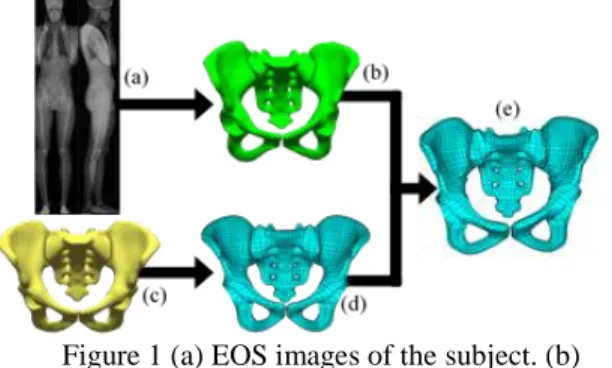
Release 15.0 (ANSYS Inc., Canonsburg, Pennsylvania, USA) and used to define cube subvolumes of the pelvic geometry. Each subvolume was meshed with linear hexahedral elements (8-node SOLID45 elements) in ANSYS. Special attention was paid to the discretisation of the NURBS so as to have a conforming mesh. The complete volume was defined as the union of the sub-volumes meshed with hexahedral elements. The ilium wing was then duplicated by symmetry and the full pelvic mesh was defined as the union of all the meshes (figure 1(c)). Mesh regularization was performed using an optimization process based on four criteria: jacobian condition number, skewness, warping factor and parallel deviation. The objective function was calculated as a weighted sum of the element metrics. This optimisation procedure was performed using conjugate gradient method and the corresponding displacement field was applied to the internal nodes at each step.
In addition, specific anatomical landmarks were identified on the surface mesh (Besnault, 1999).
2.2 Images acquisition and 3D reconstruction
Low-dose biplanar X-ray images of a 25 years old female (156 cm tall, and weighing 54.9 kg) was extracted from the database of the Institut de Biomécanique Humaine Georges Charpak. 3D reconstruction of the pelvis was performed (Figure 1(a) and Figure 1(b)) from the deformation of a generic model embedding the same anatomical landmarks as those defined on the Finite Element mesh (Mitton et al. 2006).
2.3 Personalisation of the mesh using kriging interpolation
The generic pelvic Finite Element mesh was then deformed using the kriging interpolation method (Trochu et al. 1993) to conform to the morpho-realistic
3D-reconstruction from bi-planar X-ray radiographs of the volunteer. Kriging interpolation used the above mentioned control points to obtain subject specific mesh (Figure 1 (e)). The entire process was implemented in a custom-made routine on MATLAB® Release 2014b (The MathWorks, Inc., Natick, Massachusetts, United States).
Figure 1 (a) EOS images of the subject. (b) Reconstruction of the subject geometry from X-rays.
(c) Generic model geometry. (d) Generic mesh after regularization. (e) Subject specific mesh.
2.4 Mesh quality evaluation
ANSYS® shape checking was performed over the following criteria: aspect ratio (AR), parallel deviation (PD), maximum angle (MA), jacobian ratio (JR) and warping factor (WR)
3. Results
The generic Finite Element mesh is composed of 3014 hexahedrons (Figure 1(d)). The results of the shape checking are provided in Table 1 for the generic model before regularization (GM1), for the generic model after regularization (GM2) and for the subject specific model (SSM). Our results show that the regularization process improved the quality of the generic mesh repairing all errors (GM2 vs GM1). In the end, only less than 1% of the elements remain within the warning thresholds of parallel deviation. .
Model AR PD MA JR WF Tot % GM1 0 91 265 20(2)* 249 17.23 GM2 0 19 0 0 0 0.6 SSM 0 28 1 0 45 2.4
Table 1: Type and number of warning elements per model (*elements over the error threshold)
Our results also show that the personalisation process increased the proportion of warnings but no errors occurred (SSM vs GM2). The subject specific mesh was obtained in 0.43 s. Average point-to-surface error
between the 3D reconstruction and the personalised FE mesh was 1.2 mm (max 11.6 mm).
4. Discussion
In this contribution, a new methodology has been proposed for the generation of personalised hexahedral Finite Element mesh of the pelvis from bi planar X-rays. Bi-planar X-ray imaging and the associated 3D reconstruction and clinical parameters computation provide a promising alternative to CT-scan and MR images and, as far as the authors are aware of, this is the first attempt to generate a personalised Finite Element mesh of the pelvis from this imaging modality.
Our results show that a high quality mesh (zero error) can be obtained in a fast way (less than half a second). This is promising for clinical applications. Results also show that the kriging interpolation technique seems to be well-suited for the purpose of computing a mesh deformation driven by anatomical landmarks. This proposed application.
Future works will include the definition of two generic Finite Element meshes: one for males and one for females. The robustness of the method will also be evaluated by testing the proposed method on a larger database.
Acknowledgements
The authors would like to thank the Fondation de l’Avenir for their financial support.
References
Besnault B. 1999. Modélisation par éléments finis du bassin humain en configuration de chocs automobiles. Biomécanique École nationale supérieure d’arts et métiers –ENSAM.
Mitton D, Deschênes S, Laporte S, Godbout B, Bertrand S, de Guise JA, Skalli W. 2006. 3D reconstruction of the pelvis from bi-planar raiography. Computer Methods in Biomechanics and Biomedical Engineering.9(1):1–5.
Trochu F. 1993. A Contouring Program Based on Dual Kriging Interpolation. Engineering with Computers. 9:160–17
Linder-Ganz E, Shabshin N, Itzchak Y, Yizhar Ziva, Siev-Ner I, Gefen A. 2008. Strains and stresses in sub-dermal tissues of the buttocks are greater in paraplegics than in healthy during sitting. Journal of Biomechanics. 41(2008):567-580