https://doi.org/10.1007/s40520-020-01515-1
REVIEW
Alternative and complementary therapies in osteoarthritis
and cartilage repair
N. R. Fuggle1 · C. Cooper1,2 · R. O. C. Oreffo3 · A. J. Price4 · J. F. Kaux5 · E. Maheu6 · M. Cutolo7 · G. Honvo8 · P. G. Conaghan9 · F. Berenbaum10 · J. Branco11,12 · M. L. Brandi13 · B. Cortet14 · N. Veronese15 · A. A. Kurth16 · R. Matijevic17 · R. Roth18 · J. P. Pelletier19 · J. Martel‑Pelletier19 · M. Vlaskovska20 · T. Thomas21,22 · W. F. Lems23 · N. Al‑Daghri24 · O. Bruyère8 · R. Rizzoli25 · J. A. Kanis26,27 · J. Y. Reginster8,24,28
Received: 27 December 2019 / Accepted: 14 February 2020 / Published online: 13 March 2020 © The Author(s) 2020
Abstract
Osteoarthritis (OA) is the most common joint condition and, with a burgeoning ageing population, is due to increase in prevalence. Beyond conventional medical and surgical interventions, there are an increasing number of ‘alternative’ therapies. These alternative therapies may have a limited evidence base and, for this reason, are often only afforded brief reference (or completely excluded) from current OA guidelines. Thus, the aim of this review was to synthesize the current evidence regarding autologous chondrocyte implantation (ACI), mesenchymal stem cell (MSC) therapy, platelet-rich plasma (PRP), vitamin D and other alternative therapies. The majority of studies were in knee OA or chondral defects. Matrix-assisted ACI has demonstrated exceedingly limited, symptomatic improvements in the treatment of cartilage defects of the knee and is not supported for the treatment of knee OA. There is some evidence to suggest symptomatic improvement with MSC injection in knee OA, with the suggestion of minimal structural improvement demonstrated on MRI and there are positive signals that PRP may also lead to symptomatic improvement, though variation in preparation makes inter-study comparison difficult. There is variability in findings with vitamin D supplementation in OA, and the only recommendation which can be made, at this time, is for replacement when vitamin D is deplete. Other alternative therapies reviewed have some evidence (though from small, poor-quality studies) to support improvement in symptoms and again there is often a wide variation in dosage and regimens. For all these therapeutic modalities, although controlled studies have been undertaken to evaluate effectiveness in OA, these have often been of small size, limited statistical power, uncertain blindness and using various methodologies. These deficiencies must leave the question as to whether they have been validated as effective therapies in OA (or chondral defects). The conclusions of this review are that all alternative interventions definitely require clinical trials with robust methodology, to assess their efficacy and safety in the treatment of OA beyond contextual and placebo effects. Keywords Osteoarthritis · Cartilage · Alternative · Therapy · Treatment · Herbal
Introduction
Osteoarthritis (OA) is the most common form of arthritis and the global prevalence of knee OA alone is 3.8%, affect-ing over 250 million individuals worldwide [1]. OA is an increasingly major socioeconomic and public health issue,
with the years lived with disability increasing by 64% from 1990 to 2010.
The current dogma is that OA may have differing causes but with a common, multi-tissue morphology including car-tilage fibrillation, fissure and loss, subchondral bone changes and synovitis. OA is more prevalent in females than males and, although it can affect any joint, the most common ana-tomical sites include the knee, distal interphalangeal joints and hip [2]. Clinically, OA is characterized by joint pain, significant stiffness and leads to functional decline and a reduced quality of life for the affected individual.
There are a number of different treatments for OA includ-ing non-pharmacological and pharmacological approaches.
Electronic supplementary material The online version of this article (https ://doi.org/10.1007/s4052 0-020-01515 -1) contains supplementary material, which is available to authorized users. * C. Cooper
However, despite a number of well-written and well-consid-ered guidelines [3−6], there is no direct advice regarding the application of what may be termed ‘alternative’ treatments including autologous chondrocyte implantation (ACI), autol-ogous/heterologous mesenchymal stem cells (MSCs), plate-let-rich plasma (PRP), vitamin D and other therapies (e.g. oral collagens, methylsulfonylmethane, curcumin, ginger). This lack of appropriate clinical advice and information is an issue for clinicians when considering how best to advise patients, especially as some of these therapies have a high profile in the lay press.
A current literature review was, therefore, performed and a working group of the European Society for Clinical and Economic Aspects of Osteoporosis and Osteoarthritis (ESCEO) was convened to review, evaluate and summarize current evidence regarding these putative OA treatments, and to provide expert opinion on their current role in the treatment of OA. We have classified the alternative therapies into surgical and medical approaches.
Surgical therapy for cartilage loss
Joint replacement is an established surgical technique focused on treating the end-stage of OA. For this reason, more minor surgical procedures have been developed to be used in the case of localized, traumatic or early disease with the aim of regenerating cartilage and rejuvenating the joint. In this section, we examine the evidence for the use of autologous stem cell and cartilage therapies as potential treatment options.
Autologous chondrocyte implantation in knee cartilage defects
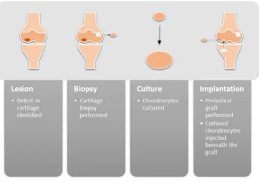
ACI has a 30 year history [7] and is an established tech-nique for the treatment of ulcerated cartilage and carti-lage defects. It involves an initial carticarti-lage biopsy, from which chondrocytes are cultured in vitro. In a second sur-gical procedure, a flap or membrane is then sutured (or glued) over the defect and the cultured chondrocytes are injected under this barrier. This process is summarized in Fig. 1. Over the last 10–15 years ACI has evolved (as bio-engineering technology has improved) and now includes matrix-assisted ACI (MACI). The patient then undergoes very careful and graded rehabilitation to prevent the patch being dislodged.
Early randomised controlled trials (RCTs) data sug-gested no significant benefit of ACI when compared to the alternative surgical option of microfracture [8] and although a histologic improvement was observed [9], the clinical relevance of this is questionable. Indeed a Cochrane review in 2011 concluded that there was insuf-ficient evidence to recommend the use of ACI [10].
The method developed over time to include collagen-covered ACI, and subsequently MACI, with the latter providing benefits including reduced size of the incision, greater surgical consistency, more consistent cell seeding, reduced periosteal hypertrophy and fewer adverse events [11−14].
Indeed, the matrix-applied method did perform sig-nificantly better than microfracture in the SUMMIT study
Fig. 1 A schematic demonstrat-ing the process of autologous chondrocyte implantation. A chondral lesion is identified and a biopsy of non-articular carti-lage is performed. The biopsy is cultured to amplify the number of chondrocytes. These are then injected under a periosteal flap (which is acquired from the proximal tibia)
(an RCT of 144 patients over 2 years) [15] in terms of clinical and functional outcomes. There was however no significant benefit over microfracture in MRI or histologi-cal outcomes. A key finding from these RCTs is that there was no correlation between the functional outcomes and evidence of structural repair when MRI is used, which is a potentially concerning finding.
ACI in combination with meniscal transplant allograft has good long-term outcomes with 75% still functioning well at 10-years (and 25% proceeding to arthroplasty). It is difficult to delineate whether the benefits of the procedure are due to ACI, meniscal transplant or indeed osteotomy (performed as part of the procedure) [16]. Similar results have been demonstrated in 57 patient with bipolar chon-dral lesions in the tibiofemoral compartment [17] with 75% having no radiographic progression at 10 years.
The cost of ACI and MACI are high, ranging from £4125 per patient to approximately £16,000 per MACI implant or £18,000 for a single vial of cells for ACI [18]. Therefore, in 2015 the technique was appraised by the National Institute for Health and Care Excellence (NICE) in the UK for the treatment of articular cartilage defects of the knee. The conclusions of this appraisal were that, while short-term clinical benefits were observed, the long-term clinical efficacy remained uncertain and the technique did not have robust evidence to demonstrate cost-effectiveness. Further research and evidence were recommended. The cost-effectiveness conclusions were considered harsh and were addressed in a consensus state-ment by UK knee surgeons, who drew attention to the estimated cost-effectiveness of ACI being between £7000 to £100,000 per Quality-Adjusted Life Year (QALY) (with the NICE threshold set at £20,000–30,000 per QALY).
There is a relative paucity of users, with, for example, only ten in the United Kingdom, which makes the develop-ment of large-scale research a challenge. MACI remains a potentially fruitful avenue for symptomatic therapy in early cartilage disease and traumatic cartilage lesions, though cru-cially not in OA.
Medical approaches
The scope of ‘non-surgical’ alternative therapies is large and, for this reason, this review focuses on the treatments which are likely to arise in clinical discussion with OA patients including autologous MSC injection, PRP, vitamin D and ‘other’ treatments.
Autologous mesenchymal stem cells
Articular cartilage is formed of a single cell type, the chon-drocyte and a stable extracellular matrix that has no vascu-lar, lymphatic or nervous supply. Subchondral bone provides mechanical and nutritional support and microfractures in this tissue can result in the release of undifferentiated mes-enchymal stem cells (MSCs) from the bone marrow to facili-tate the repair of chondral defects.
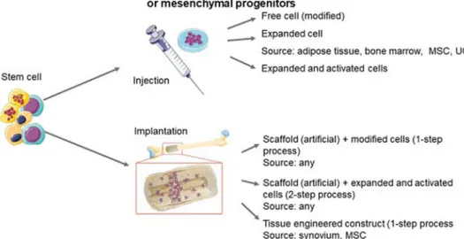
The repair capacity of MSCs has led to the development of techniques to directly inject MSCs locally into the joint following the ex vivo preparation of mesenchymal cells (Fig. 2). MSCs (also known as human or bone marrow stromal cells, multi-potent adult stem cells, mesenchymal progenitor cells and skeletal stem cells) have the ability to differentiate into the three tissue types, cartilage, bone and fat, and are invested, by definition, with an innate capacity for self-renewal and rapid proliferation. MSCs
Fig. 2 A depiction of the
inject-able and implantinject-able options for delivery of MSCs and the potential sources of MSCs which are appropriate for each method
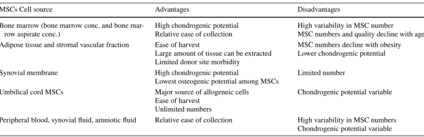
display paracrine anti-inflammatory and immunomodula-tory properties [19, 20] and can be harvested from bone marrow (biopsy or aspirate) as well as from the stromal vascular fraction in adipose tissue. (Table 1) Autologous sources avoid any immunological concerns, however, there is concern that, as MSCs are cleared from the joint rapidly, the joint may simply be experiencing the benefits afforded by a ‘wash-out’.
The use of MSCs in OA is an area of burgeoning research and, at the time of writing, there were 182 studies recorded, in various states of progress and 3 systematic reviews which best address the use of stem cells in the treatment of OA [21−23].
The review performed by Hached and colleagues ana-lysed a total of 44 trials of intra-articular injection of MSCs in the treatment of OA including bone marrow-derived, adipose tissue-derived and umbilical cord MSCs [21]. The review concluded that all three methods of acquisition of MSCs had evidence to support their use in the treatment of OA and that intra-articular injection of these cells was safe with very few side effects. An extensive review appraised 20 full-text records including systematic reviews, compre-hensive reviews, clinical reviews and meta-analyses pub-lished between 2006 and 2016 which addressed the treat-ment of cartilage lesions with MSCs [23]. In their review, the authors noted that improvements in symptoms (pain and function) were more commonly reported than structural/tis-sue improvement. There was a low level of evidence for the intervention with a mere 25 items of Level I graded evidence and subsequently concluded that it was “unclear” if stem cells were an effective treatment for OA. Broadly, stem cell therapies were effective in symptomatic (pain) relief related to chondral defects and defects (or lesions) due to OA. The authors reported, overall, limited repair and integration with extensive variability in the results presented. The main issues recorded included significant variability in MSC sources, techniques for preparation, methods of administration and
the range of co-interventions used (including micro-fracture, sub-chondral drilling, debridement, PRP).
There are very few studies which have demonstrated any degree of structural improvement in knee OA. Lamo-Espinosa and colleagues [24], reported the results of an RCT of increasing doses (10 × 106 or 100 × 106) of bone
marrow MSC, intra-articular injection against hyaluronic acid injection in 30 patients with OA (Kellgren–Lawrence grades II–IV). Participants were followed-up for 12 months and those in the MSC injection group had significant improvements in functionality and symptoms. Interest-ingly, only those in the high dose MSC injection group had statistically significant structural improvement in cartilage thickness on MRI at 12 months, opening the possibility of a dose–response. It should be noted that the inclusion of patients with such severe disease (Kellgren–Lawrence IV) suggests that the experimental group was substantially heterogeneous.
A pilot study by Orozco and colleagues [25] performed on patients with mild to severe knee OA (Kellgren–Law-rence grades II–IV) who received an intra-articular injec-tion of (40 × 106) bone marrow-derived MSCs, demonstrated
improvement in pain, function and cartilage quality at 12 months. The pain relief was maintained at 2 years, while the objective cartilage improvement (on MRI) continued on a trajectory of improvement at 2 years [26]. However, it must be noted that this study only included 12 patients and so conclusions should be tentative at best.
Allogeneic MSC injection was demonstrated to be both feasible and safe, as reported in previous studies [27], though the observed negative outcomes include the generation of fibrocartilage, injection-related pain and swelling, infec-tion post-bone marrow aspirate and a pulmonary embolus 2 weeks post-bone marrow aspirate [23]. In a further system-atic review, only 2 serious adverse events (synovial effusion and unstable angina) were observed amidst 288 patients.
Table 1 A summary of the advantages and disadvantages in OA therapy of MSCs acquired from different sources, including; bone marrow,
adi-pose tissue and stromal vascular fraction, the synovial membrane, umbilical cord and peripheral blood, synovial fluid and amniotic fluid
MSCs Cell source Advantages Disadvantages
Bone marrow (bone marrow conc. and bone
mar-row aspirate conc.) High chondrogenic potentialRelative ease of collection High variability in MSC numberMSC numbers and quality decline with age Adipose tissue and stromal vascular fraction Ease of harvest
Large amount of tissue can be extracted Limited donor site morbidity
MSC numbers decline with obesity Lower chondrogenic potential
Synovial membrane High chondrogenic potential
Lowest osteogenic potential among MSCs Limited number
Umbilical cord MSCs Major source of allogeneic cells
Ease of harvest Unlimited numbers
Chondrogenic potential variable Peripheral blood, synovial fluid, amniotic fluid Relative ease of collection High variability in MSC numbers
Pers and colleagues studied three dosages of adipose tis-sue-derived MSCs (2 × 10⁶, 10 × 106, 50 × 106 cells) in the
Adipose Derived mesenchymal stromal cells in Patients with knee Osteoarthritis (ADIPOA) trial and found 2 × 10⁶ was
optimal in terms of functionality and pain relief at 9 months [28] and postulated MSCs may operate via innate and adap-tive immune modulation [29]. This phase I trial is now in phase II (ADIPOA-2) and recruitment of 150 patients is underway.
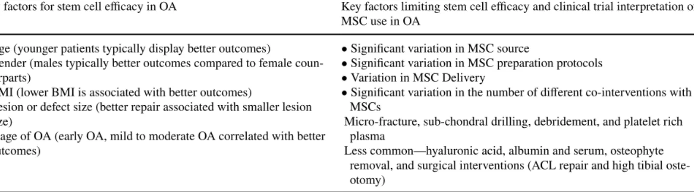
Limitations and barriers to the routine application of MSCs for OA from this plethora of clinical studies were a consequence of (i) significant variation in MSC source, (ii) significant variation in MSC preparation protocols adopted, (iii) significant variation in MSC delivery approaches adopted and, (iv) significant variation in the number of different co-interventions with MSCs including micro-fracture, sub-chondral drilling, debridement, and PRP as well as hyaluronic acid, albumin and serum, osteophyte removal, and surgical interventions (ACL repair and high tibial osteotomy).
Nevertheless, the following factors were associated with increased efficacy of MSC injection for OA:
• Younger age • Male gender • Low BMI
• Small lesion/defect
• Early/mild to moderate OA severity
It should also be considered that these MSC treatments are currently not covered by many health insurance provid-ers and the costs are high. For example, in the United States, the cost of a single stem-cell treatment for osteoarthritis was estimated at $5156 (95% CI $4550–5762) based on data from 273 centers [30].
In conclusion, the predominantly poor-quality, current literature suggests that symptoms, particularly pain, may improve with MSCs injection, however, evidence of struc-tural improvement is unconvincing and positive effects
appear to be observed in particular patient phenotypes. The overwhelming conclusion is a need to standardize the inter-vention if progress is to be made (Table 2). There is also a substantial need for phase II and III trials with the results of ADIPOA-2 being keenly awaited.
Platelet‑rich plasma
Platelets play an important role in coagulation but also inflammation and PRP is a therapy which has been used extensively in equine tendinopathy [31] and has been inves-tigated in the treatment of OA, particularly of the knee [32]. Platelet-rich plasma is a fluid which is rich in growth factors that stimulate cell proliferation, cellular migration, angiogenesis and the synthesis of the extracellular matrix including; platelet-derived growth factor (PDGF), tumor-like growth factor-β (TGF-β), fibroblast growth factor (FGF), vascular endothelial growth factor (VEGF), hepato-cyte growth factor (HGF) and insulin-like growth factor-1 (IGF-1).
It is derived through centrifugation of a patient’s blood, with the aim of separating a plasma component which is rich in platelets (> 95% platelets) from whole blood which is poor in platelets (4% platelets). The PRP is then extracted and injected into the affected joint. The intricacies of prepa-ration techniques vary and result in significantly different constituent cells (erythrocyte and leucocyte proportions), platelet concentrations and injection volumes [33] (Fig. 3). Indeed, there is a global schism in practice with Europe-ans preferring to use leukocyte-poor and AmericEurope-ans using leukocyte-rich PRP. PRP has been investigated in RCTs [34,
35] but the broad variation in preparation methods makes inter-trial comparison difficult and robust conclusions harder to ascertain and few are blinded. To emphasize this point we have synthesized and summarized some of the seminal studies below.
The issues surrounding the preparation of PRP are cov-ered in a review of the techniques utilised in a number of RCTs and systematic reviews [36]. There is substantial
Table 2 A synthesis of the main issues in MSC efficacy in the treatment of OA and limitations to adequate assessment through clinical trials
Key factors for stem cell efficacy in OA Key factors limiting stem cell efficacy and clinical trial interpretation of MSC use in OA
• Age (younger patients typically display better outcomes)
• Gender (males typically better outcomes compared to female coun-terparts)
• BMI (lower BMI is associated with better outcomes)
• Lesion or defect size (better repair associated with smaller lesion size)
• Stage of OA (early OA, mild to moderate OA correlated with better outcomes)
• Significant variation in MSC source
• Significant variation in MSC preparation protocols • Variation in MSC Delivery
• Significant variation in the number of different co-interventions with MSCs
Micro-fracture, sub-chondral drilling, debridement, and platelet rich plasma
Less common—hyaluronic acid, albumin and serum, osteophyte removal, and surgical interventions (ACL repair and high tibial oste-otomy)
variation in techniques including; the subject studied (severity of knee OA), PRP preparations, the inclusion of leukocytes, platelet count, number of injections delivered, interval/frequency of administration, volume of injection, whether fresh or freeze-thawed PRP were used, the use of anticoagulants and activating agents, separation techniques and any co-administered injections. With this in mind, a technical analysis was performed in 2017 to evaluate the similarities and differences between differing PRP formu-lations, in an attempt to determine the best preparation for the treatment of knee OA.
Filardo and colleagues [37, 38] performed a blinded trial in which they recruited participants with radiographic knee OA up to a Kellgren and Lawrence score of ≤ III, with 96 randomised to PRP and 96 to hyaluronic acid as a comparator. The PRP was centrifuged twice and PRP participants received 3 injections, once a week for three weeks and all participants were followed up for 12 months initially but extending to 5 years [39]. The key finding was that both treatments were equally effective in reducing knee OA symptoms and improving function over time but leucocyte-rich PRP was no more effective than hyaluronic acid.
To summarize the available evidence regarding PRP a number of systematic reviews have been performed [40−42]. PRP provided significant improvements in knee OA patient outcomes at 12 months and larger improvements were observed in those with milder radiographic disease (Kell-gren and Lawrence ≤ II) [40],
Significant improvements in ‘patient recorded outcomes’ were also observed with PRP as opposed to hyaluronic acid at 3–6 months (WOMAC 28.5 vs. 43.4 respectively,
p = 0.0008) and 6–12 months (WOMAC 22.8 vs. 38.1, p = 0.0062) [41].
A further systematic review published in 2018 (includ-ing 7 randomized placebo-controlled trials and 908 patients) sought to investigate the superiority of PRP over hyaluronic acid which was not demonstrated. In respect of PRP the min-imal clinically important difference (MCID) was observed in 5 of the 7 papers, and suggested that differences in clinical outcomes could be due to variation in the preparation of PRP in terms of; centrifugation (speed, frequency, time-length, activating agents), administration (frequency, volume of injection) and post-administration rehabilitation protocols [42]. From a safety point of view, no local or systemic seri-ous adverse events were noted in the reviewed articles.
Milants and colleagues used a previous definition of minimal clinically important improvement in pain (MCII) to determine whether an observed difference had any ‘mean-ingful’ effect in clinical practice. This was set at 15 out of 100 for absolute improvement and 20% for relative improve-ment for knee OA, as defined by Tubach et al. [43].
The Milants technical analysis included 19 RCTs, and studies were classified into two groups depending on out-comes with a ‘bad responder group’, defined as a response less than the minimal clinically important improvement (MCII) (n = 4 studies), and a ‘very good responder group’, defined as a response greater than twice the MCII (n = 7 studies). The reviewers contacted authors of the trials to obtain information regarding the preparation which was missing from the manuscript and PRP preparation was clas-sified according to the Mishra (a classification in which PRP is divided into 4 types depending on 3 variables; white blood cells: increased or minimal, activation: yes or no, platelet content > 5 times patient baseline or ≤ 5 times patient base-line) and PAW (Platelet concentration, Activation prior to injection, White blood cell content).
In almost all studies with a very good responder group, PRPs were leukocyte-poor, activated prior to injection and platelets < 5 times baseline or between baseline and 750,000 platelets/µL), administered according to a lower number of injections (1 or 2 rather than 3), with a longer interval between injections (2 to 3 weeks per injection rather than once weekly) and a single (as opposed to double) spinning technique. The use of leukocyte-rich PRP was only found in the bad responder group. The use of calcium chloride and citrate was common in the very good responder group.
The cost of the PRP procedure is estimated at $714 (95% CI $691–737) based on data from 179 centers from across the United States [44].
In conclusion, although PRP may have repeated mild symptomatic benefits, there is yet to be experimentally robust demonstration of symptomatic and structural effects in the current literature. Research is required to better under-stand the mechanism of action, including investigation of the survival and location of platelet-derived factors within the joint following injection. In order for PRP to be considered
Fig. 3 A comparison of platelet concentration and volume of protein-rich plasma (PRP) resulting from the extraction methods employed by 5 laboratories. [33]
within the dogma of recommended treatment for OA, at least one large, randomized, placebo-controlled trial and further investigation regarding preparation and dosage efficacy is required. This working group cannot, therefore, make a rec-ommendation to use PRP as an intervention for OA.
Vitamin D
There is a secular trend toward decreased vitamin D lev-els, with serum concentrations averaging 49 ng/mL in the mid-twentieth century to approximately 23 ng/mL now, and with over a billion individuals being vitamin D deficient or insufficient [45]. Due to the role played by sunlight in the in vivo production of vitamin D, particularly low levels are observed at the extremes of latitude [46], and in winter months. Studies of seasonal gene expression have shown that some pro-inflammatory factors, including soluble IL-6 receptor and C-reactive protein have a peak expression in winter months and vitamin D receptor expression peaks in the summer months [47]. It is, therefore, interesting that certain diseases display similar seasonality and geography, including OA. This descriptive epidemiological observation is supported by basic scientific findings including [48]: • There are receptors for vitamin D on chondrocytes which
may play a role in the regulation of matrix metallopro-teinases and prostaglandin E2 production
• Vitamin D stimulates proteoglycan synthesis in mature chondrocytes
• Vitamin D deficiency influences bone remodeling which may predispose to the development of OA
Despite these observations, four RCTs of vitamin D in OA have been performed in the United States (US) [49], India [50], the United Kingdom (UK) [51] and Australia [52]. None of these have demonstrated structural or symp-tomatic benefit in OA.
The pilot study performed in India [50] included 103 par-ticipants (59.4% females) with a baseline age of approxi-mately 50 and a baseline 25-OH vitamin D of < 20 ng/mL. They found a significant reduction in knee pain and improve-ment in function but no significant alteration in radiographic knee OA at 12 months.
The placebo-controlled trial performed by McAlindon and colleagues [49] (in the US) included 146 female partici-pants with a mean age at baseline of 62.4 years. At two years they found no improvements in knee symptoms, functional status or cartilage structure with vitamin D.
In the aforementioned UK placebo-controlled trial [51], despite an increase in serum 25-hydroxy-vitamin D (from approximately 20 to 30 µg/L) in the treatment group, no significant changes in symptomatic or radiographic knee
OA were observed after 3 years in 474 participants (over the age of 50).
In Australia, Jin and colleagues demonstrated that in 209 patients with low vitamin D (12.5–60 nmol/L) treated for 2 years with monthly oral vitamin D3 (50,000 IU), there was no significant improvement in MRI-measured tibial cartilage volume or WOMAC knee pain score [52].
The relationship between vitamin D and knee OA has been investigated in a recent systematic review of 11 stud-ies, which concluded that although vitamin D deficiency is associated with knee OA, the evidence regarding this association is inconsistent [53]. The studies included in the review were largely of cohort and cross-sectional design but also included two RCTs. The systematic review demon-strated that there was marked variation in the relationships between vitamin D and OA with a level of evidence (for an association of vitamin D deficiency with prevalent sympto-matic knee OA) of ‘moderate’, while the relationship with prevalent radiographic knee OA was graded as ‘limited’. This negative conclusion supports that of a prior systematic review [54].
It should be acknowledged that vitamin D deficiency has been associated with a range of co-morbidities, of which OA is only one. However, the adverse effect profile of the supple-ment is favorable and should be strongly considered in those at risk of deficiency. Should there be systematic screening for vitamin D deficiency or systematic supplementation of vitamin D? This question is beyond the purview of this arti-cle and is dependent on many factors which are related to local healthcare systems and economic considerations.
We conclude by recommending that, when severe defi-ciency is diagnosed (especially in winter), vitamin D should be supplemented through the evidence that such supplemen-tation ameliorates OA symptoms is inconclusive.
Other medical therapies
It should be noted that the medical therapies included in this section are very rarely mentioned in international guidelines, however, they are often the subject of discussions between patients and clinicians. Collagens, methylsulfonylmethane,
S-adenosylmethionine, curcuma, harpagophytum and ginger
are commonly used in the treatment of OA in many coun-tries [55], with polyphenols, green tea, ‘Cat’s claw’ and dairy products also being mentioned.
Collagens: oral and intra‑articular
Oral collagens are a rich source of amino acids, and, in OA, are purported to stimulate the joint to produce endogenous collagens in response to supplementation.
In 2016 a study investigated 190 patients, randomised to receive undenatured type II collagen (40 mg) or glucosamine
hydrochloride and chondroitin sulfate or placebo [56]. The primary outcome was total WOMAC change from baseline with secondary outcomes being Lequesne index and pain VAS. After 6 months they found that undenatured type II collagen led to a significantly greater reduction in WOMAC compared to placebo (551 vs. 414, p = 0.002) and compared to the glucosamine and chondroitin sulfate arm (551 vs. 454,
p = 0.009). In terms of secondary outcomes, there was a
greater reduction in Lequesne index (2.9 vs. 2.1, p = 0.009). A further RCT investigated the performance of 5, once weekly 4 mL injections of polymerized collagen type I (of porcine origin) compared to sodium hyaluronate with assess-ments at 3 and 6 months [57]. The primary outcome was Lequesne index (measuring the severity of knee OA) at the 3 month time point with a visual analogue score for pain and SF-36 questionnaire also recorded. They found no significant differences between the groups for the above outcomes at either 3 or 6 months.
Gelatin, a form of collagen-rich in proline, was assessed in 52 patients as part of a randomised, placebo-controlled trial which found significant inter-group differences in sev-eral types of pain, however, an effect size was not reported, making extrapolation to clinical benefit difficult [58]. Unde-natured collagen, an alternative form of collagen, was com-pared to glucosamine and chondroitin with 26 patients per group. This found no inter-group differences in the efficacy of the interventions [59].
In 2012, Van Vijven and colleagues published a system-atic review of a variety of oral collagens at various doses comprised of 8 trials of collagen hydrosylates (3 versus pla-cebo), gelatin (1 versus placebo) or undenatured collagen (versus glucosamine hydrochloride and chondroitin sulfate) [58]. The review concluded that there was ‘low’ grade evi-dence for the use of these in OA. Those treated with collagen hydrosylates included 313 treated patients (taking 10 g per day) against 297 on a placebo preparation and found a sig-nificant effect on symptoms (WOMAC pain (− 0.48) with a significant but small effect size of 0.17) but no effect on the joint structure as assessed by MRI scan.
There are various preparations of oral or intra-articular collagens. Although widely used in a large number of coun-tries, current data do not support a positive recommendation to treat OA patients despite a mild effect on symptoms (pain) and function.
Methylsulfonylmethane (MSM)
This dietary supplement is found in plants, fruits and veg-etables, and can be taken alone or in combination with other supplements. There have been two, notable, placebo-controlled trials of MSM in patients with knee OA, both in approximately 50 patients over 12 weeks of follow-up. The first [60] involved a dosage schedule which resulted
in 6 g per day and demonstrated a significant improvement in SF-36, WOMAC pain and function in the MSM group (p < 0.05). The second involved a dose of approximately 3 g per day demonstrated a significant improvement in WOMAC function but not in WOMAC pain or SF-36 compared to placebo [61].
A trial in knee OA randomised 118 patients between glu-cosamine, MSM, combination therapy or placebo and found a significant improvement in pain and Lequesne functional index at 12 weeks in all groups except the placebo arm.
A recent RCT examined the performance of glucosamine and chondroitin in combination with MSM versus glucosa-mine and chondroitin alone and versus placebo, in a popula-tion of 147 early knee OA patients (Kellgren and Lawrence grade I-II). This study demonstrated improvement in the groups which included MSM compared to the other treat-ment groups, in terms of pain VAS and WOMAC scores [62].
In conclusion, small trials did not demonstrate any major safety concerns for MSM treatment. Whether there is a symptomatic benefit over a short follow-up period is a ques-tion which would need to be answered through larger, well-designed trials and long term follow-up data are required. S‑adenosylmethionine (SAMe)
This is a substance produced from methionine in the liver. The treatment of hip and knee OA with SAMe was the sub-ject of a Cochrane review of 4 randomised, placebo-con-trolled trials [63]. This review included 656 patients and demonstrated a significant improvement with SAMe com-pared to placebo but with a very minor effect on pain (4 mm on a 100 mm VAS) and function (2 mm on a 100 mm VAS), which are of questionable clinical significance. There was no significant difference in adverse effects or withdrawals but it should also be noted that the methodological and reporting quality were poor and that there was a moderate degree of inter-trial heterogeneity (I2 = 54%).
Curcuma
Curcuma (or curcumin) is an extract of turmeric, a yel-low spice, and member of the ziangiberaceae family. Both curcuma and ginger have roots in Ayurvedic and Chinese medicine [64] with curcuma manifesting an anti-inflam-matory effect via cyclo-oxgenase (COX)-2, prostaglandins and leukotoxin inhibition. There is a wide variation in daily doses from 180 to 2000 mg, which makes direct, inter-study comparison problematic. Nevertheless, a meta-analysis was published in 2016 included 4 placebo-controlled trials in the context of knee OA; 2 trials versus ibuprofen and 1 versus diclofenac. In these trials of curcuma, improvements were seen in symptomatic measures and NSAID consumption (up
to 4 months) versus placebo but no significant superiority was noted versus ibuprofen or when added to diclofenac.
Although a few, idiosyncratic adverse effects were reported in the trials, the meta-analysis concluded that cur-cuma could be considered safe at daily doses of 4800 mg for 4 months [65].
A more recent meta-analysis, published in 2018, included 11 RCTs (N = 1009 patients) investigating the role of curmi-noids and boswellia (a gum-resin used in Ayurvedic medi-cine) in the treatment of knee OA [66]. There was some improvement in pain and function outcomes versus placebo, however, the conclusion was that evidence was currently too scant to allow the therapies to be included in clinical recom-mendations for treatment.
A trial of bio-optimized curcuma in the treatment of knee OA was reported in 2019 [67]. After 3 months of treatment, there were no statistical differences in intention to treat analyses for curcuma efficacy comparing the treatment to placebo arms for the co-primary endpoints (which are not included in those recommended by the European Medi-cines Agency or scientific societies). However, there was a significantly higher incidence of diarrhea in the curcuma group. In summary, experimental data regarding curcuma is sparse. The little evidence there supports a mild effect on OA symptoms. The sample sizes of the published trials are small, follow-up is short [65] and longer-term, robust studies are required before curcumin can be actively recommended from an efficacy stand-point.
Harpagophytum
Also known as ‘Devil’s claw’ Harpagophytum is an African plant which is thought to manifest anti-inflammatory effects, similar to curcuma, via inhibition of COX-2 and leukotoxin. A systematic review by Gagnier and colleagues investigated the role of Harpagophytum in the treatment of lower back pain and OA, including 3 randomised, placebo-controlled trials of hip and knee OA (385 patients) [68]. This review concluded that there was ‘moderate’ evidence of effective-ness for the use of 60 mg of harpagophytum powder, though longer, higher-quality trials are required before it can be rou-tinely recommended in clinical practice.
Ginger
Ginger, another member of the ziangiberaceae family, is thought to manifest anti-inflammatory effect via inhibition of COX, lipoxygenase, reduction in tumor necrosis factor and inflammatory prostaglandin production. There are data to suggest that ginger extract (Zintona EC®) is superior to placebo in terms of pain relief at 6 months (though efficacy was the same at 3 months) in a double-blind, randomised, placebo-controlled, crossover trial of knee OA [69].
Additionally, trials have demonstrated improvements in pain and mobility and reduced rescue medication usage intake versus placebo, though with more (mostly mild) gas-trointestinal adverse events [70].
In 2015, the trial data were summarized and analyzed in a systematic review and meta-analysis of 5 randomised placebo-controlled trials, totaling 593 patients, which found a significant reduction in pain and disability with ginger. However, twice the rate of discontinuation was observed with ginger versus placebo (Relative Risk 2.33, 95% CI 1.04–5.22) [71]. A similar finding was reported in an earlier systematic review which noted “infrequent reports of mild, and predominantly gastrointestinal, adverse effects” [72].
Conclusion
In this review, we have synthesized the current evidence regarding alternative therapies for OA. Our findings are summarised in Table 3. Publication bias may be an issue with this group of treatments, however, this is not always the case (as seen throughout this review).
In summary, for all of the interventions covered in this review, issues of study design limit the degree to which inference can be made about clinical effectiveness in symp-tomatic OA. It is clear that none of these would currently clear the required hurdle for regulatory approval, were they to be assessed in like manner to current pharmaceutical interventions. However, there is also an insufficient basis for declaring them completely ineffective. They, therefore, remain an area in which further, appropriately designed, large, blinded, RCTs are an urgent necessity.
Acknowledgements PGC is supported in part by the National Insti-tute for Health Research (NIHR) infrastructure at Leeds. The views expressed are those of the author(s) and not necessarily those of the NHS, the NIHR or the Department of Health.
Funding The ESCEO Working Group was funded by the ESCEO. The
ESCEO receives unrestricted educational grants to support its educa-tional and scientific activities from non-governmental organizations, not-for-profit organizations, non-commercial or corporate partners. The choice of topics, participants, content and agenda of the Working Groups as well as the writing, editing, submission and reviewing of the manuscript are the sole responsibility of the ESCEO, without any influence from third parties.
Compliance with ethical standards
Conflict of interest NRF, ROCO, AJP, JFK, MC, GH, JB, NV, AAK, RM, MV, WL, RRo, NA-D and RRi have no relevant conflicts of inter-est to declare. CC has received lecture fees and honoraria from Am-gen, Danone, Eli Lilly, GSK, Kyowa Kirin, Medtronic, Merck, Nestlé, Novartis, Pfizer, Roche, Servier, Shire, Takeda and UCB outside of the submitted work. EM reports personal fees from Expanscience, personal fees from Mylan - Meda, personal fees from TRB Chemed-ica, Pierre Fabre, Celgène and Fidia, and non-financial support from Pfizer, outside the submitted work. PGC reports personal fees from AbbVie, personal fees from Flexion Therapeutics, personal fees from Galapagos, personal fees from GlaxoSmithKline, personal fees from Novartis, personal fees from Pfizer, personal fees from Samumed, out-side the submitted work. FB reports personal fees from 4P Pharma, Boehringer, Bone Therapeutics, Expanscience, Galapagos, Gilead, GSK, Heel, Merck Sereno, MSD, Nordic, Novartis, Peptinov, Pfizer, Regulaxis, Roche, Sandoz, Sanofi, Servier, TRB Chemedica, UCB. MLB reports grants and honoraria from AMGEN, grants from ABIO-GEN, grants and consultancy from ALEXION, grants, consultant and honoraria from BRUNO FARMACEUTICI, grants from ELI LILLY, grants, consultancy and honoraria from KYOWA KIRIN, grants from MSD, grants from NPS, grants and consultant from SERVIER, grants and consultancy from SHIRE, outside the submitted work. BC reports grants and sponsorship from MSD, consultancy for Amgen and oc-casional interventions for Lilly, Roshe, Ferring and Expanscience.
JPP reports personal fees from Mylan, grant and personal fees from TRB Chemedica, outside the submitted work. JMP reports grant and personal fees from TRB Chemedica, outside the submitted work. TT reports personal fees from Abbvie, grants and personal fees from Am-gen, personal fees from Arrow, personal fees from BioAm-gen, personal fees from BMS, grants and personal fees from Chugai, personal fees from Expanscience, personal fees from Gilead, personal fees from Grunenthal, grants and personal fees from HAC-Pharma, personal fees from LCA, personal fees from Lilly, personal fees from Medac, grants and personal fees from MSD, grants and personal fees from Novartis, grants and personal fees from Pfizer, personal fees from Sanofi, per-sonal fees from Theramex, perper-sonal fees from Thuasne, perper-sonal fees from TEVA, grants and personal fees from UCB, grants from Bone therapeutics, outside the submitted work. OB reports grants from Bio-phytis, IBSA, MEDA, Servier, SMB, Theramex, outside the submitted work. JAK reports grants from UCB, grants from Amgen, grants from Radius Health, outside the submitted work. JYR reports grants and personal fees from IBSA-GENEVRIER, grants and personal fees from MYLAN, grants and personal fees from RADIUS HEALTH, personal fees from PIERRE FABRE, grants from CNIEL, personal fees from DAIRY RESEARCH COUNCIL (DRC), outside the submitted work.
Statement of human and animal rights For this specific review article no novel, original human participant or animal studies were performed.
Informed consent For this review article, formal consent of partici-pants was not required.
Open Access This article is licensed under a Creative Commons
Attri-bution 4.0 International License, which permits use, sharing, adapta-tion, distribution and reproduction in any medium or format, as long as you give appropriate credit to the original author(s) and the source, provide a link to the Creative Commons licence, and indicate if changes were made. The images or other third party material in this article are included in the article’s Creative Commons licence, unless indicated otherwise in a credit line to the material. If material is not included in the article’s Creative Commons licence and your intended use is not permitted by statutory regulation or exceeds the permitted use, you will need to obtain permission directly from the copyright holder. To view a copy of this licence, visit http://creat iveco mmons .org/licen ses/by/4.0/.
Table 3 A summary table of the key, take-home points for each of the interventions reviewed
Alternative therapy Key points
Autologous chondrocyte implantation • Treatment for cartilage defects and not osteoarthritis • Includes ACI and MACI
• Trial evidence to support symptomatic benefit
• Supported by NICE in the UK for specific patient group (including no previous knee surgery, limited evidence of knee osteoarthritis, large chondral defect)
Mesenchymal stem cell injection • There are multiple sources of MSCs with differing profiles of usage and limitations • Multiple sources of MSCs lead to difficulties in directly comparing clinical trials • Trial evidence to support symptomatic benefit
• Limited evidence to support structural benefit (MRI cartilage thickness) Platelet-rich plasma • Trial evidence to support symptomatic benefit
• Multiple methods of preparation lead to difficulties in directly comparing clinical trials Vitamin D • Evidence of efficacy in clinical trials is equivocal
• Recommendation to provide supplementation to those patients with evidence of depleted levels of 25OH-vitamin D
Other alternative therapies • Very limited clinical trial data to suggest the efficacy of oral collagens, MSM, SAMe, curcuma and ginger
• Adverse events: largely rare, though ginger appears to be associated with an increased risk of mild gastro-intestinal adverse events
References
1. Cross M, Smith E, Hoy D et al (2014) The global burden of hip and knee osteoarthritis: estimates from the global burden of disease 2010 study. Ann Rheum Dis 73:1323–1330. https ://doi. org/10.1136/annrh eumdi s-2013-20476 3
2. Van Saase JL, Van Romunde LK, Cats A et al (1989) Epidemiol-ogy of osteoarthritis: Zoetermeer survey. Comparison of radio-logical osteoarthritis in a Dutch population with that in 10 other populations. Ann Rheum Dis 48:271–280
3. McAlindon TE, Bannuru RR, Sullivan MC et al (2014) OARSI guidelines for the non-surgical management of knee osteoarthri-tis. Osteoarthritis Cartilage 22:363–388. https ://doi.org/10.1016/j. joca.2014.01.003
4. Bruyere O, Cooper C, Pelletier JP et al (2014) An algorithm rec-ommendation for the management of knee osteoarthritis in Europe and internationally: a report from a task force of the European Society for Clinical and Economic Aspects of Osteoporosis and Osteoarthritis (ESCEO). Semin Arthritis Rheum 44:253–263.
https ://doi.org/10.1016/j.semar thrit .2014.05.014
5. Hochberg MC, Altman RD, April KT et al (2012) American Col-lege of Rheumatology 2012 recommendations for the use of non-pharmacologic and non-pharmacologic therapies in osteoarthritis of the hand, hip, and knee. Arthritis Care Res (Hoboken) 64:465–474 6. Jordan KM, Arden NK, Doherty M et al (2003) EULAR Recom-mendations 2003: an evidence based approach to the management of knee osteoarthritis: Report of a Task Force of the Standing Committee for International Clinical Studies Including Therapeu-tic Trials (ESCISIT). Ann Rheum Dis 62:1145–1155
7. Brittberg M, Lindahl A, Nilsson A et al (1994) Treatment of deep cartilage defects in the knee with autologous chondrocyte trans-plantation. N Engl J Med 331:889–895. https ://doi.org/10.1056/ nejm1 99410 06331 1401
8. Knutsen G, Drogset JO, Engebretsen L et al (2007) A randomized trial comparing autologous chondrocyte implantation with micro-fracture. Findings at five years. J Bone Joint Surg Am 89:2105– 2112. https ://doi.org/10.2106/jbjs.g.00003
9. Saris DB, Vanlauwe J, Victor J et al (2008) Characterized chon-drocyte implantation results in better structural repair when treat-ing symptomatic cartilage defects of the knee in a randomized controlled trial versus microfracture. Am J Sports Med 36:235– 246. https ://doi.org/10.1177/03635 46507 31109 5
10. Vasiliadis HS, Wasiak J (2010) Autologous chondrocyte implan-tation for full thickness articular cartilage defects of the knee. Cochrane Database Syst Rev 10:CD003323
11. Sohn DH, Lottman LM, Lum LY et al (2002) Effect of gravity on localization of chondrocytes implanted in cartilage defects. Clin Orthop Related Res 394:254–262
12. Brittberg M (2010) Cell carriers as the next generation of cell therapy for cartilage repair: a review of the matrix-induced autol-ogous chondrocyte implantation procedure. Am J Sports Med 38:1259–1271
13. Marlovits S, Aldrian S, Wondrasch B et al (2012) Clinical and radiological outcomes 5 years after matrix-induced autologous chondrocyte implantation in patients with symptomatic, traumatic chondral defects. Am J Sports Med 40:2273–2280
14. Ebert JR, Robertson WB, Lloyd DG et al (2010) A prospective, randomized comparison of traditional and accelerated approaches to postoperative rehabilitation following autologous chondrocyte implantation: 2-year clinical outcomes. Cartilage 1:180–187 15. Saris D, Price A, Widuchowski W et al (2014) Matrix-applied
characterized autologous cultured chondrocytes versus microfrac-ture: two-year follow-up of a prospective randomized trial. Am J Sports Med 42:1384–1394. https ://doi.org/10.1177/03635 46514 52809 3
16. Ogura T, Bryant T, Minas T (2016) Biological knee reconstruc-tion with concomitant autologous chondrocyte implantareconstruc-tion and meniscal allograft transplantation: mid- to long-term out-comes. Orthop J Sports Med 4:2325967116668490. https ://doi. org/10.1177/23259 67116 66849 0
17. Ogura T, Bryant T, Mosier BA et al (2018) Autologous chondro-cyte implantation for bipolar chondral lesions in the tibiofemo-ral compartment. Am J Sports Med 46:1371–1381. https ://doi. org/10.1177/03635 46518 75697 7
18. NICE (2015) Autologous chondrocyte implantation for repair-ing symptomatic articular cartilage defects of the knee. National Institute for Health and Care Excellence, London
19. Harrell CR, Markovic BS, Fellabaum C et al (2019) Mesenchymal stem cell-based therapy of osteoarthritis: Current knowledge and future perspectives. Biomed Pharmacother 109:2318–2326. https ://doi.org/10.1016/j.bioph a.2018.11.099
20. Pastides P, Chimutengwende-Gordon M, Maffulli N et al (2013) Stem cell therapy for human cartilage defects: a systematic review. Osteoarthr Cartil 21:646–654. https ://doi.org/10.1016/j. joca.2013.02.008
21. Hached F, Vinatier C, Le Visage C et al (2017) Biomaterial-assisted cell therapy in osteoarthritis: From mesenchymal stem cells to cell encapsulation. Best Pract Res Clin Rheumatol 31:730–745. https ://doi.org/10.1016/j.berh.2018.05.002
22. Jevotovsky DS, Alfonso AR, Einhorn TA et al (2018) Osteo-arthritis and stem cell therapy in humans: a systematic review. Osteoarthritis Cartilage 26:711–729.https ://doi.org/10.1016/j. joca.2018.02.906
23. Hart D (2017) Mesenchymal Stem Cells: The hope, the hype and the reality in the treatment of osteoarthritis, A knowledge synthe-sis of clinical research (2010-2016) emphasizing the safety and efficacy of stem cell treatment for osteoarthritis. Bone and joint health strategic clinical network (BJH SCN)
24. Lamo-Espinosa JM, Mora G, Blanco JF et al (2016) Intra-articular injection of two different doses of autologous bone marrow mes-enchymal stem cells versus hyaluronic acid in the treatment of knee osteoarthritis: multicenter randomized controlled clinical trial (phase I/II). J Transl Med 14:246. https ://doi.org/10.1186/ s1296 7-016-0998-2
25. Orozco L, Munar A, Soler R et al (2013) Treatment of knee osteo-arthritis with autologous mesenchymal stem cells: a pilot study. Transplantation 95:1535–1541. https ://doi.org/10.1097/TP.0b013 e3182 91a2d a
26. Orozco L, Munar A, Soler R et al (2014) Treatment of knee osteoarthritis with autologous mesenchymal stem cells: two-year follow-up results. Transplantation 97:e66–e68. https ://doi. org/10.1097/tp.00000 00000 00016 7
27. Vega A, Martin-Ferrero MA, Del Canto F et al (2015) Treatment of knee osteoarthritis with allogeneic bone marrow mesenchy-mal stem cells: a randomized controlled trial. Transplantation 99:1681–1690. https ://doi.org/10.1097/tp.00000 00000 00067 8
28. Pers YM, Rackwitz L, Ferreira R et al (2016) Adipose mesen-chymal stromal cell-based therapy for severe osteoarthritis of the knee: a phase I dose-escalation trial. Stem Cells Transl Med 5:847–856. https ://doi.org/10.5966/sctm.2015-0245
29. Pers YM, Quentin J, Feirreira R et al (2018) Injection of adipose-derived stromal cells in the knee of patients with severe osteoar-thritis has a systemic effect and promotes an anti-inflammatory phenotype of circulating immune cells. Theranostics 8:5519– 5528. https ://doi.org/10.7150/thno.27674
30. Piuzzi NS, Ng M, Chughtai M et al (2018) The stem-cell market for the treatment of knee osteoarthritis: a patient perspective. J Knee Surg 31:551–556. https ://doi.org/10.1055/s-0037-16044 43
31. Ortved KF (2018) Regenerative medicine and rehabilitation for tendinous and ligamentous injuries in sport horses. Vet Clin
N Am Equine Pract 34:359–373. https ://doi.org/10.1016/j. cveq.2018.04.012
32. Mascarenhas R, Saltzman BM, Fortier LA et al (2015) Role of platelet-rich plasma in articular cartilage injury and disease. J Knee Surg 28:3–10. https ://doi.org/10.1055/s-0034-13846 72
33. Kaux JF, Le Goff C, Seidel L et al (2011) Comparative study of five techniques of preparation of platelet-rich plasma. Pathol Biol (Paris) 59:157–160. https ://doi.org/10.1016/j.patbi o.2009.04.007
34. Lin KY, Yang CC, Hsu CJ et al (2019) Intra-articular Injection of platelet-rich plasma is superior to hyaluronic acid or saline solution in the treatment of mild to moderate knee osteoarthritis: a randomized, double-blind, triple-parallel, placebo-controlled clinical trial. Arthroscopy 35:106–117. https ://doi.org/10.1016/j. arthr o.2018.06.035
35. Huang Y, Liu X, Xu X et al (2019) Intra-articular injections of platelet-rich plasma, hyaluronic acid or corticosteroids for knee osteoarthritis: a prospective randomized controlled study. Ortho-pade 48:239–247. https ://doi.org/10.1007/s0013 2-018-03659 -5
36. Milants C, Bruyère O, Kaux J-F (2017) Responders to platelet-rich plasma in osteoarthritis: a technical analysis. BioMed Res Int 2017:7538604. https ://doi.org/10.1155/2017/75386 04
37. Filardo G, Kon E, Di Martino A et al (2012) Platelet-rich plasma vs hyaluronic acid to treat knee degenerative pathol-ogy: study design and preliminary results of a randomized con-trolled trial. BMC Musculoskelet Disord 13:229. https ://doi. org/10.1186/1471-2474-13-229
38. Filardo G, Di Matteo B, Di Martino A et al (2015) Platelet-rich plasma intra-articular knee injections show no superiority versus viscosupplementation: a randomized controlled trial. Am J Sports Med 43:1575–1582. https ://doi.org/10.1177/03635 46515 58202 7
39. Di Martino A, Di Matteo B, Papio T et al (2018) Platelet-rich plasma versus hyaluronic acid injections for the treatment of knee osteoarthritis: results at 5 years of a double-blind, randomized controlled trial. Am J Sports Med. https ://doi.org/10.1177/03635 46518 81453 2
40. Campbell KA, Saltzman BM, Mascarenhas R et al (2015) Does intra-articular platelet-rich plasma injection provide clinically superior outcomes compared with other therapies in the treatment of knee osteoarthritis? A systematic review of overlapping meta-analyses. Arthroscopy 31:2213–2221. https ://doi.org/10.1016/j. arthr o.2015.03.041
41. Meheux CJ, McCulloch PC, Lintner DM et al (2016) Efficacy of intra-articular platelet-rich plasma injections in knee osteoar-thritis: a systematic review. Arthroscopy 32:495–505. https ://doi. org/10.1016/j.arthr o.2015.08.005
42. Di Y, Han C, Zhao L et al (2018) Is local platelet-rich plasma injection clinically superior to hyaluronic acid for treatment of knee osteoarthritis? A systematic review of randomized controlled trials. Arthritis Res Ther 20:128. https ://doi.org/10.1186/s1307 5-018-1621-0
43. Tubach F, Ravaud P, Martin-Mola E et al (2012) Minimum clini-cally important improvement and patient acceptable symptom state in pain and function in rheumatoid arthritis, ankylosing spondylitis, chronic back pain, hand osteoarthritis, and hip and knee osteoarthritis: Results from a prospective multinational study. Arthritis Care Res (Hoboken) 64:1699–1707. https ://doi. org/10.1002/acr.21747
44. Piuzzi NS, Ng M, Kantor A et al (2019) What is the price and claimed efficacy of platelet-rich plasma injections for the treat-ment of knee osteoarthritis in the United States? J Knee Surg 32:879–885. https ://doi.org/10.1055/s-0038-16699 53
45. Barger-Lux MJ, Heaney RP (2002) Effects of above average sum-mer sun exposure on serum 25-hydroxyvitamin D and calcium absorption. J Clin Endocrinol Metab 87:4952–4956. https ://doi. org/10.1210/jc.2002-02063 6
46. Koch T (2014) 1831: the map that launched the idea of global health. Int J Epidemiol 43:1014–1020. https ://doi.org/10.1093/ ije/dyu09 9
47. Dopico XC, Evangelou M, Ferreira RC et al (2015) Widespread seasonal gene expression reveals annual differences in human immunity and physiology. Nat Commun 6:7000. https ://doi. org/10.1038/ncomm s8000
48. Tetlow LC, Woolley DE (2001) Expression of vitamin D recep-tors and matrix metalloproteinases in osteoarthritic cartilage and human articular chondrocytes in vitro. Osteoarthr Cartil 9:423– 431. https ://doi.org/10.1053/joca.2000.0408
49. McAlindon T, LaValley M, Schneider E et al (2013) Effect of vitamin D supplementation on progression of knee pain and car-tilage volume loss in patients with symptomatic osteoarthritis: a randomized controlled trial. JAMA 309:155–162. https ://doi. org/10.1001/jama.2012.16448 7
50. Sanghi D, Mishra A, Sharma AC et al (2013) Does vitamin D improve osteoarthritis of the knee: a randomized controlled pilot trial. Clin Orthop Relat Res 471:3556–3562. https ://doi. org/10.1007/s1199 9-013-3201-6
51. Arden NK, Cro S, Sheard S et al (2016) The effect of vitamin D supplementation on knee osteoarthritis, the VIDEO study: a ran-domised controlled trial. Osteoarthr Cartil 24:1858–1866. https ://doi.org/10.1016/j.joca.2016.05.020
52. Jin X, Jones G, Cicuttini F et al (2016) Effect of vitamin D supple-mentation on tibial cartilage volume and knee pain among patients with symptomatic knee osteoarthritis: a randomized clinical trial. JAMA 315:1005–1013. https ://doi.org/10.1001/jama.2016.1961
53. Vaishya R, Vijay V, Lama P et al (2019) Does vitamin D defi-ciency influence the incidence and progression of knee osteoar-thritis? A literature review. J Clin Orthop Trauma 10:9–15. https ://doi.org/10.1016/j.jcot.2018.05.012
54. Diao N, Yang B, Yu F (2017) Effect of vitamin D supplementation on knee osteoarthritis: a systematic review and meta-analysis of randomized clinical trials. Clin Biochem 50:1312–1316. https :// doi.org/10.1016/j.clinb ioche m.2017.09.001
55. Lim YZ, Hussain SM, Cicuttini FM et al (2019) Bioactive food as dietary interventions for arthritis and related inflammatory dis-eases. In: Watson RR, Preedy VR (eds) Bioactive foods in chronic disease states. Academic Press, Boston, pp 97–137. https ://doi. org/10.1016/B978-0-12-81382 0-5.00006 -4
56. Lugo JP, Saiyed ZM, Lane NE (2016) Efficacy and tolerability of an undenatured type II collagen supplement in modulating knee osteoarthritis symptoms: a multicenter randomized, double-blind, placebo-controlled study. Nutr J 15:14. https ://doi.org/10.1186/ s1293 7-016-0130-8
57. Martin Martin LS, Massafra U, Bizzi E et al (2016) A double blind randomized active-controlled clinical trial on the intra-articular use of Md-Knee versus sodium hyaluronate in patients with knee osteoarthritis ("Joint"). BMC Musculoskelet Disord 17:94. https ://doi.org/10.1186/s1289 1-016-0948-4
58. Van Vijven JP, Luijsterburg PA, Verhagen AP et al (2012) Symp-tomatic and chondroprotective treatment with collagen derivatives in osteoarthritis: a systematic review. Osteoarthr Cartil 20:809– 821. https ://doi.org/10.1016/j.joca.2012.04.008
59. Crowley DC, Lau FC, Sharma P et al (2009) Safety and efficacy of undenatured type II collagen in the treatment of osteoarthritis of the knee: a clinical trial. Int J Med Sci 6:312–321
60. Kim LS, Axelrod LJ, Howard P et al (2006) Efficacy of methyl-sulfonylmethane (MSM) in osteoarthritis pain of the knee: a pilot clinical trial. Osteoarthr Cartil 14:286–294. https ://doi. org/10.1016/j.joca.2005.10.003
61. Debbi EM, Agar G, Fichman G et al (2011) Efficacy of methyl-sulfonylmethane supplementation on osteoarthritis of the knee: a randomized controlled study. BMC Complement Altern Med 11:50. https ://doi.org/10.1186/1472-6882-11-50
62. Lubis AMT, Siagian C, Wonggokusuma E et al (2017) Com-parison of Glucosamine-Chondroitin Sulfate with and without Methylsulfonylmethane in Grade I-II Knee Osteoarthritis: A Double Blind Randomized Controlled Trial. Acta Med Indones 49:105–111
63. Rutjes AW, Nuesch E, Reichenbach S et al (2009) S-Adenosylme-thionine for osteoarthritis of the knee or hip. Cochrane Database Syst Rev 4:CD007321. https ://doi.org/10.1002/14651 858.CD007 321.pub2
64. Goel A, Kunnumakkara AB, Aggarwal BB (2008) Curcumin as "Curecumin": from kitchen to clinic. Biochem Pharmacol 75:787– 809. https ://doi.org/10.1016/j.bcp.2007.08.016
65. Daily JW, Yang M, Park S (2016) Efficacy of turmeric extracts and curcumin for alleviating the symptoms of joint arthritis: a systematic review and meta-analysis of randomized clinical trials. J Med Food 19:717–729. https ://doi.org/10.1089/jmf.2016.3705
66. Bannuru RR, Osani MC, Al-Eid F et al (2018) Efficacy of cur-cumin and Boswellia for knee osteoarthritis: systematic review and meta-analysis. Semin Arthritis Rheum 48:416–429. https :// doi.org/10.1016/j.semar thrit .2018.03.001
67. Henrotin Y, Malaise M, Wittoek R et al (2019) Bio-optimized Curcuma longa extract is efficient on knee osteoarthritis pain: a double-blind multicenter randomized placebo controlled three-arm study. Arthritis Res Ther 21:179. https://doi.org/ 10.1186/ s1307 5-019-1960-5
68. Gagnier JJ, Chrubasik S, Manheimer E (2004) Harpgophytum procumbens for osteoarthritis and low back pain: a system-atic review. BMC Complement Altern Med 4:13. https ://doi. org/10.1186/1472-6882-4-13
69. Wigler I, Grotto I, Caspi D et al (2003) The effects of Zintona EC (a ginger extract) on symptomatic gonarthritis. Osteoarthr Cartil 11:783–789
70. Altman RD, Marcussen KC (2001) Effects of a ginger extract on knee pain in patients with osteoarthritis. Arthritis Rheum 44:2531–2538
71. Bartels EM, Folmer VN, Bliddal H et al (2015) Efficacy and safety of ginger in osteoarthritis patients: a meta-analysis of randomized placebo-controlled trials. Osteoarthr Cartil 23:13–21. https ://doi. org/10.1016/j.joca.2014.09.024
72. Leach MJ, Kumar S (2008) The clinical effectiveness of Gin-ger (Zingiber officinale) in adults with osteoarthritis. Int J Evid Based Healthc 6:311–320. https ://doi.org/10.111 1/j.1744-1609.2008.00106 .x
Publisher’s Note Springer Nature remains neutral with regard to
jurisdictional claims in published maps and institutional affiliations.
Affiliations
N. R. Fuggle1 · C. Cooper1,2 · R. O. C. Oreffo3 · A. J. Price4 · J. F. Kaux5 · E. Maheu6 · M. Cutolo7 · G. Honvo8 · P. G. Conaghan9 · F. Berenbaum10 · J. Branco11,12 · M. L. Brandi13 · B. Cortet14 · N. Veronese15 · A. A. Kurth16 · R. Matijevic17 · R. Roth18 · J. P. Pelletier19 · J. Martel‑Pelletier19 · M. Vlaskovska20 · T. Thomas21,22 · W. F. Lems23 · N. Al‑Daghri24 · O. Bruyère8 · R. Rizzoli25 · J. A. Kanis26,27 · J. Y. Reginster8,24,28
1 MRC Lifecourse Epidemiology Unit, University
of Southampton, Tremona Road, Southampton SO16 6YD, UK
2 NIHR Musculoskeletal Biomedical Research Unit, University of Oxford, Oxford, UK
3 Bone and Joint Research Group, Centre for Human Development, Stem Cells and Regeneration, Institute of Developmental Sciences, University of Southampton, Southampton, UK
4 Nuffield Department of Orthopaedics, Rheumatology and Musculoskeletal Sciences, University of Oxford, Oxford, UK
5 Department of Physical and Rehabilitation Medicine & Sports Traumatology, FIFA Medical Centre of Excellence, IOC Research Centre for Prevention of Injury and Protection of Athlete Health, FIMS Collaborative Center of Sports Medicine, University Hospital and University of Liège, Liege, Belgium
6 Rheumatology Department, AP-HP, Saint-Antoine Hospital, 4 Blvd. Beaumarchais, Paris, France
7 Research Laboratory and Academic Division of Clinical Rheumatology, Department of Internal Medicine, University of Genoa, Genoa, Italy
8 WHO Collaborating Centre for Public Health Aspects of Musculoskeletal Health and Aging, Liege, Belgium
9 Leeds Institute of Rheumatic and Musculoskeletal Medicine, University of Leeds & NIHR Leeds Biomedical Research Centre, Leeds, UK
10 Department of Rheumatology, Sorbonne Université, INSERM CRSA, AP-HP Saint-Antoine Hospital, Paris, France
11 Centro Hospitalar de Lisboa Ocidental- Hospital Egas Moniz, Lisbon, Portugal
12 CEDOC / NOVA Medical School, Nova University of Lisbon, Lisbon, Portugal
13 Department of Surgery and Translational Medicine, University of Florence, Florence, Italy
14 Department of Rheumatology and EA 4490, Lille University Hospital, Lille, France
15 National Research Council, Neuroscience Institute, Aging Branch, Padua, Italy
16 Department of Orthopaedic Surgery, Themistocles Gluck Hospital, Ratingen, Germany
17 Faculty of Medicine, Clinic for Orthopedic Surgery and Traumatology, Clinical Center of Vojvodina, University of Novi Sad, Novi Sad, Serbia
18 Institute of Outdoor Sports and Environmental Science, German Sport University, Cologne, Germany
19 Osteoarthritis Research Unit, University of Montreal Hospital Research Centre (CRCHUM), Montreal, QC, Canada 20 Medical Faculty, Department of Pharmacology, Medical
University Sofia, 2, Zdrave Str, 1431 Sofia, Bulgaria 21 Department of Rheumatology, Hôpital Nord, CHU de
Saint-Etienne, Saint-Étienne, France
22 INSERM U1059, Université de Lyon, Saint-Étienne, France 23 Location VU Medical Center, Department of Rheumatology
and Immunology, Amsterdam University Medical Center, Amsterdam, The Netherlands
24 Chair for Biomarkers Research, Biochemistry Department, College of Science, King Saud University, Riyadh, Kingdom of Saudi Arabia
25 Division of Bone Diseases, Geneva University Hospitals and Faculty of Medicine, Geneva, Switzerland
26 Mary McKillop Health Institute, Australian Catholic University, Melbourne, Australia
27 Centre for Metabolic Bone Diseases, University of Sheffield Medical School, Sheffield, UK
28 Department of Public Health, Epidemiology and Health Economics, University of Liège, CHU Sart Tilman B23, 4000 Liege, Belgium