Shunting the intervillous space: New concepts in human uteroplacental
vascularization
1Jean-Pierre Schaaps,a,b* Vassilis Tsatsaris,a,c Frederic Goffin,a Jean-François Brichant,d Katty Delbecque,e Malek Tebache,f Laurent Collignon,f Maria Christina Retz,a Jean-Michel Foidarta
Department of Obstetrics and Gynecology, CHR de la Citadelle, Liège, Belgium,a Department of Human Embryology, University of Liège,
Liège, Belgium,b Department of Obstetrics and Gynecology, Hôpital Cochin, Maternité Port-Royal, Paris, France,c Department of Anesthesiology, CHR de la Citadelle,d Department of Pathology, Centre Hospitalier Universitaire, Liège, Belgium,e Department of
Radiology, CHR de la Citadelle, Liège, Belgiumf
Abstract
Objective: It is supposed that the intervillous space is not perfused by maternal blood during the first trimester, suggesting vascular shunts in the myometrium. We therefore attempted to provide arguments for a functional vascular anastomotic network located in the placental bed during human pregnancy.
Study design: Three-dimensional (3D) sonography, laboratory analyses, and anatomic studies (hysterectomy specimens, uteroplacental vascular cast) were performed.
Results: Color Doppler showed a vascular network with anastomotic aspect located in the placental bed. A vascular cast of a uterus, obtained after postpartum hemorrhage, demonstrated a vascular anastomotic network in the myometrium. Higher Po2 levels in the uterine vein compared with the intervillous space confirmed the
functional nature of this shunt. Low resistances in the uterine arteries during the first week after delivery suggested that this vascular network remains functional after placental expulsion.
Conclusion: Our studies have yielded functional and anatomic evidence of an arteriovenous shunt located in the subplacental myometrium.
Key Words: Physiology; Placenta; Arteriovenous; Shunt; Uteroplacental circulation; Three-dimensional vascular Doppler; Pregnancy
Until the 18th century it was thought that the maternal and fetal circulations were in continuity. However, in 1754 John Hunter demonstrated that wax injected via the uterine arteries found its way into the intervillous space but not into the fetal circulation.1,2 Major advances in the physiology of uteroplacental vascularization were made in the 20th century, notably by Ramsey et al.2-8 The current model resembles a "series" system in which maternal blood enters the uterus via arcuate, radial, and spiral uterine arteries before "spouting" into the intervillous space. After circulating around the villi, the maternal blood is drained by the uterine veins and re-enters the maternal circulation. In this model the intervillous space behaves as an arteriovenous shunt.
Some authors have described intramyometrial arteriovenous anastomoses but have been unable to integrate them into the current model of uteroplacental vascularization.9 Advances in ultrasonographic techniques now make it possible to study the human uteroplacental circulation in vivo. Three-dimensional (3D) reconstruction based on color Doppler images of the placental insertion site revealed a rich, apparently anastomotic intramyometrial vascular network. The aim of this study was to integrate this subplacental anastomotic network into a new physiologic model of uteroplacental vascularization.
Material and methods Patients
This is a cross-sectional study. Each patient was included in 1 experimental part of the study. All the patients included in this study had uncomplicated singleton pregnancies and gave birth to healthy infants. The protocol
1 Supported by grants from the Fond National pour la Recherche Scientifique (FNRS, Belgium) and from the College National des
was approved by the Ethics Committee of our university (Université de Liege, Liege, Belgium), and informed consent was obtained from the patients before inclusion to the study.
Ultrasound examinations
Ultrasound examinations were performed with an HDI 5000 device (ATL Advanced Technology Laboratories, Bothell, Wash) and a 12.5-MHz linear-array transducer. Harmonic imaging was used for conventional 2-dimensional (2D) examination. High-sensitivity color Doppler imaging (PRF 700 Hz) was activated to analyze uteroplacental vascularization. Three-dimensional reconstruction was performed by manual scanning on an Advanced 3DI workstation (SGI 02 Silicon Graphics Model 1000, ATL Advanced Technology Laboratories). Pulsed Doppler was used to calculate the uterine artery resistance index.
Thirty pregnant patients were prospectively enrolled in this study, 10 between 8 and 14 weeks of gestation, 10 between 20 and 24 weeks, and 10 between 38 and 40 weeks. Examinations and 3D reconstructions focused on the center of the implantation site and on myometrium not involved in placentation.
After delivery, 22 patients were prospectively enrolled for postpartum ultrasound studies. The myometrium was analyzed by using tissue harmonic imaging. Color Doppler was used to study myometrial vascularization at 2 sites, namely, the implantation site and a site not involved in placentation. Uterine artery resistance was determined by calculating the resistance index (RI) by pulsed Doppler by using the formula RI = S — D/S, where D is the end-diastolic velocity and S is the peak systolic velocity.
Oxygen and hemoglobin measurements
This part of the study involved 9 pregnant women delivered by elective cesarean section between 37 and 40 weeks of gestation. The indications for cesarean section were breech presentation or at least 2 previous cesarean deliveries. The placental location and uteroplacental vascularization were determined by ultrasound examination the day before delivery. All the women, breathing room air, were delivered under neuraxial anesthesia. Uterine blood was collected during cesarean section, just before opening the inferior segment. The uterine vein was sampled on the side of placentation, or on either side if the placenta was located in the mid-portion of the uterus. Blood was collected from the intervillous space immediately after the infant was born, before the placenta was expelled. Intervillous blood was sampled in situ with a 21-gauge needle inserted through the chorial plate. Blood was also collected from the umbilical vein just after the placenta was expelled. Blood pH, Po2, Pco2, and
hemoglobin levels were measured within 5 minutes on a blood gas analyzer (Omni9 Modular System, AVL Scientific Corp, Ga).
Hysterectomy
The pathology department of our institution provided 4 uteruses from women who had undergone early
postpartum hysterectomy (3 for postpartum hemorrhage and 1 for uterine rupture). After hysterectomy, the uteri were immersed in 4% formalin solution during 24 hours and sent to the pathology department. Anatomic examination was then performed. Photographs were taken before dissection. The uterus was measured and weighted. First, an anatomic description was performed. Then, the uterus was opened from the left side and an anatomic description of the cervix, the uterine cavity, and the placental insertion was performed. Sections of the uterus were also taken for anatomic examination. Photographs of these sections were taken. Samples from the cervix, the uterine wall, and the implantation site were taken for histologic examination.
All hysterectomies were performed within 24 hours after delivery, and at gestational ages between 38 and 40 weeks. One of these cases was used for vascular casting.
Vascular casting
The uterus obtained after postpartum hysterectomy because of severe hemorrhage as a result of uterine rupture was perfused with 500 mL of heparin saline solution (500 IU of heparin/mL) via the uterine arteries and veins to avoid clotting and to obtain complete blood washout. The casting compound was prepared immediately before use by mixing 2 volumes of methyl methacrylate solution (Technovit 7143, Kulzer, Wehrheim, Germany) with 1 volume of the catalyzer (Technovit 2060, Kulzer). Arteries and veins were catheterized, and the casting medium was injected manually. To avoid extravasation of the casting compound, the vessels were clamped after each injection.
The uterus was kept at room temperature for 24 hours to permit complete polymerization and was then immersed in 20% KOH solution for 1 week to digest the tissue. The KOH solution was changed every day. After complete tissue digestion, the cast was cleansed and left at room temperature for 24 hours to dry. Thin and superficial vessels were removed, leaving only the large vessels. Magnetic resonance imaging (MRI) of the cast was used to study the vascular anatomy.
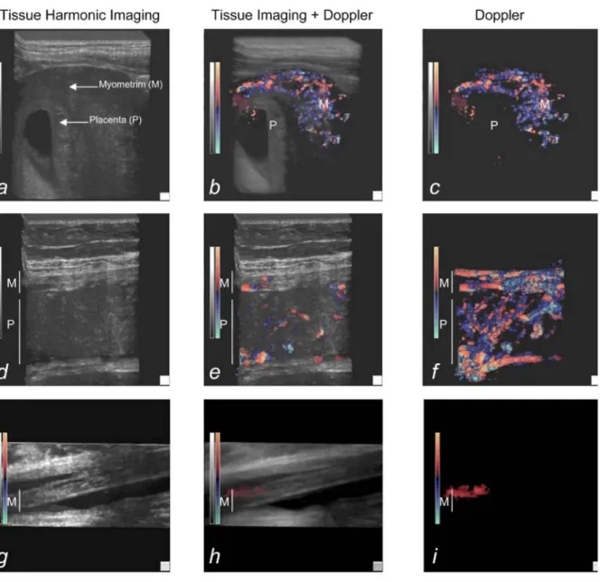
Figure 1: Representative aspect of the uterus and placenta by 3D sonography, a, b, and c, First-trimester pregnancies (7 weeks), d, e, and f, Third-trimester pregnancies; 3D reconstruction focused on the uteroplacental unit (30 weeks), g, h, and i, Third-trimester pregnancies; 3D reconstruction focused on the uterine wall opposite the placenta (30 weeks). Three views of the myometrium and the placenta are presented for each study: High-resolution 3D tissue imaging (a, d, and g), 3D tissue imaging coupled to color Doppler (b, e, and h), and 3D color Doppler (c, f, and i). The myometrium (M) and placenta (P) are indicated on each picture.
Figure 2: Anatomic description of vascular shunts in the myometrium. A term human uterus was obtained after delivery and perfused with polymerizing solution (frontal view of the anterior side of the uterus [a] and cross-section of the uterus [b]). The vascular network was isolated after tissue digestion (frontal view of the anterior side of the uterus [c] and cross-section of the uterus [d]). Thin and superficial vessels (the capillaries) were removed, revealing large and anastomotic vessels (e, original magnification x5). An MRI cross-section of the cast showed that the vascular network predominated in the anterior myometrium (AM) (f).
Statistical analysis
Data are expressed as median and interquartiles. Values in the different groups were compared by using the Kruskall-Wallis test, and significant differences were further analyzed by pairwise comparisons with the Mann-Whitney U test. P values less than .05 were considered to denote significant differences.
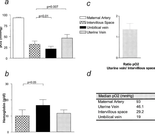
Figure 3: Intramyometrial shunting is functional during pregnancy, a, Oxygen measurements in maternal arterial blood, maternal uterine venous blood, blood form the intervillous space, and blood from the umbilical vein. Samples were taken from 9 patients undergoing cesarean section at term because of breech presentation or a history of multiple cesarean delivery, b, Hemoglobin levels in uterine venous blood, blood from the intervillous space, and blood from the umbilical vein, c, Representative ratio between oxygen levels in the uterine vein and intervillous space.
Results
Three-dimensional color Doppler imaging was used to study uteroplacental vascularization from first trimester to term. Blood flow higher than 3 cm/s could be detected with the Doppler settings used here. No vascular signal was detected in the placenta before 11 weeks of gestation (6/10 patients in the group before 14 weeks of gestation), whereas a rich vascular network was present in the myometrium, next to the placenta (Figure 1,, a, b, and c). Fetal and maternal blood flow was detected in the placenta from the 12th week of gestation. The
maternal/ fetal origin was determined on the basis of the pulsation rate obtained by pulsed Doppler. In the myometrium located below the placental bed, 3D reconstruction that used power Doppler showed a dense, apparently anastomotic vascular network (Figure 1, d, e, and f). No such vascular system was detected in myometrium not involved in placentation (Figure 1, g, h, and i). Thus, 3D power Doppler sonography revealed a dense vascular network in the myometrium below the placental bed, but not in the myometrium located opposite the placental bed.
Vascular anastomoses in the myometrium
We then prepared casts of the uterine vascularization to confirm the presence of vascular anastomoses in the myometrium. After postdelivery hysterectomy for severe hemorrhage, the uterus was perfused with a
polymerizing solution (Figure 2, a and b). The placenta was located on the anterior myometrium (AM). The cast of uterine vessels was obtained after tissue digestion (Figure 2, c and d). Superficial and thin vessels were removed, and the large vessels located within the myometrium were found to constitute an anastomotic network (Figure 2,e).
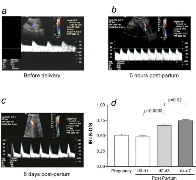
Figure 4: Intramyometrial shunting remains functional after delivery, a, Representative uterine Doppler aspect in an uncomplicated pregnancy at term, b, Representative uterine Doppler aspect 5 hours after delivery, c, Representative uterine Doppler aspect 6 days after delivery, d, Uterine Doppler aspect in normal term
pregnancies (n = 25) and postpartum (n = 22), within 48 hours after delivery, on day 2 or 3 after delivery, and on day 4 to day 7 after delivery.
Arteriovenous shunts are functional in vivo
To confirm that the vascular network was composed of functional arteriovenous anastomoses, we analyzed blood gases in maternal blood drawn from the femoral artery, the uterine vein, and the intervillous space. Fetal blood was sampled from the umbilical vein.
The Po2 value in maternal arterial blood was 93 mm Hg (90-95.6). The Po2 value in the uterine vein (46.15 mm
Hg [38-50.6]) was significantly higher than that in the intervillous space (29.20 mm Hg [28-33.9]), P = .007; Figure 3, a and d). Moreover, the Po2 value in the uterine vein was consistently higher than that in the
intervillous space, with a ratio of 1.48 between the two compartments (Figure 3, c). The Po2 value in the
umbilical vein (19 mm Hg [15.4-17.2]) was significantly lower than that in the intervillous space (29.20 mm Hg [28-33.9] P = .01; Figure 3 a and d). To rule out fetal blood contamination of maternal blood drawn from the
intervillous space, we measured the hemoglobin level in fetal blood and in maternal blood from the uterine vein and intervillous space (Figure 3, b). There was no significant difference between the uterine vein and the intervillous space (11.60 ± 2.24 g/dL vs 10.84 ± 2.50 g/dL, P = .6), whereas the hemoglobin level in fetal blood was significantly higher than that in maternal blood drawn from the intervillous space (16.55 ± 2.23 g/dL vs 10.84 ± 2.50 g/dL, P = .02).
The myometrial anastomotic network remains functional after delivery
Postdelivery uterine artery Doppler values and spectra (Figure 4, b and c) were compared with those obtained during normal third-trimester pregnancies (Figure 4, a).
A RI was used to assess vascular resistance. During the first 48 hours after delivery, resistances were low in the uterine arteries (RI = 0.48 ± 0.07) and similar to those found during pregnancy. Resistance in the uterine vessels gradually increased during the first week after delivery. Vascular resistance was higher on days 2 and 3 than on days 0 and 1 (RI = 0.67 ± 0.06 and 0.48 ± 0.07, respectively; P = .0007). Vascular resistance then increased on days 4 to 7 relative to days 2 and 3 (RI = 0.75 ± 0.05 and 0.67 ± 0.06, respectively; P = .03).
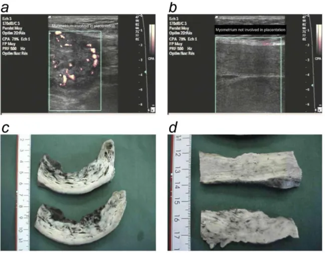
Figure 5: Shunts are located in the basal myometrium but not in the parietal myometrium, a and b,
Representative power Doppler sonographs of the uterus 5 hours after delivery, (a) Power Doppler aspect of the basal myometrium (myometrium involved in placentation). (b) Power Doppler aspect of parietal myometrium (myometrium not involved in placentation). c and d, Anatomic aspect of the uterus 2 hours after delivery, (c) Basal myometrium. Arrowheads show the vascular network, (d) Parietal myometrium.
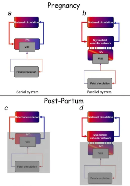
Figure 6: Comparison with the classical model of uteroplacental vascularization, a and b, Schematic drawings of uteroplacental vascularization, (a) Classical model of uteroplacental vascularization. The maternal
circulation and intervillous chamber (IVC) are connected "in series." (b) Proposed model of uteroplacental vascularization. The intervillous chamber and myometrial anastomotic vascular network are connected "in parallel" to the maternal circulation. Large red arrows represent the uterine arterial system. Large blue arrows represent the uterine venous system, c and d, Postpartum modifications in both systems. The gray shading represents the part of the system that is removed after delivery.
The vascular network is located in the placental bed
Several lines of evidence suggested that the vascular network described in this study was located in the myometrium involved in placentation but not in the opposite uterine wall. With the use of 3D color Doppler, marked blood flow was detected in the placental bed (Figure 1, d, e, and ƒ) but not in the opposite uterine wall (Figure 1, g, h, and i). Power Doppler performed a few hours after delivery revealed vascular flow in the myometrium that was involved in placentation (Figure 5, a) but not in the rest of the myometrium (Figure 5, b). Similarly, anatomic data obtained with postpartum hysterectomy specimens showed large vascular lakes in the
placental bed (Figure 5, c) but not in other parts of the uterus (Figure 5, d). The cast experiments also demonstrated that myometrial vascularization was predominant in the placental bed (Figure 3,f). Comment
Current knowledge of human uteroplacental vascularization is mainly based on works by Ramsey and
Donner.2,4,5,8 Vascularization of the pregnant uterus by the maternal circulation can be assimilated to a "series" system in which the maternal blood enters the uterus via the uterine arcuate, radial, and spiral arteries before "spouting" into the intervillous space. After circulating around the villi, maternal blood is drained by the uterine veins and re-enters the maternal circulation. The intervillous space therefore acts as an arteriovenous shunt (Figure 6, a). Recent works, especially in the first trimester of pregnancy, have thrown new light on the organization of uteroplacental vascularization. After its implantation, the trophoblast begins to induce vascular remodeling, first in the endometrium and then in the adjacent myometrium, which can be detected by Doppler imaging.10,11 The stimuli thus triggered create a very rich and dense intramyometrial vascular network located in the basal plate of the uterus. This vascular architecture coincides with changes in the terminal portions of spiral arteries, because of their invasion by extravillous trophoblastic cells.12-14.Trophoblastic villi obtain their
metabolic and gaseous substrates from a fluid that does not contain maternal red cells, the maternal blood having percolated through the trophoblastic plugs. From the end of the first trimester, the hemostatic plugs sealing the uteroplacental arteries gradually dissolve and the maternal blood enters the intervillous space; the placenta becomes hemochorial, and the classical arteriovenous shunt is composed of the intervillous space.19 This mode of circulation persists until delivery, and uteroplacental blood flow increases until term. After placental expulsion, the uterus retracts and the resulting muscle tension creates a physiologic tourniquet that allows clots to form at the detachment site. It is also thought that the conversion of the spiral arteries does not involve their whole length, but that a portion of the radial artery remains vasoreactive, and constriction of this segment prevents postpartum hemorrhage. Uterine artery resistance values should thus increase sharply after delivery. However, our results paradoxically show the persistence of low-resistance uterine circulation, even though the uterine muscle is retracted, the placental shunt disappears, and postpartum hemorrhage is avoided.
Our studies, based on sonography, laboratory analyses (serial blood gas assays), and anatomic investigations (of hysterectomy specimens and uteroplacental vascular casts), have yielded functional and anatomic evidence of an arteriovenous shunt located in the subplacental myometrium. Contrary to the classical model of circulatory communication in "series" in the proposed model, the intervillous space is connected "in parallel" with the uterine circulation (Figure 6, b). Three-dimensional color Doppler images show a rich vascular network with an anastomotic aspect located in the myometrium of the placental bed but not in the parietal myometrium. To confirm this finding, we prepared a vessel cast of a uterus obtained immediately after placental expulsion. Vascular anastomoses 2 to 3 mm in diameter were found throughout the thickness of the myometrium. Such anastomoses had already been described by Heckel and Tobin,9 who demonstrated their arteriovenous nature by injecting colored resins intra-arterially (red resin) and intravenously (blue resin).
We confirmed the functional nature of the subplacental arteriovenous shunt by showing an oxygen gradient between uterine venous blood and the intervillous space, the latter being significantly more desaturated than the former. Similar data were recently published,20 but they were based on specimens of intervillous spaces taken after delivery. In our study, in which the specimens of intervillous spaces were obtained before delivery, fetal blood contamination of the intervillous space during sampling may have explained the lower partial oxygen pressure in the intervillous space than in uterine venous blood. However, the difference in the hemoglobin concentration between the intervillous space (10.84 g/dL) and fetal venous blood (16.55 g/dL) showed that any such contamination would have been minimal and could not have explained the difference in Po2.
Finally, the lack of hemodynamic changes in the postpartum period confirms the persistence of the
intramyometrial arteriovenous shunt. Indeed, according to the "series" model (Figure 6, c), which is challenged by our findings, as the uterus contracts immediately after delivery to prevent postpartum hemorrhage, resistance in the uterine arteries should also increase. In contrast, in the model we propose, the subplacental shunt persists after delivery, ensuring continuity with the maternal systemic circulation (Figure 6, d) and avoiding postpartum hemorrhage. Uterine Doppler studies performed during the first week after delivery showed that uterine resistance remained low for 48 hours after delivery, gradually increasing toward values observed outside pregnancy. Moreover, after delivery, power Doppler studies showed blood flow within vascular lacunae located solely in the part of the myometrium involved in placentation. These vascular connections were confirmed on postpartum hysterectomy specimens. Maternofetal exchanges therefore occur within a system in which the uterine vascular network plays an important role by supplying blood to the intervillous space, which is
high-flow vascular shunt. This system does not exclude previous concepts since the intervillous space remains connected to the uterine vascularization via spiral arteries. The anastomotic network protects the fetal circulation from maternal hypertensive events. The slow intervillous circulation of maternal red cells is atraumatic and facilitates gas transport to the intervillous space. Placental detachment after delivery does not open up high-flow vessels connected directly to the maternal circulation, but rather vessels that are easily compressed by uterine retraction and are in diversion relative to high flow. The human hemochorial placenta is therefore functionally connected "in parallel" to the principal anastomotic vascular network located in the myometrium.
Acknowledgments
We thank Professor P. Kaufmann (Department of Pathology, Aachen, Germany) for technical assistance with vascular casting.
References
1. Hunter W. The gravid uterus. Birmingham; 1774.
2. Ramsey EM. The story of the spiral arteries. J Reprod Med 1981;26:393-9.
3. Ramsey EM, Martin CB Jr, McGaughey HS Jr, Kaiser IH, Donner MW. Venous drainage of the placenta in rhesus monkeys: radiographic studies. Am J Obstet Gynecol 1966; 95:948-55.
4. Ramsey EM. Uteroplacental circulation during labor. Clin Obstet Gynecol 1968;11:78-95. 5. Ramsey EM. Maternal and foetal circulation of the placenta. Ir J Med Sci 1971;140:151-68.
6. Ramsey EM, Chez RA, Doppman JL. Radioangiographic measurement of the internal diameters of the uteroplacental arteries in rhesus monkeys. Am J Obstet Gynecol 1979;135:247-51.
7. Ramsey EM. Excitement. In: Moawad AH, Lindheimer MD, editors. Uterine and placental blood flow. Vol 1. New York: Masson; 1981. 8. Ramsey EM. What we have learned about placental circulation. J Reprod Med 1985;30:312-7.
9. Heckel GP, Tobin CE. Arteriovenous shunts in the myometrium. Am J Obstet Gynecol 1956;71:199-205.
10. Jaffe R, Woods JR Jr.. Color Doppler imaging and in vivo assessment of the anatomy and physiology of the early uteroplacental circulation. Fertil Steril 1993;60:293-7.
11. Jaffe R. Development of early uteroplacental circulation. Early Pregnancy 2001;5:34-5.
12. Damsky CH, Librach C, Lim KH, Fitzgerald ML, McMaster MT, Janatpour M, et al. Integrin switching regulates normal tropho-blast invasion. Development 1994;120:3657-66.
13. Genbacev O, McMaster MT, Fisher SJ. A repertoire of cell cycle regulators whose expression is coordinated with human cytotro-phoblast differentiation. Am J Pathol 2000;157:1337-51.
14. Zhou Y, Fisher SJ, Janatpour M, Genbacev O, Dejana E, Wheelock M, et al. Human cytotrophoblasts adopt a vascular phenotype as they differentiate: a strategy for successful endovas-cular invasion? J Clin Invest 1997;99:2139-51.
15. Jaffe R, Jauniaux E, Hustin J. Maternal circulation in the first-trimester human placenta—myth or reality? Am J Obstet Gynecol 1997;176:695-705.
16. Burton GJ, Jauniaux E. Maternal vascularisation of the human placenta: does the embryo develop in a hypoxic environment? Gynecol Obstet Fertil 2001;29:503-8.
17. Burton GJ, Jauniaux E, Watson AL. Maternal arterial connections to the placental intervillous space during the first trimester of human pregnancy: the Boyd collection revisited. Am J Obstet Gynecol 1999;181:718-24.
18. Hustin J, Schaaps J-P. Echo(cardio)graphic and anatomic studies of the maternotrophoblastic border during the first trimester of pregnancy. Am J Obstet Gynecol 1987; 157(1): 162-8.
![Figure 2: Anatomic description of vascular shunts in the myometrium. A term human uterus was obtained after delivery and perfused with polymerizing solution (frontal view of the anterior side of the uterus [a] and cross-section of the uterus [b])](https://thumb-eu.123doks.com/thumbv2/123doknet/6202109.160065/4.892.128.770.230.921/anatomic-description-vascular-myometrium-perfused-polymerizing-solution-anterior.webp)