A Comparison of 2 Mitral Annuloplasty Rings
for Severe Ischemic Mitral Regurgitation:
Clinical and Echocardiographic Outcomes
Khalil Fattouch, MD, PhD,
*,†Marco Moscarelli, MD, PhD,
‡,§Sebastiano Castrovinci, MD,
*Francesco Guccione, MD, PhD,
*Pietro Dioguardi, MD,
*Giuseppe Speziale, MD, PhD,
‡and
Patrizio Lancellotti, MD, PhD
¶,#Controversies regarding the choice of annuloplasty rings for treatment of ischemic mitral regurgitation still exist. Aim of the study is to compare early performance of 2 different rings in terms of rest and exercise echocardio-graphic parameters (transmitral gradient, systolic pulmonary artery pressure, and mitral valve area), clinical outcomes, and recurrence of mitral regurgita-tion. From January 2008 till December 2013, prospectively collected data of patients who underwent coronary artery bypass grafting and undersizing mitral valve annuloplasty for severe chronic ischemic mitral regurgitation at our Institution were reviewed. A total of 93 patients were identified; among them 44 had semirigid Memo 3D ring implanted (group A) whereas 49 had a rigid profile 3D ring (group B). At 6 months, recurrent ischemic mitral regurgitation, equal or more than moderate, was observed in 4 and 6 patients in the group A and B, respectively (P ¼ 0.74). Group A showed certain improved valve geometric parameters such as posterior leaflet angle, tenting area, and coaptation depth. Transmitral gradient was significantly higher at rest in the group B (P o 0.0001). During exercise, significant increase of transmitral gradient and systolic pulmonary artery pressure was observed in group B (Po 0.0001). Mitral valve area was not statistically significantly smaller at rest in between groups (P¼ 0.09); however, it significantly decreased with exercise in group B (P ¼ 0.01). At midterm follow-up, patients in group B were more symptomatic. In patients with chronic ischemic mitral regurgitation, use of semirigid Memo 3D ring when compared to the rigid Profile 3D may be associated with early improved mitral valve geometrical conformation and hemodynamic profile, particularly during exercise. No difference was observed between both groups in recurrent mitral regurgitation.
Semin Thoracic Surg]:]]]–]]] I 2016 Elsevier Inc. All rights reserved. Keywords: Mitral Regurgitation (MR), Ischemic mitral regurgitation (IMR), Mitral Valve Repair (MVR), Coronary artery bypass grafting (CABG)
INTRODUCTION
Chronic ischemic mitral regurgitation (IMR) is a secondary mitral regurgitation (MR) characterized by apparently normal leaflets and subvalvular apparatus. The mitral incompetence is the consequence of a systolic restrictive motion of the leaflets (leaflet tethering, type IIIb) or annular dilatation (type I) according to Carpentier's classification, possibly exacerbated by ventricular remodeling.1When dealing with IMR, echocardiog-raphy plays a major role in identifying the types of leaflet tethering and the change of mitral valve apparatus geometry.2 Annular dimension, coaptation depth (CD), tenting area (TA) or volume, interpapillary distance, and leaflet angles are the most important echocardiographic parameters to report, representing
*Department of Cardiovascular Surgery, GVM Care and Research,
Maria Eleonora Hospital, Palermo, Italy
†Department of Surgery and Cancer, University of Palermo, Palermo, Italy ‡Department of Cardiovascular Surgery, GVM Care and Research, Anthea
Hospital, Bari, Italy
§Nationale Heart Lung Institute, London UK
¶Department of Cardiology, Heart Valve Clinic, CHU Sart Tilman, GIGA
Cardiovascular Sciences, University of Liege Hospital, Liege, Belgium
#GVM Care and Research, E.S. Health Science Foundation, Lugo (RA), Italy
Abstract presented at AATS Mitral Conclave 2015, 23-25 April, NY, USA. Address reprint requests to Khalil Fattouch, MD, PhD, Department of Cardiovascular Surgery, GVM Care and Research, Maria Eleonora Hospital, Viale Regione siciliana, 1571, 90100, Palermo, Italy. E-mail: khalilfattouch@hotmail.com
Changes in mean trans-mitral gradient after surgery and at 6 months follow-up.
Central Message
The semirigid annuloplasty ring may show better early performance than a rigid saddle-shape ring in hemodynamic profile at rest and with exercise. No effect of recurrent MR was found.
Perspective Statement
In a context of chronic ischemic mitral regur-gitation, the use of a semirigid ring (memo 3D) can be associated with early improved rest and stress echocardiographic parameters and reduced NYHA class at midterm follow-up. However, it does not add benefits in reduction of recurrent mitral regurgitation when com-pared with a rigid ring.
predictors of recurrent MR after restrictive mitral valve annuloplasty (MVA).2 To date, restrictive MVA rep-resents the most performed techniques used to treat patients with secondary MR.3 Several studies have shown that the recurrence of MR at 1-year remains high (10%-30%).4-6 Moreover, it has been reported that restrictive MVA could be associated with impaired hemodynamic profile with higher trans-mitral gradient and systolic pulmonary artery pressure (PAP) at exercise and worsening in functional New York Heart Association (NYHA) class.7Several types of prosthetic rings exist to remodel the mitral annulus. Complete prosthetic rings are often preferred over a band because of the lower recurrent MR rate.8 Com-plete rigid or semirigid rings have different shapes, orifice areas, and septolateral dimensions, and further-more, they have controversial results in recurrent MR and mitral valve conformation changes.9Main aim of this study is to compare early performance of a semirigid and a saddle-shaped rigid annuloplasty rings at rest and with exercise in patients with IMR under-going restrictive MVA associated with coronary artery bypass surgery and to assess midterm clinical status. MATERIALS AND METHODS
Population
The present study is a retrospective analysis of prospectively recorded data. From January 2008 to
December 2013, patients who underwent restrictive MVA for severe chronic IMR in our institutions were reviewed. Patients fulfilling the following inclusion criteria were eligible to be included in this study: (1) history of ischemic cardiomyopathy, chronic severe IMR owing to systolic restrictive leaflet motion with or without mitral annulus dilatation, (2) sinus rhythm, (3) narrow QRS (o120 ms), and (4) absence of other mitral valve pathology. Further, 93 patients were identified; among them, 44 patients had semirigid memo 3D (Sorin) ring implanted (group A) and 49 had a rigid Profile 3D (Medtronic) ring (group B). Ethical board approved the study, and individual consent was waived. Demographics, clinical, and echocardiographic data are illustrated in
Table 1.
Operative Technique
All procedures were performed through median sternotomy on normothermic cardiopulmonary bypass with intermittent antegrade blood cardio-plegia. All patients underwent coronary artery bypass grafting concomitant to restrictive MVA. Mitral annuloplasty ring size was determined by standard measurement of the intertrigonal distance and anterior leaflet surface, and then downsizing by one for the Memo 3D group was performed. For the Profile 3D group, size of ring was equal to the
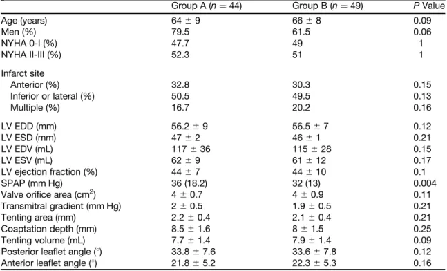
Table 1. Baseline Clinical and Echocardiographic Characteristics
Group A (n¼ 44) Group B (n¼ 49) P Value
Age (years) 64⫾ 9 66⫾ 8 0.09 Men (%) 79.5 61.5 0.06 NYHA 0-I (%) 47.7 49 1 NYHA II-III (%) 52.3 51 1 Infarct site Anterior (%) 32.8 30.3 0.15 Inferior or lateral (%) 50.5 49.5 0.13 Multiple (%) 16.7 20.2 0.16 LV EDD (mm) 56.2⫾ 9 56.5⫾ 7 0.12 LV ESD (mm) 47⫾ 2 46⫾ 1 0.21 LV EDV (mL) 117⫾ 36 115⫾ 28 0.15 LV ESV (mL) 62⫾ 9 61⫾ 12 0.17 LV ejection fraction (%) 44⫾ 7 44⫾ 10 0.1 SPAP (mm Hg) 36 (18.2) 32 (13) 0.004
Valve orifice area (cm2) 4⫾ 0.7 4⫾ 0.9 0.11
Transmitral gradient (mm Hg) 2⫾ 0.5 1.9⫾ 0.5 0.21
Tenting area (mm) 2.2⫾ 0.4 2.1⫾ 0.4 0.21
Coaptation depth (mm) 8.5⫾ 1.6 8⫾ 1.5 0.25
Tenting volume (mL) 7.7⫾ 1.4 7.9⫾ 1.4 0.09
Posterior leaflet angle (1) 33.8⫾ 7.6 33.6⫾ 7.8 0.12
Anterior leaflet angle (1) 21.8⫾ 5.2 22.3⫾ 5.3 0.16
Group A, semirigid memo 3D; group B, rigid profile 3D. Values are expressed in mean ⫾ standard deviation or median and IQR. EDD, end-diastolic diameter; ESD, end-systolic diameter; EDV, end-diastolic volume; ESV, end-systolic volume; LV, left ventricle; NYHA, New York Heart Association; SPAP, systolic pulmonary arterial pressure.
surface of anterior leaflet. This was a single-surgeon series (K.F.).
Echocardiographic Examination
All patients underwent preoperative and post-operative (same day of hospital discharge) trans-thoracic echo examinations. Intraoperative transesophageal examination was also performed routinely. All echocardiographic examinations were performed using the Philips Healthcare IE 33 echo-cardiograph system. All Doppler-echoecho-cardiographic recordings were stored on a dedicated workstation for off-line subsequent analysis. Measurements were performed blind of the surgical data. For each measurement, a minimum of 2 cardiac cycles was averaged. Left ventricle (LV) diastolic and end-systolic dimensions were measured from parasternal acquisitions. The biplane method of discs' summa-tion (modified Simpson's rule) was applied to quantify LV end-diastolic and end-systolic volumes and ejection fraction. The severity of MR appreciated using an integrated approach. MR was quantitated using the proximal isovelocity surface area and10 using the Doppler method, and the measurement of vena contracta and effective regurgitant orifice area ofZ20 mm2was the threshold of severity.11 Peak and mean transmitral pressure gradients were calcu-lated using the modified Bernoulli equation. Systolic PAP was derived from the regurgitant jet of tricuspid regurgitation using systolic transtricuspid pressure gradient and with the addition of 10 mm Hg for right atrial pressure.11Septolateral annulus diameters were measured from parasternal long-axis view. CD and TA were measured from the 4-chamber view. Mitral valve area was determined by 2-dimensional planim-etry. 3D echocardiographic measurements were performed using the MV Q Lab software. Results from preopeative and postoperative 2D transthoracic echocardiographic examinations were compared regarding the mitral valve geometric changes (pos-terior leaflet angle, TA, and CD). Heart rate and systolic blood pressure value were also recorded at rest and with exercise.
Exercise Echocardiography
Beta-blockers were withdrawn the day of the test. A symptom-limited, graded bicycle exercise test was performed in the semisupine position on a tilting exercise table in all patients at follow-up. After an initial workload of 25 W maintained for 2 minutes, the workload was increased every 2 minutes by 25 W. Blood pressure and a 12-lead electro-cardiogram were recorded every 2 min. Both the
transmitral pressure gradient and the systolic PAP were recorded.10,12
Outcome Data
All survived patients underwent a semisupine exercise echocardiography test to assess changes in systolic PAP and transmitral pressure gradient after 6 months after the operation at our main echo laboratory. In-hospital morbidity (renal failure, res-piratory distress, bleeding, mediastinitis, etc) and mortality were evaluated in all patients. The follow-up was obtained from patient interviews, hospital record reviews, or personal communications with the patient's physician. Death, NYHA functional class, and readmission for heart failure to hospital represented the collected data for midterm outcome. Cardiac-related death was defined as a sudden death or death related to myocardial infarction, congestive heart failure, or cardiac arrhythmias.
Statistical Analysis
Patients' demographic, clinical, and operative data were summarized as mean⫾ standard deviation for normally distributed continuous variables, otherwise median and interquartile range (IQR) for nonpara-metric continuous variables or proportion or prev-alence for categorical variables was used. Differences between subgroups were compared using theχ2 test (2-tailed) for categorical variables and the Student's t-test or the Mann-Whitney U test or Kruskal-Wallis test, as appropriate, for continuous variables. Comparisons of the data over time were performed by analysis of variance between groups. Values of P less than 0.05 were considered to indicate statistical significance. All statistical analyses were performed with STATISTICA version 10 (StatSoft Inc, Tulsa, Okla).
RESULTS
Patients' Characteristics
Patients' characteristics are listed inTable 1. Mean age was 65⫾ 10 years and 70% were men. The site of infarction was inferior or lateral in 50%, anterior in 31.5%, and multiple myocardial necrotic sites in 18.5%. In a context of a small sample size, no statistically significant differences were found between groups regarding baseline clinical and echocardiographic variables, except for systolic PAP being significantly higher in the group A (P ¼ 0.004). Interestingly, mitral valve geometric param-eters such as TA and volume, CD, and posterior and anterior leaflet angles, were similar in both groups (P ¼ 0.21, 0.09, 0.25, 0.12, and 0.16, respectively).
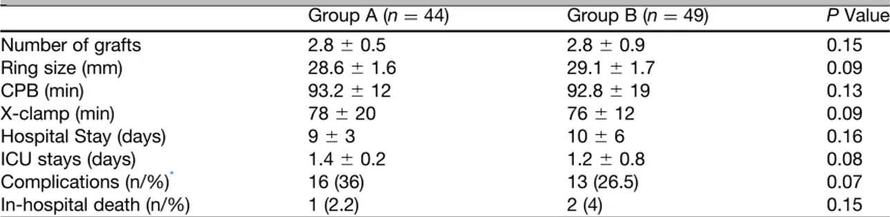
Surgical Data and Postoperative Outcomes The mean time of cardiopulmonary bypass was 93.2 ⫾ 12 minutes in the group A vs 92.8 ⫾ 19 minutes in the group B (P ¼ 0.13). The mean time of aortic cross-clamp was 78⫾ 20 minutes in the group A vs 76⫾ 12 minutes in the group B (P ¼ 0.09). The mean number of grafts was 2.8 ⫾ 0.5 for group A and 2.8⫾ 0.9 for group B (P ¼ 0.15). Hospital stay was 9⫾ 3 days in the group A vs 10 ⫾ 6 days in the group B (P ¼ 0.16). No significant differences between groups were found on postoperative complications and in-hospital deaths (Table 2). All survived patients were discharged with trivial or without MR.
Follow-Up Data
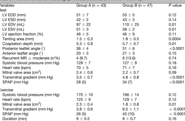
At 6 months follow-up, rest and exercise echo-cardiographic examination was obtained in all patients. Recurrent Z moderate MR was observed in 4 (9.7%) patients in the group A and 6 (13.6%) in the group B (P ¼ 0.74). Semirigid Memo 3D annuloplasty ring group showed superior positive reduction of CD and TA than the Profile group (Table 3). Of note, the posterior leaflet angle was significantly worsened in the rigid Profile 3D annu-loplasty ring (361 ⫾ 41 group A vs 511 ⫾ 61 group B, P o 0.0001) but not the anterior leaflet angle (P ¼ 0.15). There were no significant differences between groups in LV diameters and LV ejection fraction (Table 3). The transmitral pressure gradient was higher at discharge and at 6 months rest echo in the group B (3.5 ⫾ 0.7 mm Hg group A vs 4.8 ⫾ 0.8 mm Hg group B,P o 0.0001) (Fig. 1). Systolic PAP was not statistically different at discharge but became higher at 6 months follow-up rest in group B (28 [IQR ¼ 5] vs 34 (5) mm Hg, P o 0.0001) (Fig. 2). The exercise-induced increase in transmitral pressure gradient (3.8⫾ 0.6 mm Hg group A vs 6.5 ⫾ 1.1 mm Hg group B, P o 0.0001), and the resulting increase in systolic PAP (26 [IQR 5] mm
Hg group A vs 40 [IQR¼ 10] mm Hg group B, P o 0.0001) (Fig. 2) were also greater in the group B. The mitral valve area was smaller in group B at rest (2.4⫾ 0.6 cm2 vs 2.2 ⫾ 0.7 cm2, P ¼ 0.09) and signifi-cantly decreased with exercise (2.3⫾ 0.4 cm2vs 1.8 ⫾ 0.8 cm2,P ¼ 0.01). Midterm telephonic follow-up
was 100% complete (median¼ 36 months, IQR ¼ 24, max 68 months); 2 (4.6%) and 3 (6.3%) patients died in group A and B, respectively. At midterm follow-up, 37 (90.2%) and 27 (61.3%) patients were in NYHA class 0-I and 4 (9.8%) and 17 (38.7%) in class II-III (group A and B, respectively,P ¼ 0.02), and the hospital readmission for congestive heart failure was significantly higher in group B (2.3% vs 8.8% group A and B, respectively P o 0.0001) (Table 4).
DISCUSSION
Mitral Valve Repair and Recurrent MR Chronic IMR remains one of the most complex and unresolved aspects of the treatment of valvular heart disease.2 Restrictive MVA, which was first introduced by Bolling et al3 in patients with end-stage ischemic and nonischemic dilated cardiomy-opathy, has become a standard procedure for treat-ing secondary MR. Undersized rtreat-ings may reduce the leaflet area necessary to cover the orifice, move the leaflets closer, together by reducing the anteropos-terior (septolateral) annular diameter, and thus facilitate effective coaptation.13-16 Furthermore, restrictive annuloplasty has been associated with a high recurrence rate of MR (10%-30%), partly owing to further distortion of the mitral valve apparatus (ie, increased posterior leaflet tethering) and continuous LV remodeling.17The group of Dion et al15stressed that sufficient coaptation reserve might prevent recurrent MR, although this therapeutic approach does not directly address tethering by the remodeled LV.18,19 It has been well know that recurrent MR depends not only from failure in surgical techniques
Table 2. Surgical Data and Postoperative Outcomes
Group A (n¼ 44) Group B (n¼ 49) P Value
Number of grafts 2.8⫾ 0.5 2.8⫾ 0.9 0.15
Ring size (mm) 28.6⫾ 1.6 29.1⫾ 1.7 0.09
CPB (min) 93.2⫾ 12 92.8⫾ 19 0.13
X-clamp (min) 78⫾ 20 76⫾ 12 0.09
Hospital Stay (days) 9⫾ 3 10⫾ 6 0.16
ICU stays (days) 1.4⫾ 0.2 1.2⫾ 0.8 0.08
Complications (n/%)* 16 (36) 13 (26.5) 0.07
In-hospital death (n/%) 1 (2.2) 2 (4) 0.15
Group A, semirigid memo 3D; group B, rigid profile 3D; CPB, cardiopulmonary bypass; ICU, intensive care unit.
*Mediastinitis, low cardiac output syndrome, atrialfibrillation, renal failure, respiratory failure, major bleeding. X-clamp: cross-clamp time.
Table 3. Rest and Stress Echocardiographic Data Obtained at 6-Month Follow-Up
Variables Group A (n¼ 43) Group B (n¼ 47) P value
Rest LV EDD (mm) 51⫾ 7 53⫾ 5 0.12 LV ESD (mm) 42⫾ 2 43⫾ 3 0.14 LV EDV (mL) 97⫾ 22 110⫾ 25 0.01 LV ESV (mL) 51⫾ 3 58⫾ 2 0.01 LV ejection fraction (%) 46⫾ 5 46⫾ 9 0.11 Tenting area (mm) 1.5⫾ 0.3 1.8⫾ 0.5 0.0004 Coaptation depth (mm) 5.3⫾ 0.8 5.7⫾ 0.7 0.01
Posterior leaflet angle (1) 36⫾ 4 51⫾ 6 o0.0001
Anterior leaflet angle (1) 20⫾ 3 21⫾ 3 0.15
Recurrent MRZ moderate (n/%) 4 (9.7) 6 (13.6) 0.74
Systolic blood pressure (mm Hg) 128⫾ 7 127⫾ 9 0.18
Heart rate (bpm) 70⫾ 5 71⫾ 7 0.16
Mitral valve area (cm2) 2.4⫾ 0.6 2.2⫾ 0.7 0.09
Transmitral gradient (mm Hg) 3.5⫾ 0.7 4.8⫾ 0.8 o0.0001
SPAP (mm Hg) 28 (5) 34 (7) o0.0001
Exercise
Systolic blood pressure (mm Hg) 170⫾ 10 166⫾ 14 0.12
Heart rate (bpm) 125⫾ 9 129⫾ 7 0.12
Mitral valve area (cm2) 2.3⫾ 0.4 1.8⫾ 0.8 0.01
Transmitral gradient (mm Hg) 3.8⫾ 0.6 6.5⫾ 1.1 o 0.0001
SPAP (mm Hg) 26 (5) 45 (10) o 0.0001
Duration (min) 9⫾ 0.5 8⫾ 0.7 0.16
Group A, semirigid memo 3D; Group B, rigid profile 3D. EDD, diastolic diameter; ESD, systolic diameter; EDV, end-diastolic volume; ESV, end-systolic volume; pulmonary arterial pressure.
Figure 1. Changes in mean transmitral gradient after surgery and at 6 months follow-up both at rest and during exercise echocardiography. Group A memo 3D semirigid (light blue) vs Group B profile 3D rigid (dark blue). Preoperative, 2⫾ 0.5 vs 1.9 ⫾ 0.5 (P ¼ 0.21); postoperative (discharge) 2 ⫾ 0.5 vs 3.4 ⫾ 0.9 (P o 0.001); follow-up rest (6 months) 3.5⫾ 0.7 vs 4.8 ⫾ 0.8 (P o 0.001); follow-up stress 3.8 ⫾ 0.6 vs 6.5 ⫾ 1.1 (P o 0.001). (Color version offigure is available online athttp://www.semthorcardiovascsurg.com.)
Figure 2. Changes in pulmonary systolic artery pressure after surgery and at 6 months follow-up both at rest and during exercise echocardiography. Group A memo 3D semirigid (light blue) vs group B profile 3D rigid (dark blue). Preoperative 36(18.2) vs 32(13), (P¼ 0.004); postoperative (discharge) 28 (5) vs 30(10) (P o 0.001); follow-up rest (6 months) 28 (5) vs 34 (5) (Po 0.001); follow-up stress 25(5) vs 45 (10) (P o 0.001). Data are presented with median and IQR. (Color version offigure is available online athttp://www.
and patients selection but also from ongoing LV remodeling in the setting of ischemic mitral diseases. However, in our series, there were no differences in recurrent MR between the 2 groups.
Hemodynamic Performance After Undersized Mitral Annuloplasty
It is known that the annulus restriction may reduce effective orifice areas and increase transmitral pres-sure gradients, resulting in functional mitral steno-sis.20 This is traditionally attributed to a potentially excessive reduction of mitral annular area by the ring and abnormal leaflet opening function (diastolic tethering of anterior mitral leaflet).6,7 Whether the degree of mitral stenosis created by restrictive mitral annuloplasty limits patients' exercise capacity and compromises the patient outcome remains contro-versial.21,22However, most studies have based steno-sis grading on the mean transmitral pressure gradient at rest that largely underestimates the hemodynamic effect of stenosis.23,24Exercise Doppler echocardiog-raphy may provide a comprehensive evaluation of the consequences of stenosis and of its dynamic nature as for IMR.10The more the valve opening is restricted during exercise, the higher is the increase in trans-mitral pressure gradient and in pulmonary pressure, and the lower is the exercise tolerance. Bertrand et al7 have recently confirmed that indexed effective orifice area at peak exercise is an independent predictor of exercise capacity and clinical outcome. In our series, the transmitral pressure gradient was already higher at rest in the group B (3.5⫾ 07 mm Hg group A vs 4.8 ⫾ 0.8 mm Hg group B, P o 0.0001). The exercise-induced increase in transmitral pressure gradient (3.8 ⫾ 0.6 mm Hg group A vs 6.5 ⫾ 1.1 mm Hg group B,P o 0.0001) and the resulting increase in systolic PAP (26 (5) mm Hg vs 45 (10) mm Hg,P o 0.0001) (Figs. 1and2) were also greater in the group B. Furthermore, we speculate that the high incidence of hospital readmission for congestive heart failure symptoms was observed in group B (2.3% group A vs 8.8% group B, P o 0.0001). Moreover, we observed that mitral valve area was lower in group B with exercise (2.3⫾ 0.4 mm in group A vs 1.8⫾ 0.8 mm in group B, P = 0.01).
Semirigid vs Saddle-Shaped Rigid Annuloplasty Ring
The techniques of mitral valve reconstruction have been well established, but controversies remain regarding the types of annuloplasty rings.9 The available annuloplasty rings are rigid,flexible, com-plete, partial, or semirigid orflexible. Saddle-shaped annuloplasty rings are being increasingly used dur-ing mitral valve repair. They might provide a more favorable and uniform annular force distribution when compared to theflat rings.25,26By diminishing mitral leaflet strain and improving leaflet coaptation geometry, saddle-shaped annuloplasty may thus enhance repair durability. Therefore, the mitral valve reconstruction primarily aims at restoring not only a normal valve anatomy but also a normal valve function.1
The design and structure of annuloplasty rings are constantly evolving. The semirigid Memo 3D annulo-plasty ring is capable of mimicking the physiological 3D motion of the native mitral annulus and accom-modating the anatomical shape while remodeling the mitral annulus and ensuring the leaflet coaptation during systole. The rigid Profile 3D annuloplasty ring has a unique design based on the saddle-shaped geometry of normal human mitral annuli. Although the general goal of these devices is the same, namely, to increase leaflet coaptation and to support the posterior annulus against dilation, the extent of change in mitral valve configuration is not uniform. For instance, the septolateral distance is shorter in the rigid Profile 3D annuloplasty ring than in the semirigid Memo 3D annuloplasty ring (ie, for 28 mm size ring the septo-lateral distance is 15.6 mm and 17.8 mm, respectively, for Memo 3D and Profile 3D; data by manufacturing company), and the orifice area is smallest. In our study, we confirmed that the magnitude and direction of improvement in mitral valve geometric parameters was considerably different between groups. With the semi-rigid Memo 3D annuloplasty ring, except for the anterior leaflet angle, all the parameters improved. Conversely, with the rigid Profile 3D annuloplasty ring, the posterior leaflet angle worsened after the implanta-tion probably because of the smallest septolateral distance with respect to Memo 3D ring (increased posterior leaflet tethering). Moreover, the CD and TA
Table 4. Clinical Midterm Follow-Up
Variables Group A (n¼ 43) Group B (n¼ 47) P Value
Death 2 (4.6) 3 (6.3) 0.12
NYHA 0-I 37 (90.2) 27 (61.3) 0.2
NYHA II-III 6 (9.8) 20 (38.7) 0.002
Hospital readmission for CHF symptoms 2.3⫾ 0.2 8.8⫾ 1.6 o0.001
are smaller in Memo 3D vs 3D profile ring. Therefore, we can speculate that these observations are because of the fact that the Memo 3D ring can mimic the native motion of the mitral annulus specifically in end-systole and improve the coaptation between the leaflets. However, in this series, restrictive annuloplasty proved to be effective regardless of the ring used, as there was no significant difference in recurrent MR between the 2 groups.
Previous studies have highlighted the potential risk of functional mitral stenosis after restrictive annulo-plasty with the Carpentier-Edwards Physio ring (Edwards Lifesciences Corporation), but not with the asymmetric IMR ETlogix ring (Edwards Life-sciences Corporation).27The different geometric and functional characteristics of the IMR ETlogix ring have been largely incriminated in these differences. Whether semirigid rings provide more reliable func-tional repair results than saddle-shaped rigid rings have not been evaluated. In our study, the semirigid Memo 3D annuloplasty ring was associated with a better hemodynamics (lower transmitral pressure gradient and systolic PAP) at rest and during exercise and clinical status (lower NYHA functional class). Moreover, the mitral valve area was larger rather than in rigid Profile 3D annuloplasty ring. The hemody-namic benefit of the semirigid Memo 3D annulo-plasty ring has been recently outlined.28Conversely, the rate of exercise-induced increase in transmitral pressure gradient and the resulting rise in systolic PAP was significantly higher with the rigid Profile 3D annuloplasty ring. These data suggest that the semi-rigid Memo 3D annuloplasty ring has less potential risk of postoperative functional stenosis. This could possibly be related to a structural advantage of this ring that is capable to mimicking the normal motion of the annulus, which preserves both the anteropos-terior movement and folding dynamics.29However, we believe that the improvement in tenting param-eters observed in Memo 3D group is not necessary attributed to the use of ring in itself but also depend on LV reverse remodeling and subsequent geometric mitral apparatus modifications.
LIMITATIONS
We can identify several limitations of our study. First, this was a single-center study with limited
sample size. The complexity of the disease and the potential heterogeneity of the patients enrolled might have introduced some bias. This might explain some subtle differences between the 2 study groups at study baseline. However, our population repre-sented the patients seen in our Heart Valve Clinic. The right atrial pressure was estimated at 10 mm Hg both at rest and during exercise. Resting right atrial pressure is extensively variable between subjects. In addition, this estimation may also miss the potential influence of exercise-induced changes in right atrial pressure. Nevertheless, the noninvasive evaluation of right atrial pressure during exercise (ie, when venous compliance is known to decrease) with noninvasive methods such as Doppler echocardiography remains difficult, is probably subject to low accuracy, and is not validated. Moreover, right atrial pressure is frequently assumed to be 5 mm Hg in normal subjects30 and 10 mm Hg in patients with heart disease.31Though we have demonstrated higher risk of functional mitral stenosis in patients receiving a Profile rigid ring, this cannot be generalized to all category or rigid rings; moreover, we cannot say whether or not this actually results in a poorer clinical outcomes or survival rate in the long run. Long-term follow-up data and more investigations are needed to address this issue.
CONCLUSION
Undersized MVA is still the most popular techni-que used to treat IMR despite that recurrent MR is still high and functional mitral stenosis should occur. In our study, the semirigid Memo 3D annuloplasty ring was associated with enhanced hemodynamic per-formance at rest and during exercise, which was translated by a reduced risk of postoperative func-tional stenosis and a better clinical funcfunc-tional status. Despite the improvement in tenting parameters in the semirigid group, no difference was observed between the 2 groups for MR recurrence. Up till now, no data may suggest that semirigid rings could be preferred to rigid one for treating IMR. For a future perspective in valve-in-ring implantation, the use of semirigid ring would be preferred. However, continuous investiga-tions are mandatory to clarify thesefindings.
1. Carpentier A: Cardiac valve surgery—The French correction. J Thorac Cardiovasc Surg 86:323-337, 1983
2. Lancellotti P, Zamorano JL, Vannan MA: Imag-ing challenges in secondary mitral regurgitation:
Unsolved issues and perspectives. Circ Cardio-vasc Imaging 7:735-746, 2014
3. Bolling SF, Pagani FD, Deeb GM, et al: Intermediate-term outcome of mitral recon-struction in cardiomyopathy. J Thorac
Cardiovasc Surg 115:381-386, 1998 [discus-sion 387-388]
4. LaPar DJ, Acker MA, Gelijns AC, et al: Repair or replace for severe ischemic mitral regur-gitation: Prospective randomized multicenter
data. Ann Cardiothorac Surg 4:411-416, 2015
5. Acker MA, Dagenais F, Goldstein D, et al: Severe ischemic mitral regurgitation: Repair or replace?J Thorac Cardiovasc Surg 150: 1425-1427, 2015
6. Kron IL, Hung J, Overbey JR, et al: Predicting recurrent mitral regurgitation after mitral valve repair for severe ischemic mitral regurgitation. J Thorac Cardiovasc Surg 149(752-761):e751, 2015
7. Bertrand PB, Gutermann H, Smeets CJ, et al: Functional impact of transmitral gradients at rest and during exercise after restrictive annu-loplasty for ischemic mitral regurgitation. J Thorac Cardiovasc Surg 148:183-187, 2014
8. McGee EC, Gillinov AM, Blackstone EH, et al: Recurrent mitral regurgitation after annulo-plasty for functional ischemic mitral regurgita-tion. J Thorac Cardiovasc Surg 128:916-924, 2004
9. Wan S, Lee AP, Jin CN, et al: The choice of mitral annuloplastic ring—Beyond surgeon's preference. Ann Cardiothorac Surg 4:261-265, 2015
10. Lancellotti P, Magne J: Stress echocardiography in regurgitant valve disease. Circ Cardiovasc Imaging 6:840-849, 2013
11. Nishimura RA, Otto CM, Bonow RO, et al: 2014 aha/ACC guideline for the management of patients with valvular heart disease: Executive summary: A report of the American college of cardiology/American heart association task force on practice guidelines. J Am Coll Cardiol 63:2438-2488, 2014
12. Tumminello G, Lancellotti P, Lempereur M, et al: Determinants of pulmonary artery hyper-tension at rest and during exercise in patients with heart failure. Eur Heart J 28:569-574, 2007
13. Schwammenthal E: Undersized and over-stretched: Mitral mechanics after restrictive
annuloplasty. J Am Coll Cardiol 65:462-464, 2015
14. Gorman 3rd JH, Gorman RC, Jackson BM, et al: Annuloplasty ring selection for chronic ische-mic mitral regurgitation: Lessons from the ovine model. Ann Thorac Surg 76:1556-1563, 2003
15. Bax JJ, Braun J, Somer ST, et al: Restrictive annuloplasty and coronary revascularization in ischemic mitral regurgitation results in reverse left ventricular remodeling. Circulation 110: II103-II108, 2004
16. Otsuji Y, Gilon D, Jiang L, et al: Restricted diastolic opening of the mitral leaflets in patients with left ventricular dysfunction: Evi-dence for increased valve tethering. J Am Coll Cardiol 32:398-404, 1998
17. Hung J, Solis J, Handschumacher MD, et al: Persistence of mitral regurgitation following ring annuloplasty: Is the papillary muscle out-side or inout-side the ring?J Heart Valve Dis 21: 218-224, 2012
18. Lancellotti P, Marwick T, Pierard LA: How to manage ischaemic mitral regurgitation. Heart 94:1497-1502, 2008
19. Fattouch K, Murana G, Castrovinci S, et al: Mitral valve annuloplasty and papillary muscle relocation oriented by 3-dimensional transeso-phageal echocardiography for severe functional mitral regurgitation. J Thorac Cardiovasc Surg 143:S38-S42, 2012
20. Magne J, Pibarot P, Dagenais F, et al: Preoper-ative posterior leaflet angle accurately predicts outcome after restrictive mitral valve annulo-plasty for ischemic mitral regurgitation. Circu-lation 115:782-791, 2007
21. Kainuma S, Taniguchi K, Daimon T, et al: Does stringent restrictive annuloplasty for functional mitral regurgitation cause functional mitral stenosis and pulmonary hypertension?Circula-tion 124:S97-106, 2011
22. Deja MA, Zak A, Malinowski M, et al: Restric-tive mitral annuloplasty does not limit exercise
capacity. Ann Thorac Surg 100:1326-1332, 2015
23. Williams ML, Daneshmand MA, Jollis JG, et al: Mitral gradients and frequency of recurrence of mitral regurgitation after ring annuloplasty for ischemic mitral regurgitation. Ann Thorac Surg 88:1197-1201, 2009
24. Rubino AS, Onorati F, Santarpia G, et al: Impact of increased transmitral gradients after undersized annuloplasty for chronic ischemic mitral regurgitation. Int J Cardiol 158:71-77, 2012
25. Jensen MO, Jensen H, Smerup M, et al: Saddle-shaped mitral valve annuloplasty rings experi-ence lower forces compared with flat rings. Circulation 118:S250-S255, 2008
26. Mahmood F, Gorman 3rd JH, Subramaniam B, et al: Changes in mitral valve annular geometry after repair: Saddle-shaped versusflat annulo-plasty rings. Ann Thorac Surg 90:1212-1220, 2010
27. Martin CE, Castano M, Gomez-Plana J, et al: Mitral stenosis after imr etlogix ring annulo-plasty for ischemic regurgitation. Asian Cardi-ovasc Thorac Ann 20:534-538, 2012 28. Bruno PG, Leva C, Santambrogio L, et al: Early
clinical experience and echocardiographic results with a new semirigid mitral annulo-plasty ring: The sorin memo 3D. Ann Thorac Surg 88:1492-1498, 2009
29. Ryomoto M, Mitsuno M, Yamamura M, et al: Is physiologic annular dynamics preserved after mitral valve repair with rigid or semirigid ring? Ann Thorac Surg 97:492-497, 2014
30. Mahjoub H, Levy F, Cassol M, et al: Effects of age on pulmonary artery systolic pressure at rest and during exercise in normal adults. Eur J Echocardiogr 10:635-640, 2009
31. Schwammenthal E, Vered Z, Agranat O, et al: Impact of atrioventricular compliance on pul-monary artery pressure in mitral stenosis: An exercise echocardiographic study. Circulation 102:2378-2384, 2000