Université de Sherbrooke
“Acid-spike” effect in spurs/tracks of the low/high linear energy transfer radiolysis of water: Potential implications for radiobiology and nuclear industry
Par
Vanaja KANIKE
Département de médecine nucléaire et radiobiologie
Mémoire présenté à la Faculté de médecine et des sciences de la santé en vue de l’obtention du diplôme de maître ès sciences (M.Sc.)
en "sciences des radiations et imagerie biomédicale" Sherbrooke, Québec, Canada
November 2016
Jury
Pr Klaus Klarskov Examinateur, Département de pharmacologie-physiologie,
Faculté de médecine et des sciences de la santé Pr Richard J. Wagner Examinateur, Département de médecine nucléaire et
radiobiologie, Faculté de médecine et des sciences de la santé Pr Jean-Paul Jay-Gerin Directeur de recherche, Département de médecine nucléaire
et radiobiologie, Faculté de médecine et des sciences de la santé
Résumé
Effet de "pic acide" dans les grappes / trajectoires de la radiolyse de l’eau à faible / haut transfert d'énergie linéaire : implications potentielles pour la radiobiologie et
l’industrie nucléaire
Par
Vanaja KANIKE
Département de médecine nucléaire et radiobiologie
Mémoire présenté à la Faculté de médecine et des sciences de la santé en vue de l’obtention du diplôme de maître ès sciences (M.Sc.) en "sciences des radiations et imagerie biomédicale", Faculté de médecine et des sciences de la santé, Université de Sherbrooke,
Sherbrooke, Québec, Canada J1H 5N4
Les ions hydronium (H3O+) sont formés, à temps courts, dans les grappes ou le long des trajectoires de la radiolyse de l'eau par des rayonnements ionisants à faible transfert d’énergie linéaire (TEL) ou à TEL élevé. Cette formation in situ de H3O+ rend la région des grappes/trajectoires du rayonnement temporairement plus acide que le milieu environnant. Bien que des preuves expérimentales de l’acidité d’une grappe aient déjà été signalées, il n'y a que des informations fragmentaires quant à son ampleur et sa dépendance en temps. Dans ce travail, nous déterminons les concentrations en H3O+ et les valeurs de pH correspondantes en fonction du temps à partir des rendements de H3O+ calculés à l’aide de simulations Monte Carlo de la chimie intervenant dans les trajectoires. Quatre ions incidents de différents TEL ont été sélectionnés et deux modèles de grappe/trajectoire ont été utilisés : 1) un modèle de grappe isolée "sphérique" (faible TEL) et 2) un modèle de trajectoire "cylindrique" (TEL élevé). Dans tous les cas étudiés, un effet de pH acide brusque transitoire, que nous appelons un effet de "pic acide", est observé immédiatement après l’irradiation. Cet effet ne semble pas avoir été exploré dans l'eau ou un milieu cellulaire soumis à un rayonnement ionisant, en particulier à haut TEL. À cet égard, ce travail soulève des questions sur les implications possibles de cet effet en radiobiologie, dont certaines sont évoquées brièvement. Nos calculs ont ensuite été étendus à l’étude de l'influence de la température, de 25 à 350 °C, sur la formation in situ d’ions H3O+ et l’effet de pic acide qui intervient à temps courts lors de la radiolyse de l’eau à faible TEL. Les résultats montrent une augmentation marquée de la réponse de pic acide à hautes températures. Comme de nombreux processus intervenant dans le cœur d’un réacteur nucléaire refroidi à l'eau dépendent de façon critique du pH, la question ici est de savoir si ces fortes variations d’acidité, même si elles sont hautement localisées et transitoires, contribuent à la corrosion et l’endommagement des matériaux.
Mots clés : Eau liquide, radiolyse, transfert d’énergie linéaire (TEL), structure de trajectoire, grappe, simulations Monte Carlo de la chimie des trajectoires, ion hydronium (H3O+), rendement radiolytique (valeur G), pH, pic acide, température, radiobiologie, radiothérapie, nucléaire réacteur refroidi à l’eau.
Summary
“Acid-spike” effect in spurs/tracks of the low/high linear energy transfer radiolysis of water: Potential implications for radiobiology and nuclear industry
By
Vanaja KANIKE
Département de médecine nucléaire et radiobiologie
Thesis presented at the Faculty of Medicine and Health Sciences in order to obtain the Master of Sciences (M.Sc.) degree in “Radiation Sciences and Biomedical Imaging”, Faculty of Medicine and Health Sciences, Université de Sherbrooke, Sherbrooke, Québec,
Canada J1H 5N4
Hydronium ions (H3O+) are formed within spurs or tracks of the low or high linear energy transfer (LET) radiolysis of pure, deaerated water at early times. The in situ radiolytic formation of H3O+ renders the spur and track regions temporarily more acid than the surrounding medium. Although experimental evidence for an acidic spur has already been reported, there is only fragmentary information on its magnitude and time dependence. In this work, spur or track H3O+ concentrations and the corresponding pH values are obtained from our calculated yields of H3O+ as a function of time, using Monte Carlo track chemistry simulations. We selected four impacting ions and we used two different spur and track models: 1) an isolated “spherical” spur model characteristic of low-LET radiation and 2) an axially homogeneous “cylindrical” track model for high-LET radiation. Very good agreement was found between our calculated time evolution of G(H3O+) in the radiolysis of pure, deaerated water by 300-MeV incident protons (which mimic 60Co /fast electron irradiation) and the available experimental data at 25 °C. For all cases studied, an abrupt transient acid pH effect, which we call an “acid spike”, is observed during and shortly after the initial energy release. This acid-spike effect is virtually unexplored in water or in a cellular environment subject to the action of ionizing radiation, especially high-LET radiation. In this regard, this work raises a number of questions about the potential implications of this effect for radiobiology, some of which are briefly evoked. Our calculations were then extended to examine the effect of temperature from 25 to 350 °C on the yield of H3O+ ions that are formed in spurs of the low-LET radiolysis of water. The results showed an increasingly acidic spike response at higher temperatures. As many in-core processes in a water-cooled nuclear reactor critically depend on pH, the question here is whether these variations in acidity, even highly localized and transitory, contribute to material corrosion and damage.
Keywords : Liquid water, radiolysis, linear energy transfer (LET), track structure, spur, Monte Carlo track chemistry simulations, hydrogen ion (H3O+), radiation chemical yield (G-value), pH, acid spike, temperature, radiobiology, radiotherapy, water-cooled nuclear reactor.
Table of Contents
Résumé...iii
Summary ... iv
Table of contents ...v
List of figures ...vii
List of tables ... xiii
List of abbrevations ... xiv
1 - Introduction ... 1
1.1 Radiolysis of water ... 3
1.1.1 The track structure in radiation chemistry and radiobiology ... 6
i) Low-LET radiation tracks ... 6
ii) High-LET radiation tracks ... 8
1.1.2 Time scale of events and formation of primary free-radical and molecular products in neutral water radiolysis...11
1.1.3 Spurs/Tracks are acidic ... 18
1.2 Many cellular processes critically depend on pH...23
1.3 pH in nuclear reactors...24
1.4 Research objectives...27
2 - Monte Carlo simulations ... 29
2.1 The IONLYS code ... 30
2.2 The IRT code ... 33
2.3 Simulation of the effects of temperature... 36
3 - Article No. 1 ... 45
V. Kanike, J. Meesungnoen, and J.-P. Jay-Gerin “Transient acid pH effect in tracks in the radiolysis of water: Does this effect contribute to biological damage caused by ionizing radiation?” Austin J. Nucl. Med. Radiother., 2015, 2(1), 1011-1016 4 - Article No. 2 ... 65
V. Kanike, J. Meesungnoen, and J.-P. Jay-Gerin
“Acid-spike effect in spurs/tracks of the low/high linear energy transfer radiolysis of water: Potential implications for radiobiology”
5 - Article No. 3 ... 93
V. Kanike, J. Meesungnoen, S. Sanguanmith, D.A. Guzonas, C.R. Stuart, and J.-P. Jay-Gerin “Generation of ultrafast transient acid spikes in high-temperature water irradiated with low linear energy transfer radiation” CNL Nucl. Rev., 2016, in press Manuscript No. CNLNR-D-15-00052R1 (accepted for publication: February 2nd, 2016) 6 - Discussion ... 123
6.1 Examples of molecular processes intervening in cells in an acidic environment ... 124
6.1.1 Superoxide radical anion (O2•) ... 125
6.1.2 Nitric oxide (•NO)...126
6.1.3 Activity of enzymes...127
6.1.4 Loss of bases (DNA abasic sites by hydrolytic, acid-catalyzed N-C bond cleavage)...129
6.2 Comparison of our results with liquid electron microscopy simulations...130
6.3 Application to radiotherapy: Conventional, FLASH, and filamentation irradiations...133
7 - Conclusion ... 137
Acknowledgments ... 139
List of Figures
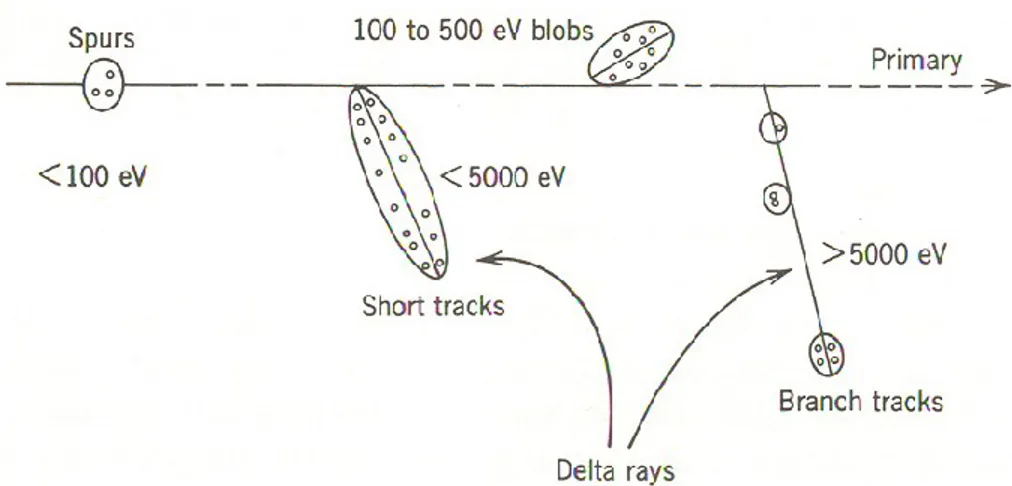
Chapter 1Figure 1.1 Classification of energy deposition events in water .………....…………..…..8
by track structure entities so-called spurs (spherical entities, up to 100 eV), blobs (spherical or ellipsoidal, 100-150 eV), and short tracks (cylindrical, 500 eV-5 keV) for a primary high-energy electron (not to scale). Short and branch tracks are, collectively, described as -rays. From BURTON (1969), with permission.
Figure 1.2 Projections into the XY-plane of figure of track segments …………...……..9
of 300 (a) and 0.15 (b) MeV protons (LET ~ 0.3 and 70 keV/m, respectively) incident on liquid water at 25 °C, calculated (at ~10-13 s) with our Monte Carlo simulation code (KANIKE et al., 2015a). The two irradiating protons are generated at the origin and start moving along the Y axis. Dots represent the energy deposited at points where an interaction occurred.
Figure 1.3 Primary energy-loss events in high-LET radiation tracks…...9
(FERRADINI, 1979).
Figure 1.4 Projections over the XY-plane of track segments calculated ……….……...10
(at ~10-13 s) for (a) H+ (0.15 MeV), (b) 4He2+ (1.75 MeV/nucleon), (c) 12C6+ (25.5 MeV/nucleon), and (d) 20Ne10+ (97.5 MeV/nucleon) impacting ions. Ions are generated at the origin and along the Y axis in liquid water under identical LET conditions (~70 keV/μm). Dots represent the energy deposited at points where an interaction occurred. From MUROYA et al. (2006), with permission.
Figure 1.5 Time scale of events in the radiolysis of water by low-LET radiation...12
The time scale of chemical reactions leading to generation of specific radiolytic products is also shown.
Figure 1.6 Frequency of a given energy loss for 150-keV incident electrons...22 in liquid water at 25 °C. Electrons are followed over their whole track until their energy is lower than ~7.3 eV (threshold for electronic excitation). The corresponding average energy loss per event is ~47 eV. 104-105 different track histories were used in the simulations.
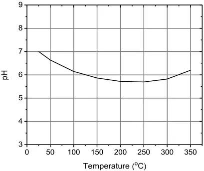
Figure 1.7 Temperature dependence of pH in pure water...26
(ELLIOT and BARTELS, 2009).
Chapter 2
Figure 2.1 Temperature dependence of the (average) electron thermalization...39
25-350 °C used in this study (MEESUNGNOEN and JAY-GERIN, 2005;
SANGUANMITH et al., 2011; MUROYA et al., 2012).
Figure 2.2 Temperature dependence of DH3O+, DOH-, and DH2O used in the...40 simulations over the range of 25-350 °C (ELLIOT and BARTELS, 2009).
Figure 2.3 Temperature dependence of the spur lifetime (s) for the...43 low-LET radiolysis of pure liquid water in the range of 25-350 °C
(SANGUANMITH et al., 2012).
Chapter 3 – Article No. 1
Figure 1 Projections over the XY-plane of track segments of 300 (a) and...62
0.15 (b) MeV protons (LET ~ 0.3 and 70 keV/m, respectively) incident on liquid water at 25 °C, calculated (at ~10-13 s) with our IONLYS Monte Carlo track-structure simulation code (see text). The two irradiating protons are generated at the origin and start traveling along the Y-axis. Dots represent the energy deposited at points where an interaction occurred. Note that the penetration range of 1H+ in liquid water, at the considered energy of 0.15 MeV, amounts to ~2.3 m (ref. 18).
Figure 2 Time evolution of the yield (in molecule/100 eV) of hydrogen...63
ions for the radiolysis of pure, deaerated liquid water by 300- and 0.15-MeV incident protons (LET ~ 0.3 and 70 keV/m, respectively) at 25 °C from ~1 ps to 1 ms. The solid and dashed lines show the corresponding values of
G(H3O+) obtained from our Monte Carlo simulations (see text). Experimental data for 60Co /fast electron (~0.3 keV/m) irradiation: (□) ref. 30, (▼) ref. 31, (∆) ref. 32, (●) ref. 33, and (○) ref. 34. There are no experimental data available for 0.15-MeV irradiating protons with which to compare our results.
Figure 3 Variation of pH with time calculated for 300-MeV incident……..…...64
protons (LET ~ 0.3 keV/m) using the isolated “spherical” spur model (solid line), characteristic of low-LET radiation, and for 0.15-MeV incident protons (LET ~ 70 keV/m) using the axially homogeneous “cylindrical” track model (dashed line), characteristic of high-LET radiation, at 25 °C from ~1 ps to 1 ms (see text).
Chapter 4 – Article No. 2
Figure 1 Time evolution of G(H3O+) (in molecule/100 eV)...75 for the radiolysis of pure, deaerated liquid water by 300-MeV incident protons (LET ~ 0.3 keV/m) at 25 °C from ~1 ps to 1 ms. The red solid line shows the hydrogen ion yield values obtained from our Monte Carlo simulations (see text). Experimental data for 60Co /fast electron irradiation are: (□) ref. 76,
(▼) ref. 77, (∆) ref. 78, (●) ref. 79, and (○) ref. 80. For the sake of reference, our simulated time-dependent yields of eaq and •OH (see ref. 81), H• and OH are also included in the figure. Note that the hydroxide ion OH, which is formed largely by the reaction: eaq + •OH OH (k = 3.55 × 10¹⁰ M-1 s-1) as the spur expands, contributes to an alkaline spur and consequently counteracts the acid-spike effect discussed in this work. However, as we can see from the figure, G(OH) remains much smaller than G(H3O+) over the time range of interest. As a result, its effect only slightly modifies the quantitative features of the pH and can be ignored to a good approximation. Finally, the (dotted) line shown at ~0.2 s indicates the end of spur expansion (ref. 47), i.e., the time required to observe the transition from nonhomogeneity to homogeneity in the distribution of the radiolytic species.
Figure 2 Time dependence of the extents G(H3O+) (in molecule/100 eV)...76
of the different reactions that are involved in the decay of H3O+, calculated from our Monte Carlo simulations of the radiolysis of pure, deaerated water by 300-MeV incident protons (LET ~ 0.3 keV/m) at 25 °C, in the interval of ~1 ps to 1 ms. Other reactions, such as H3O+ + O• •OH + H2O (k = 5 1010 M-1 s-1)and H3O+ + HO2 H2O2 + H2O (k = 5 1010 M-1 s-1), contribute only little to the decay of G(H3O+). The (dotted) line shown at ~0.2 s indicates the end of spur expansion (ref. 47).
Figure 3 Time dependences of H3O+ yields (in molecule/100 eV)...77 calculated from our Monte Carlo simulations of the radiolysis of pure, deaerated liquid water at 25 °C and in the interval of ~1 ps to 1 ms, for impacting 300-MeV (~0.3 keV/m) and 150-keV (~70 keV/m) protons, and 1.75-MeV/nucleon (~70 keV/m) and 0.6-MeV/nucleon (~146 keV/m) 4He2+ ions. It is worth noting here that G(OH), in all high-LET ion tracks considered, remains at a nearly constant level well below 1 G-unit, and therefore much smaller than G(H3O+), during the lifetime of the tracks (not shown in the figure). Consequently, as mentioned in the caption of Fig. 1, the formation of OH ions only slightly modifies the quantitative features of the pH and can simply be ignored.
Figure 4 Time evolution of pH in a spur calculated for 300-MeV...78 incident protons in pure, deaerated liquid water (LET ~ 0.3 keV/m) using the isolated “spherical” spur model, characteristic of low-LET radiation, at 25 °C (see text). The solid and dashed lines show the pH values obtained for two different spur radii ro = 11.7 and 8.3 nm, respectively. The (dotted) line shown at ~0.2 s indicates the end of spur expansion (ref. 47).
Figure 5 Simulated track history (at ~10-13 s, projected into the XY-plane...80 of figure) of a 150-keV proton (LET ~ 70 keV/m) traversing through liquid water at 25 °C. The irradiating proton is generated at the origin and starts traveling along the Y-axis. Dots represent the energy deposited at points where an interaction occurred. The track can be described as two coaxial cylindrical volumes centered on the path of the proton. The inner cylindrical volume (i.e.,
the region adjacent to the trajectory) is the track “core” with radius rc. Surrounding the core is a much larger region called the “penumbra” where all of the energy is deposited by energetic secondary electrons (-rays) created in knock-on collisions by the primary proton. The total time for penumbra formation may be as long as ~1 ps, and its radius extends to the limit of the range of knock-on electrons.
Figure 6 Simulated track history (at ~10-13 s, projected into the XY-plane...81 of figure) of a 1.75-MeV/nucleon helium ion (LET ~ 70 keV/m) incident on liquid water at 25 °C. Irradiating conditions are the same as in Fig. 5.
Figure 7 Simulated track history (at ~10-13 s, projected into the XY-plane...81 of figure) of a 0.6-MeV/nucleon helium ion (LET ~ 146 keV/m) incident on liquid water at 25 °C. Irradiating conditions are the same as in Fig. 5.
Figure 8 Variation of pH with time calculated for 150-keV incident...82 protons (LET ~ 70 keV/m) using the axially homogeneous cylindrical track model, characteristic of high-LET radiation, for different physical core radii between 2 and 25 nm, at 25 °C from ~1 ps to 1 ms (see text).
Figure 9 Variation of pH with time calculated for pure, deaerated liquid...83
water at 25 °C and in the interval of ~1 ps to 1 ms, for irradiating 300-MeV protons (LET ~ 0.3 keV/m) (dotted line) using the isolated spherical spur model (characteristic of low-LET radiation) and for impacting 150-keV protons (LET ~ 70 keV/m), and 1.75-MeV/nucleon (LET ~ 70 keV/m) and 0.6-MeV/nucleon (LET ~ 146 keV/m) helium ions using the axially homogeneous cylindrical track model (characteristic of high-LET radiation) (see text).
Chapter 5 – Article No. 3
Figure 1 Time evolution of G(H3O+) (in molecule per 100 eV)...116 for the radiolysis of pure, deaerated liquid water by 300-MeV incident protons at 25 °C (a) and 350 °C (b). The red solid lines show the hydronium ion yield values obtained from our Monte Carlo simulations. Experimental data: (□) 66, (▼) 67, (Δ) 68, (●) 69, and (○) 70. For the sake of reference, our simulated time-dependent yields of eaq, •OH, H•, and OH are also included. The dotted lines shown at 2 × 10⁻⁷ s at 25 °C and at 3.5 × 10⁻⁸ s at 350 °C indicate the end of spur expansion 45, i.e., the time (s) required for the changeover from nonhomogeneous spur kinetics to homogeneous kinetics in the bulk solution (thus defining the so-called “primary” radical and molecular yields of radiolysis).
Figure 2 Time dependence of the extents G(H3O+)...117 (in molecule per 100 eV) of the different reactions that are involved in the decay of H3O+, calculated from our Monte Carlo simulations of the radiolysis
of pure, deaerated water by 300-MeV incident protons at 25 °C (a) and 350 °C (b). The dotted lines shown at 2 × 10⁻⁷ s at 25 °C and 3.5 × 10⁻⁸ s at 350 °C indicate the time (s) at which spur expansion is complete 45.
Figure 3 Time dependences of H3O+ yields (in molecule per 100 eV)...118 calculated from our Monte Carlo simulations of the radiolysis of pure, deaerated liquid water in the interval of 1 ps to 1 s for impacting 300-MeV protons at different temperatures between 25 and 350 °C. The long-dashed line indicates the time required to observe, at a given temperature, the transition from nonhomogeneity to homogeneity in the distribution of the radiolytic species.
Figure 4 (a) Temperature dependence of the (average) electron...119 thermalization distance (rth) of subexcitation electrons in liquid water over the range of 25-350 °C 17,35,51; (b) Variation of the diffusion coefficient for the hydronium ion, D(H3O+), in water as a function of temperature 10 used in this work.
Figure 5 Temperature dependence of the concentration of H3O+ ions...120 (in M) arising through water’s autoprotolysis 10.
Figure 6 Time evolution of pH in a spur calculated for pure, ...121 deaerated liquid water at different temperatures between 25 and 350 °C and in the interval of 1 ps to 10 s, for irradiating 300-MeV protons using the isolated spherical spur model, characteristic of low-LET radiation.
Figure 7 Variation of pH with temperature over the range of 25-350 °C...122
calculated for pure, deaerated liquid water for irradiating 300-MeV protons using the isolated spherical spur model, at three different times during spur expansion: 1 ps, 1 ns, and 1 s.
Chapter 6 – Discussion
Figure 6.1 Effect of pH on the activity of enzymes...128
(PARK and ZIPP, 2000).
Figure 6.2 N-C bond cleavage leads to base release in acidic pH...129 (guanine is taken here as an example). Acid-catalyzed hydrolysis at the C1’ position of the N-glycosidic bond of deoxyguanosine results in release of guanine and formation of an AP site (1). This abasic site can lead to DNA single strand scission by -elimination of the adjacent 3’ phosphate residue (2). The subsequent elimination of the phosphate on the 5’-side of the abasic site is slow under physiological conditions, but occurs readily under alkaline conditions. From SHEPPARD et al. (2000), with permission.
Figure 6.3 Steady state pH of pure, deaerated water irradiated by...………..132
irradiated solution decreases from the initial pre-irradiation value of 7 to a value approaching pH = 3.25 at the highest dose rates used in liquid cell electron microscopy experiments. From SCHNEIDER et al. (2014), with permission, and private communication.
List of Tables
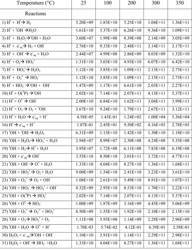
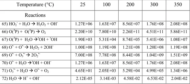
Table 1 Main spur/track reactions and rate constants...41 (k in M-1 s-1; for first-order reactions, the value of k is given in s-1) used in our simulations of the radiolysis of pure liquid water in the temperature range of 25-350 °C (ELLIOT and BARTELS, 2009).
List of Abbreviations
CONV Conventional
D Diffusion coefficient
DEA Dissociative electron attachment DNA Deoxyribonucleic acid
e-aq Hydrated electron
eV Electron-volt
fs Femtosecond
GX or g(X) Primary yield of the radiolytic species X G(X) Experimental yield of the final product X Gy/s Gray/second (dose rate)
iNOS Inducible nitric oxide synthase
IR Infrared
IRT Independent reaction times
k Reaction rate constant
keV Kilo-electron-volts
LET Linear energy transfer
MC Monte Carlo
MeV Mega-electron-volts
µm micrometer
s microsecond
pH -log10 H+, where the square brackets denote concentration pKa -log10 Ka, where Ka is the acid dissociation constant
ps Picosecond
SOD Superoxide dismutase
60Co Cobalt-60
1 - INTRODUCTION
Radiation chemistry is a mature branch of radiation science which is continually evolving and finding wider applications. The radiation chemistry of water is of considerable importance, for the intrinsic scientific interest it generates. This is particularly apparent in the study of the role of free radicals in biology generally, and radiation biology specifically because living cells and tissue consist mainly of water (70%-85% by weight). Therefore, it is important to know how ionizing radiation interacts with water and aqueous solutions, what the subsequent water decomposition products are and how they are spatially distributed (what is commonly referred to as the “track structure”), depending on the radiation type and energy (a measure of which is given by the “linear energy transfer” or LET) and the irradiation conditions. Aqueous radiation chemistry is also of great importance in various areas of nuclear science and technology such as water-cooled nuclear power reactors where water, used both as moderator and as a heat transport medium, is circulating around the reactor core at temperatures of ~250-350 °C, and where the radiolytic processes need to be carefully controlled to avoid the deleterious effects of water radiolysis and minimize corrosion. For a detailed account of the history and present status of aqueous radiation chemistry, see, for example: ALLEN (1961), DRAGANIĆ and DRAGANIĆ (1971), FERRADINI and PUCHEAULT (1983), BUXTON (1987), KROH (1989), SPINKS and WOODS (1990), JONAH (1995), FERRADINI and JAY-GERIN (1999), ZIMBRICK (2002), LAVERNE (2004), ELLIOT and BARTELS (2009), WARDMAN (2009), and MEESUNGNOEN
and JAY-GERIN (2011).
In a biological system, the cell is damaged by ionizing radiation. In this regard, a thorough knowledge of the radiation chemistry of water is critical to our understanding of early stages in the complicated chain of radiobiological events that follow the absorption of radiation. Indeed, in a cellular environment, reactive species generated by water radiolysis are likely to cause chemical modifications and changes in cells, which subsequently may act as triggers of signalling or damaging effects (MUROYA et al.,
2006; AZZAM et al., 2012; O’NEILL and WARDMAN, 2009). Ultimately, this can lead
biomolecules (e.g., nucleic acids, proteins, and lipids), DNA and its associated water molecules are considered to be the most important target in defining the radiobiological response. Damage is caused either directly or indirectly through chemical attack by radiolytic products as the radiation track passes through and deposits energy near to (mostly bulk water) or in the DNA. Interestingly, it is the spatial distribution of the DNA lesions rather than their number that is most biologically relevant. Such damage can be repaired or, if unrepaired or mis-repaired, may result in cytotoxic and mutagenic effects and chromosomal instability, all of which can contribute to tumorigenesis, cell death, or long-term stressful effects in surviving cells (BECKER and SEVILLA, 1993; BECKER et al., 2011; CADET et al., 1997; von SONNTAG, 2006; O’NEILL, 2001; AZZAM et al., 2012). A goal of radiobiology research is to understand how radiation exposure deregulates molecular pathways that are important in maintaining genomic integrity.
Monte Carlo simulation methods are well suited to take into account the stochastic nature of the complex sequence of events that are generated in aqueous systems following the absorption of ionizing radiation. Simulations allow the reconstruction of the intricate action of radiation. This is a powerful tool for studying the relationship between the initial radiation track structure, the ensuing chemical processes, and the stable end products formed in the radiolysis of both pure water and water-containing solutes. Stochastic simulation codes employing Monte Carlo procedures have been used with success by a number of investigators to model the entire water radiolysis process as a function of time, LET of the radiation, pH, presence or absence of oxygen, temperature,
etc. (for reviews, see, for example: BALLARINI et al., 2000; UEHARA and NIKJOO,
2006; KREIPL et al., 2009; MEESUNGNOEN and JAY-GERIN, 2011). In particular,
these model calculations provide the user with detailed information on the early physicochemical track structure (i.e., the physical and chemical events that occur in the “native” radiation track) and the spatio-temporal development of the track (i.e., how the initial, spatially nonhomogeneous distribution of reactive species relaxes in time toward a homogeneous distribution). This information can then be used to develop a realistic description of all reactive fragment species created at early times and involved as precursors to radiobiological damage. Such knowledge is critical to unravel the
fundamental biochemical mechanisms leading to the biological consequences of ionizing radiation.
In such a perspective, we used in this study Monte Carlo track chemistry simulations to calculate, at 25 °C, the pH values prevailing in the track regions of the radiolysis of pure, deaerated water during and shortly after irradiation. The concentrations of hydronium ions (H3O+) generated in situ in these regions and the corresponding pH values were obtained from our calculated yields (or G-values) of H3O+ as a function of time (in the interval of 1 ps to 1 ms), assuming two different track structure models: (1) an isolated “spherical” spur model characteristic of low-LET radiation and (2) an axially homogeneous “cylindrical” track model for high-LET radiation. For all cases studied, an abrupt transient acid pH effect, which we called an “acid spike”, was observed at times immediately after the initial energy release (KANIKE et al., 2015a,b). Following this work, calculations were then extended to examine the effect of temperature on the magnitude and duration of this acid spike response in the range from ambient up to 350 °C (KANIKE et al., 2016). To the best of our knowledge, this early-time, acid-spike effect has never been explored in water either at ambient or at elevated temperatures, or in living cells subject to ionizing radiation. In this respect, the present study prompts a number of important questions about the potential implications of these in situ variations in acidity, even if transitory, for radiobiology or for water-cooled nuclear reactors in terms of biological damage or material corrosion and damage.
1.1 Radiolysis of water
Water radiolysis is the chemical decomposition of water molecules due to the action of ionizing radiation. A thorough understanding of the radiolysis of water involves knowledge of the early physical energy deposition around the path of the incident radiation (mainly through ionization, electronic excitation, and dissociation of the water molecules), and the subsequent physicochemical and nonhomogeneous chemical evolution of the system (PLATZMAN, 1958; KUPPERMANN, 1959) to times, at room temperature, on the order of the microsecond. The products obtained after nonhomogeneous stage are usually regarded as homogeneously distributed in the bulk of the solution. Indeed, in a cellular environment, reactive species generated by water
radiolysis are likely to cause chemical modifications and changes in cells, which subsequently may act as triggers of signalling or damaging effects (MUROYA et al., 2006; O’NEILL AND WARDMAN, 2009; WARDMAN, 2009; AZZAM et al., 2012).
Ultimately, this can lead to observable biological responses.
It is noteworthy that the extent and nature of cellular radiobiological damage depends not only on the absorbed dose but also on the quality of radiation. The “linear energy transfer” (LET) (also called “stopping power” by physicists, in units of keV/m) represents, to a first approximation, the nonhomogeneity of energy deposition on a sub-microscopic scale. Briefly, for low-LET, sparsely ionizing radiation (e.g., -rays from 60Co and 137Cs, hard X-rays, or high-energy charged particles, such as fast electrons or protons generated by a particle accelerator: LET ~ 0.3 keV/µm), the radiolysis of pure deaerated liquid water principally leads to the formation of the radicals and molecular products e-aq (hydrated electron), H• (hydrogen atom), H2 (molecular hydrogen), •OH (hydroxyl radical), H2O2 (hydrogen peroxide), HO2•/O2•- (hydroperoxyl/superoxide anion radicals, pKa = 4.8 in water at 25 °C), H+ or equivalently, H3O+ or H+aq (hydrogen ion), OH- (hydroxide ion), etc. (for a review, see: BUXTON, 1987; SPINKS and WOODS,
1990; FERRADINI and JAY-GERIN, 1999; MEESUNGNOEN and JAY-GERIN, 2011).
Under ordinary irradiation conditions (i.e., at modest dose rates), these species are generated nonhomogeneously on subpicosecond time scales in small, spatially isolated regions of dense ionization and excitation events, commonly referred to as “spurs”
(MAGEE, 1953), along the track of the incident radiation. At ~1 ps, all the species begin
to diffuse away from the site where they were originally produced. The result is that a fraction of them react together within the spurs as they develop in time while the remainder escape into the bulk solution. At ambient temperature and pressure, the so-called “spur expansion” is essentially complete by ~0.2 µs after the initial energy deposition (SANGUANMITH et al., 2012). At this time, the species that have escaped from spur reactions become homogeneously distributed throughout the bulk of the solution and the track of the radiation no longer exists. The yields per 100 eV of absorbed energy of the species that remain after spur expansion and become available to react with dissolved solutes (if any) present in low or moderate concentrations, are the so-called
“primary” (or “escape”) yields. They are denoted by g(e-aq), g(H•), g(H2), g(•OH), g(H2O2), etc.1
For low-LET radiation, the radiolysis of pure, deaerated (air-free) liquid water can be represented conceptually by the following global equation, written for an absorbed energy of 100 eV (FERRADINI and JAY-GERIN, 1999):
g(H2O) H2O g(e-aq) e-aq + g(H•) H• + g(H2) H2 + g(H+) H+ + g(OH-) OH- + g(•OH) •OH + g(H2O2) H2O2
+ g(HO2•/O2•-) HO2•/O2•- + ... [1] where the symbol is used to distinguish reactions brought about by the absorption of ionizing radiation, the coefficients g(X) are the primary yields of the various radiolytic species X, and g(H2O) denotes the corresponding yield for net water decomposition. For 60Co γ-irradiated neutral water at 25 °C, g(e
-aq) = 2.65, g(H•) = 0.6, g(H2) = 0.45, g(•OH) = 2.8, and g(H2O2) = 0.68 molecules per 100 eV (ELLIOT et al., 1993; FERRADINI and
JAY-GERIN, 2000; ELLIOT and BARTELS, 2009).
These product yields are connected by the following equations:
g(e-aq) + g(OH-) = g(H+)
g(e-aq) + g(H•) + 2g(H2) = g(•OH) + 2g(H2O2) + 3g(HO2•/O2•-) [2] expressing the charge conservation (electro-neutrality) and material balance (stoichiometry) of Eq. [1]. We should note that, for low-LET radiolysis, HO2•/O2•- has an extremely small yield in comparison to the other radiolytic species (about less than 1%) and can be usually ignored, although in aerated solution the yields of O2•- is high due to solvated electrons react with oxygen.
1 The number of species produced (or consumed) per unit of energy absorbed is termed the G-value and is used to express the radiation chemical yield. G-values are quoted as
g(X) (some authors prefer to use GX) for the so-called primary yields of the species X (normally measured at the time after which spurs have dissipated) and G(product) for experimentally measured or final yields. Throughout this work, G-values are quoted in units of molecules per 100 eV. For conversion into SI units (mol J-1), 1 molecule per 100 eV ≈ 1.0364 × 10⁻⁷ mol J-1 (or 0.10364 mol J-1).
With in the lifetime of a spur, the yields of free radical and molecular species formed by the irradiation vary with time, and also depend on the LET of the incident radiation and the concentration of added solutes or scavengers. One of the main goals in the study of the radiation chemistry of water is to determine those yields and their time dependences, under different irradiation conditions.
1.1.1 The track structure in radiation chemistry and radiobiology
The quantities and proportions of the chemical products formed in the radiolysis of water are highly dependent on the distances separating the primary radiolytic species from each other along the track of the ionization radiation. The distribution of separations, i.e., the “track structure”, is determined to a large extent by the distribution of the physical energy deposition events and their geometrical dispositions, or, in other words, by the radiation type and energy. The radiation track structure is of crucial importance in specifying the precise spatial location and identity of all the radiolytic species and free-radical intermediates generated in the tracks, and their subsequent radiobiological action at the molecular and cellular levels. Tracks are dynamic and are constantly expanding in time due to diffusion of the reactive species (LAVERNE, 2000, 2004). Track structure, combined with a reaction scheme and yields of primary species, forms the basis of radiation-chemical theory (MOZUMDER, 1999). It is now well accepted that differences in the biochemical and biological effects (e.g., damage to DNA, changes in cell signalling, etc.) of different qualities (LET) of radiation must be analyzed in terms of track structure (CHATTERJEE and HOLLEY, 1993; MUROYA et al., 2006). Two different radiation track structures are generally considered as a function of LET:
i. Low-LET radiation tracks ii. High-LET radiation tracks.
i) Low-LET radiation tracks
Ionizing radiations such as fast electrons generated from X- or -ray beams have high energy and low LET. For example, the average LET of a 1-MeV Compton electron in water is ~0.3 keV/µm. The track-averaged mean energy loss per collision event by such an electron is in the region ~47-56.8 eV (COBUT, 1993; LAVERNE and
PIMBLOTT, 1995; COBUT et al., 1998; AUTSAVAPROMPORN, 2006; MIRSALEH
KOHAN et al., 2013). This means that the energy-loss events are, on the average,
separated by distances of 200 nm. This nonhomogeneous distribution of energy deposition events in space gives rise to the “spur” theory for low-LET track structure
(ALLEN, 1948; MAGEE, 1953; MOZUMDER and MAGEE, 1966a,b), according to
which the entire track is to be viewed as a random succession of (more or less spherical) spurs, or spatially localized energy-loss events. The few tens of electron-volts deposited in a spur cause a secondary electron to be ejected from a molecule. As the ejected electron moves away, it undergoes collisions with surrounding water molecules, loses its excess energy, and becomes thermalized (~0.025 eV at 25 °C) within 8-12 nm of its geminate positive ion (GOULET et al., 1990, 1996; PIMBLOTT and MOZUMDER,
2004; MEESUNGNOEN and JAY-GERIN, 2005a; UEHARA and NIKJOO, 2006). This
average “electron thermalization distance” or “penetration range” (rth) can be viewed as an estimate of the spur’s initial radius, prior to spur expansion. Thus, the individual spurs produced by low-LET radiation are so far apart along the track that they are not initially overlapping (but they will overlap somewhat later as they develop in time).
In their pioneering work to model the radiation-chemical consequences of different energy-loss processes, MOZUMDER and MAGEE (1966a,b) considered, somewhat arbitrarily, a low-LET track as composed of a random sequence of three types of essentially non-overlapping entities: “spurs, blobs, and short tracks” (Figure 1.1). The spur category contains all track entities created by the energy losses between the lowest excitation energy of water and 100 eV; in most cases, there are one to three ion pairs in such isolated spatial areas and about the same number of excited molecules (PIMBLOTT
and MOZUMDER, 1991). Blobs were defined as track entities with energy transfers
between 100-500 eV, and short tracks as those with energy transfers between 500 eV and 5 keV. Secondary electrons produced in energy transfers above 5 keV were considered as “branch tracks”. Short and branch tracks are, collectively, described as -rays. This old concept of track entities proved to be very helpful in greatly facilitating the visualization of track processes and in modeling radiation-chemical kinetics. It is still a useful approach for the classification of track structures, since it takes into account the spatial arrangements of initial species, which affect their subsequent reactions.
Figure 1.1 Classification of energy deposition events in water by track structure entities so-called spurs (spherical entities, up to 100 eV), blobs (spherical or ellipsoidal, 100-500 eV), and short tracks (cylindrical, 500 eV-5 keV) for a primary high-energy electron (not to scale). Short and branch tracks are, collectively, described as -rays. From BURTON (1969), with permission.
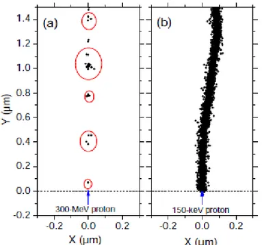
Figure 1.2 shows typical two-dimensional representations of the track segments of
300- and 0.15-MeV irradiating protons (LET 0.3 and 70 keV/m, respectively) on liquid water at 25 °C, calculated with our Monte Carlo simulation code (KANIKE et al.,
2015a). It illustrates the non homogeneity of the energy deposition on a sub-microscopic
scale. At the lowest LET (Figure 1.2a), tracks are formed initially by well-separated “spurs” (spherical in shape) that develop independently in time (without interference from the neighbouring spurs). As LET increases, the mean separation distance between the spurs decreases and the isolated spur structure changes to a situation in which the spurs overlap and form a dense continuous column (cylindrical shape) (Figure 1.2b).
ii) High-LET radiation tracks
The column of species defined initially by the overlapping spurs along the path of a high-LET particle makes up what is referred to as the “track core”. It is surrounded by a coaxial region traversed by large numbers of emergent, comparatively low-LET secondary electrons (-rays), called the “penumbra” (for example, see: PUCHEAULT,
1979; MAGEE and CHATTERJEE, 1980, 1987; PARETZKE, 1987; MOZUMDER,
1999; LAVERNE, 2000, 2004). Such a “high-LET” radiation track structure can actually
be seen in heavy-ion irradiations (PLANTE et al., 2005; MUROYA et al., 2006). It is illustrated schematically in Figure 1.3.
Figure 1.2 Projections into the XY-plane of figure of track segments of 300 (a) and 0.15
(b) MeV protons (LET ~ 0.3 and 70 keV/m, respectively) incident on liquid water at 25 °C, calculated (at ~10-13 s) with our Monte Carlo simulation code (KANIKE et al., 2015a). The two irradiating protons are generated at the origin and start moving along the Y axis. Dots represent the energy deposited at points where an interaction occurred.
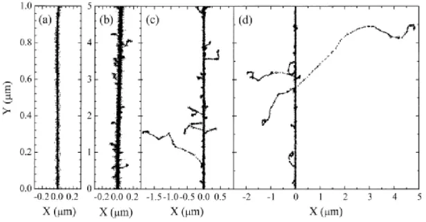
Figure 1.4 Projections over the XY-plane of track segments calculated (at ~10-13 s) for (a) H+ (0.15 MeV), (b) 4He2+ (1.75 MeV/nucleon), (c) 12C6+ (25.5 MeV/nucleon), and (d) 20Ne10+ (97.5 MeV/nucleon) impacting ions. Ions are generated at the origin and along the Y axis in liquid water under identical LET conditions (~70 keV/μm). Dots represent the energy deposited at points where an interaction occurred. From MUROYA et al. (2006), with permission.
Figure 1.4 illustrates typical two-dimensional representations of short (1-5 m) track segments of H+, 4He2+, 12C6+, and 20Ne10+ ions, calculated with our own Monte Carlo simulation code under the same LET conditions (70 keV/m). As one can see, these tracks can be considered as straight lines with the ejected high-energy secondary electrons travelling to a greater average distance away from the track core as the velocity of the incident ion increases, from protons to neon ions. In other words, even though all those particles are depositing the same amount of energy per unit path length, that energy is lost in a volume that increases in the order H+ < 4He2+ < 12C6+ < 20Ne10+, indicating that the higher-Z particle (where Z is the ion charge number) has the lower mean density of reactive species (MUROYA et al., 2006; MEESUNGNOEN and JAY-GERIN, 2011). The fact that tracks of different ions with the same LET have different radial distributions of energy deposited by -rays is in accord with Bethe’s theory of stopping power
(BETHE, 1930; BETHE and ASHKIN, 1953) and indicates that LET is not a unique
(SCHULER and ALLEN, 1957; SAUER et al., 1977; LAVERNE and SCHULER, 1987; KAPLAN and MITEREV, 1987; FERRADINI, 1990; FERRADINI and JAY-GERIN,
1999; LAVERNE, 2000, 2004). Attempts have been made to introduce other comparative
characteristics of radiation track effects to replace LET like, for example, the (Z*/)2 factor (where Z* is an energy-dependent effective charge of the ion and is the ratio of its velocity to that of light) (KATZ, 1970; WALIGÓRSKI et al., 1986; YAMASHITA et
al., 2008) or yet the parameter MZ2/E (where M is the ion mass and E = ½MV2 its kinetic
energy) (LAVERNE, 2004). Several sets of radiation chemical data appear to be better unified using these phenomenological parameters instead of LET, others do not. Following PIMBLOTT and LAVERNE (2002), it should be recognized, however, that no deterministic parameterization can realistically represent a phenomenon that is stochastic in nature. Nevertheless, despite its limitations, LET still remains the most useful single parameter in the radiation chemistry of heavy ions.
1.1.2 Time scale of events and formation of primary free-radical and molecular products in neutral water radiolysis
From the viewpoint of pure aqueous radiation chemistry, the successful prediction of the effects of radiation type and energy in radiolysis not only requires a realistic description of the early physical aspects of the radiation track structure, but also an accurate modelling of the temporal development of the track, in which the various radiolytic species are specified and allowed to diffuse from their initial positions and react with one another (or with the environment) (MUROYA et al., 2006). Therefore, it is critical to understand how the radiation quality (LET) and the irradiation conditions affect the subsequent water decomposition products, their space distribution and thereby the observed yields. Finally, it is also important to know how the initial, spatially nonhomogeneous distribution of reactive species relaxes in time toward a homogeneous distribution.
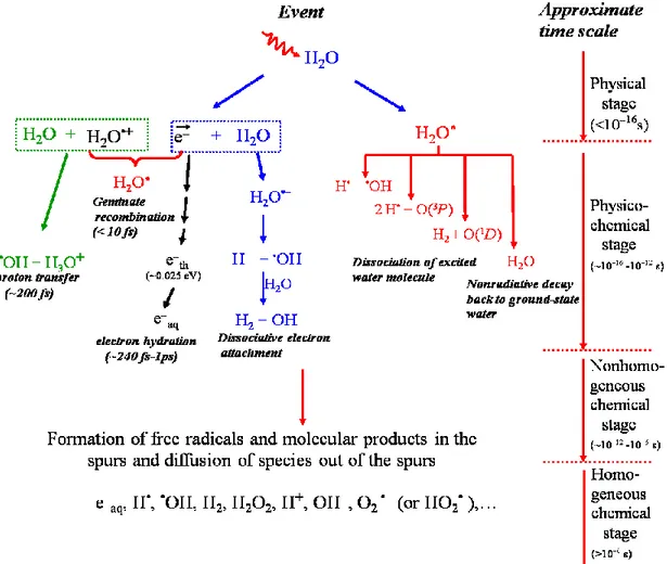
The complex events that accompany the absorption of high-energy photons or the passage of fast charged particles in liquid water can usually be divided into three, more or less clearly demarcated, consecutive, temporal stages: physical, physicochemical, and chemical (PLATZMAN, 1958; KUPPERMANN, 1959). These stages correspond with
the initial dissipation of energy in the system, the establishment of thermal equilibrium, and the establishment of chemical equilibrium, respectively (Figure 1.5). In a physiologic system, there follows a biological stage in which the cells respond to the damage resulting from the products formed in the preceding stages. These four different stages are briefly described below (for recent reviews on the subject, see: MEESUNGNOEN
and JAY-GERIN, 2011; AZZAM et al., 2012).
Figure 1.5 Time scale of events in the radiolysis of water by low-LET radiation. The
time scale of chemical reactions leading to generation of specific radiolytic products is also shown.
(i) The “physical” stage
The physical stage consists of the phenomena by which energy is transferred from the incident high-energy radiation to the water. Its duration is less than ~10-16 s. The result of this energy absorption is the production, along the path of the radiation, of a large number of ionized and electronically excited water molecules, denoted H2O•+ and H2O*elec, respectively, and secondary electrons are generated:
H2O H2O•+ + e- (ionization) [3]
H2O H2O*elec (excitation) [4]
Note that H2O*elec represents here many excited states, including the so-called “superexcited” states (PLATZMAN, 1962a) and the excitations of collective electronic oscillations of the “plasmon” type (HELLER et al., 1974; KAPLAN and MITEREV,
1987; LAVERNE and MOZUMDER, 1993; WILSON et al., 2001).
Generally, the electron ejected in the ionization event has sufficient energy either to ionize or excite one or more other water molecules in the vicinity, and this leads, as mentioned above, to the formation of track entities, commonly known as “spurs”, that contain the products of the events.
(ii) The “physicochemical” stage
The ions and excited-state water molecules formed during the physical stage are extremely unstable and undergo fast reorganization in this second or physicochemical stage, which lasts not more than 10-12 s after the initial energy deposition. These processes produce radical and molecular products of the radiolysis that are distributed in a highly nonhomogeneous track structure.
In the time scale of 200 fs (LI et al., 2013), the positive ion H2O•+ decomposes to form an •OH radical by transferring a proton to a neighbouring water molecule:
H2O•+ + H2O H3O+ + •OH , [5]
where H3O+ (or equivalently, H+aq) represents the hydrated proton. However, before reaction [5] occurs, H2O•+ may undergo a random walk via a sequence of resonant
electron transfers (about 21, on the average) from neighbouring water molecules to the H2O•+ hole (or electron-loss center) (OGURA and HAMILL, 1973; MOZUMDER and
MAGEE, 1975). The ranges of a migrating hole are a few molecular diameters (COBUT
et al., 1998).
The secondary (“dry”) electron ejected from an ionized water molecule undergoes scattering as it moves away from its parent cation. It can cause further ionization and excitation to occur if it has sufficient kinetic energy. Eventually, its energy falls below the first electronic excitation threshold of water (~7.3 eV; MICHAUD et al., 1991), forming the so-called “subexcitation electron” (e-sub) (PLATZMAN, 1955). The latter loses the rest of its energy relatively slowly by exciting vibrational and rotational modes of water molecules. Once it is thermalized (e-th) (after 10-40 fs at 25 °C; see GOULET
et al., 1990, 1996; MEESUNGNOEN et al., 2002a), it can get localized or “trapped” (e-tr)
in a pre-existing potential energy well of appropriate depth in the liquid (then forming the so-called “wet” electron whose exact physicochemical nature is still the subject of investigation) before reaching a fully relaxed, hydrated state (e-aq) as the dipoles of the surrounding molecules orient in response to the negative charge of the electron. In liquid water at room temperature, thermalization, trapping, and hydration can then follow in quick succession (on the time scale of ~240 fs-1 ps, as revealed from time-resolved femtosecond laser spectroscopic studies) (MOZUMDER, 1999; JAY-GERIN et al., 2008;
MEESUNGNOEN and JAY-GERIN, 2011):
e- e-sub e-th e-tr e-aq [6]
In the course of its thermalization, the slowing-down electron can be recaptured by its parent cation (prior to the occurrence of reaction [5]) due to the Coulomb attraction of the latter which tends to draw them back together to undergo electron-cation “geminate” recombination:
e- + H2O•+ H2O*vib . [7]
As the electron is recaptured, the parent ion is transformed into a (vibrationally) excited neutral molecule.
In the course of its thermalization, the ejected electron can also be temporarily captured resonantly by a water molecule to form a transient molecular anion:
e- + H2O H2O- . [8]
This anion then undergoes dissociation mainly into H- and •OH according to
H2O- H- + •OH , [9]
followed by the reaction of the hydride anion (H-) with another water molecule through a fast proton transfer reaction:
H- + H2O H2 + OH- [10]
Reactions [8]-10] correspond to the so-called “dissociative electron attachment” or DEA process, which has been observed in amorphous solid water at ~20 K for electron energies between about 5 and 12 eV (ROWNTREE et al., 1991). It has been suggested that DEA to water was responsible, at least in part, for the yield of “nonscavengeable” molecular hydrogen observed experimentally in the radiolysis of liquid water at early times (PLATZMAN, 1962b; FARAGGI and DÉSALOS, 1969; GOULET and JAY-GERIN, 1989; KIMMEL et al., 1994; COBUT et al., 1996; MEESUNGNOEN et al.,
2015). Recent experiments have sustained this proposed mechanism for the production of
H2, by showing that the previously accepted nonscavengeable yield of H2 is due to precursors of e-aq (i.e., “dry” or “pre-hydrated” electrons) and it can be lowered with suitable scavengers in sufficiently high concentrations (PASTINA et al., 1999).
Excited water molecules may be produced directly in an initial act (reaction [4]) or by neutralization of an ion (reaction [7]). Very little is known about the decay channels for an excited water molecule in the liquid phase and the branching ratios associated with each of them. Fortunately, the contribution of the water excited states to the primary radical and molecular products in water radiolysis is of relatively minor importance in comparison with that of the ionization processes, so that the lack of information about their decomposition has only limited consequences. Hence, the competing de-excitation mechanisms of H2O* are generally assumed to be essentially the same as those reported
for an isolated water molecule,2 namely (for example, see: SWIATLA-WOJCIK and BUXTON, 1995; COBUT et al., 1998; MEESUNGNOEN and JAY-GERIN, 2005a;
SANGUANMITH et al., 2011a; KANIKE et al., 2015b),
H2O* H• + •OH [11a]
H2O* H2 + O(1D) [11b]
H2O* 2H• + •O•(3P) [11c]
H2O* H2O + release of thermal energy [11d] where O(1D) and •O•(3P) represent the oxygen atom in its singlet 1D first excited state and
triplet 3P ground state, respectively (see Figure 1.5). Specific to the liquid phase, the following dissociation reaction:
H2O* e-aq + H2O•+ [11e]
also needs to be considered in the menu of possibilities that can lead to the decay of H2O*. Its threshold is at ~6.5 eV (NIKOGOSYAN et al., 1983; MIGUS et al., 1987;
BERNAS et al., 1997).
It is believed that reaction [11a] is the main source of the “initial” (i.e., at ~10-12 s, prior to spur/track expansion) yield of hydrogen atoms. Note also that the O(1D) atoms
produced in reaction [11b] react very efficiently with water to form H2O2 (or probably also 2•OH) (TAUBE, 1957; BIEDENKAPP et al., 1970). By contrast, the ground-state O(3P) atoms in aqueous solution are rather inert to water but react with most additives
(AMICHAI and TREININ, 1969). As for the values of the branching ratios (or decay
probabilities) used for the different decay channels [11a-e], they are chosen in order to consistently match the observed picosecond G-values of the various spur species
(MUROYA et al., 2002; MEESUNGNOEN and JAY-GERIN, 2005a).
By ~1 ps following the passage of the radiation, the various initial radiolysis products are: e-aq, H•, H2, •OH, H2O2, H+ (or H3O+), OH-, O2•- (or HO2•, depending on the pH), •O•(3P), etc. At this time, these species begin to diffuse away from the position
where they were originally produced. The result is that a fraction of them react together
2 It should be noted that the same decay processes have been reported to occur for the electronically and vibrationally excited H2O molecules in the gas phase.
within the spurs/tracks as they develop in time while the remainder escape into the bulk solution in the chemical stage.
(iii) The “chemical” stage
The third or chemical stage consists of diffusion and reactions of the reactive species present at the end of the physicochemical stage and initially distributed nonhomogeneously with high concentrations in the center of spurs or along the axis of tracks. This stage is usually divided into two parts. The first part corresponds to the stage of “nonhomogeneous chemistry”, which consists of the period after ~10-12 s, during which spurs or tracks develop in time. A number of like radicals will combine to form the molecular products H2 and H2O2; a number will combine to re-form H2O, while the remainder will diffuse out into the bulk of the solution. At 25 °C, the spur/track expansion is essentially complete by ~10-7-10-6 s (for example, see: BUXTON et al.,
1987; SANGUANMITH et al., 2012). At this time, the species that have escaped from
spur or track reactions become homogeneously distributed throughout the bulk solution (i.e., the system at large) (PLANTE et al., 2005; MUROYA et al., 2006). Beyond a few microseconds, the reactions which occur in the bulk solution can usually be described with conventional homogeneous chemistry methods. This is the second part of the chemical stage, the so-called stage of “homogeneous chemistry”. The radical and molecular products which emerge from the spurs/tracks are then available for reaction with dissolved solutes (if any) present (in low or moderate concentrations) at the time of irradiation.
(iv) The “biological” stage
The biological stage is the final stage in a physiologic system, the cells responding to the damage resulting from the products formed in the preceding stages. During this stage (~10-3 s or longer, depending very much upon the medium), the biological responses affecting the long-term consequences of radiation exposure are induced.
In air-saturated solutions (where the concentration of dissolved oxygen in the water is ~2.5 × 10⁻⁴ M at 25 °C), e-aq and H• atoms are rapidly (on a time scale of a few
tenths of a microsecond) converted to superoxide radical anion (O2•-)/hydroperoxyl (HO2•) radicals, according to:
e-aq + O2 O2•- k12 = 2.34 1010 M-1 s-1 [12] H• + O2 HO2• k13 = 1.31 1010 M-1 s-1 [13] where k12 and k13 are the rate constants for the two individual reactions (ELLIOT and
BARTELS, 2009). Accordingly, in an aerobic cellular environment at pH 7, the major reactive species at homogeneity include O2•-, •OH, and H2O2 (the other molecular product, H2, is relatively inert and normally plays only little part in the radiolysis of aqueous solutions, most of it escaping from solution) (SPINKS and WOODS, 1990).
In biological systems, ionizing radiation can also stimulate inducible nitric oxide synthase (iNOS) activity in hit cells (MIKKELSEN and WARDMAN, 2003), thereby generating large amounts of nitric oxide •NO (officially called nitrogen monoxide). Although •NO is chemically inert toward most cellular constituents (except for heme), it reacts quickly with O2•- to form the peroxynitrite anion (ONOO-) with a rate constant (1.9 × 10¹⁰ M-1 s-1) that is larger than that for the copper/zinc-superoxide dismutase (SOD)-catalyzed disproportionation of O2•-(4 × 10⁹ M-1 s-1) (KOPPENOL, 1998; JAY-GERIN
and FERRADINI, 2000). Like •OH radicals, ONOO- and its conjugate acid,
peroxynitrous acid ONOOH (pKa = 6.8 at 37 °C) (PRYOR and SQUADRITO, 1995), are powerful oxidizing agents. They are capable of attacking a wide range of cellular targets, including lipids, thiols, proteins, and DNA bases (for example, see: HALLIWELL and
GUTTERIDGE, 2015).
1.1.3 Spurs/tracks are acidic
The major reducing radical formed in neutral solutions during water radiolysis was shown experimentally to bear a unit negative charge (CZAPSKI and SCHWARZ,
1962; COLLINSON et al., 1962), a result that contributed to the discovery of the
“hydrated electron” in 1962 (HART and ANBAR, 1970). This suggests that an ejected electron can escape from its parent H2O•+ ion and that H2O•+ ions temporarily exist in a spur. The formation of H3O+ via the proton transfer reaction [5] therefore renders the spur
more acid than the body of the solution (SPINKS and WOODS, 1990). Some experimental evidence for this acid pH effect has been reported by several authors. For example, SMITH and STEVENS (1963) irradiated aqueous solutions of 1,1-diethoxyethane CH3CH(OC2H5)2 buffered at pH 7 with 50-kVp X-rays and showed that hydrolysis catalyzed by H3O+ ions formed during the primary radiolytic processes in water:
occurred. Assuming a spherically symmetric spur with a radius of 3 nm, the authors estimated that the pH in the spur would need to be 1.4 to account for the observed hydrolysis. Another experiment indicative of an acid spur was the observation of a transient absorption attributed to Cl2•- in the pulse radiolysis of neutral aqueous sodium chloride solutions at Cl- concentrations of 0.1 M or greater (ANBAR and THOMAS, 1964). The formation of Cl2•- normally requires an acid medium. The results suggested the importance of H3O+ ions in the pH-dependent reaction of radiation-induced •OH radicals with chloride ions (MATSUYAMA and NAMIKI, 1965):
Cl- + •OH + H3O+ → Cl• + 2H2O [15]
followed by the combination of the Cl• atom with Cl- to form Cl2•-:
Cl• + Cl- → Cl2•-. [16]
in the “spur” regions at early time.
Apart from these few experiments aiming at demonstrating this transient acid pH effect in a spur, there is only fragmentary information on its magnitude and time dependence following energy deposition. Moreover, the influence of the quality (or LET) of the radiation on the pH has not been investigated. In this work, our objective is to calculate quantitatively the pH values prevailing in the spur or track regions, using the general relationship:
C = ρ D G , [17] where C is the concentration of species, ρ is the density of the solution (1 g/cm3 for liquid water at 25 °C), D is the radiation dose, and G is the chemical yield (for example, see:
HUMMEL, 1995). Note that with C in mol/dm3, D in J/kg (or Gy), and G in mol/J, the
density is to be expressed in kg/dm3 in order to have a consistent set of units.
Keeping in mind that the pH is defined as the negative logarithm (base 10) of the concentration of H3O+ ions:
t log
H O
t
pH 3 , [18]
we thus need to estimate the concentration of hydronium ions generated in situ in the spur or track regions as a function of time as well as the time evolution of G(H3O+) produced in the radiolysis of pure, deaerated water. As for the calculation of the radiation dose, we selected two different spatio-temporal models of a spur or track:
i. An isolated “spherical” spur model characteristic of low-LET radiation ii. An axially homogeneous “cylindrical” track model for high-LET radiation which are described below.
Spherical spur model
For low-LET radiation (for example, 300-MeV irradiating protons, LET ~ 0.3 keV/µm), we assume that the hydronium ions are produced evenly in an isolated spherical spur. The spur’s initial radius ro, prior to spur expansion, is equal to the average electron thermalization distance (rth) obtained from our Monte Carlo simulations (~11.7 nm at 25 °C) (GOULET et al., 1990, 1996; MEESUNGNOEN and JAY-GERIN, 2005a). The low-LET spur concentrations of H3O+ are derived from
3 3 3 3 4 event loss energy Mean O H O H t r t G t , [19where the mean energy loss in a single energy deposition event (i.e., the mean energy deposited in a spur) in liquid water is taken to be ~47 eV (COBUT, 1993; COBUT et al.,
r(t)2 = ro2 + 6 D t [20] represents the change with time of ro due to the three-dimensional diffusive expansion of the spur. Here, t is time and D is the diffusion coefficient of H3O+ in water (D = 9.46 × 10⁻⁹ m2 s-1 at 25 °C) (FRONGILLO et al., 1998; TIPPAYAMONTRI et al., 2009).
Figure 1.6 shows the distribution of energy-loss events calculated for 150-keV incident electrons in liquid water at 25 °C using our Monte Carlo simulation code
(AUTSAVAPROMPORN, 2006; MIRSALEH KOHAN et al., 2013). In the calculations,
each simulation typically involved ~104-105 different primary tracks. The most probable energy loss in a single event was 15-20 eV, while the mean energy loss was ~47 eV/event. These values are in good agreement with those (22-23 and 56.8 eV, respectively) calculated previously for electrons with 1 MeV incident energy in liquid water (LAVERNE and PIMBLOTT, 1995).3 They clearly indicate that most energy-loss events by fast electrons involve small transfers of energy (MOZUMDER, 1999). Note also that the various minima observed in the energy-loss distribution below 35 eV are associated with the abrupt changes in the total cross section due to the thresholds for electronic excitations and ionizations (or for multiple-scattering events involving these electronic energy losses) in this energy range (COBUT et al., 1998; MEESUNGNOEN et al., 2002b).
3 It has been demonstrated that the probability of a given energy loss in a collision shows very little dependence on the incident electron energy from 10 keV to 1 MeV (for example, see: COBUT et al., 1998; PIMBLOTT et al., 1990; LAVERNE and PIMBLOTT,
0 20 40 60 80 100 0.000 0.005 0.010 0.015 0.020 0.025 P ro ba bil ity o f e ve nt p er e V
Energy loss (eV)
150-keV incident electrons Liquid water
Figure 1.6 Frequency of a given energy loss for 150-keV incident electrons in liquid water at 25 °C. Electrons are followed over their whole track until their energy is lower than ~7.3 eV (threshold for electronic excitation). The corresponding average energy loss per event is ~47 eV. 104-105 different track histories were used in the simulations.
Cylindrical track model
For high-LET radiation, we consider the track as being an axially homogeneous cylinder, of length L = 1 µm and initial radius rc equal to the radius of the physical track “core” (which corresponds to the tiny radial region within the first few nanometers around the impacting ion trajectory). In this region the energy density of deposition is very high (CHATTERJEE and HOLLEY, 1993; MEESUNGNOEN and JAY-GERIN,
2011; MOZUMDER, 1999; MAGEE and CHATTERJEE, 1980, 1987). For the sake of
illustration, we have considered track segments of three different high-LET irradiating ions: i) 150-keV protons (LET ~ 70 keV/µm), ii) 1.75-MeV/nucleon helium ions (LET ~ 70 keV/µm), and iii) 0.6-MeV/nucleon helium ions (LET ~ 146 keV/µm) in liquid water at 25 °C. The simulated track segments were calculated (at 10-13 s) with our Monte Carlo simulation code (KANIKE et al., 2015b, figures 5-7). In this case, the high-LET track concentrations of H3O+ are simply obtained from (MEESUNGNOEN and