Identification of Unknown Carboxydovore Bacteria Dominant in
Deciduous Forest Soil via Succession of Bacterial Communities, coxL
Genotypes, and Carbon Monoxide Oxidation Activity in Soil
Microcosms
Isabelle Lalonde, Philippe Constant INRS-Institut Armand-Frappier, Laval, Québec, Canada
Surveys of the coxL gene, encoding the large subunit of the CO dehydrogenase, are used as a standard approach in ecological
studies of carboxydovore bacteria scavenging atmospheric CO. Recent soil surveys unveiled that the distribution of coxL
se-quences encompassing the atypical genotype coxL type I group x was correlated to the CO oxidation activity. Based on
phyloge-netic analysis including the available coxL reference genome sequences, this unusual genotype was assigned to an unknown
member of the Deltaproteobacteria, with the coxL sequence from Haliangium ochraceum being the sole and closest reference
sequence. Here we seek to challenge the proposed taxonomic assignation of the coxL group x genotype through the monitoring
of CO consumption activity and microbial community successions during the colonization of sterile soil microcosms inoculated
with indigenous microorganisms. In our study, we established that the estimated population density of Deltaproteobacteria was
too small to account for the abundance of the coxL group x genotype detected in soil. Furthermore, we computed a correlation
network to relate 16S rRNA gene profiles with the succession of coxL genotypes and CO uptake activity in soil. We found that
most of the coxL genotypes for which the colonization profile displayed covariance with CO uptake activity were related to
po-tential carboxydovore bacteria belonging to Actinobacteria and Alphaproteobacteria. Our analysis did not provide any evidence
that coxL group x genotypes belonged to Deltaproteobacteria. Considering the colonization profile of CO-oxidizing bacteria and
the theoretical energy yield of measured CO oxidation rates in soil microcosms, we propose that unknown carboxydovore
bacte-ria harboring the atypical coxL group x genotype are mixotrophic K-strategists.
C
arbon monoxide (CO) is a trace gas in the atmosphere, with a
typical mixing ratio of 60 to 150 ppb by volume (ppbv) (
1
).
Combustion of fossil fuels and biomass burning are the main
sources of this gas (
2
). The hydroxyl radicals (OH˙) are
responsi-ble for 85% of the global sink of CO, while specialized aerobic
microorganisms in the soil consume the remaining 15% (
3
,
4
).
These CO-oxidizing bacteria represent the most uncertain term
for the budget of atmospheric CO. The diversity and
ecophysiol-ogy of this ubiquitous functional group present in the
environ-ment are poorly docuenviron-mented and impair the fate prediction of the
biological sink and the atmospheric burden CO in response to
global change.
CO-oxidizing bacteria possess a CO dehydrogenase enzyme
(CODH) catalyzing the following reaction: CO
⫹ H
2O ¡ CO
2⫹
2H
⫺⫹ 2e
⫹. The enzyme is composed of a dimer of heterotrimers
with CoxS, CoxM, and CoxL as small, medium, and large
sub-units, respectively (
5
). The CoxL subunit contains the active site of
the enzyme and is used as a molecular marker to assess
CO-oxi-dizer diversity in the environment (
6
). Furthermore, the coxL gene
sequences encompass two different phylogenetic groups: the type
I group that has been genetically and biochemically characterized
and the putative type II group. There is compelling evidence
sug-gesting that type II CODH is not functional or uses another
sub-strate than CO (
7
,
8
), and thus coxL sequences encompassing this
group are not considered in an environmental survey of
CO-oxi-dizing bacteria. CO-oxiCO-oxi-dizing bacteria harboring coxL type I
com-prise two different physiological groups. They are composed of
the carboxydotrophic bacteria, displaying low affinity for CO and
the ability to grow using CO as the sole carbon and energy source,
and the carboxydovore bacteria, displaying high affinity for CO,
enabling the capacity to scavenge the trace amounts present in the
atmosphere (
9
). Only a few carboxydovore bacteria have been
extensively characterized, encompassing Alphaproteobacteria
(Bradyrhizobium spp., Stappia spp., Aminobacter sp., and Ruegeria
pomeroy DSS-3) (
10–12
), Betaproteobacteria (Burkholderia spp.)
(
13
), Gammaproteobacteria (Alkalilimnicola ehrlichii and
Strepto-monas sp. strain LUP) (
10
,
14
), Actinobacteria (Mycobacterium
spp.) (
10
), and Chloroflexi (Ktedonobacteria) (
15
). However, the
environmental factors determining their activity and distribution
remain elusive (
9
).
Previously published environmental surveys of coxL sequences
have been done in volcanic deposits (
6
,
16
,
17
), hot springs (
18
),
and aquatic environments (
19
). Even though forest ecosystems
exert a dominant role in the global sink of atmospheric CO (
20
),
the carboxydovore bacteria thriving in forest soil have received
Received 3 November 2015 Accepted 12 December 2015 Accepted manuscript posted online 18 December 2015
Citation Lalonde I, Constant P. 2016. Identification of unknown carboxydovore bacteria dominant in deciduous forest soil via succession of bacterial communities, coxL genotypes, and carbon monoxide oxidation activity in soil microcosms. Appl Environ Microbiol 82:1324 –1333.doi:10.1128/AEM.03595-15. Editor: F. E. Löffler, University of Tennessee and Oak Ridge National Laboratory Address correspondence to Philippe Constant, [email protected]. Supplemental material for this article may be found athttp://dx.doi.org/10.1128 /AEM.03595-15.
Copyright © 2016, American Society for Microbiology. All Rights Reserved.
on June 23, 2016 by INRS-Institut Armand-Frappier
http://aem.asm.org/
little attention. Recently, a soil survey of coxL type I sequences in
deciduous forest have led to the identification of a new coxL group,
referred to as coxL group x in this paper, for which the distribution
was correlated with CO soil uptake activity. A phylogenetic
anal-ysis identified the reference coxL sequence from Haliangium
ochraceum as the closest relative of the coxL group x genotype
suggesting the presence of unknown CO-oxidizing
Deltaproteo-bacteria in soil (
21
). However, this assumption is highly uncertain
because of the potential lateral transfer of the coxL gene and the
relatively low identity score (less than 75%) between coxL group x
sequences retrieved from soil and the reference coxL sequence in
H. ochraceum. To address the problematic nature of this
assump-tion, our study aimed to challenge the hypothetical affiliation of
the coxL group x genotype with Deltaproteobacteria. The
succes-sion of bacterial communities, coxL genotypes, and the CO
oxida-tion activity were monitored in sterile deciduous forest soil
micro-cosms inoculated with the indigenous microorganisms. Also, we
verified the hypothesis that the maturation of the CO oxidation
activity would be related to the concomitant emergence of the
coxL group x genotype and 16S rRNA gene sequences from
Delta-proteobacteria.
MATERIALS AND METHODS
Soil sample and incubation. The soil sample was collected from a decid-uous forest located about 40 km from Montreal on the south shore of the St. Lawrence River (45°67=N, 73°32=W). Soil texture was determined by the hydrometer method, and particle size distribution assigned soil sam-ples to the silt loam textural class (22). The same site was visited to inves-tigate the impact of land use change, soil nutrients, and physicochemical properties on CO-oxidizing bacteria (21). The soil sample was homoge-nized (2-mm-pore sieve) and used for the preparation of the colonization substrates and inocula. For the colonization substrate, soil was sterilized two times, separated by a 24-h interval. Sterilization was done in 500-ml Wheaton glass bottles closed with a foam cap. Three bottles were prepared containing 72 g of soil. After the sterilization procedure, the bottles were kept on the laboratory bench with a foam cap for a 5-day period in order to evacuate CO produced during autoclaving. Sterile soil microcosms were inoculated with 8 g of homogenized nonsterile soil (i.e., indigenous soil microorganisms), corresponding to a 10% (vol/vol) inoculum. Three bottles were treated in the same manner with 80 g of sterile soil without inoculation for the controls. The sterile foam cap was kept during the incubation at 20°C to allow gaseous exchanges in the microcosms. After 0, 4, 9, 12, 15, 22, 36, and 55 incubation days, subsamples of 5.5 g were collected in the six microcosms for DNA extraction (0.5 g), and moisture content (5 g) was determined using the standard gravimetric method. Soil water content was maintained at approximately 35% during the incuba-tion period. This water level was selected to avoid diffusion limitaincuba-tion of the CO uptake activity, while supporting bacterial metabolism and growth (23,24). Soil samples dedicated to DNA extraction were frozen in a 2-ml microtube containing 700l TPM buffer (50 mM Tris-HCl [pH 7], 1.7% polyvinylpolypyrrolidone, 20 mM MgCl2), 35l 20% SDS, and 0.5 g glass beads (0.1-mm diameter).
CO oxidation rate in soil. Before measurement of CO oxidation ac-tivity, the foam cap was replaced with a butyl septum cap. A CO gas mixture (508⫾ 10 ppm by volume [ppmv] CO [GTS-Welco, PA, USA]) was injected to get an approximately 4-ppmv initial concentration in the static headspace. Headspace samples (10 ml) were collected with a Pres-sure Lok gastight glass syringe (VICI Precision Sampling, Inc., Baton Rouge, LA, USA) and injected in a gas chromatograph equipped with a reduction gas detector (ta3000R; Ametek Process Instruments, DE, USA). The first-order oxidation rates were calculated by integrating the CO mole fraction time series measured over a 2-h period, using at least four con-centration points for data integration. After each oxidation rate
measure-ment, a foam cap was put back onto the bottles for incubation. Reproduc-ibility of the CO analyses was assessed before each set of measurements by repeated analyses of certified CO standard gas (2.05⫾ 0.10 ppmv [GTS-Welco, PA, USA]), and standard deviations were lower than 5%.
DNA extraction and qPCR assays. Genomic DNA was extracted us-ing a combination of chemical and mechanical cell lysis procedures (25). DNA was precipitated with 2 volumes 96% ethanol, and a polyvinylpoly-pyrrolidone spin column was used for final purification (26). Purified DNA extracts were eluted in 200l nuclease-free water. Purified DNA was used for quantitative PCR (qPCR) of bacterial 16S rRNA gene, coxL type I, and coxL type I Deltaproteobacteria (represented as coxL group x in this article) using previously described procedures (21). The reactions were performed using 5l of diluted genomic DNA (1:50).
coxL genotyping. Genomic DNA extracted after 4, 22, 36, and 55 incubation days was used for coxL pyrosequencing. PCR amplification of coxL genes using the universal assay (21), library preparation, and 454 pyrosequencing were performed at McGill University and Génome Qué-bec Innovation Centre. A total of 45,000 raw sequences were obtained from the 12 libraries. The sequences were analyzed using the software mothur (27). Sequences with a quality score (“qaverage”) lower than 25, with more than 8 consecutive homopolymers, and with ambiguous bases were all removed. The average length of sequences was 336 bases. Se-quences were aligned against a coxL seSe-quences database previously built (21). An arbitrary identity cutoff of 0.04 was used for the clusterization of nucleotide sequences into operational taxonomic units (OTU) represent-ing coxL sequences belongrepresent-ing to the same bacterial species (21). Libraries were standardized to the sequencing effort of the smallest coxL gene li-brary (i.e., 641 sequences) to avoid biases in comparative analyses intro-duced by the sampling depth. At this stage, hypothetical coxL type II OTU identified by the canonical amino acid signature of their active site (AYR GAGR) were removed from the data set. The resulting OTU table was used for rarefaction and diversity analyses. OTU with less than 7 se-quences (0.1% of total sese-quences) were eliminated, and 136 OTU were left for phylogenetic and correlation network analysis. Nucleic acid sequences were imported in the software MEGA (28) for phylogenetic analysis using the reference database and procedure previously described (21) to which sequences were added.
Bacterial 16S rRNA gene profiling. Genomic DNAs extracted after 4, 22, 36, and 55 incubation days were used for 16S rRNA gene profiling. PCR amplification of the V6 to V8 regions of 16S rRNA, library prepara-tion, and Illumina MiSeq 250-bp paired-end sequencing reactions were performed by the technical staff of McGill University and Génome Qué-bec Innovation Centre. The two paired-end reads (forward and reverse) were merged using Flash (29) with a minimum overlap length between the two reads of 20 bases and a maximum overlap of 250 bases. The maximum allowed ratio between the number of mismatched base pairs and the over-lap length was set to 0.3. Reads were truncated to a uniform length of 420, corresponding to the length of more than 97.5% of the total reads. Reads with a low-quality score were removed using a maximum expected error value of 2.0 (30). The remaining reads (4,058,447) were processed using the software QIIME version 1.8.0 (31). Reads were dereplicated and then sorted by abundance. Unique reads (1,214,627) were then clustered at 97% identity using USEARCH (30) as the OTU picking method, with a minimum of two sequences for the OTU to be considered. The resulting OTU were given an extra check for chimeras using the Gold reference database (http://drive5.com/uchime/uchime_download.html), and those that were flagged as chimeric (10,339) were removed. The remaining 2,060 nonchimeric OTU were assigned using the “assign taxonomy” com-mand (QIIME) run against the Greengenes database (version gg_13_8) (32). Reads were rarefied to the sequencing effort of the smallest 16S rRNA gene library (206,201 reads) to avoid biases in comparative analysis intro-duced by the sampling depth, giving a final number of 613 OTU.
Statistical analysis. Statistical analyses were all performed using the software R (33). The package “Hmisc” was used to measure Spearman correlation between coxL/16S rRNA gene ratio estimated by qPCR and
on June 23, 2016 by INRS-Institut Armand-Frappier
http://aem.asm.org/
soil CO uptake (34). The package “Vegan” was used to compute␣ diver-sity indexes and diversity with homogeneity multivariate dispersion (35, 36). The package Weighted Gene Co-Expression Network Analysis (WGCNA) was used to identify modules of highly correlated 16S rRNA OTU (37). The data matrix used for this analysis was expressed in 16S rRNA gene copies per gram of soil dry weight. For this purpose, the rela-tive frequency of individual 16S rRNA gene OTU as determined through sequencing was multiplied by the absolute copy number of the 16S rRNA gene as determined by qPCR. A soft power of 12 was used to raise the Spearman correlation matrix into an adjacency matrix. This matrix was then converted into a topological overlap (TO) matrix by the TOM sim-ilarity algorithm, and genes were hierarchically clustered based on TO similarity. Modules comprising less than 10 OTU were not considered, and modules having an eigengene dissimilarity of⬍0.45 were merged. Spearman correlations were computed between the modules eigengene and CO uptake rate and the absolute abundance of coxL OTU. The abso-lute abundance of coxL OTU (expressed in copies per gram of soil dry weight) was computed by multiplying the relative abundance of individ-ual OTU obtained from pyrosequencing profiles by the absolute abun-dance of coxL genes determined by qPCR.
Nucleotide sequence accession number. Raw sequences have been deposited in the Sequence Read Archive of the National Center for Bio-technology Information under BioProject no.PRJNA299500.
RESULTS
Maturation of soil CO uptake activity. The maturation of CO
uptake activity was detected after a lag phase of 10 days (
Fig. 1
).
We observed that the activity then increased continuously,
reach-ing 3,466
⫾ 800 pmol g dry weight
⫺1h
⫺1after 55 days. This
activity is comparable to the oxidation rate of 3,243 pmol g dry
weight
⫺1h
⫺1measured in the native soil (
21
), indicating that
activity was restored at the end of the incubation period.
Further-more, we found that no significant CO uptake activity was
ob-served in the control experiment comprising noninoculated
ster-ile soil (
Fig. 1
).
Abundance of bacterial 16S rRNA and the coxL gene. Using
qPCR, we determined that the abundance of 16S rRNA gene
in-creased from (3.3
⫾ 0.5) ⫻ 10
9copies g dry weight
⫺1at the
beginning of the experiment to (4.4
⫾ 1.4) ⫻ 10
10copies g dry
weight
⫺1after 4 days of incubation (see Fig. S1 in the
supplemen-tal material). No significant increase was observed between 22 and
55 incubation days, with (2.6
⫾ 0.5) ⫻ 10
11copies g dry weight
⫺1at the end of the incubation period. Quantification of the coxL
gene followed the same trend as the 16S rRNA gene, while coxL
group x increased exponentially after a lag phase of 15 days (see
Fig. S1). The relative abundance of coxL genotypes in soil was
estimated by computing the coxL/16S rRNA gene ratio (
Fig. 2
).
We established that the relative abundance of coxL increased by
5.0%
⫾ 0.94% after 4 days and then decreased exponentially,
reaching 2.2%
⫾ 0.37% at the end of the incubation. On the other
hand, the relative abundance of coxL group x increased
exponen-tially from 0.05%
⫾ 0.01% at day 15 to 0.7% ⫾ 0.09% after 55 days
of incubation. The relative abundance of coxL group x was
posi-tively correlated with CO uptake rate (Spearman, P
⬍ 0.001),
while the abundance of neither the coxL gene nor 16S rRNA gene
showed a significant relationship to CO oxidation activity.
Bacterial 16S rRNA genotypes. The sequencing effort was
suf-ficient to cover most of the diversity of 16S rRNA genes, as assessed
by rarefaction curves that reached a saturation plateau (data not
shown). In total, 613 OTU were identified. Interestingly, the
in-cubation of soil microcosms resulted in an overall decrease in
species richness and species evenness (
Table 1
), indicating that a
fraction of the bacteria present in the native soil inoculum
dem-onstrated the ability to colonize sterilized soil with some
dispro-portionate single-species contribution to diversity. The
 diversity
analysis showed that the structure of microbial communities was
significantly more heterogeneous at the beginning of the
incuba-tion than observed in the last three subsamples, indicating
repro-ducible enrichment of the subset of the initial bacterial
commu-0 10 20 30 40 50 60 5000 4000 3000 2000 1000 -1000 0
Incubation Time (Days)
CO uptake (pmol h
-1 g
(dw)
-1)
FIG 1 Time series of the CO oxidation activity measured in inoculated (black dots) and sterile (gray dots) soil microcosms. Averages are presented with standard deviations derived from three independent replicates. The gray line represents the CO oxidation rate measured in native soil. The black arrows indicate selected soil samples for high-throughput sequencing of PCR-ampli-fied coxL and 16S rRNA genes. dw, dry weight.
7 6 5 4 3 2 1 0 10 20 30 40 50 60 0 10 20 30 40 50 60 1.0 0.8 0.6 0.4 0.2 0.0 coxL / 16S rRN A gene (%) coxL (group x) / 16S rRN A gene (%)
Incubation Time (Days)
A
B
FIG 2 Time series of the relative abundance of (A) coxL and (B) coxL group x genotypes in soil microcosms as estimated by qPCR.
on June 23, 2016 by INRS-Institut Armand-Frappier
http://aem.asm.org/
nity in replicated microcosms (see Fig. S2A in the supplemental
material). At the end of the incubation period, we found that soil
microbial communities were dominated by the phyla
Actinobac-teria (65%
⫾ 4%), Acidobacteria (17% ⫾ 1%), and Proteobacteria
(16%
⫾ 3%). Proteobacteria were dominated by
Alphaproteobac-teria (16%
⫾ 2.6%), followed by Betaproteobacteria (0.44% ⫾
0.43%), Gammaproteobacteria (0.12%
⫾ 0.12%), and
Deltapro-teobacteria (0.008%
⫾ 0.002%). Considering the abundance of the
16S rRNA gene determined by qPCR and assuming three rRNA
gene copies per genome (
38
), we evaluated that the population
density of Deltaproteobacteria reached 7.0
⫻ 10
4cell g dry
weight
⫺1in the soil microcosm. This is well below the absolute
abundance of coxL group x sequences determined by qPCR (1.8
⫻
10
9copies g dry weight
⫺1).
coxL genotypes. Rarefaction analysis showed that the
sequenc-ing effort was sufficient to cover coxL diversity (data not shown),
with 136 OTU identified. As observed in the 16S rRNA gene
pro-file, coxL richness and evenness decreased during soil colonization
(
Table 1
). Homogenization of coxL genotype profiles in replicated
microcosms was slower than that in 16S rRNA genes, and they
exhibited more heterogeneity in soil subsamples collected after 4
and 22 days than was observed in the replicated microcosms
sam-pled at days 36 and 55 (see Fig. S2B in the supplemental material).
The coxL OTU were assigned to one of the three reference
geno-types in a phylogenetic analysis (
Fig. 3
). At the end of the
incuba-tion, 53%
⫾ 3% of coxL sequences were assigned to coxL group x,
while Actinobacteria and Alphaproteobacteria represented 14%
⫾
3% and 31%
⫾ 3% of retrieved coxL sequences. We noticed that
few OTU dominated the three genotypes. One OTU closely
re-lated to Bradyrhizobium japonicum USDA (Alphaproteobacteria
[OTU 1]) represented 70% of total sequences encompassing
Alp-haproteobacteria genotype and 18% of total coxL sequences (see
Fig. S3A in the supplemental material). The most abundant OTU
belonging to Actinobacteria was closely related to Arthrobacter sp.
strain FB24 (OTU 4) and represented 24% of the sequences
com-prising this genotype (see Fig. S3B). OTU 2 and OTU 3
repre-sented 21 and 11% of total sequences encompassing the coxL
group x genotype, respectively (see Fig. S3C). The only reference
bacterium present in the cluster coxL group x defined by the
phylogenetic analysis is H. ochraceum. The percentage of identity
between this reference sequence and the closest OTU was 80%.
All of the other OTU assigned to coxL group x were more
dis-tantly related to H. ochraceum, with an identity score of between
46 and 59%.
Correlation network analysis. The CO oxidation activity,
coxL genotypes, and 16S rRNA gene profile databases were
com-bined to correlate the soil genetic profile with the CO oxidation
activity and to test the hypothesis that coxL group x sequences
positively correlated to activity belonged to Deltaproteobacteria.
For this purpose, a correlation network was first constructed using
the time series of 16S rRNA profiles. This analysis led to a
classi-fication of the OTU into five modules according to the covariance
of their soil colonization profile (
Fig. 4
). The first module (module
1) was dominated by OTU encompassing Actinobacteria (62%),
Acidobacteria (22%), and Proteobacteria (15%). The second
mod-ule (modmod-ule 2) was mostly composed of Firmicutes OTU (90%).
Module 3 was the most taxonomically diverse, comprising 12
dif-ferent phyla. Module 4 was mainly represented by OTU affiliated
with Actinobacteria (60%), Proteobacteria (26%), and
Actinobac-teria (9%). Finally, module 5 was composed mostly of
Proteobac-teria (35%), ActinobacProteobac-teria (26%), and Chlamydia (16%).
Corre-lation of modules’ eigengenes with the CO oxidation rates
measured during the incubation of soil microcosms unveiled that
the relative abundance of OTU encompassing module 1 was
sig-nificantly correlated to the maturation of CO uptake activity in
soil microcosms (Spearman, P
⬍ 0.001). Thus, the OTU
consti-tuting module 1 were from presumptive carboxydovore bacteria
and other bacteria displaying a similar colonization profile but
incapable of CO oxidation activity.
The 16S rRNA gene OTU assigned as Acidobacteriaceae,
Acti-nospicaceae, Microbacteriaceae, Bradyrhizobiaceae, and
Acetobac-teraceae dominated the composition of module 1 with 22, 36, 21,
7, and 3% of total sequences in the module, respectively (
Table 2
).
Most of the taxonomic groups (family level) comprising module 1
are represented by at least a reference genome or bacterium
pos-sessing coxL sequence (
Table 2
). Even though no coxL genes were
found in the reference genomes from Actinospicaceae,
Microbac-teriaceae, and Acetobacteraceae, the colonization profile of OTU
assigned to these taxonomic groups was significantly correlated
with CO oxidation activity (
Table 2
).
Correlation of modules’ eigengenes with time series of coxL
genotypes led to the identification of 42 coxL OTU whose
coloni-zation profiles were significantly correlated with those of 16S
rRNA gene OTU comprising module 1 (
Fig. 5
). Among these
OTU, 27 encompassed coxL group x, 5 belonged to Actinobacteria,
8 comprised Alphaproteobacteria, and 2 were outside these
groups and affiliated with a coxL sequence from
Deinococcus-Thermus. Convergence of the taxonomic affiliation and the
rel-ative abundance of some rRNA gene OTU in module 1 and
coxL OTU (i.e., Bradyrhizobium spp.) supported the phylogenetic
analysis of coxL genes (see Table S1 in the supplemental material).
No members of the Deltaproteobacteria were present in module
1, impairing the assignation of coxL group x genotype to this
taxonomic group. At the end of the incubation, the absolute
abundance of the 27 coxL group x OTU correlated with the CO
uptake rate was (2.4
⫾ 0.34) ⫻ 10
9bacteria g dry weight
⫺1, as
TABLE 1 Specific richness and evenness of coxL and 16S rRNA gene OTU in soil microcosms
Incubation time (day)
Diversity by index:
16S rRNA gene coxL
Shannon index Simpson index Shannon index Simpson index
4 4.57⫾ 0.10 0.980⫾ 0.001 4.43⫾ 0.14 0.978⫾ 0.004
22 3.45⫾ 0.08 0.920⫾ 0.007 4.07⫾ 0.21 0.953⫾ 0.004
36 3.66⫾ 0.04 0.930⫾ 0.006 3.87⫾ 0.23 0.924⫾ 0.018
55 3.84⫾ 0.12 0.937⫾ 0.006 3.91⫾ 0.23 0.930⫾ 0.018
on June 23, 2016 by INRS-Institut Armand-Frappier
http://aem.asm.org/
Group X [75; 2673]
Actinobacteria [36; 869]
Dinoroseobacter shibae (ABV 92954) Ruegeria pomeroyi (AAV 95654) Roseobacter litoralis (AE I93739) Otu 296 [7]
Nisaea denitrificans (W P_028465373) Burkholderia xenovorans (ABE 30822) Burkholderia xenovorans (ABE 35958)
Otu 79 [30] Otu 100 [7]
Alkalilimnicola ehrlichii (ABI56911)
Alphaproteobacteria [20; 1283]
Ktedonobacter racemifer (W P_007909428) Edaphobacter aggregans (W P_035348617) Thermaerobacter marianensis (ADU51603)
Streptomyces thermoautotrophicus (AHL20252) Alicyc lobacillus herbarius (W P_026962960)
Sulfobacillus acidophilus (AEW04034) Sulfobacillus acidophilus (AEJ40384)
Meiothermus ruber (ADD28140) Otu 18 [36]
Otu 58 [12] Sphaerobacter thermophilus (ACZ40190)
Rhodothermus marinus (ACY49636)
Haliscomenobacter hydrossis (AEE 53112)
Niastella koreensis (AEW01397) 0.2
0 20 40 60 80 100 0 10 20 30 40 50 60 0 20 40 60 80 100 0 10 20 30 40 50 60 0 20 40 60 80 100 0 10 20 30 40 50 60
A
B
C
D
Incubation Time (Days) Incubation Time (Days)
Incubation Time (Days)
Sequences (%)
Sequences (%)
Sequences (%)
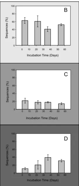
FIG 3 Maximum likelihood phylogenetic analysis of coxL-inferred amino acid sequences of 136 OTU and reference sequences from NCBI (147 residues). (A) The three main subclasses of sequences belonging to coxL are represented in the three clusters. The number of sequences of each OTU or a cluster is indicated in square brackets as “[OTU number; number of sequences].” The percentages of replicated trees in which the associated CoxL sequences clustered together in the bootstrap test (1,000 replicates) are shown for nodes supported byⱖ50% of the replicates. (B to D) Time series of coxL sequence relative abundance in (B) coxL group x, (C) coxL Actinobacteria, and (D) coxL Alphaproteobacteria clusters.
on June 23, 2016 by INRS-Institut Armand-Frappier
http://aem.asm.org/
Acidobacteria Actinobacteria AD3 Chlamydiae Chloroflexi Cyanobacteria Firmicutes Gemmatimonadetes Planctomycetes Proteobacteria TM6 Verrucomicrobia WPS Acidobact eriaceae Koribact eraceae Unknown Ellin6513 Unknown Solibact erales Act inospicaceae Cellulomonadaceae Conexibact eraceae EB1017 Frankiaceae Gaiellaceae Intrasporangiaceae Microbact eriaceae Micromonosporaceae Myc obact eriaceae Nocardiaceae Pseudonocardiaceae Streptomyc etaceae Streptosporangiaceae Thermomonosporaceae Unknown Acidimicrobiales Unknown Act inomyc etales Unknown Solirubrobact erales Unknown J G37-AG-4 Amoebophilaceae Chitinophagaceae Sphingobact eriaceae Parachlamy diaceae Rhabdochlamy diaceae Unknown C hlamy diales Unknown C hlamy diia Ktedonobact eraceae Thermogemm atisporaceae Unknown M LE1-12 Unknown 258ds10 Alicyc lobacillaceae Bacillaceae Clost ridiaceae Paenibacillaceae Peptococc aceae SBYG_4172 Unknown Gemm -1 Unknown Gemm atimonadetes Unknown W D2101 Acetobact eraceae Beijerinck iaceae Bradyrhizobiaceae Caulobact eraceae Hy phomicrobiaceae Methylocyst aceae Rhodospirillaceae Unknown Ellin329 Unknown R hizobiales Unknown Burkholderiaceae Comamonadaceae Procabact eriaceae Unknown Burkholderiales Haliangiaceae Myx ococc aceae Polyangiaceae Syntrophobact eraceae Unknown MIZ 46 Unknown Myx ococc ales Coxiellaceae Enterobact eriaceae Legionellaceae Pseudomonadaceae Sinobact eraceae Xanthomonadaceae Unknown Legionellales Unknown SJA Unknown SBRH 58 auto67_4W Chthoniobact eraceae Unknown S-BQ2-57 Unknown Unass igned
Modules
1 2 3 4 5 0 30 60Number of OTUs
Bacteroidetes Fibrobacteres 16S rRNA families1
2
3
4
5
Number of sequences in the module (%) 66,589 24,606 564,791 26,217 Color keyA
B
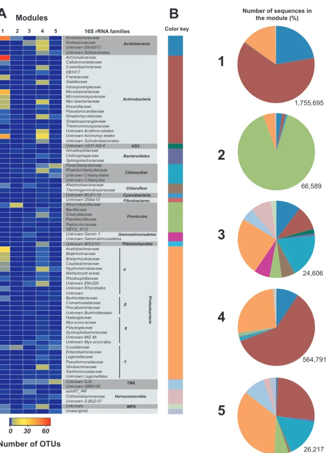
1,755,695FIG 4 Taxonomic composition of the five modules defined by the covariance of the 16S rRNA OTU time series in the soil. (A) The heat map shows the number of OTU representing each family detected in the modules. (B) The OTU assigned to the same bacterial phyla were clustered together, and the number of sequences comprising each group was computed to report their relative abundance in the five modules. The color key used to identify each bacterial phylum in the pie charts is defined along the side of the heat map. The total number of sequences comprised in the modules is shown at the bottom of each pie chart.
on June 23, 2016 by INRS-Institut Armand-Frappier
http://aem.asm.org/
estimated by multiplying the relative abundance of the coxL
OTU obtained from pyrosequencing profiles by the absolute
abundance of coxL group x genes determined by qPCR.
Con-sidering the specific richness and the abundance of OTU
en-compassing the coxL group x genotype, module 1 was searched
for potential 16S rRNA gene OTU candidates to infer a
taxo-nomic affiliation with unknown presumptive carboxydovore
bacteria. 16S rRNA OTU belonging to Actinospicaceae and
Mi-crobacteriaceae were the best coxL group x candidates, as predicted
using the correlation network analysis. These two bacterial families
were the sole candidates for which the absolute abundance and
spe-cies richness of 16S rRNA OTU were congruent with those of coxL
group x OTU in module 1 (see Table S1).
DISCUSSION
Enrichment of microorganisms utilizing atmospheric trace gas is
challenging because the energy potential of these substrates
usu-ally supports mixotrophic survival metabolism, where reduced
inorganic and organic compounds are simultaneously used as
en-ergy sources for maintenance or survival. Furthermore, increase
of energy potential through increased gas concentration is not
recommended because it will result in the enrichment of microbes
that are incapable of scavenging atmospheric trace gas due to their
low substrate affinity. Most high-affinity CO-oxidizing bacteria
were identified through screening of isolates obtained from
envi-ronmental samples or public collections after detection on coxL in
sequenced genome. One notable exception was the isolation of the
first carboxydovore bacterium, Aminobacter sp. strain cox1, after
long-term exposure of soil to 40 to 400 ppmv CO (
39
). Recent
investigations into the diversity of soil carboxydovore bacteria led
to the identification of an atypical coxL group x genotype for
which the distribution was significantly related to the CO uptake
rate in soil (
21
). Before investment of cultivation efforts to isolate
these unknown carboxydovore bacteria, we sought to challenge
the hypothetical taxonomic affiliation of atypical coxL genotype to
Deltaproteobacteria through the monitoring of microbial
succes-sion and maturation of CO uptake activity in soil microcosms.
Successions of 16S rRNA and coxL genes analyzed by qPCR
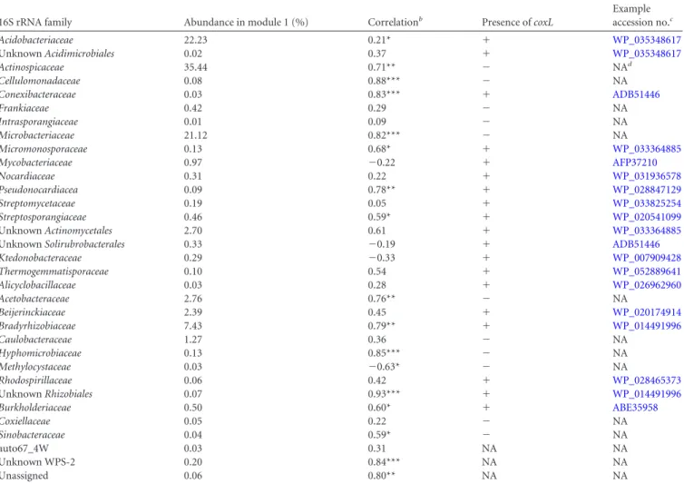
TABLE 2 Composition of module 1 comprising 16S rRNA OTU displaying colonization profiles related to maturation of CO oxidation activity in soila
16S rRNA family Abundance in module 1 (%) Correlationb Presence of coxL
Example accession no.c Acidobacteriaceae 22.23 0.21* ⫹ WP_035348617 Unknown Acidimicrobiales 0.02 0.37 ⫹ WP_035348617 Actinospicaceae 35.44 0.71** ⫺ NAd Cellulomonadaceae 0.08 0.88*** ⫺ NA Conexibacteraceae 0.03 0.83*** ⫹ ADB51446 Frankiaceae 0.42 0.29 ⫺ NA Intrasporangiaceae 0.01 0.09 ⫺ NA Microbacteriaceae 21.12 0.82*** ⫺ NA Micromonosporaceae 0.13 0.68* ⫹ WP_033364885 Mycobacteriaceae 0.97 ⫺0.22 ⫹ AFP37210 Nocardiaceae 0.31 0.22 ⫹ WP_031936578 Pseudonocardiacea 0.09 0.78** ⫹ WP_028847129 Streptomycetaceae 0.19 0.05 ⫹ WP_033825254 Streptosporangiaceae 0.46 0.59* ⫹ WP_020541099 Unknown Actinomycetales 2.70 0.61 ⫹ WP_033364885
Unknown Solirubrobacterales 0.33 ⫺0.19 ⫹ ADB51446
Ktedonobacteraceae 0.29 ⫺0.33 ⫹ WP_007909428 Thermogemmatisporaceae 0.10 0.54 ⫹ WP_052889641 Alicyclobacillaceae 0.03 0.28 ⫹ WP_026962960 Acetobacteraceae 2.76 0.76** ⫺ NA Beijerinckiaceae 2.39 0.45 ⫹ WP_020174914 Bradyrhizobiaceae 7.43 0.79** ⫹ WP_014491996 Caulobacteraceae 1.27 0.36 ⫺ NA Hyphomicrobiaceae 0.13 0.85*** ⫺ NA Methylocystaceae 0.03 ⫺0.63* ⫺ NA Rhodospirillaceae 0.06 0.42 ⫹ WP_028465373 Unknown Rhizobiales 0.07 0.93*** ⫹ WP_014491996 Burkholderiaceae 0.50 0.60* ⫹ ABE35958 Coxiellaceae 0.05 0.22 ⫺ NA Sinobacteraceae 0.04 0.59* ⫺ NA auto67_4W 0.03 0.31 NA NA Unknown WPS-2 0.20 0.84*** NA NA Unassigned 0.06 0.80** NA NA a
The relative abundance of sequences encompassing each family and the presence or the absence of the coxL gene in the reference genome of each taxonomic group are presented. Spearman correlations between the absolute abundance of OTU grouped at the family level and CO oxidation rate are presented.
b
Correlation significance levels are indicated by asterisks: *, P⬍ 0.05; **, P ⬍ 0.01; and ***, P ⬍ 0.001.
cFound by sequence identity with blastp (49) using the coxL sequence in Burkholderia xenovorans LB400 (accession no.ABE35958). d
NA, not applicable.
on June 23, 2016 by INRS-Institut Armand-Frappier
http://aem.asm.org/
(
Fig. 2
) unveiled that coxL carboxydovore bacteria generally are
opportunistic r-strategist bacteria using labile nutrients in soil.
They also increase their relative abundance early in the incubation
period. Our data showed that their enrichment was transient,
sug-gesting that their populations were not maintained upon soil
nu-trient depletion. Interestingly, the CO-oxidizers represented by
coxL group x displayed the K-strategist colonization profile, with
slow growth of their population sustained by the efficient use of
limited resources in soil. At the end of the incubation, total coxL
and coxL group x genotypes represented 2.2% and 0.7% of the
total bacterial population, respectively. It is believed that
atmo-spheric CO can support mixotrophic metabolism of these
car-boxydovore bacteria (
9
). To address that question, we combined
our qPCR time series with thermodynamic models. The
theoret-ical population size of high-affinity CO-oxidizing bacteria in soil
for which CO oxidation fully supplies maintenance energy
re-quirements was estimated based on the theoretical maintenance
energy consumption rate and the free energy of atmospheric CO
oxidation, using the following model formulated by Conrad (
40
):
N
⫽ [1.4 ⫻ 10
14(⫺⌬G) d]/mE, where N is the theoretical
popu-lation size of CO-oxidizing bacteria in soil (expressed as the
num-ber of cells per gram of soil dry weight), 1.4
⫻ 10
14is a constant
expressing the density of bacterial cells containing 1 mol carbon
(number of cells per mole of C biomass),
⌬G is the Gibbs free
energy of atmospheric CO oxidation (
⫺235 kJ per mol of CO), d
is the measured CO oxidation rate (moles of CO per gram of soil
dry weight), and mE (4.5 kJ per mol of C biomass) is the energy
maintenance requirement of the population (
41
). This model
es-timated that measured CO uptake activity would provide
suffi-cient energy to support the survival of a maximal population 2.5
⫻
10
7carboxydovore bacteria g dry weight
⫺1. According to our
qPCR results, the abundance of total coxL and coxL group x
se-quences reached (5.5
⫾ 0.97) ⫻ 10
9and (1.8
⫾ 0.20) ⫻ 10
9copies
g dry weight in soil at the end of the incubation, respectively.
Taken together, these observations imply that oligotrophic
culti-vation media comprising both organic carbon and CO would be
necessary to isolate unknown carboxydovore bacteria harboring
the atypical coxL group x genotype that are expected mixotrophic
K-strategists.
Although microbial succession was shaped by stochasticity in
the colonial growth dynamic of individual cells (
42
) and alteration
of the chemical structure and bioavailability of soil organic carbon
due to autoclaving (
43
), the taxonomic affiliation of coxL
geno-types inferred from phylogenetic analysis resulted in similar
dis-tributions compared with the observations made using native
de-ciduous soil (
21
). Actinobacteria, Alphaproteobacteria, and coxL
group x were the dominating coxL genotypes, representing 31%
⫾
3%, 14%
⫾ 3%, and 53% ⫾ 3% of the total sequences,
respec-tively. Very few studies have reported the taxonomic composition
of carboxydovore bacteria in soil. A soil survey along a succession
of volcanic deposits unveiled an enrichment of coxL sequences
closely related to Burkholderia isolates known to consume
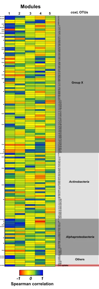
atmo-FIG 5 Spearman correlations between the coxL OTU with the eigengenes of the five modules comprising 16S rRNA OTU displaying a similar soil coloni-zation profile. Only the eigengene of module 1 was correlated with the CO uptake rate measured in soil microcosms. Significant correlations between the time series of individual coxL OTU and the eigengene of module 1 are marked with asterisks along the side of the heat map (*, P⬍ 0.05; **, P ⬍ 0.01; ***, P ⬍ 0.001). 2 3 5 6 8 11 12 13 14 16 17 21 22 24 25 26 27 28 29 30 32 33 34 36 37 38 39 42 45 47 50 53 56 60 62 69 71 72 77 78 85 86 88 89 91 95 101 102 103 104 108 111 112 115 116 117 118 125 137 138 143 146 150 154 162 166 169 173 183 193 234 259 264 267 289 4 7 10 19 23 35 40 41 43 46 48 54 55 63 64 65 66 67 75 76 81 83 92 97 98 109 123 126 133 134 135 140 160 186 200 283 1 9 15 20 31 49 51 52 59 68 70 73 84 87 93 94 110 122 182 197 18 58 79 100 296
Modules
1 2 3 4 5-1 0 1
Spearman correlation
Group X Alphaproteobacteria Actinobacteria Others CO uptake *** coxL OTUs *** *** ***** ** *** ** * ** ** * ** ** * * ** ** * *** * * ** ** ** * * ** ** ** * ***** ** *** * * ** *** *** ***on June 23, 2016 by INRS-Institut Armand-Frappier
http://aem.asm.org/
spheric CO (
10
) as a function of vegetation regeneration and CO
uptake activity (
17
,
44
). In this study, two coxL OTU (OTU 79 and
100) related to Burkholderia xenovorans LB400 coxL sequence
were detected, but their temporal distribution profile was not
re-lated to the maturation of CO uptake activity in soil. It has been
reported that some species of mycobacteria from the
Mycobacte-riaceae family in the Actinobacteria phylum are capable of
con-suming atmospheric CO (
45
,
46
). In this study, 14 coxL OTU
closely related to Mycobacterium sequences were detected, but
none displayed significant correlation with the CO oxidation rate
in soil. The high abundance of coxL sequences closely related to
Bradyrhizobium spp. and the correlation of their distribution
pro-file with CO uptake activity suggest implication of these
Alphapro-teobacteria in the activity measured in the microcosms. Although
they were not dominant in previous soil surveys, their capacity to
consume atmospheric CO has already been reported using
envi-ronmental isolates (
10
). Sequences encompassing coxL group x
were abundant, with H. ochraceum as the only representative. CO
uptake activity has been confirmed in this bacterium (
21
), but no
coxL sequences have been noticed in sequenced genomes of the
other aerobic Deltaproteobacteria encompassing the orders
Myxo-coccales and Bdellovibrionales. The combination of qPCR and
high-throughput sequencing of 16S rRNA and coxL genes led to
the conclusion that it is very unlikely that Deltaproteobacteria
har-bor the coxL group x genotype. If unknown Deltaproteobacteria
harbored the atypical coxL genotype, then a strong primer bias has
impaired their detection or considerably underestimated their
abundance.
Network correlation analysis led to the identification of
taxo-nomic groups of bacteria for which the distribution was
signifi-cantly correlated with the distribution of OTU encompassing coxL
group x. Although the analysis does not imply any causal effect, it
supported the suggestion that unknown carboxydovore bacteria
are mixotrophic K-strategists, which is critical for the
establish-ment of the best isolation strategy. Among the taxonomic groups
detected in module 1 for which the eigengene was correlated to
CO uptake rate, bacteria encompassing the Actinospicaceae and
Microbacteriaceae families were the most probable taxonomic
as-signment of unknown carboxydovore bacteria harboring the coxL
group x genotype. This taxonomic inference will need validation
through isolation efforts since no representative of these two
fam-ilies was reported as CO-oxidizing bacteria. Interestingly,
micro-organisms that belong to these taxonomic groups are generally
K-strategists, with some of them having the ability to use
recalci-trant carbon such as cellulose (
47
). Furthermore, soil amendment
experiments unveiled that cellulose stimulates CO uptake activity
in soil, while glucose exerts the inverse effect (I. Lalonde,
unpub-lished data). Bioprospection efforts are currently being used by the
authors to identify and characterize unknown carboxydovore
bac-teria harboring the coxL group x genotype. Alternatively,
single-cell sequencing (
48
) could be envisaged if cultivation efforts are
unsuccessful.
ACKNOWLEDGMENTS
I.L. is grateful to the Fondation Universitaire Armand-Frappier INRS for her M.Sc. scholarship.
We are grateful to the personnel staff of McGill University and Gé-nome Québec Innovation Centre for preparation of 16S rRNA gene li-braries and sequencing services. Mondher Khdhiri is acknowledged for
technical assistance in quality control, classification, and taxonomic affil-iation of 16S rRNA gene sequences.
FUNDING INFORMATION
Gouvernement du Canada | Natural Sciences and Engineering Research Council of Canada (NSERC) provided funding to Philippe Constant un-der grant number 418252-2012.
The NSERC funding was via a Discovery grant.
REFERENCES
1. Seiler W. 1974. The cycle of atmospheric CO. Tellus 26:116 –135.http:
//dx.doi.org/10.1111/j.2153-3490.1974.tb01958.x.
2. Park K, Emmons LK, Wang Z, Mak JE. 2015. Joint application of concentration and␦18O to investigate the global atmospheric CO budget. Atmosphere 6:547–578.http://dx.doi.org/10.3390/atmos6050547. 3. Monks PS. 2005. Gas-phase radical chemistry in the troposphere. Chem
Soc Rev 34:376 –395.http://dx.doi.org/10.1039/b307982c.
4. Inman RE, Ingersoll RB, Levy EA. 1971. Soil: a natural sink for carbon monoxide. Science 172:1229 –1231. http://dx.doi.org/10.1126/science
.172.3989.1229.
5. Dobbek H, Gremer L, Kiefersauer R, Huber R, Meyer O. 2002. Catalysis at a dinuclear [CuSMo(⫽O)OH] cluster in a CO dehydrogenase resolved at 1.1-A resolution. Proc Natl Acad Sci U S A 99:15971–15976.http://dx
.doi.org/10.1073/pnas.212640899.
6. Dunfield KE, King GM. 2004. Molecular analysis of carbon monoxide-oxidizing bacteria associated with recent Hawaiian volcanic deposits. Appl Environ Microbiol 70:4242– 4248. http://dx.doi.org/10.1128/AEM.70.7
.4242-4248.2004.
7. Lorite MJ, Tachil J, Sanjuan J, Meyer O, Bedmar EJ. 2000. Carbon monoxide dehydrogenase activity in Bradyrhizobium japonicum. Appl En-viron Microbiol 66:1871–1876.http://dx.doi.org/10.1128/AEM.66.5.1871
-1876.2000.
8. Cunliffe M. 2011. Correlating carbon monoxide oxidation with cox genes in the abundant marine Roseobacter clade. ISME J 5:685– 691.http://dx
.doi.org/10.1038/ismej.2010.170.
9. King GM, Weber CF. 2007. Distribution, diversity and ecology of aerobic CO-oxidizing bacteria. Nat Rev Microbiol 5:107–118.http://dx.doi.org
/10.1038/nrmicro1595.
10. King GM. 2003. Molecular and culture-based analyses of aerobic carbon monoxide oxidizer diversity. Appl Environ Microbiol 69:7257–7265.http:
//dx.doi.org/10.1128/AEM.69.12.7257-7265.2003.
11. Weber CF, King GM. 2007. Physiological, ecological, and phyloge-netic characterization of Stappia, a marine CO-oxidizing bacterial genus. Appl Environ Microbiol 73:1266 –1276.http://dx.doi.org/10.1128/AEM
.01724-06.
12. Cunliffe M. 2013. Physiological and metabolic effects of carbon monoxide oxidation in the model marine bacterioplankton Ruegeria pomeroy DSS-3. Appl Environ Microbiol 79:738 –740. http://dx.doi.org/10.1128/AEM
.02466-12.
13. Weber CF, King GM. 2012. The phylogenetic distribution and ecological role of carbon monoxide oxidation in the genus Burkholderia. FEMS Mi-crobiol Ecol 79:167–175.http://dx.doi.org/10.1111/j.1574-6941.2011
.01206.x.
14. Hoeft SE, Blum JS, Stolz JF, Tabita FR, Witte B, King GM, Santini JM, Oremland RS. 2007. Alkalilimnicola ehrlichii sp. nov., a novel, arsenite-oxidizing haloalkaliphilic gammaproteobacterium capable of chemoau-totrophic or heterotrophic growth with nitrate or oxygen as the electron acceptor. Int J Syst Evol Microbiol 57:504 –512.http://dx.doi.org/10.1099
/ijs.0.64576-0.
15. King CE, King GM. 2014. Description of Thermogemmatispora carbox-idivorans sp. nov., a carbon-monoxide-oxidizing member of the class Kt-edonobacteria isolated from a geothermally heated biofilm, and analysis of carbon monoxide oxidation by members of the class Ktedonobacteria. Int J Syst Evol Microbiol 64:1244 –1251. http://dx.doi.org/10.1099/ijs.0
.059675-0.
16. King GM, Weber CF. 2008. Interactions between bacterial carbon monoxide and hydrogen consumption and plant development on re-cent volcanic deposits. ISME J 2:195–203.http://dx.doi.org/10.1038
/ismej.2007.101.
17. Weber CF, King GM. 2010. Distribution and diversity of carbon mon-oxide-oxidizing bacteria and bulk bacterial communities across a
on June 23, 2016 by INRS-Institut Armand-Frappier
http://aem.asm.org/
sion gradient on a Hawaiian volcanic deposit. Environ Microbiol 12: 1855–1867.http://dx.doi.org/10.1111/j.1462-2920.2010.02190.x. 18. Yang J, Zhou E, Jiang H, Li W, Wu G, Huang L, Hedlund BP, Dong H.
2015. Distribution and diversity of aerobic carbon monoxide-oxidizing bacteria in geothermal springs of China, the Philippines, and the United States. Geomicrobiol J 32:903–913.http://dx.doi.org/10.1080/01490451
.2015.1008605.
19. Cunliffe M, Schafer H, Harrison E, Cleave S, Upstill-Goddard R, Murrell JC. 2008. Phylogenetic and functional gene analysis of the bacte-rial and archaeal communities associated with the surface microlayer of an estuary. ISME J 2:776 –789.http://dx.doi.org/10.1038/ismej.2008.28. 20. Badr O, Probert SD. 1995. Sinks and environmental impacts for
atmo-spheric carbon monoxide. Appl Energy 50:339 –372.http://dx.doi.org/10
.1016/0306-2619(95)98803-A.
21. Quiza L, Lalonde I, Guertin C, Constant P. 2014. Land-use influences the distribution and activity of high affinity CO-oxidizing bacteria associ-ated to type I-coxL genotype in soil. Front Microbiol 5:271.http://dx.doi
.org/10.3389/fmicb.2014.00271.
22. Elghamry W, Elashkar M. 1962. Simplified textural classification trian-gles. Soil Sci Soc Am J 26:612– 613.http://dx.doi.org/10.2136/sssaj1962
.03615995002600060028x.
23. King GM, Hungria M. 2002. Soil-atmosphere CO exchanges and micro-bial biogeochemistry of CO transformations in a Brazilian agricultural ecosystem. Appl Environ Microbiol 68:4480 – 4485.http://dx.doi.org/10
.1128/AEM.68.9.4480-4485.2002.
24. Moxley JM, Smith KA. 1998. Factors affecting utilisation of atmospheric CO by soils. Soil Biol Biochem 30:65–79.http://dx.doi.org/10.1016/S0038
-0717(97)00095-3.
25. Constant P, Poissant L, Villemur R. 2008. Isolation of Streptomyces sp. PCB7, the first microorganism demonstrating high-affinity uptake of tropospheric H2. ISME J 2:1066 –1076. http://dx.doi.org/10.1038
/ismej.2008.59.
26. Berthelet M, Whyte LG, Greer CW. 1996. Rapid, direct extraction of DNA from soils for PCR analysis using polyvinylpolypyrrolidone spin columns. FEMS Microbiol Lett 138:17–22.http://dx.doi.org/10.1111/j
.1574-6968.1996.tb08128.x.
27. Schloss PD, Westcott SL, Ryabin T, Hall JR, Hartmann M, Hollister EB, Lesniewski RA, Oakley BB, Parks DH, Robinson CJ, Sahl JW, Stres B, Thallinger GG, Van Horn DJ, Weber CF. 2009. Introducing mothur: open-source, platform-independent, community-supported software for describing and comparing microbial communities. Appl Environ Micro-biol 75:7537–7541.http://dx.doi.org/10.1128/AEM.01541-09.
28. Tamura K, Dudley J, Nei M, Kumar S. 2007. MEGA4: Molecular Evo-lutionary Genetics Analysis (MEGA) software version 4.0. Mol Biol Evol 24:1596 –1599.http://dx.doi.org/10.1093/molbev/msm092.
29. Magocˇ T, Salzberg SL. 2011. FLASH: fast length adjustment of short reads to improve genome assemblies. Bioinformatics 27:2957–2963.http://dx
.doi.org/10.1093/bioinformatics/btr507.
30. Edgar RC, Haas BJ, Clemente JC, Quince C, Knight R. 2011. UCHIME improves sensitivity and speed of chimera detection. Bioinformatics 27: 2194 –2200.http://dx.doi.org/10.1093/bioinformatics/btr381.
31. Caporaso JG, Kuczynski J, Stombaugh J, Bittinger K, Bushman FD, Costello EK, Fierer N, Pena AG, Goodrich JK, Gordon JI. 2010. QIIME allows analysis of high-throughput community sequencing data. Nat Methods 7:335–336.http://dx.doi.org/10.1038/nmeth.f.303.
32. McDonald D, Price MN, Goodrich J, Nawrocki EP, DeSantis TZ, Probst A, Andersen GL, Knight R, Hugenholtz P. 2012. An improved Green-genes taxonomy with explicit ranks for ecological and evolutionary
anal-yses of bacteria and archaea. ISME J 6:610 – 618.http://dx.doi.org/10.1038
/ismej.2011.139.
33. R Core Development Team. 2008. R: a language and environment for statistical computing. R Foundation for Statistical Computing, Vienna, Austria.
34. Harrell FEJ. 2015. R package version 3.16-0. Hmisc: Harrell Miscella-neous.http://CRAN.R-project.org/package⫽Hmisc.
35. Oksanen J, Blanchet FG, Kindt R, Legendre P, Minchin PR, O’Hara RB, Simpson GL, Solymos P, Stevens MHH, Wagner H. 2012. Vegan: community ecology package. R package version 2.0-4. http://cran.r
-project.org/package⫽vegan.
36. Anderson MJ, Ellingsen KE, McArdle BH. 2006. Multivariate dispersion as a measure of beta diversity. Ecol Lett 9:683– 693.http://dx.doi.org/10
.1111/j.1461-0248.2006.00926.x.
37. Langfelder P, Horvath S. 2008. WGCNA: an R package for weighted correlation network analysis. BMC Bioinformatics 9:559.http://dx.doi
.org/10.1186/1471-2105-9-559.
38. Vetrovsky T, Baldrian P. 2013. The variability of the 16S rRNA gene in bacterial genomes and its consequences for bacterial community analyses. PLoS One 8:e57923.http://dx.doi.org/10.1371/journal.pone.0057923. 39. Hardy KR, King GM. 2001. Enrichment of high-affinity CO oxidizers in
Maine forest soil. Appl Environ Microbiol 67:3671–3676.http://dx.doi
.org/10.1128/AEM.67.8.3671-3676.2001.
40. Conrad R. 1999. Soil microorganisms oxidizing atmospheric trace gases (CH4, CO, H2, NO). Indian J Microbiol 39:193–204.
41. Tijhuis L, Van Loosdrecht MCM, Heijnen JJ. 1993. A thermodynami-cally based correlation for maintenance Gibbs energy requirements in aerobic and anaerobic chemotrophic growth. Biotechnol Bioeng 42:509 –
519.http://dx.doi.org/10.1002/bit.260420415.
42. Alonso AA, Molina I, Theodoropoulos C. 2014. Modeling bacterial population growth from stochastic single-cell dynamics. Appl Environ Microbiol 80:5241–5253.http://dx.doi.org/10.1128/AEM.01423-14. 43. Berns AE, Philipp H, Narres HD, Burauel P, Vereecken H, Tappe W.
2008. Effect of gamma-sterilization and autoclaving on soil organic matter structure as studied by solid state NMR, UV and fluorescence spectros-copy. Eur J Soil Sci 59:540 –550.http://dx.doi.org/10.1111/j.1365-2389
.2008.01016.x.
44. Weber CF, King GM. 2010. Quantification of Burkholderia coxL genes in Hawaiian volcanic deposits. Appl Environ Microbiol 76:2212–2217.http:
//dx.doi.org/10.1128/AEM.01861-09.
45. King GM. 2003. Uptake of carbon monoxide and hydrogen at envi-ronmentally relevant concentrations by mycobacteria. Appl Environ Microbiol 69:7266 –7272.http://dx.doi.org/10.1128/AEM.69.12.7266
-7272.2003.
46. Kim YM, Park SW. 2012. Microbiology and genetics of CO utilization in mycobacteria. Antonie Van Leeuwenhoek 101:685–700.http://dx.doi.org
/10.1007/s10482-012-9698-y.
47. Lynd LR, Weimer PJ, van Zyl WH, Pretorius IS. 2002. Microbial cellulose utilization: fundamentals and biotechnology. Microbiol Mol Biol Rev 66:506 –577. http://dx.doi.org/10.1128/MMBR.66.3.506-577
.2002.
48. Ottesen EA, Hong JW, Quake SR, Leadbetter JR. 2006. Microfluidic digital PCR enables multigene analysis of individual environmental bac-teria. Science 314:1464 –1467.http://dx.doi.org/10.1126/science.1131370. 49. Altschul SF, Gish W, Miller W, Myers EW, Lipman DJ. 1990. Basic local alignment search tool. J Mol Biol 215:403– 410.http://dx.doi.org/10.1016
/S0022-2836(05)80360-2.