Université de Montréal
L’impact des résections de l’insula sur la personnalité
par
Benjamin Hébert-Seropian
Département de psychologie Faculté des arts et des sciences
Mémoire présenté à la Faculté des arts et des sciences en vue de l’obtention du grade de Maîtrise en sciences (M. Sc.)
en psychologie
Août, 2016
i
Résumé
La recherche montre que l’insula est impliquée dans le traitement d’informations intéroceptives, émotionnelles et relevant de fonctions exécutives de haut niveau. L’hypothèse des marqueurs somatiques propose que ces fonctions vraisemblablement séparées travaillent plutôt de concert au sein d’un système neural dont le rôle consiste à extraire les messages émotionnels des signaux corporels. Si l’insula exerce effectivement un rôle de modulateur des sensations corporelles et des processus cognitifs découlant de ceux-ci, des lésions au cortex insulaire risquent d’occasionner des altérations au niveau de l’expérience émotionnelle, des fonctions exécutives et de la personnalité. La présente étude a pour but de mesurer ces changements chez 19 patients ayant subi une insulectomie unilatérale dans le cadre de leur traitement de l’épilepsie. Ces patients ont été comparés à un groupe contrôle composé de 19 patients épileptiques ayant subi une résection du lobe temporal. Les participants ont été évalués par l’entremise du Iowa Scales of Personality Change (ISPC), rempli par un proche du patient. Les résultats montrent que les patients du groupe insulaire exhibent des changements qui dénotent une dérégulation émotionnelle à long terme, caractérisée par une augmentation modérée de l’irritabilité, de la labilité émotionnelle, de l’anxiété et de la frugalité, tous des changements qui, outre l’anxiété, n’ont pas été observés chez les patients temporaux. Cependant, pour ce qui est des fonctions exécutives, aucun changement significatif n’a été noté. De plus, la comparaison pré- et post opératoire des scores des deux groupes aux items de l’ISPC ne s’est pas avérée significative. Globalement, les résultats suggèrent que l’insula joue probablement un rôle accessoire au sein du modèle proposé par l’hypothèse des marqueurs somatiques et que les résections unilatérales partielles ou complètes de l’insula ne risquent pas d’occasionner de changements prononcés de la personnalité.
ii
Abstract
Research has shown that the insula is involved in the processing of information relating to interoceptive, emotional and executive functions. It was proposed that these two seemingly separate functions may work conjointly as part of a large neural circuit tasked with the extraction of emotional information from bodily signals. It was hypothesized that, if the insula does indeed modulate feelings and the cognitive processes which derive from them, insular damage would result in alterations of emotional experience, executive functions and personality. To that effect, we examined such changes in a group of patients (n = 19) who underwent epilepsy surgery involving partial or complete resection of the insula, and compared them to a group of patients who underwent temporal lobe epilepsy surgery (n = 19) as a lesion-control group. Participants were assessed on the Iowa Scales of Personality Change, filled by a close relative at least six months after surgery. While pre- vs. post-surgery changes did not significantly differ between groups on any of the outcome variables, insular resections were associated with mild but significant increases in irritability, emotional lability, anxiety, and frugality postoperatively, which, with the exception of increased anxiety, were not found among temporal patients. Against our initial prediction, the surgery did not lead to executive functioning deficits. Overall, our results support the notion that the insula most likely holds an accessorial role in the model proposed by the somatic marker hypothesis, and that there isn’t a risk of dramatic personality change as a result of the partial or complete unilateral surgical removal of the insula.
iii
Table des matières
Résumé ... i
Abstract ... ii
Table des matières... iii
Liste des tableaux ... v
Article ... v
Liste des figures ... vi
Article ... vi
Liste des sigles ... vii
Remerciements ... viii
Introduction ... 1
L’insula ... 1
Études fonctionnelles du rôle de l’insula ... 1
L’hypothèse des marqueurs somatiques ... 3
L’impact des lésions de l’insula sur la personnalité ... 6
Le traitement chirurgical de l’épilepsie ... 8
Hypothèses ... 9 Objectifs ... 9 Article ... 10 Abstract ... 11 1. Introduction ... 12 2. Method ... 14
2.1 Participants and procedure ... 14
2.2 Assessment ... 15
2.3 Supplementary measures ... 16
2.4 Statistical analyses ... 17
iv
3.1 Sample characteristics ... 19
3.2 ISPC score changes following unilateral resection of the insula ... 19
3.3 ISPC score changes following temporal lobe resection ... 20
3.4 Comparison between the insular and the temporal groups ... 20
3.5 Relationship between type of surgery and worsening on ISPC ratings ... 21
3.6 Relationship between time elapsed since surgery and severity of symptoms ... 22
3.7 Hemisphere of lesion as a predictor of ISPC scores ... 22
3.8 Relationship between type of insulectomy and worsening on ISPC dimensions ... 22
4. Discussion ... 23
5. Conclusion ... 29
Disclosure of conflicts of interests ... 30
References ... 31 Tables ... 40 Figures... 51 Figure 1 ... 51 Discussion ... 52 Conclusion ... 58 Bibliographie... 60
v
Liste des tableaux
Article
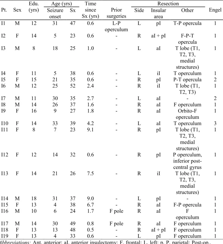
Table 1. Demographic and surgery-related characteristics of the insular group (n = 19). ... 40
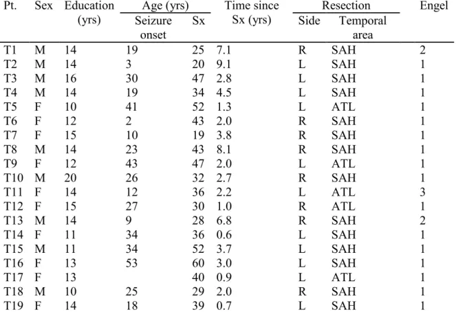
Table 2. Demographic and surgery-related characteristics of the temporal group (n = 19). ... 41
Table 3. Effect of time on mean ISPC scores for the insular group (n = 19). ... 42
Table 4. Effect of time on mean ISPC scores for the temporal group (n =19). ... 43
Supplemental Table S1. Pearson’s correlations exploring the relationship between ISPC ratings and scores on the supplementary measures (N = 49). ... 44
Supplemental Table S2. Effect of time and of the site of lesion on mean ISPC scores for the insular and temporal groups (N =38). ... 45
Supplemental Table S3. Chi-square tests examining the relationship between the type of surgery and the frequency of significantly lower postoperative ISPC scores. ... 46
Supplemental Table S4. Chi-square tests examining the relationship between anterior insulectomy and the frequency of significantly lower postoperative ISPC dimension scores (N = 35). ... 47
Supplemental Table S5. Chi-square tests examining the relationship between posterior insulectomy and the frequency of significantly lower postoperative ISPC dimension scores (N = 35). ... 48
Supplemental Table S6. Pearson’s correlation tests exploring the relationship between time and severity of symptoms. ... 49
Supplemental Table S7. Measuring the effect of the hemisphere on ISPC scores for the insular group (n = 19). ... 50
vi
Liste des figures
Article
Figure 1. Percentages of individuals whose ISPC ratings improved or worsened following
vii
Liste des sigles
ISPC : Iowa Scales of Personality Change AVC : Accident vasculaire cérébral BAI : Beck Anxiety Inventory
BDI-II : Beck Depression Inventory-II CPFvm : Cortex préfrontal ventromédian IGT : Iowa Gambling Task
IRI : Interpersonal Reactivity Index
IRMf : Imagerie par résonance magnétique fonctionnelle RM-ANOVA : Repeated measures analysis of variance VLSM : Voxel-based lesion-symptom mapping
viii
Remerciements
La réalisation de ce mémoire a été rendue possible grâce à la contribution de plusieurs personnes à qui je voudrais témoigner toute ma gratitude. Tout d’abord, je tiens à remercier mes parents et ma sœur, sur qui j’ai toujours pu compter ; leur support inconditionnel m’a permis de poursuivre mes rêves avec ténacité. Je suis également reconnaissant envers ma directrice de recherche, Carole Sénéchal, qui me guide et m’offre un soutien indéfectible depuis notre toute première rencontre, il y a cinq ans de cela déjà. Au-delà de ses qualités de chercheuse et de professeure, j’estime beaucoup Carole pour son altruisme. Ce projet n’aurait pu non plus voir le jour sans la participation de mon codirecteur de recherche, Franco Lepore, qui m’a chaleureusement accueilli dans son équipe avec son entregent d’exception pour ensuite m’accompagner à chaque étape du parcours en me prodiguant de judicieux conseils. Merci à Olivier Boucher, ce mentor qui m’a transmis la piqûre pour la recherche grâce à son dévouement à la cause scientifique ainsi que sa grande bonté envers ses collègues. Tu es une grande source d’inspiration pour moi. Aux Drs Nguyen et Bouthillier ainsi qu’à la professeure Isabelle Rouleau,
merci infiniment pour vos précieux commentaires et votre support. Finalement, j’ai une pensée toute particulière pour mes amis et amies proches qui m’ont lu, écouté, supporté, voire subi pendant la longue phase de rédaction de ce mémoire. Je tiens notamment à souligner la contribution de Sarah, David, Zakaria et Simon. Je ne saurais conclure sans exprimer ma reconnaissance envers tous les autres acteurs qui m’ont également prêté main-forte et que je ne peux citer nommément par souci de concision. Sans vous tous, il va sans dire que ce projet qui m’a tant passionné n’aurait pu se réaliser.
1
Introduction
L’insula
Le cortex insulaire est une zone du cortex cérébral profondément dissimulée à l’intersection des lobes du cerveau, au creux de la fissure sylvienne. Cet emplacement central lui a valu son nom d’« insula », du terme latin signifiant « île ». Cette aire cérébrale est aussi nommée île de Reil, en l’honneur du physicien allemand Johann Christian Reil, qui l’a décrite pour la première fois en 1796 (Binder, Schaller, & Clusmann, 2007). Il a toutefois fallu attendre jusqu’en 1955 pour qu’on soit en mesure d’émettre une hypothèse concrète quant à son rôle, et ce, grâce aux travaux du Dr Penfield, un neurochirurgien montréalais. Celui-ci a induit des courants électriques dans les portions inférieures de l’insula, alors exposées chez des patients ayant subi une résection du lobe temporal afin de traiter une forme d’épilepsie réfractaire à la médication (Penfield & Faulk, 1955). Tandis qu’ils étaient stimulés à l’aide d'un faible courant électrique, 40 % des 36 patients étudiés ont rapporté avoir ressenti des sensations somatosensorielles, surtout au niveau de la bouche et des membres supérieurs. Un autre 40 % des patients ont indiqué que les stimulations électrocorticales induisaient des sensations au niveau des viscères, incluant entre autres des douleurs à l’abdomen, de la nausée et une impression de mouvement gastro-intestinal. Cette constatation a valu à l’insula le surnom de cerveau viscéral, décrivant une fonction qui, on le découvrira plus tard, ne constitue en fait qu’une parcelle du rôle de l’insula.
Études fonctionnelles du rôle de l’insula
Avec l’émergence de techniques d’imagerie cérébrale sophistiquées, on a découvert que l’insula joue un rôle central au sein de plusieurs fonctions. Cette zone est notamment activée de
2
façon constante lors du traitement d’informations intéroceptives, socioémotionnelles et lors de la prise de décisions risquées.
L’intéroception réfère à la sensibilité d’un sujet à l’égard des signaux sensoriels provenant des viscères, des tendons, des muscles et des articulations. L’imagerie par résonance magnétique fonctionnelle (IRMf) a permis l’observation d’une activité insulaire particulièrement forte chez les individus ayant une meilleure sensibilité intéroceptive, un constat qui vient appuyer les données colligées par le Dr Penfield et son équipe (Critchley, Wiens, Rotshtein, Ohman, & Dolan, 2004; Terasawa, Shibata, Moriguchi, & Umeda, 2013). Aujourd’hui, la perception de ces signaux peut être mesurée objectivement, par exemple lors d’un test au cours duquel le sujet tente de détecter les battements de son cœur (Schandry, 1981).
Plusieurs études en IRMf présentent de manière récurrente une activation de l’insula lors du traitement d’informations émotionnelles et socioémotionnelles (Kurth, Zilles, Fox, Laird, & Eickhoff, 2010; Lamm & Singer, 2010; Phan, Wager, Tyalor, & Liberzon, 2002). Qui plus est, la recherche a mis en évidence que la sensibilité intéroceptive est liée à la disposition des individus à faire l’expérience de certaines émotions. Cette interaction relèverait d’un mécanisme ayant une incidence sur l’intensité de l’émotion ressentie, et ce, en fonction de l’attention que prête un individu aux sensations corporelles qui sont associées à cette émotion (Critchley et al., 2004; Wiens, Mezzacappa, & Katkin, 2000). Dans le même ordre d’idées, plusieurs études ont rapporté qu’une activité anormale de l’insula était associée à des troubles émotionnels tels que la dépression, l’anxiété et l’alexithymie (Ernst et al., 2014; Naqvi & Bechara, 2009; Paulus & Stein, 2006; Sliz & Hayley, 2012). L’alexithymie est un trouble caractérisé par une incapacité à identifier et à exprimer ses émotions, et est souvent accompagnée d’une grande difficulté à différencier les sensations corporelles des émotions (Taylor, 2000). Le fait qu’il y ait à la fois une dérégulation de
3
l’activité insulaire, une réactivité émotionnelle amoindrie ainsi qu’une sensibilité intéroceptive accrue chez les alexithymiques laisse présager que l’insula joue un rôle important dans la traduction de sensations corporelles en expériences émotionnelles identifiables (Bernhardt et al., 2013; Ernst et al., 2014; Reker et al., 2010; Werner, Duschek, Mattern, & Schandry, 2009).
En plus de son rôle au sein d’un réseau alliant intéroception et émotions, l’insula est impliquée dans les processus de prises de décisions risquées. Des études d’IRMf ont rapporté une activation particulièrement forte de l’insula précédant une prise de décision comportant un risque (Kuhnen & Knutson, 2005) ainsi qu’après avoir perçu les conséquences découlant d’une prise de décision risquée (Paulus, Rogalsky, Simmons, & Murray, 2003). On a alors suggéré que la portion antérieure de l’insula se charge d’estimer le risque associé à un choix lors de la prise de décision, pour ensuite intégrer l’information provenant des erreurs de prédictions afin de parfaire son modèle prédictif (Preuschoff, Quartz, & Brossaerts, 2008).
Si on ignore actuellement l’étendue du lien entre les différentes fonctions de l’insula, soit sur le plan de l’intéroception, du traitement de signaux socioémotionnels et dans le cadre de la prise de décision risquée, il existe néanmoins un modèle qui propose d’unifier ces différentes composantes : il s’agit de l’hypothèse des marqueurs somatiques (Damasio, 1994).
L’hypothèse des marqueurs somatiques
L’hypothèse des marqueurs somatiques explique que les signaux en provenance du corps (en grec, sôma signifie « corps », d’où l’adjectif « somatique ») font partie d’un vaste réseau neural permettant l’intégration de l’information intéroceptive et émotionnelle lors de certains processus cognitifs de haut niveau, tels que le raisonnement et la prise de décision (Bechara & Damasio, 2005; Damasio, 1994). Essentiellement, cette théorie stipule que le comportement, les attitudes et
4
la prise de décision d’un individu sont grandement influencés par un processus d’échange d’informations entre les sensations corporelles et la cognition. L’inspection constante – souvent inconsciente – de l’état physiologique peut influencer le comportement d’un individu par le biais d’un système qui vise à prédire un gain ou une perte potentielle face à un stimulus émotionnel particulier. Les changements physiologiques engendrés par le stimulus peuvent ne pas être perceptibles par un observateur externe (relâchement d’hormones, accélération du rythme cardiaque), ou au contraire, évidents (posture, expression faciale, comportements spécifiques de type fight or flight). Si l’état corporel est catégorisé comme négatif (accélération du rythme cardiaque, sudation, serrement de la gorge), la situation particulière est alors étiquetée comme négative, ce qui a pour effet d’alerter l’organisme d’une situation fâcheuse à éviter. À l’opposé, si l’état corporel est perçu comme potentiellement favorable, le marquage positif encourage la prise d’une décision facilitant le rapprochement de l’objet en question.
Cette théorie a vu le jour afin d’expliquer des changements graves de la personnalité observés chez des individus à la suite d’un dommage causé à leur cortex préfrontal ventromédian (CPFvm). La particularité de ces changements comportementaux négatifs réside dans le fait que cette altération de la personnalité n’est pas attribuable à une réduction des capacités cognitives ni physiques; la majorité des cas recensés ne présentent aucune défaillance au niveau des connaissances, de l’expression et de la compréhension du langage, de la mémoire de travail ou de l’attention. Néanmoins, ceux-ci présentent une détérioration de leur capacité à exprimer leurs émotions et faire l’expérience de sentiments appropriés selon une mise en situation donnée. De plus, ces individus au cortex préfrontal lésé ont souvent la particularité de ne plus être en mesure de gérer efficacement leur temps et leurs activités : effectuer un choix banal devient particulièrement difficile, puisque les décisions dépendent désormais d’un processus d’analyse de
5
type coût-bénéfice vis-à-vis d’une gamme d’options souvent conflictuelles et impliquant une pléthore de conséquences potentielles (Bechara & Damasio, 2005). Le neurologue Antonio Damasio a alors proposé que ce déficit nouvellement acquis soit attribuable aux lésions du CPFvm, une zone cérébrale nécessaire au bon fonctionnement de ce système biorégulatoire usant de signaux émotionnels afin de prendre des décisions de façon intuitive plutôt que par un raisonnement purement logique. Tout comme plusieurs penseurs et chercheurs l’ayant précédé, Damasio rejette l’idée que chaque décision soit prise à la suite d’un processus cognitif rationnel, puisque cela nécessiterait du temps, des connaissances et une capacité de traitement de l’information quasi infinis (Damasio, 1994).
L’hypothèse des marqueurs somatiques est notamment corroborée par les résultats découlant d’une tâche expérimentale nommée Iowa Gambling Task (IGT), qui consiste à observer la stratégie d’un participant dont les choix sont influencés par le marquage émotionnel subi à la suite de pertes et de gains d’argent fictif. Les sujets ayant une lésion au CPFvm éprouvent plus de difficulté à choisir une stratégie gagnante. Tout comme le CPFvm, l’importance de l’insula a été mis en évidence pour le bon fonctionnement de ce système puisqu’elle permettrait la représentation des marqueurs émotionnels dérivés de l’information en provenance du corps (Damasio, Damasio, & Tranel, 2013). Anatomiquement parlant, cette piste est appuyée par la façon dont l’insula est connectée bidirectionnellement à presque tous les centres majeurs du traitement des émotions et de la cognition : le thalamus, l’amygdale, l’hippocampe, le cortex cingulaire antérieur, le cortex préfrontal et les noyaux gris centraux (Flynn, 1999; Körding, 2007).
Vu l’étendue des données colligées jusqu’à présent qui suggèrent que l’insula est impliquée dans de multiples tâches combinant les processus émotionnels — de l’ordre de la sensibilité intéroceptive ainsi que de la prise de décision —, il est possible de postuler que l’endommagement
6
ou la destruction du cortex insulaire puisse causer l’altération de la personnalité, ou du moins un certain degré de détérioration sur le plan affectif, comportemental et cognitif. Dans le domaine de la neuropsychologie cognitive, l’utilisation d’une technique nommée lesion method qui consiste à recenser l’information relative aux déficits nouvellement acquis à la suite de l’endommagement ou à la destruction d’une zone du cerveau est fréquemment utilisée pour démontrer la validité d’une hypothèse. Si les données sont en nombre suffisant, il est alors possible d’établir une corrélation entre de nouvelles défaillances et des locus de lésions spécifiques, ce qui permet ultimement d’inférer une fonction aux zones concernées (Kosslyn & Intriligator, 1992).
L’impact des lésions de l’insula sur la personnalité
Un cas largement cité pour illustrer les effets des lésions sur la personnalité est celui de Phineas Gage. Dans une étude, on rapportait que cet individu qui menait auparavant une vie conventionnelle est devenu instable psychologiquement et fortement asocial après un accident du travail au cours duquel son lobe frontal gauche fut détruit par une barre de fer (Harlow, 1868). S’il s’agit là d’un cas très populaire, il demeure néanmoins que les perturbations au niveau de la personnalité sont dans bien des cas la règle plutôt que l’exception chez ceux ayant subi une lésion au niveau du cortex préfrontal (Barrash, Tranel, & Anderson, 2000). D’ailleurs, la majorité des cas rapportant des changements de la personnalité traitent uniquement de patients présentant des lésions du cortex préfrontal, plus particulièrement dans la portion ventromédiane (DeLuca & Diamond, 1995; Eslinger & Damasio, 1984; Steinman & Bigler, 1986; Tranel, Anderson, & Benton, 1994).
En contrepartie, très peu d’études se sont penchées sur l’impact qu’une lésion insulaire peut avoir sur le comportement, les émotions, les habiletés sociales, et les fonctions exécutives.
7
Vraisemblablement, cette omission est largement due au faible taux de lésions affectant le cortex insulaire pris isolément (Cereda, Ghika, Maeder, & Bogousslavsky, 2002). L’une des seules études de cas ayant traité de l’impact d’une lésion au cortex insulaire sur la personnalité rapporte qu’une patiente est devenue considérablement moins émotive, ne ressentant plus de tristesse ni de joie dans des contextes où elle pouvait reconnaître cognitivement qu’il serait normalement approprié de ressentir de telles émotions (Borg et al., 2013). Elle développa notamment des comportements phobiques ainsi qu’une obsession pour la peinture. Un membre de sa famille immédiate l’a également décrite comme était devenue plus irritable et impulsive à la suite de la lésion. Toujours selon le témoignage de ses proches, elle présentait aussi une humeur plus labile et démontrait une insensibilité face aux autres (Borg et al., 2013). Les limites les plus importantes de cette étude reposent d’une part sur le fait qu’elle ne porte que sur un seul patient, et d’autre part, sur l’absence de destruction bilatérale de l’insula et la présence d’autres zones lésées. En effet, cette femme de 36 ans a souffert d’un AVC ischémique ayant uniquement détruit le côté postérieur gauche de l’insula ainsi que les portions médianes du cortex somatosensoriel secondaire.
Une autre série d’études ayant examiné les changements à la suite de lésions insulaires sont basées sur un vaste échantillon de vétérans de la guerre du Vietnam ayant survécu à un traumatisme crânien au cours de leur service militaire. En utilisant une méthode d’analyse à base de voxels visant à corréler des symptômes à un type de lésion au sein d’une large banque de données
(voxel-based lesion-symptom mapping, ou VLSM), on a observé que les lésions insulaires étaient liées à
des déficits au niveau de la reconnaissance des émotions chez autrui, de l’empathie et de l’intelligence émotionnelle (Barbey, Koenigs, & Grafman, 2012; Dal Monte et al., 2013; Driscoll, Dal Monte, Solomon, Krueger, & Grafman, 2012), ainsi qu’à une augmentation de l’apathie et de l’anxiété (Barbey et al., 2012; Knutson et al., 2013). Tout comme pour l’étude de cas
8
précédemment citée, les patients issus de ces études VLSM n’ont pas subi de lésions affectant l’insula de façon exclusive; cette zone étant dissimulée profondément au centre du cortex cérébral, un trauma dû à un objet pénétrant le cerveau ne peut faire autrement que de détruire des portions adjacentes. Conséquemment, toute tentative d’inférer un rôle à l’insula à partir de telles études doit être prudente.
Le traitement chirurgical de l’épilepsie
L’insula constitue le foyer épileptique chez un nombre non négligeable de patients aux prises avec une épilepsie pharmacorésistante (Isnard, Guénot, Sindou, & Mauguière, 2004; Nguyen et al., 2009), et sa résection chirurgicale a été associée à un taux d’efficacité très élevé pour le contrôle des crises (Kaido et al., 2006; Malak et al., 2009; von Lehe et al., 2009). Dans une récente revue de la littérature visant à évaluer les risques et les avantages de ce type de chirurgie, il a été rapporté que 81 % des patients (N = 299) ne faisaient plus de crises épileptiques à la suite de leur opération (Malak et al., 2009). D’autre part, les patients épileptiques n’acquièrent généralement pas de nouveaux déficits aux tests neuropsychologiques conventionnels à la suite de l’insulectomie unilatérale partielle ou complète (Boucher, Rouleau, Escudier, et al., 2015). Cependant, une récente étude dont les données sont issues du même échantillon a mis en évidence que les patients insulaires présentent des déficits légers dans l’identification d’émotions chez autrui (Boucher, Rouleau, Lassonde, et al., 2015). À première vue, ces résultats semblent indiquer que la résection de l’insula peut être effectuée sans compromettre les capacités cognitives des patients, ou du moins, sans les aggraver considérablement. Toutefois, les conséquences de ces résections sur la personnalité demeurent aujourd’hui très peu étudiées. En effet, les cas rapportés jusqu’à présent se sont concentrés presque uniquement sur la performance cognitive, et ce, en employant
9
des batteries de tests cognitifs qui ne traitent pas spécifiquement des conséquences émotionnelles, cognitives et comportementales plus subtiles d’une telle opération.
Hypothèses
L’hypothèse sous-jacente à cette étude propose que, compte tenu du rôle de l’insula dans le traitement d’informations intéroceptives, émotionnelles et des processus cognitifs découlant de ceux-ci, une lésion au cortex insulaire risque d’occasionner des altérations de l’expérience émotionnelle, des fonctions exécutives et, plus généralement, de la personnalité. Un groupe contrôle a été recruté afin de vérifier l’hypothèse secondaire voulant que ces altérations émotionnelles et comportementales soient dues principalement à la résection de l’insula, plutôt qu’au fait d’avoir subi une chirurgie majeure dans le cadre du traitement de l’épilepsie.
Objectifs
Le présent projet de recherche a pour objectif de mesurer les changements de la personnalité chez des patients ayant subi une insulectomie unilatérale complète ou partielle à l’aide d’un questionnaire évaluant quatre dimensions: les fonctions exécutives/la prise de décision, le comportement social, l’irascibilité, et la motivation/l’hypoémotionalité. Les patients du groupe insulaire (n = 19) ont été comparés à un groupe de patients épileptiques (n = 19) ayant subi une résection du lobe temporal épargnant le cortex insulaire
10
Article
Does Unilateral Insular Damage Disturb Personality? A study with epileptic patients
Authors:
Benjamin Hébert-Seropian a, Olivier Boucher a, Carole Sénéchal b, Isabelle Rouleau c,d, Alain
Bouthillier c, Franco Lepore a, Dang Khoa Nguyen c
Affiliations:
a Centre de recherche en neuropsychologie et cognition (CERNEC), Département de psychologie,
Pavillon Marie-Victorin, Université de Montréal, CP 6128, succursale Centre-Ville, Montréal, QC, H3C 3J7
b Faculté d’éducation, Université d'Ottawa, 145 Jean-Jacques-Lussier Private, Ottawa, ON,
K1N 9A7
c Centre hospitalier de l’Université de Montréal – Hôpital Notre-Dame, Montréal, QC, Canada. d Département de psychologie, Université du Québec à Montréal, Montréal, QC, Canada.
11
Abstract
The insula is increasingly acknowledged as a potential seizure focus in drug-resistant epilepsy, and the advent of microsurgical techniques has allowed insular cortectomy to become a surgical procedure of choice when the insular cortex is involved in epileptogenesis. However, considering neuropsychological evidence of an insular role in socio-emotional processing, it remains unknown whether such cortical resections involving the insula disturb personality and social behavior as experienced in daily life. We examined such changes in a group of patients (n = 19) who underwent epilepsy surgery involving partial or complete resection of the insula, and compared them to a group of patients who underwent temporal lobe epilepsy surgery (n = 19) as a lesion-control group. Participants were assessed on the Iowa Scales of Personality Change, filled by a close relative at least six months after surgery. While pre- vs. post-surgery changes did not significantly differ between groups on any of the ISPC items, insular resections were associated with mild but significant increases in irritability, emotional lability, anxiety, and frugality postoperatively, which, with the exception of increased anxiety, were not found among temporal patients. To our knowledge, this study is the first to systematically assess changes in personality in such a relatively large sample of patients with insular damage. Given the moderate nature of these changes and considering that they do not significantly differ from that of temporal lobe epilepsy surgery, our result should not constitute a contraindication for unilateral insulectomy when the insula is involved in the genesis of drug-resistant seizures.
12
1. Introduction
The insula is located at the intersection of the frontal, temporal, and parietal lobes, entrenched deep within the Sylvian fissure. It is also called the “hidden” fifth lobe of the brain, in consideration for its size and restricted accessibility. The insula has long been perceived solely as a somatic-visceral region based on early reports of somatosensory and visceral responses elicited by direct electro-cortical stimulation of the exposed inferior portion of the insular cortex after temporal lobe resection for the treatment of drug-refractory epilepsy (Penfield & Faulk, 1955). However, more recent investigations indicate that the insula’s omnipresent role in interoception – the perception of afferent information from bodily signals – may in fact be part of a much broader neural circuit allowing for the integration of emotional information in decision-making and reasoning processes (Bechara & Damasio, 2005; Damasio, 1994). According to this “somatic marker hypothesis”, the unconscious assessment of one’s own physiological state influences behavior by forming the basis for prediction of potential gain or harm associated with a particular emotionally-competent stimulus. The insula has been highlighted as an imported neural substrate of this system by allowing the representation of emotional markers derived from bodily feelings (Damasio et al., 2013).
Consistent with a central role in affective processes, functional neuroimaging studies have shown that abnormal levels of insular activation are linked with emotional dysregulation, such as anxiety, depression, and drug addiction (Naqvi & Bechara, 2009; Paulus & Stein, 2006). Furthermore, there is converging neuroimaging evidence of insular activation during social information processing (Kurth et al., 2010; Phan et al., 2002). Studies with healthy participants have shown activation of the insula immediately before risk-averse decision-making (Kuhnen &
13
Knutson, 2005) and after outcomes from risky decisions (Paulus et al., 2003), while rare studies involving patients with isolated insular damage have reported impaired risky decision-making on gambling tasks (Clark et al., 2008; Clark, Bettina, Bruss, Tranel, & Bechara, 2014; Weller, Levin, Shiv, & Bechara, 2009). Results from a series of studies using voxel-based lesion-symptom mapping (VLSM) in a vast sample of Vietnam War veterans with focal penetrating traumatic brain injury provided additional support for a role of the insula in emotion recognition and empathy (Dal Monte et al., 2013; Driscoll et al., 2012), as well as in apathy, anxiety, and emotional intelligence (Barbey et al., 2012; Knutson et al., 2013).
Given the increasing evidence relating the insula to socio-emotional processes, it is sensible to postulate that damage to the insular cortex can result in a certain degree of affective, personality and social behavior disturbances. However, presumably because of the low prevalence of cerebral damage confined to this particular area of the brain (Cereda et al., 2002), very few studies have examined whether or not insular lesions can alter attitudinal, behavioral and emotional characteristics, and if these changes translate into poorer social abilities and decision-making in daily life. In a rare case-study, Borg et al. (2013) (Borg et al., 2013) did report impaired emotional processing and personality changes in a patient with damage to the left posterior insula and medial portion of the SII cortices following ischemic stroke. This 36-year-old woman evaluated her emotions as less intense and was rated by her family as less sensitive to others, as well as more irritable and impulsive, as measured with the Iowa Scales of Personality Changes (ISPC).
With the advent of state of the art microsurgical techniques, there has been a multiplication of partial and complete insular resections for epilepsy control purposes as the insula is involved in the epileptogenic zone of a non-negligible proportion of drug-resistant epileptic patients (Isnard et al., 2004; Nguyen et al., 2009). This procedure has been associated with excellent outcomes in
14
terms of seizure control (Kaido et al., 2006; Malak et al., 2009; von Lehe et al., 2009), and standard neuropsychological testing have revealed only weak evidence of cognitive decline following surgery, with significant deterioration being only found for speech outcomes (Boucher, Rouleau, Escudier, et al., 2015). However, using experimental neurobehavioral paradigms, we recently showed that socio-emotional processing in these patients was characterized by impairments in emotion recognition and risky decision-making after surgery (Boucher, Rouleau, Lassonde, et al., 2015). While this is consistent with accumulating evidence linking the insula to socio-emotional processes, whether insular resection in drug-resistant patients with insular cortex epilepsy leads to emotional, social behavior and personality disturbances as experienced in day-to-day life remains unknown. In the present study, we sought to shed light on this very issue with a behavioral measure specifically designed to assess personality changes following brain injury.
2. Method
2.1 Participants and procedure
All of the 20 adult patients who underwent partial or complete insular resection for drug-resistant epilepsy in our epilepsy service during the period extending from November 2004 to July 2014 were invited to participate in this study, with the exception of one patient who already suffered from major behavioral disorders prior to surgery. Participants were part of a larger study involving neurobehavioral testing of socio-emotional function (Boucher, Rouleau, Lassonde, et al., 2015). All patients accepted to participate. After completing the neurobehavioral tests, participants were given three questionnaires to fill out in person. Those questionnaires were administered to collect individual data on measures of depression, anxiety and empathy. When a
15
close relative was also present, and provided that the patient consented, the former was given the option to fill out the ISPC in the waiting room. The patient was advised that the close relative had to know them for at least one year preoperatively and be in frequent contact with them. When a close relative was not present, the questionnaire was sent by mail to the close-relative’s address. Patients were provided a monetary compensation of $50 for participation to this study. In addition, a lesion control group (n = 34) was formed by recruiting by telephone adult patients who underwent a temporal lobe resection for the treatment of drug-refractory epilepsy the period extending from November 2005 to February 2015. Upon the reception of all required
questionnaires, a monetary compensation of $20 was sent to the patient. Five participants (1 from the insular group, 4 from the temporal group) were excluded from the study for failing to provide the completed questionnaire.
2.2 Assessment
Personality and socio-emotional changes were assessed using the ISPC (Barrash,
Anderson, Hathaway-Nepple, Jones, & Tranel, 1997) filled by a partner, a family member, or a friend of each patient. The French version of the questionnaire (Juillerat, Peter-Favre, & Van der Linden, 1998) was used for all participants, except for four epileptic patients (two in each group) whose first language was English. The ISPC is a 30-item questionnaire containing 26
characteristics that are likely to change as a result of a neurological condition, including emotional functioning and social behavior, plus four control characteristics that are considered unlikely to change following brain injury. For each item, the characteristic is briefly defined to insure comprehension by the reader. In the original questionnaire, informants are asked to rate each characteristic according to how the patient was prior to his/her neurological condition on a
16
“before” scale, and how he has been behaving since then on a “now” scale. To adapt the questionnaire to our study population, we modified the latter so that respondents were asked to rate the patients before and after epilepsy surgery. Each characteristic is rated on a 7-point scale where “1” reflects very good functioning, “3” reflects average level of the characteristics, “5” indicates that the characteristic is problematic, and “7” indicates a severe problem. Previous investigations using the ISPC with patients presenting focal brain damage revealed the existence of five dimensions: Executive/Decision-Making Deficits, Disturbed Social Behavior, Irascibility, Diminished Motivation/Hypo-emotionality, and Distress (Barrash et al., 2011). Three additional characteristics which are not part of these dimensions (Lack of stamina, Suspiciousness, and Obsessiveness), as well as two control scales (Negative and Positive), are also measured. The ISPC was shown to be sensitive to behavioral changes associated with ventromedial prefrontal cortex damage (Barrash et al., 2011; Barrash et al., 2000), neurosurgery for brain tumors
(Jenkins, Drummond, & Andrewes, 2016) and traumatic brain injury (Rochat, Ammann, Mayer, Annoni, & Van der Linden, 2009).
2.3 Supplementary measures
The following measures were used to verify the consistency of the observer-rated changes in relation to the patients’ self-reported scores. Depressive symptoms were assessed with the Beck Depression Inventory-II (BDI-II; Beck et al., 1996), a self-reported instrument composed of 21 items representing a clinical symptom of depression, which must be answered on a scale ranging from “0” (no problem) to “3” (severe problem). The severity of anxiety symptoms was assessed using the Beck Anxiety Inventory (BAI; Beck, Epstein, Brown, Steer, 1988). This questionnaire is composed of 21 items representing anxiety symptoms, and the participants are asked to indicate
17
the extent to which they were bothered by each item during the preceding week, on a scale from “0” (not at all) to “3” (severely). Finally, empathy was assessed using the Interpersonal Reactivity Index (IRI; Davis, 1983). This self-administered questionnaire is composed of 28 items answered on a 5-point Likert scale ranging from “Does not describe me very well” to “Describes me very well”. The questionnaire has four scales, each derived from seven different items: Perspective Taking, Fantasy, Empathic concern and Personal Distress .
2.4 Statistical analyses
The validity of the score ranges was confirmed and potential outliers were verified using an absolute Z score of 3.29 as the acceptable limit (Field, 2013). For each variable, the normality of distribution was inspected visually using a skewness and kurtosis range of -2.0 to 2.0 (George & Mallery, 2010). Construct validity of peers’ ratings on the ISPC was explored by running Pearson correlations between post-surgery scores on each item and each supplementary measure from self-administered questionnaires (BDI-II, BAI, and IRI scales). Between-group differences on basic demographic and surgery-related variables were tested using a series of analyses of variance (ANOVAs; age at the time of diagnosis, age at the time of surgery, time elapsed since surgery) and non-parametric chi-square tests (category variables; gender, hemisphere of lesion, seizure control outcome). The only difference between both groups lied at the level of the time elapsed since surgery variable. To account for such differences, each of the 19 insular participants was matched with a participant of the temporal lobe group using the number of years since surgery as the matching criteria in IBM’s FUZZY case control matching extension command.
Differences between mean preoperative and postoperative ISPC ratings were first assessed separately for each group by using repeated measures analysis of variance (RM-ANOVA) with
18
Time (pre- vs. post-surgery score) as a within-subject factor. Then, in order to examine whether these changes differed between groups, the RM-ANOVAs were rerun by adding Group (insular vs. temporal resection) as a between-subject factor in order to test interaction effects between Time and site of resection. Frequency of individual improvement and worsening on each ISPC (≥ 1-point change) and dimension (> 1 S.D.) was also compared between groups using a set of chi-square analyses.
Supplemental Pearson’s correlations were used to examine the relationship between the time elapsed since surgery and the ISPC items that showed Time effects (at p < 0.10) in the RM-ANOVA. These tests were performed separately for the insular and the temporal groups. Secondly, RM-ANOVAs were performed to explore the interaction between the specific site of insulectomy (anterior vs. posterior) and the proportion of participants with an important deterioration on ISPC dimensions. These analyses were conducted once for each independent variable by first including the group of patients with insular lesions only, and subsequently conducting them with both groups at the same time. Finally, a RM-ANOVA was performed for the insular group only to explore the interaction between Time and Hemisphere of resection and the ISPC items that were found to be different between the pre-surgery and post-surgery ratings (at p < 0.10) in the RM-ANOVA performed with the insular group.
All tests were carried out using IBM SPSS Statistics 23.0 and differences were considered significant at p < 0.05.
19
3. Results
3.1 Sample characteristics
Characteristics of the final sample are described in Table 1 and Table 2. When comparing the insular group (n = 19) to the matched temporal sample (n = 19), no significant group
differences were found in terms of age, gender, years of education, hemisphere of lesion, seizure control outcome, age at the time of surgery or time elapsed since surgery (all ps > 0.10). In the final sample, both groups are characterized by a majority of females (insula, 57%; temporal, 53%) and right-handed individuals (insula, 84%; temporal, 79%).
Pearson correlations between post-surgery scores and each supplementary measure revealed significant associations between BDI-II scores and ISPC items from the Executive/Decision-Making Deficits dimension (in order of importance: items #22, #2, #10, #3, #12 and #9) as well as the Distress dimension (items #29, #4, #13) (Supplemental Table S1). Similarly, BAI scores were significantly correlated with ISPC items from the Executive/Decision-Making Deficits dimension (items #3, #2, #22) and with the Distress dimension (items #29, #13). The IRI-Personal Distress subscale showed a similar trend, its scores being significantly associated with items from the Executive/Decision-Making Deficits dimension (items #22, #3, #2, #12, #9) as well as the Distress dimension (items #29, #13, #4).
3.2 ISPC score changes following unilateral resection of the insula
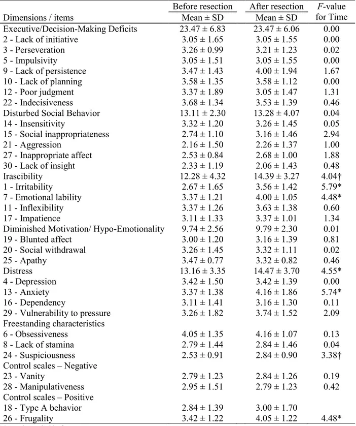
Mean ISPC scores for each item and results from the RM-ANOVAs for the insular group are reported in Table 2. Significant Time effects were found for Irritability (p = 0.028), Lability/Moodiness (p = 0.048), Anxiety (p = 0.028), Frugality (p = 0.048) and for the Distress
20
dimension (p = 0.047). For all significant Time effects, the postoperative rating was higher than the preoperative rating (Irritability, ΔM = 0.89; Lability/Moodiness, ΔM = 0.63; Anxiety, ΔM = 0.79; Frugality, ΔM = 0.63; Distress, ΔM = 1.31). Overall, 53 % of patients had increased postoperative rating for irritability, 42 % for emotional lability, 37 % for anxiety and 26 % for frugality. A trend (p < 0.10) of higher postoperative scores was also observed for the ISPC items Suspiciousness (p = 0.083) and the Irascibility dimension (p = 0.061).
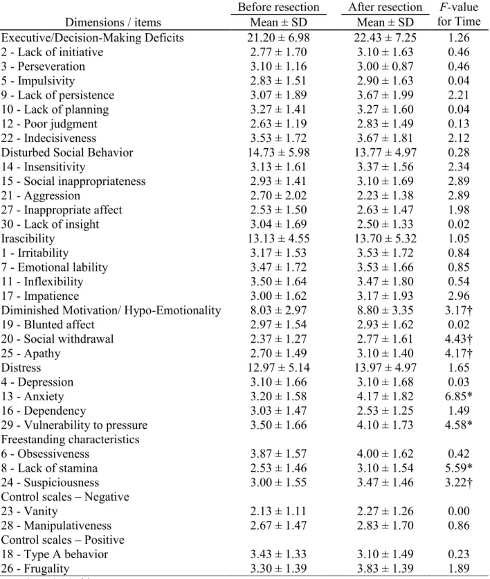
3.3 ISPC score changes following temporal lobe resection
Mean ISPC scores for each item and results from the RM-ANOVAs for the temporal group are reported in Table 3. Significant Time effects were found for Anxiety (p = 0.016), Lack of stamina (p = 0.030) and Vulnerability to pressure (p = 0.046). For all significant Time effects, the postoperative score was higher (Anxiety, ΔM = 0.97; Lack of stamina, ΔM = 0.63; Vulnerability to pressure, ΔM = 0.90). Overall, 53 % of patients had an increased postoperative rating for Anxiety, 42 % for Lack of stamina, and 42 % for Vulnerability to pressure. A trend (p < 0.10) for higher postoperative scores was also observed for Social withdrawal (p = 0.050), Suspiciousness (p = 0.090), Apathy (p = 0.056) and the Diminished motivation / hypo-emotionality dimension (p = 0.092).
3.4 Comparison between the insular and the temporal groups
RM-ANOVAs comparing the insular patients to the temporal group revealed no significant Group effects (Supplemental Table S2). There were significant Time effects for Irritability (F (1, 35) = 4.85; p = 0.034), Lability/moodiness (F (1, 36) = 4.35; p = 0.044), Anxiety (F (1, 36) = 12.33;
21
= 0.046), Suspiciousness (F (1, 36) = 5.84, p = 0.021), Frugality (F (1, 36) = 5.50, p = 0.025),
Vulnerability to pressure (F (1, 36) = 6.64, p = 0.014), and for the Irascibility dimension (F (1, 36) =
4.38, p = 0.044). Again, for all significant Time effects, the group average for the postoperative score was higher than the preoperative score. The only significant Time × Group interaction was found for Apathy (F (1, 36) = 4.41; p = 0.043). Bonferroni-adjusted pairwise comparisons showed
that preoperatively, the temporal group presented a significantly lower level of apathy when compared to the insular group (p = 0.006). For the temporal group only, the postoperative score was deemed significantly higher than the preoperative score (p = 0.018).
3.5 Relationship between type of surgery and worsening on ISPC
ratings
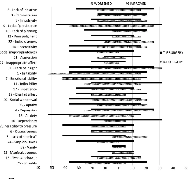
Percentages of individuals whose ISPC ratings improved or worsened following the two types of surgeries can be visualized in Figure 1. Chi-square tests testing for differences in the proportion of patients who improved or worsened on the ISPC items between both types of surgery (insular vs temporal) showed a significant difference in Lack of stamina (2(1,
N = 38) = 7.13, p
= 0.019), with the temporal group presenting a higher number of cases where participants worsened following surgery. An opposite trend was observed for Dependency (2(1,
N = 38) = 4.38,
p = 0.090), with the temporal group presenting a higher number of cases where participants
improved following surgery.
A second set of chi-square calculations examining the relationship between the type of surgery (insular vs temporal) and the number of participants with an important deterioration on ISPC dimensions (≥ 1 SD) showed a significant difference for the Diminished Motivation/
Hypo-22 Emotionality dimension (2(1,
N = 38) = 5.76, p = .046), with the temporal group presenting a
higher number of cases where participants worsened following surgery (Supplemental Table S3).
3.6 Relationship between time elapsed since surgery and severity of
symptoms
Pearson’s correlation tests performed between time elapsed since surgery and outcomes showed that, for the temporal group only, a trend was observed for a moderate negative correlation between the distress dimension of the ISPC and the time elapsed since surgery (r = -0.44, p = 0.069; Supplemental Table S6), suggesting decreasing distress as time since surgery increases. For both groups, there was no correlation between time since surgery and anxiety, lability/moodiness, irritability or frugality.
3.7 Hemisphere of lesion as a predictor of ISPC scores
Analyses examining whether postoperative score changes in the insular patients varied according to the hemisphere of resection showed no significant Time × Hemisphere interaction (Supplemental Table S7).
3.8 Relationship between type of insulectomy and worsening on
ISPC dimensions
Another set of chi-square calculations examining the relationship between the specific site of insulectomy (anterior vs. posterior) and the proportion of participants with an important deterioration on ISPC dimensions scores did not reveal any group differences approaching
23
significance (Supplemental Tables S4 and S5). Given the small number of patients in each group, however, these results should be interpreted with caution.
4. Discussion
This study assessed the emotional, social behavior and personality changes in 19 patients whose epilepsy surgery involved partial or complete resection of the insula, and compared the latter to a control group of 19 patients who underwent temporal lobe epilepsy surgery matched for socio-demographic and epilepsy-related factors. Results indicate that surgical resections involving the insular region are associated with increased irritability, emotional lability, anxiety, and frugality. These changes, although relatively mild for the vast majority of patients, are in line with previous reports of personality disturbances following insular damage (Borg et al., 2013; Dal Monte et al., 2013; Driscoll et al., 2012) and sometimes persist years after surgery. With the exception of increased anxiety, these changes were not significant among the temporal lobe patients, although the pre- vs. post-surgery changes did not significantly differ between groups on any of the outcome variables.
Functional neuroimaging studies have identified the insula as the main cortical target for interoceptive signals and for emotion processing (A. D. Craig, 2009; Critchley et al., 2004; Damasio et al., 2000). The insula’s crucial role in feeling state modulation may contribute, at least partially, to the mild postoperative psychological disturbances observed in some patients with insular damage. The prevailing view concerning the insula’s role posits that the insula translates interoceptive signals into a recognizable feeling state by constantly assessing bodily changes at the visceral and somatosensory level (A. D. Craig, 2009; Damasio & Carvalho, 2013). With the help of fine-grained representations of interoceptive states, cortical maps of the ever-changing somatic
24
landscape is communicated bidirectionally to brain regions related to emotion processing, language, memory and reasoning, including the thalamus, amygdala, hippocampus, prefrontal cortex and anterior cingulate cortex (Stephani, Fernandez-Baca Vaca, Maciunas, Koubeissi, & Lüders, 2011). This extensive connectivity could be essential for an efficient crosstalk between cognition and feeling, thus promoting the rational control of drives and emotions through emotionally informed decision-making (Damasio & Carvalho, 2013).
The increase in post-operational irascibility and anxiety – with about two insular patients out of five showing increased anxiety and/or emotional lability following surgery, and half of insular patients showing increased irritability – support the notion that a mechanism tasked with the regulation of mood and emotions could be compromised due to insular damage, altering the otherwise effective communication between body states and cognition (Paulus & Stein, 2006). Furthermore, while the anxiety experienced by the temporal patients tended to resorb with the passage of time, anxiety was independent from time spent since surgery in the insular patients, suggesting a potentially long-lasting post-lesion anxiogenic effect.
“Anxious sensitivity” is a term used to refer to the degree to which certain individuals perceive interoceptive signals as threatening (Reiss, Peterson, Gursky, & McNally, 1986). Paulus & Stein (2006) (Paulus & Stein, 2006) proposed that the insula could mediate anxious sensitivity through its pivotal role played in interoception, monitoring stimuli that can affect internal body state and relaying information to systems of the limbic and executive functioning areas that manage attention allocation, context evaluation and action planning. Functional neuroimaging showed that anxiety-prone individuals have increased insular activation during emotion processing tasks (Stein, Simmons, Feinstein, & Paulus, 2007) and that lorazepam, a drug often prescribed to relieve anxiety, dose-dependently reduce anterior insula activation bilaterally (Paulus, Feinstein,
25
Castillo, Simmons, & Stein, 2005). In the case of insular damage, the postoperative increase in anxiety could be attributable to the alteration of perceived somatic states, or to the inability to assess them.
Given the evidence linking insular damage to poorer decision-making (Critchley, Mathias, & Dolan, 2001; Kuhnen & Knutson, 2005; Paulus et al., 2003), it may appear somewhat surprising that lesions to the insular cortex in our sample did not lead to executive and decision-making deficits. However, we did observe increased postoperative frugality, a finding that may relate to the involvement of the insula in emotional decision-making, whereby this failure to properly assess somatic states while choosing between options could lead to indecisiveness and risk avoidance (Paulus & Stein, 2006), resulting in a conservative behavior with regard to money. In that vein, a study conducted by Markett et al. (2016) (Markett, Heeren, Montag, Weber, & Reuter, 2016) showed that higher loss aversion – the greater sensitivity to losses than gains – was associated with reduced volumes of bilateral insulae. This suggests that structural differences in this region could alter the processing of aversive bodily states (Canessa et al., 2013), ultimately biasing decision-making towards the avoidance of negative outcomes. Concordantly, increased frugality following insular lesions may reflect a greater sensitivity to monetary losses than to material gains in a purchasing decision situation. Further research should be conducted in order to verify this hypothesis.
Despite existing evidence relating insular damage to hypo-emotionality and to impaired social cognition (Borg et al., 2013; Boucher, Rouleau, Lassonde, et al., 2015; Calder, Keane, Manes, Antoun, & Young, 2000; Driscoll et al., 2012; Gu et al., 2012), in the present study, we found no evidence of such changes following insular resection. As suggested by Papagno et al. (Papagno et al., 2016), sudden lesions of the insular cortex occurring in otherwise healthy patients
26
(as it can be the case in the event of ischemic strokes) may differ from that of epileptic patient whose life-long pathology directly affected the insular cortex, allowing sufficient time for alternative circuits to develop during the progression of the disease. Alternatively, given the well-established functional segregation within the insula [7], it is also possible that certain symptoms of personality change predominantly occur when a particular subregion of the insula is damaged. For instance, the increased insensitivity, apathy and social withdrawal reported in the case-study conducted by Borg et al. (2013) (Borg et al., 2013) was associated with damage to the left-posterior insula, a lesion comparable to only two of the participants from our insular group. Additionally, it is probable that bilateral insular lesions lead to much more severe behavioral disturbances compared to unilateral lesions, but such bilateral lesions are extremely rare and typically involve other structures as well. Nevertheless, in a rare patient whose insular cortices were destroyed bilaterally, feelings and emotions remained intact (Damasio et al., 2013). As a result, it was suggested that the insula may not be essential for feeling states, but may rather be involved in modulating feeling states and in relating them to decision-making processes on account of its richly connected topographical organization and finer-grained representations of interoceptive information.
While the close relatives of patients from the temporal group did report heightened postoperative anxiety and vulnerability to pressure, no other significant changes on items linked to emotional and mood regulation were observed. It is now well established that temporal lobe epilepsy surgery can lead to an increase in postoperative anxiety, especially in patients with a history of anxiety disorder (Malmgren, Starmark, Ekstedt, Rosen, & Sjoberg-Larsson, 2002; Vazquez & Devinsky, 2003). Nonetheless, there has also been reports of anxiety disorders surfacing in up to 50% of patients with no pre-existing psychiatric history (Blumer, Wakhlu,
27
Davies, & Hermann, 1998). Our results also showed a mild increase in postoperative fatigability (lack of stamina), a finding which is in line with a study of 63 patients that had undergone resective temporal lobe epilepsy surgery and reported having limited energy, even 12 years after their operation (Ring, Moriarty, & Trimble, 1998; Tanriverdi, Poulin, & Olivier, 2007).
The fact that the two groups tended to display a differentiated set of postoperative psychological changes raises the question regarding the origin of these disturbances. Conceptually speaking, personality is more trait dependent than state dependent. However, in epileptic patients, many dynamic factors can alter mood, emotion and personality. As such, it is often difficult to determine if a particular behavior is trait-like or simply transitory (Helmstaedter & Witt, 2012). It has been shown that on average, epileptic patients display mild behavioral abnormalities before undergoing surgery, and factors that can affect emotionality and behavior include the lateralization of the epileptogenic zone, the presence of psychiatric comorbidities, of pre-existing cognitive dysfunctions and the psychotropic side effects of anti-epileptic drugs (Andelman, Fried, & Neufeld, 2001; Glosser, Zwil, Glosser, O’Connor, & Sperling, 2000; Helmstaedter & Witt, 2012; Hessen, Lossius, Reinvang, & Gjerstad, 2007). Following surgery, other factors need to be taken into account, including the locus of lesion, postoperative clinical support as well as seizure outcome (Hermann, Wyler, & Somes, 1992; Kellett, Smith, Baker, & Chadwic, 2015). With regard to seizure outcome, research has shown that seizure-free individuals are more prone to postoperative improvements (Fraser & Thorbecke, 1997; Kellett et al., 2015; Malmgren, Sullivan, Ekstedt, Kullberg, & Kumlien, 1997; Tanriverdi et al., 2007). However, even in the case of seizure freedom, there can be an adjustment period characterized by a worsening of mood or behavior often lasting up to two years, during which patients learn to live without epilepsy by abolishing learnt maladapted behavior or relationships (Wilson, Bladin, & Saling, 2001).
28
Ferguson and Rayport (Ferguson & Rayport, 1965) described the transitioning process from being ill to becoming ‘normal’ as disarming due to the loss of emotional and social benefits that chronic epilepsy may otherwise grant by excusing inadequate behavior, by keeping the patient from having to comply with daily commitments or by allowing recognition despite poor work performance. Wilson, Bladin and Saling (Wilson et al., 2001) coined the phrase ‘burden of normality’ to describe the potentially deleterious psychological impact of being cured from a chronic and disabling illness such as epilepsy. For instance, epileptic patients will often hope for the surgery to act as a cure, absolving them of all of their problems (Ferguson & Rayport, 1965; Wheelock, Peterson, & Buchtel, 1998). If not met, the high expectations of postoperative life may increase psychological distress. For instance, the rearrangement of the individual’s identity from sick to well can generate the need to prove to themselves and others of their normalcy, a process that can lead to greater distress (Tedman, Thornton, & Baker, 1995; Wilson et al., 2001). On a sociological level, close relatives can sometimes hold the patient back in their recovery by resisting to the patient’s attempts at becoming increasingly independent and this can sometimes result in the dissolution of relationships and additional postoperative distress to the patient (Carran, Kohler, & O’Connor, 2000; Wilson et al., 2001). As such, it is plausible that the psychological impact of the ‘burden of normality’ contributed, in addition to the locus of lesion, to the postoperative changes observed in both of our groups.
Among the limitations of our study is the fact that patients were assessed with a questionnaire filled retroactively. Thus, the assessment of presurgical behavior may have been affected by recall biases. Additionally, while the observer-rated ISPC items correlated well with their equivalent construct on the supplementary measures obtained from the patients, it is possible that the ISPC is not always sensitive enough to detect subtle changes, especially for patients who
29
are less demonstrative of their symptoms, or for relatives who are less perceptive. As such, future research would benefit from an additional subjective evaluation obtained directly from the patient. Another limitation pertains to the modest sample size. A larger sample size would have permitted comparisons according to the specific subregion involved in insular resection, while also granting sufficient statistical robustness to highlight significant differences between the types of surgery performed and pre- vs postoperative ratings. This would have been of particular interest in consideration of the wealth of data that has demonstrated a functional segregation within the insula (Chikama, McFarland, Amaral, & Haber, 1997; Flynn, 1999). Similarly, other epilepsy and surgery related variables such as the pre- and postoperative intake of anticonvulsants could have been controlled for with a larger sample size. Finally, one last limitation concerns the fact that most of our resections were not purely insular, as they often included one or more of the adjacent opercula. As a result, some of the results may be partially attributable to damage to the opercula.
5. Conclusion
To our knowledge, this study is the first to assess personality changes in a group of epileptic patients who underwent partial or complete insular resection for drug-resistant epilepsy. Our study has highlighted some of the mild changes that can occur following surgical removal of the insula, including higher postoperative irritability, emotional lability and anxiety. These alterations are in line with previous research from lesion and neuroimaging studies, and support the notion that the insula plays an active role in emotion regulation. However, while disturbances in emotional regulation were observed for most patients following surgery, those were mild in nature and expected deficits in executive and social functioning as well as symptoms of hypo-emotionality were not observed. Thus, the mild changes observed in our sample of insular patients do not
30
constitute a contraindication for unilateral insular resections in the context of epilepsy surgery when the insula is demonstrated to be actively involved in epileptogenesis. Nevertheless, prior to undergoing insular resection, patients should probably be informed of this mild risk of increased irritability, emotional lability and anxiety following surgery, in order for them to make an informed decision.
Disclosure of conflicts of interests
31
References
Andelman, F., Fried, I., & Neufeld, M. Y. (2001). Quality of life self-assessment as a function of lateralization of lesion in candidates for epilepsy surgery. Epilepsia, 42(4), 549-555. doi:10.1046/j.1528-1157.2001.19100.x
Barbey, A. K., Koenigs, M., & Grafman, J. (2012). An integrative architecture for general intelligence and executive function revealed by lesion mapping. Brain, 135, 1154-1164. doi:10.1093/brain/aws021
Barrash, J., Anderson, S. W., Hathaway-Nepple, J., Jones, R. D., & Tranel, D. (1997). The Iowa
Scales of Personality Change. Iowa City.
Barrash, J., Asp, E., Markon, K., Manzel, K., Anderson, S. W., & Tranel, D. (2011). Dimensions of personality disturbance after focal brain damage: Investigation with the Iowa Scales of Personality Change. Journal of Clinical and Experimental Neuropsychology, 33(8), 833-852. doi:doi: 10.1080/13803395.2011.561300
Barrash, J., Tranel, D., & Anderson, S. W. (2000). Acquired personality disturbances associated with bilateral damage to the ventromedial prefrontal region. Developmental
Neuropsychology, 18(3), 355-381. doi:10.1207/S1532694205Barrash
Bechara, A., & Damasio, A. R. (2005). The somatic marker hypothesis: A neural theory of economic decision. Games and Economic Behavior, 52(2), 336-372.
doi:10.1016/j.geb.2004.06.010
Beck, A. T., Epstein, N., Brown, G., & Steer, R. A. (1988). An inventory for measuring clinical anxiety: Psychometric properties. Journal of Consulting and Clinical Psychology, 56, 893-897. doi:10.1037/0022-006X.56.6.893
32
Beck, A. T., Steer, R. A., & Brown, G. K. (1996). Manual for Beck Depression Inventory-II. San Antonio, TX: Psychological Corp.
Blumer, D., Wakhlu, S., Davies, K., & Hermann, B. P. (1998). Psychiatric outcome of temporal lobectomy for epilepsy: Incidence and treatment of psychiatric complications. Epilepsia,
39(5), 478-486.
Borg, C., Bedoin, N., Peyron, R., Bogey, S., Laurent, B., & Thomas-Antérion, C. (2013). Impaired emotional processing in a patient with a left posterior insula-SII lesion.
Neurocase, 19(6), 592-603. doi:10.1080/13554794.2012.713491
Boucher, O., Rouleau, I., Escudier, F., Malenfant, A., Denault, C., Charbonneau, S., . . . Nguyen, D. K. (2015). Neuropsychological performance before and after partial or complete insulectomy in patients with epilepsy. Epilepsy & Behavior, 43, 53-60.
doi:10.1016/j.yebeh.2014.11.016
Boucher, O., Rouleau, I., Lassonde, M., Lepore, F., Bouthillier, A., & Nguyen, D. K. (2015). Social information processing following resection of the insular cortex.
Neuropsychologia, 71, 1-10. doi:10.1016/j.neuropsychologia.2015.03.008
Calder, A. J., Keane, J., Manes, F., Antoun, N., & Young, A. W. (2000). Impaired recognition and experience of disgust following brain injury. Nature Neuroscience, 3(11), 1077-1078. Canessa, N., Crespi, C., Motterlini, M., Baud-Bovy, G., Chierchia, G., Pantaleo, G., . . . Cappa,
S. F. (2013). The functional and structural neural basis of individual differences in loss aversion. Journal of Neuroscience, 33(36), 14307-14317.
doi:10.1523/JNEUROSCI.0497-13.2013
Carran, M. A., Kohler, C. G., & O’Connor, M. J. (2000). Marital status after epilepsy surgery.