The right temporal lobe variant of frontotemporal dementia: cognitive and
neuroanatomical profile of three patients
Sven Joubert1,2, Olivier Felician2,3, Emmanuel Barbeau2,4, Jean-Philippe Ranjeva5, Marion Christophe2, Mira Didic2,6, Michel Poncet2,6, Mathieu Ceccaldi2,6
1 Centre de recherche de l’Institut universitaire de gériatrie de Montréal, Quebec, Canada 2
Service de Neurologie et de Neuropsychologie, AP-HM Timone, Marseille, France 3
Laboratoire de Neurobiologie intégrative et Adaptative, CNRS UMR 6612, Marseille, France
4
CERCO UMR-5549, Toulouse, France 5
Centre de Résonance Magnétique Biologique et Médicale (CRMBM) CNRS UMR 6612, Faculté de Médecine, Marseille, France
6
Laboratoire de Neurophysiologie et de Neuropsychologie, INSERM EMI-U 9926, Université de la Méditerranée, Marseille, France
Correspondence should be addressed to: Dr. Sven Joubert, Centre de recherche de l’IUGM, 4565, chemin Queen Mary, Montreal (Quebec), H3W 1W5, Canada; Tel.: (514) 340-3540, ext.3551; Fax: (514) 340-3548; sven.joubert@umontreal.ca
Abstract
The right temporal variant of frontotemporal lobar degeneration (Rtv-FTLD) is a focal degenerative condition affecting predominantly the right temporal lobe. The aim of this study was to further characterize the profile of cognitive impairment and the neuroanatomical basis of Rtv-FTLD patients without behavioural disturbances. A group of three patients with this syndrome had a detailed neuropsychological assessment, along with Voxel-Based Morphometry (VBM) of their brain to determine location of cortical atrophy. VBM analyses showed a pattern of atrophy that was predominant in the right hemisphere and concerned primarily the right anterior temporal lobe region. Patients carried out a test of famous people in which their ability to recognize, name and provide semantic information about famous persons from their faces, their voices and their names was investigated. They all showed a severe defect in recognizing, naming and identifying famous people irrespective of modality. Therefore, their inability to recognize famous people resulted from a multimodal defect (semantic). These results highlight the semantic nature of the defect, and suggest that the anterior right temporal lobe may have a prominent role in processing person-based semantic knowledge. This study helps in further understanding the neuropsychological profile of patients with Rtv-FTLD.
Abbreviations: Rtv-FTLD = Right temporal variant frontotemporal lobar degeneration; MMSE = Mini-Mental State Examination; SD = Semantic dementia
INTRODUCTION
Focal cortical atrophies are degenerative conditions that can affect selectively one domain of mental function, while other domains remain relatively spared. Focal cortical atrophies such as frontotemporal lobar degeneration [1] can disrupt selectively spatial and perceptual skills, gesture, language, episodic memory and semantic memory [2, 3, 4, 5, 6, 7]. The right temporal variant of frontotemporal lobar degeneration (Rtv-FTLD) is one of the presentations of such focal degenerative processes [8, 9, 10, 11, 12, 13, 14]. Typically, the few reported cases of patients affected by this condition presenting with cognitive deficits showed slowly progressive difficulties in recognizing and identifying familiar people in their surroundings and famous people seen on television or in magazines. This condition has also been referred to as progressive prosopagnosia or cross-modal agnosia for familiar people. In most of these case studies, but not all [13], the deficit not only affected the recognition of faces but also affected the recognition of names [9, 10, 15] and voices [10, 11, 12]. This problem was characterized by a failure to access person-specific semantic information allowing identifying a person, and this occurred irrespective of the modality of presentation. Thus, most cases of Rtv-FTLD can be considered to reflect a cross-modal person-based recognition defect. In most reports, the locus of atrophy predominated in the right anterior temporal lobe [8, 9, 10, 14, 15].
In a prior study, we reported patient F.G., a 71-year-old man who had slowly developed progressive difficulties recognizing people he knew (including his ex-wife) and celebrities on television. Using a test devised to assess a person’s ability to process subtle modifications in facial geometry [16], it was demonstrated that the patient’s face recognition deficit resulted from a high-level visuoperceptual deficit that affected his ability to access the global configurations of faces and other visually complex entities (e.g. famous monuments) [13]. In contrast, his ability to identify these same entities through their names was entirely preserved.
Using MRI volumetry, we demonstrated that the most important atrophy was found in the right fusiform gyrus and parahippocampal cortex. Two years later, FG became unable to recognize familiar people from their names and from their voices in his natural environment, and a test of famous people confirmed that at this point he was unable to identify celebrities from their faces but also from their names [14]. At this stage SPECT showed severe cortical perfusion in the right anterior temporal lobe, and damage to this region was also confirmed by MRI volumetry. At this point, the neuropsychological profile of FG was similar to other cases of Rtv-FTLD [9, 10, 11]. Interestingly, the evolution of F.G. was strikingly similar to that of V.H. [9], progressing from isolated face recognition deficits to person recognition deficits later on during the disease.
Recently, Gainotti et al. [12] reported the case of patient CO, a 49-year-old man who also showed a selective deficit in the recognition of familiar people, including the friends of his sons and long-time customers of the bank where he worked. Prevalent atrophy was found in the right anterior temporal lobe. This longitudinal study brought a substantial contribution to our comprehension of this syndrome, because it showed that CO had severe difficulties to access person-specific information about people not only upon presentation of their faces but also through their voices. It is interesting that both patients Emma [11] and Maria [10] were also reported to have had voice recognition deficits in addition to their face and name recognition deficits, although this was not systematically tested and the observation was retrospective in nature. This was also the case with patient BD [17], who presented with associative prosopagnosia following right anterior temporal lobe lesions consequent to Herpes Simplex Encephalitis (HSE).
In the present article, we report a group study of three patients with Rtv-FTLD who came to our Neurology and Neuropsychology Unit, all presenting with prominent difficulties in recognizing familiar and famous people (this was their primary complaint). The first goal of
this study is to test the hypothesis that this difficulty stems from semantic disturbances, affecting recognition in all modalities of input. The perceptual and cognitive processes involved in the multimodal recognition and identification of known persons are illustrated in a model in Figure 1. In order to test our hypothesis, we devised a clinical protocol evaluating a person’s ability to name and provide semantic information about famous people upon presentation of their faces, their voices and their names. The second aim of this study is to verify the hypothesis that the common area of damage in the three patients is the right anterior temporal lobe and that this region is significantly more affected than other regions of the brain when using quantitative volumetry. In order to test this hypothesis, we use Voxel-Based Morphometry to quantitatively assess the location and extent of brain damage in the group of patients. The third aim of this study was to better characterize the pattern of neuropsychological impairment in Rtv-FTLD and compare it with the classical clinical features of SD reported in the literature.
Figure 1. A model of multimodal person recognition, adapted from Bruce and Young’s influential
model of face perception [24]. We propose to use this model for understanding the cognitive processes involved in multimodal person recognition and at what level they are affected in the three patients investigated in the present report. In the first stage, following primary sensory analysis, facial, lexical and vocal stimuli are processed at a level of “structural encoding”. The facial, vocal and lexical stimuli will then activate their corresponding “recognition units”, which correspond to pre-existing modality-specific representations of these stimuli and which allow to express a sense of familiarity. Finally, the last stage consists in accessing person-specific semantic information (“person identity nodes”). Access to this biographical knowledge will lead in turn to the ability to name and provide explicit information about the famous person in question. The three routes in the model are assumed to cooperate in the sense of multimodal interactions. We hypothesize that the three patients reported in the present study have a defect at the level of “Person identity nodes”, reflecting inaccessible or degraded semantic knowledge about familiar and famous persons.
METHODS
Subjects
Patient 1. Patient CM was 61 when she came to our service. She first came to medical attention with her husband, because she became increasingly unable to recognize many people she had known previously. She did not recognize many familiar people such as acquaintances, friends and some relatives. When she first came to our service, CM was a newly-retired school teacher. Her husband recalls that she often met the parents of her students but was unable to recognize them, although she had known them quite well over a period of several years. She did not recognize their faces, and when she heard them speak or when her husband told her their names, this did not help her recognize them. According to her and her husband, this problem had started about 2-3 years before and had been gradually worsening. She found it also increasingly difficult to recognize famous people she saw on television. She also complained of more general memory problems, although she had difficulty providing specific examples. She was spoken, her speech was fluent, carried meaning and was well-organized, with no paraphasic intrusions. She remained entirely independent in daily life.
Patient 2. Patient FC was 59 years of age at the time of the initial evaluation. For the last two years, she complained of increasing difficulties recognizing people she knew. This included some relatives and acquaintances, and occurred particularly when she had not seen them for some time. She also complained of not recognizing children related to her who had been growing up. When she watched television, she could not recognize many actors and TV commentators. She did not complain specifically about not recognizing familiar voices. She also complained of other difficulties with her memory, such as forgetting the names of familiar people and street names. Her spoken language was preserved, except for occasional word-finding difficulties. She remained extremely independent in her life. There were no
changes in behavior. She was very insightful of her difficulties. According to herself and her family, her problems had started about 2 years before she first came to our service.
Patient 3. Patient FG was reported in two previous neuropsychological investigations [13, 14]. He first came to our service in 2001, because of increasing difficulties recognizing familiar and famous people. His deficit was initially limited to the recognition of faces [13]. Two years later, at age 73, he showed severe memory disturbances in addition to the aggravation of his initial primary face recognition defect [14]. At this stage, he was unable to recognize any face, including his own. He also could not recognize people from their names or their voices, including his friends and the neurologists and neuropsychologists in our service, whom he knew quite well. Throughout the course of his disease, he did not present any behavioral changes. He was always very aware of his difficulties, and often joked about them.
Neuropsychological evaluation
All patients underwent a comprehensive neuropsychological evaluation. Testing included general tests of intellectual efficiency (WAIS-R) [18] and memory (WMS-R) [19], as well as a range of more detailed neuropsychological tests covering domains such as language, memory, executive functions, visuoperceptual and visuospatial abilities. Results of the 3 patients are presented in Table 1. Language tests are of particular interest here because they may allow discriminating different patterns of impairment between the Rtv-FTLD patients presented in this study from classic language impairments reported in semantic dementia patients [7]. Furthermore, visuoperceptual tests such as the VOSP [19] may be useful in determining whether patients in this study have basic visuoperceptual defects that may impair their recognition of objects and persons. It is particularly important when evaluating semantic
memory to ensure that semantic defects affecting the recognition of objects and people may not be due, at least in part, to underlying visuoperceptual disturbances.
Neuropsychological testing
The group of 3 Rtv-FTLD patients (education=12 years, SD=0) was compared with a group of 20 healthy age-matched seniors matched for age and level of education (M=58.9 years; SD=9.1; 12 males and 8 females; education=11.8 years, SD=3.8). The Test of famous people was devised to assess a person’s ability to recognize, name and provide semantic information about famous persons (political figures, actors, singers, athletes, etc.) upon presentation of their faces, their names and their voices. Within the context of this study, we use the term ‘identification’ to refer to a person’s ability to provide accurate semantic information about famous persons from questions. The battery is composed of seven subtests. The battery is computerized and stimuli are presented using E-Prime software (v1.1, PST Inc, PA, USA). Responses are provided on a keyboard and error rates and reaction times are measured. Responses are not time-limited. All three patients and their families gave informed consent before testing in accord with institutional guidelines.
Familiarity of faces. 24 faces of famous people and 24 unknown faces are presented on a computer screen, one by one. Stimuli are randomly mixed and presented successively. The subject is instructed that he will be presented a series of randomly mixed faces of celebrities and unknown people. The subject has to judge whether each face is familiar or unfamiliar by pressing the appropriate key. One point is awarded for each correct answer and zero point for an incorrect answer. Within the framework of the model presented in Figure 1, this familiarity task would test the integrity of the “face recognition unit” level of processing.
Familiarity of voices. 24 excerpts of the voices of famous people and 24 excerpts of unknown voices are presented to the subject. The excerpts last approximately 10 seconds and are controlled for semantically as best as possible. Famous phrases were excluded and phrases in which the content could provide information to the listener about the identity of the famous person were excluded (for example, a political speech for a president). One repeat presentation was allowed if this was requested by the subject. The subject has to determine whether each voice excerpt corresponds to the voice of a famous person (familiar) or an unknown person (unfamiliar). One point is awarded for each correct answer. In the model we present in Figure 1, this task would test the integrity of the “voice recognition units” level.
Familiarity of names. 24 names of famous people and 24 unknown names are presented on a computer screen, one by one. Once again, the subject is instructed to determine whether each name is familiar or unfamiliar. One point is awarded for each correct answer and zero point for an incorrect answer. In the model we present in Figure 1, this familiarity task would test the integrity of the “name recognition units” stage.
Naming and identification of famous faces (providing semantic information). 20 photographs of famous faces are presented successively. First, the subject has to provide the full name of the person. Identification then consists of a series of 4 semantic questions concerning each famous person. Two of these questions are general semantic questions: 1) What is the profession of this person? and 2) Is this person alive or deceased? Then, two other questions specific to each individual are asked: 3) What information can you tell me that is specific about this person? and 4) What other information can you tell me that is specific about this person? Each question appears below the photograph, so that the face remains present during the simultaneous presentation of the questions. In the naming task, one point is awarded for
each correct answer (total score of 20). In the identification task, half a point is awarded for each correct answer (total score of 40). Within the context of the model we present in Figure 1, naming and providing semantic information about a famous person from face, voice or name requires access to multimodal “person identity nodes”, in other words the biographical knowledge we hold about an individual.
Naming and identification of famous voices. 20 excerpts of famous voices (10-second long, similar to those in the voice familiarity task) are presented to the subject. As in the previous subtest, each celebrity has to be named. Then, identification from voice consists of 4 semantic questions, identical to the ones presented above. Scoring is also identical as above.
Identification of famous names. 20 full names of famous people are presented to the subject, who has to answer the same four semantic questions as above concerning these people. Half a point is awarded for each correct answer (total score of 40).
Face-name matching (inter-modal semantic matching). In this condition, the subject is required to match the full name of a famous person with the corresponding face presented in a set of three horizontally aligned famous faces. The subject is shown 20 sets of name-face associations. In each set, the name of a famous person appears in the centre top part of the screen. Below, the faces of three famous persons are presented simultaneously, matched for gender and approximate age. Positions of targets and foils are randomized in the set of three faces. The subject is instructed to match the famous name with the corresponding face. One point is awarded for each correct answer and zero point for an incorrect answer (total score of 20). The inter-modal semantic matching tasks (face-name and face-voice) can be assumed to test the multimodal interactions that take place between modality-specific levels of processing
(i.e., within facial, vocal and lexical recognition units and between recognition units and PINs).
Face-voice matching. This condition is identical to the one above, but here the voice of a famous person has to be matched with its corresponding face, presented in a set of 3 famous faces. A 10-second voice excerpt of a famous person is presented during the simultaneous presentation of a set of three famous faces, and the subject has to match the voice with the corresponding face. Positions of targets and foils are randomized. One point is awarded for each correct answer and zero point for an incorrect answer (total score of 20).
Neuroimaging
MRI exploration of the patients was performed at the time of the neuropsychological evaluation and was conducted on a 1.5T Magnetom Vision Plus MR imager (Siemens, Erlangen, Germany). Morphological 3D T1-weighted magnetization prepared rapid gradient echo (MPRAGE) was acquired in the 3 patients using the following parameters (TE/TR = 4.7ms/9.7ms, flip angle 12°, 128 contiguous slices, matrix=2562, isotropic voxel 1.253 mm3). The same exploration was performed in a group of 28 controls with no neurological or psychiatric medical history, included in a larger neuroimaging study (mean age=64.6 years, SD=6.9).
Statistical analyses
For the Test of famous people, nonparametric Mann-Whitney tests were performed for each subtest in order to compare the performance of the group of patients (n=3) with the group of healthy controls (n=20). Level of significance was set to p=0.05.
Voxel based morphometry (VBM) is useful in analyzing changes in the local grey matter densities of the brain and in comparing the local values in a patient group on a pixel-to-pixel basis relative to similar data obtained in a control population. Voxel-Based Morphometry (VBM) analysis in this study was based on the general linear model to compare grey matter (GM) fraction maps from patients and controls. For each subject, 3D T1-weighted images were spatially normalized into the MNI space using the anatomical template provided in the SPM2 program (Welcome institute, London). After performing spatial normalization preserving voxel intensities (concentrations) irrespective of the volumes of the regions that were stretched by warping, the normalized T1-weighted images were segmented using the mutual information procedure (SPM2). The resulting GM fraction maps were smoothed using a 12mm Gaussian filter to minimize spatial difference between subjects and to better satisfy conditions of the random field theory by creating a sufficient spatial dependency between neighboring pixels. After global normalization based on GM intensities (proportional scaling), GM fraction maps from the 3 patients (considered as a group) were compared to the GM fraction maps of the 28 controls using a fixed effect procedure (Ancova with age as confounding covariate, p<0.01 corrected for multiple comparisons, extend threshold k>5). Local maxima coordinates of significant clusters in the MNI space were transformed into Talairach coordinates using a nonlinear transformation (Brett, 2000 http://www.mrc-cbu.cam.ac.uk/Imaging/mnispace.html) to locate clusters and assign them to Brodmann areas.
RESULTS
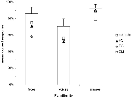
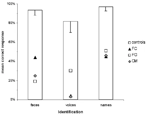
Test of famous people. All results are presented in detail in Figures 2a to 2d (including standard deviations expressing individual differences in the control group). In the familiarity tasks, the group of patients was significantly impaired on the famous/unknown faces (z=2.614, p<0.01) and famous/unknown voices (z=2.475, p<0.01) tasks when compared with
the group of controls, but this difference marginally did not reach significance on the famous/unknown names test (z=1.953, p=0.06). In the second part of the test, the patient group’s ability to name famous faces and famous voices was severely impaired when compared with controls (faces: z=2.758, p<0.01; voices: z=2.747, p<0.01). Moreover, the patients’ ability to identify famous people upon presentation of their faces, their voices and their names was also severely affected (faces: z=2.749, p<0.01; voices: z=2.745, p<0.01; z=2.905, names: p<0.01). Thus, the patients were unable to provide accurate semantic information about famous people irrespective of the modality of presentation (see Fig 2c). Finally, their ability to carry out inter-modal matching tasks was also significantly compromised when compared with controls (name matching: z=3.037, p<0.01; face-voice matching: z=2.869, p<0.01), although to a lesser extent than in the naming and identification tasks (see Fig 2d). In summary, results of the group of patients indicate impaired performance across the different subtests of the Test of famous people.
Figure 2. Performance of the three patients on the Test of famous people, compared to a group of
age-matched healthy control subjects. Significant differences between the group of controls and the group of patients are represented by a *. Group results as well as individual results for each patient are presented.
Figure 2a. Familiarity (famous/unknown)
Figure 2b. Naming famous people
Figure 2c. Identification of famous people (providing semantic information)
Neuropsychological evaluation
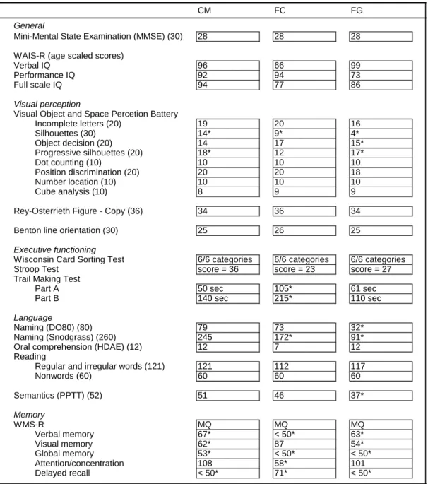
Patient CM. CM had a MMSE score of 28/30 at the time of the neuropsychological evaluation [20]. Her intellectual efficiency, such as measured by the WAIS-R, [18] was within the lower normal range (global IQ = 94). Anterograde memory was severely impaired (global MQ = 53), although attention/concentration remained normal (score=108) [19]. Language, executive functions, praxis, primary visuoperceptual and visuoperceptual skills were preserved. Reading and writing were preserved, including irregular words. CM was able to name 245/260 line drawings from the Snodgrass and Vanderwart battery [21] depicting various exemplars of biological entities and man-made objects. She was also able to provide accurate definitions for these same exemplars during a subsequent evaluation when given their names. Therefore, these results indicate that CM’s semantic breakdown appears to involve primarily person-based knowledge, sparing other main categories of exemplars. Results of CM’s neuropsychological evaluation are presented in Tables 1 and 2.
Patient FC. FC had a MMSE score of 28/30 at the time of the assessment. [20]. She had a full-scale IQ of 77 [18]. Anterograde memory was also severely impaired, with a global MQ of 50 [19]. Visuoperceptual and visuospatial skills were intact [22]. Some aspects of executive functioning were impaired. For instance, she obtained a subnormal score on parts A and B of the Trail Making test, highlighting some difficulties at the attentional level and in mental flexibility. Her performance on the DO80 picture naming test was just below cut-off score, confirming her mild word finding difficulties [23]. Reading and writing skills were intact. She was able to name 172/260 line drawings from the Snodgrass and Vanderwart battery [21]. The profile of naming impairment illustrated in Table 2 shows greater difficulty for naming biological exemplars than man-made exemplars. During an ensuing evaluation, she was able to provide little semantic information about these same entities when given their names.
These results seem to suggest that her problems identifying biological exemplars highlight a semantic defect.
Patient FG. FG’s neuropsychological evaluation was reported in two previous studies. In the initial stages of his condition [13], he presented with an inability to recognize the faces of familiar and famous persons, along with a mild apperceptive agnosia. Semantic memory was preserved. Two years later, his visuoperceptual deficit had deteriorated. More importantly, there had been a severe decline in both semantic and anterograde memory [14]. Within the context of this study, the neuropsychological assessment of patient FG (73-years-old at the time of the evaluation) indicates a full scale IQ of 94 [18], preservation of executive functions, spontaneous speech (although halted by intermittent word finding difficulties), reading and writing abilities and visuospatial skills. In contrast, anterograde memory is severely impaired (global MQ = 53) [19], as well as visuoperceptual skills. His ability to name various categories of exemplars at this stage is severely impaired (see Table 2).
In summary, the general neuropsychological evaluation suggests that the three patients have a similar profile of neuropsychological impairment. Results show relative preservation of intellectual functions, oral and written language, executive functions, visuoperceptual (except for patient FG) and visuospatial functions in the 3 patients. They are also well spatially and temporally oriented and are independent in everyday life. In contrast, they all have a significant anterograde memory defect upon testing, affecting the acquisition and retention of newly-learned information.
CM FC FG General
Mini-Mental State Examination (MMSE) (30) 28 28 28
WAIS-R (age scaled scores)
Verbal IQ 96 66 99
Performance IQ 92 94 73
Full scale IQ 94 77 86
Visual perception
Visual Object and Space Percetion Battery
Incomplete letters (20) 19 20 16 Silhouettes (30) 14* 9* 4* Object decision (20) 14 17 15* Progressive silhouettes (20) 18* 12 17* Dot counting (10) 10 10 10 Position discrimination (20) 20 20 18 Number location (10) 10 10 10 Cube analysis (10) 8 9 9
Rey-Osterrieth Figure - Copy (36) 34 36 34
Benton line orientation (30) 25 26 25
Executive functioning
Wisconsin Card Sorting Test 6/6 categories 6/6 categories 6/6 categories
Stroop Test score = 36 score = 23 score = 27
Trail Making Test
Part A 50 sec 105* 61 sec
Part B 140 sec 215* 110 sec
Language
Naming (DO80) (80) 79 73 32*
Naming (Snodgrass) (260) 245 172* 91*
Oral comprehension (HDAE) (12) 12 7 12
Reading
Regular and irregular words (121) 121 112 117
Nonwords (60) 60 60 60 Semantics (PPTT) (52) 51 46 37* Memory WMS-R MQ MQ MQ Verbal memory 67* < 50* 63* Visual memory 62* 87 54* Global memory 53* < 50* < 50* Attention/concentration 108 58* 101 Delayed recall < 50* 71* < 50*
* indicates impaired scores
maximum score for each test is indicated in brackets
Table 1. Results of the neuropsychological assessment. Maximum score for each test is indicated in
brackets. Part of FG’s neuropsychological evaluation is reproduced with permission from Lippincott, Williams & Wilkins: Joubert S, Felician O, Barbeau E, Sontheimer A, Guedj E, Ceccaldi M, Poncet M.. Progressive prosopagnosia: clinical and neuroimaging results. Neurology, 2004; 63(10):1962-1965.
CM FC FG controls Living entities Insects 88% 13% 0% 89,6% Vegetables 92% 31% 0% 99,4% Fruits 100% 73% 0% 87,3% Animals 86.7% 57% 10% 94,5% Birds 100% 56% 0% 90,1% Non-living entities
Parts of the body 100% 92% 75% 89,2%
Articles of furniture 100% 86% 50% 87,3% Articles of clothing 95% 90% 68% 86,3% Tools 91% 55% 58% 91,4% Transportation 100% 90% 90% 90,2% Toys 92% 42% 0% 84,6% Musical instruments 89% 44% 0% 92,5%
Table 2. Picture naming performance on the Snodgrass and Vanderwart battery (1980).(21)
Neuroimaging
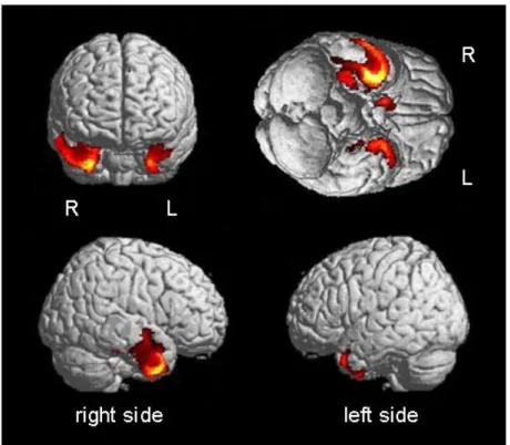
VBM results of the group of three patients are illustrated in Table 3 and Figure 3a. Compared with controls, the group of patients showed predominant atrophy in the right anterior temporal lobe, particularly in the inferior lateral and medial aspects of this region. The pattern of atrophy was asymmetrical, affecting to a much greater degree the right hemisphere. On the global analyses, the extent of regions showing GM density abnormalities in patients were four fold larger inside the right temporal lobe (5515 voxels) relative to the left temporal lobe (1360 voxels) (see Table 3). In addition, individual analyses have shown a right lateralization of GM abnormalities for the three patients with Right/Left GM ratios of 3.24 , 2.78 and 5.0 for FC, FG and CM respectively (see Figure 3b). These results taken together indicate that the right anterior temporal lobe is severely damaged in cross-modal agnosia for familiar people. The hippocampus is also significantly affected. Interestingly, atrophy also extends into the right medium temporal gyrus, nearby regions involved in the processing of human voices [31].
Figure 3a. Common local grey matter atrophy observed in the group of 3 patients compared to 28
healthy age-matched controls using voxel-based morphometry (VBM). Results indicate predominant atrophy in the right anterior temporal lobe, particularly in its inferior and lateral portion, and in the medial temporal lobe (hippocampus). Images on the left, from top to bottom, represent front view and right lateral view. Images on the right, from top to bottom, represent bottom view and left lateral view.
Figure 3b. Common local grey matter atrophy observed in each individual patient vs. controls (N=28,
Ancova with age as covariate, p0.001, corrected). Images on the left, from top to bottom, represent front view and right lateral view. Images on the right, from top to bottom, represent bottom view and left lateral view.
X Y Z
Right hemisphere
Right inferior temporal gyrus -30 -2 -35 18.97 <0.0001
Right hippocampus -30 -18 -13 14.59
Right medial temporal gyrus -50 7 -23 12.25
Right caudate -9 11 -6 12.95 546 <0.0001
Left hemisphere
Left superior temporal gyrus 28 4 -38 12.70
41 16 -29 9.36
36 9 -22 9.31
1360 <0.0001 5515
Locations Talairach coordinates Voxel (Z-score)
P (corrected) Cluster
(k)
Table 3. Local grey matter atrophy observed in the 3 patients compared to 28 controls (Ancova;
p<0.001, corrected for multiple comparisons, with age as confounding covariate). Talairach coordinates reported here represent local maxima.
DISCUSSION
We report a group of three Rtv-FTLD patients who came to our service for increasing difficulties in recognizing familiar and famous persons in their environment. Upon testing, their performance was significantly impaired on all subtests of the Famous persons test, except for the famous names familiarity test where performance was marginally non-significant. These results taken together demonstrate that these patients’ failure to recognize famous people in this test reflects a cross-modal defect and does not merely concern the recognition of faces. The data offer empirical evidence that patients presenting with cross-modal agnosia for people following right anterior temporal lobe lesions are significantly impaired at recognizing familiar voices.
Results of this study can be interpreted within the context of person recognition models. In the initial versions of the model developed by Bruce and collaborators [24, 25], the authors describe four main levels of processing. The first level of analysis is the structural encoding
level: this is where facial percepts are formed. The second level of analysis involves the activation of stored representations of known faces, coined ‘face recognition units’ (FRUs). The third level of analysis involves person identity nodes (PINs), which refers to the general semantic information we have about a person. Finally, the last stage of processing is providing the name of the person. Similar computations can be processed from name (‘name recognition units’) and voices (‘voice recognition units’). This model served in the context of the present study as a conceptual framework to explain the pattern of cognitive impairment observed in the three patients reported in the present study (see Figure 1). Within the context of this model, the patients’ cognitive deficit is thus likely to occur at the level of the PINs (amodal semantic knowledge), considering their severe inability to provide semantic information about famous people irrespective of modality. Yet, considering that their ability to judge famous faces and voices as being familiar stood between chance and performance of controls, there could also be a mild degree of impairment at the level of face recognition units and voice recognition units.
Neural correlates of person-based knowledge
In most of the reported cases of progressive cross-modal loss of knowledge for familiar people [8, 9, 10, 12], but not all [11, 13], the syndrome was associated with a locus of atrophy that predominated in the infero-lateral portion of the right temporal lobe. In this study, VBM indicated that the right anterior and inferior temporal lobe was the region found to be commonly damaged in the three patients. Relatively circumscribed damage to the right anterior temporal lobe has led some authors to suggest that this region plays a critical role in person-based semantic knowledge [9, 10]. Similarly, based on the longitudinal observation of patient CO, Gainotti et al. [12] recently proposed that this region played a critical integrative role in the convergence of unimodal face, voice and name recognition subsystems into a
multimodal person-recognition system. Their interpretation of a bilaterally distributed although asymmetrical network of anterior temporal regions involved in conceptual knowledge (left temporal lobe for verbal information and RTL for non-verbal information) are in line with Damasio’s proposal of convergence zones, which suggests that the temporal poles act as higher-order convergence areas involved in integrating the multiple aspects of a semantic representation [27, 28, 29]. In our view, the right anterior temporal lobe may play an important role in integrating incoming information from modality-specific areas devoted to unimodal person-based processing (for example, selective face processing in the occipitotemporal region and human voice processing in the superior temporal gyrus) [30, 31, 32]. Left homologous structures in turn would be devoted to lexical and phonological aspects of person knowledge (i.e. proper names [32]). Greater difficulty for recognizing people in Rtv-FTLD may reflect damage to both the stored semantic representations of persons as well interruption of the more direct connections between right hemisphere-based face and voice processing channels and more upstream right anterior temporal integrative zones involved in binding the various traits of a person into a coherent well-structured conceptual representation.
The relation between cross-modal agnosia for familiar people and semantic dementia According to some authors, cross-modal agnosia for familiar people has been hypothesized to reflect a right-hemisphere variant of semantic dementia (SD) [9, 14, 26]. This is based mainly on the observation that the right hemisphere lesions observed in this syndrome correspond precisely to the left inferior and anterolateral lesions observed in SD. Moreover, the pattern of semantic memory breakdown in Rtv-FTLD is highly reminiscent of SD, although the nature of the semantic deficit itself is expressed differently in the two syndromes. The expression of semantic breakdown may reflect the lateralization of the lesion, affecting to greater extent
person-specific information in the right hemisphere variant, and more general knowledge (e.g. objects) in the more classical left hemisphere presentation (SD). Thompson et al. [33], for instance, showed a double dissociation between two patients. The patient with the predominantly right-sided presentation (J.P.) presented with severely impaired person-specific semantic knowledge, with relative preservation of knowledge about objects and animals, while the patient with the predominantly left-sided presentation (M.A.) exhibited exactly the opposite pattern of performance. In the present study, CM’s object knowledge was entirely preserved despite severe person-based semantic knowledge, which corroborates the view that person-based semantic knowledge may be selectively impaired. In another study comparing LTV vs. RTV patients [34], the same authors showed that the two groups presented with distinct cognitive and behavioural patterns of impairment. Of particular interest, the most significant complaint found in the RTV group was the difficulty in recognizing familiar people (91%), including friends, family and famous people seen on television. In contrast, the most significant complaints of the LTV group were word-finding difficulties. Snowden et al [26] also investigated the integrity of person-based semantic knowledge in predominantly left-sided vs. predominantly right-left-sided presentations of semantic dementia. This study showed that SD patients with predominantly left-sided atrophy had greater problems retrieving biographical knowledge from famous names than from famous faces, while SD patients with predominantly right-sided atrophy showed the reverse pattern. This was interpreted in terms of the left and right temporal regions contributing respectively to separate verbal and visual semantic knowledge. This study suggests that various aspects of person-based semantic knowledge may be differentially affected based on the lateralization of atrophy. In summary, the variations in the distribution of hemispheric atrophy are likely to account for the differential patterns of semantic impairment. Language is more affected in SD, accounting for the often severe word finding, naming and comprehension difficulties, while semantic
information pertaining to visual and vocal stimuli (e.g. faces and voices) is more disrupted in Rtv-FTLD. Nevertheless, because atrophy is always invariably bilateral, naming and word finding difficulties can be observed in Rtv-FTLD [8, 14] and face recognition deficits are also reported in SD [1].
In addition, there are a number of neuropsychological features in the patients reported in this study that appear to be distinct from SD. The first is the preservation of reading abilities. During the general neuropsychological evaluation, the three patients were perfectly able to read a list low, average and high frequency regular and irregular words (see Table 1). This contrasts with the pattern of surface dyslexia that is typically observed in SD [6]. Preserved reading of irregular words in the three patients may be here interpreted as reflecting the relative sparing of left hemisphere structures essential to reading and the relatively preserved comprehension of underlying linguistic knowledge. Another difference between Rtv-FTLD and SD concerns speech. While speech is very fluent in SD, it is marked by persistent word-finding difficulties. Its content is often empty and egocentric, referring most of the time to personal experience and anecdotes. This was not the case in the three patients reported here. Speech was always very articulate and was not self-centered, and topics covered many different domains not necessarily related to their own experiences. Word-finding difficulties only became apparent with time, progressing slowly over the course of the disease (as was the case with FG). Finally, in contrast with SD, high-level comprehension remained intact, even in the context of listening to complex conversations and stories, evidenced during neuropsychological testing but also during lengthy conversations with the patients and their families. Another notable feature in the three patients reported here was the severity of the anterograde memory disorder. Some previously reported cases of patients with cross-modal agnosia for familiar people were documented to have anterograde memory defects [8, 11, 15, 33] while others were not [9, 10, 12]. This suggests that the defective ability to acquire new
information is not a consistent feature of Rtv-FTLD but occurs in some cases. Finally, some authors have found certain behavioural disorders characteristic of Rtv-FTLD [34, 35, 36], such as loss of insight, social awkwardness, physical negligence, irritability, fixed ideas and impulsiveness (these neuropsychiatric symptoms occur much less frequently in the Ltv-FTLD) [34]. Similar features were reported in previous case studies of cross-modal agnosia for familiar people, including physical negligence [11, 15], uninhibited behaviour [10], mental rigidity and reduction of social contacts [12], changes in eating behaviour [12, 15] and increased social withdrawal and irritability [33]. In the study by Tyrrell et al. [8] and in patients FC and FG of the present study, no behavioural changes were reported. CM did not show behavioural modifications at the time of the neuropsychological evaluation, but some months after testing she started showing some modifications including singing and uninhibited verbal and physical behaviour such as putting her feet on the examiner’s desk or making sexually-oriented comments. Specific behavioural changes thus appear to be common in cross-modal agnosia for familiar people, but may emerge in some instances later in the course of the disease. Taken together, these neuropsychological features may be helpful to differentiate right-sided presentations from the more common left-sided presentations of SD. In summary, Rtv-FTLD and SD share common neuropsychological impairments (differentially distributed patterns of semantic impairment), but they also differ on a number of presenting features reflecting the hemispheric distribution of atrophy.
The question remains as to why left-sided presentations are more common than right-sided presentations. One possible answer is that current diagnostic and neuropsychological tools are not developed sufficiently at this point to evaluate adequately a person’s aptitude to identify familiar people, particularly from different modalities. Another explanation is that language disturbances are more evident and are easier to pinpoint than difficulties with face recognition or person recognition. Consequently, the patients with predominant right temporal
lobe atrophy may seek medical advice only when the language problems start to emerge [8, 26]. Language problems are also more perceptible by the patients’ relatives and friends, which can increase the patient’s awareness of the problem and influence the decision to seek medical attention.
Acknowledgements
We are grateful as well to CM, FC and FG who kindly contributed to this study. We would like to thank them for their longstanding enthusiasm and cooperation.
Competing interests None
References
1. Neary D, Snowden JS, Gustafson L, Passant U, Stuss D, Black S, Freedman M, Kertesz A, Robert PH, Albert M, Boone K, Miller BL, Cummings J, Benson DF (1998)
Frontotemporal lobar degeneration: a consensus on clinical diagnostic criteria. Neurology 51:1546-1554
2. Didic M, Ali Cherif A, Gambarelli D, Poncet M, Boudouresques J (1998) A permanent pure amnestic syndrome of insidious onset related to Alzheimer's disease. Ann Neurol 43:526-530
3. Mesulam MM (1982) Slowly progressive aphasia without generalized dementia. Ann Neurol 11:592-598
4. Benson DF, Davis RJ, Snyder BD (1988) Posterior cortical atrophy. Arch Neurol 45:789-793
5. De Renzi E (1986) Slowly progressive visual agnosia or apraxia without dementia. Cortex 22:171-180
6. Snowden JS, Goulding PJ, Neary D (1989) Semantic dementia: a form of circumscribed cerebral atrophy. Behavioural Neurology 2:167-182
7. Hodges JR, Patterson K, Oxbury S, Funnell E (1992) Semantic dementia. Progressive fluent aphasia with temporal lobe atrophy. Brain 115:1783-1806
8. Tyrrell PJ, Warrington EK, Frackowiak RS, Rossor MN (1990) Progressive
degeneration of the right temporal lobe studied with positron emission tomography. J Neurol Neurosurg Psychiatry 53:1046-1050
9. Evans JJ, Heggs AJ, Antoun N, Hodges JR (1995) Progressive prosopagnosia associated with selective right temporal lobe atrophy. A new syndrome? Brain 118:1-13
10. Gentileschi V, Sperber S, Spinnler H (2001) Crossmodal agnosia for familiar people as a consequence of right infero-polar temporal atrophy. Cognitive Neuropsychology 18:439-463
11. Gentileschi V, Sperber S, Spinnler H (1999) Progressive defective recognition of familiar people. Neurocase 5:407-424
12. Gainotti G, Barbier A, Marra C (2003) Slowly progressive defect in recognition of familiar people in a patient with right anterior temporal atrophy. Brain 126:792-803 13. Joubert S, Felician O, Barbeau E, Sontheimer A, Barton JJ, Ceccaldi M, Poncet M
(2003) Impaired configurational processing in a case of progressive prosopagnosia associated with predominant right temporal lobe atrophy. Brain 126:2537-2550
14. Joubert S, Felician O, Barbeau E, Sontheimer A, Guedj E, Ceccaldi M, Poncet M (2004) Progressive prosopagnosia: clinical and neuroimaging results. Neurology 63:1962-5. 15. Barbarotto R, Capitani E, Spinnler H, Trivelli C (1995) Slowly progressive semantic
impairment with category specificity. Neurocase 1:107-119
16. Barton JJ, Press DZ, Keenan JP, O'Connor M (2002) Lesions of the fusiform face area impair perception of facial configuration in prosopagnosia. Neurology 58:71-78 17. Hanley JR, Young AW, Pearson NA (1989) Defective recognition of familiar people.
Cognitive Neuropsychology 6:179-210
18. Weschler D (1989) Echelle d'intelligence de Weschler pour Adultes, forme révisée. In: Les editions du Centre de Psychologie appliquee, Paris
19. Weschler D (2001) Echelle clinique de memoire de Weschler MEM III (WMS-III). Les editions du Centre de Psychologie appliquee, Paris
20. Folstein MF, Folstein SE, McHugh PR (1975) "Mini-mental state". A practical method for grading the cognitive state of patients for the clinician. J Psychiatr Res 12:189-198
21. Snodgrass JG, Vanderwart M (1980) A standardized set of 260 pictures: norms for name agreement, image agreement, familiarity, and visual complexity. J Exp Psychol [Hum Learn] 6:174-215
22. Warrington EK, James M (1991) The visual object and space perception battery. Thames Valley Test Company, Bury St. Edmunds
23. Deloche G, Hannequin D (1997) Test de dénomination orale d'images: DO80. Les editions du Centre de Psychologie appliquée, Paris
24. Bruce V, Young A (1986) Understanding face recognition. Br J Psychol 77: 305-27. 25. Burton AM, Bruce V, Johnston RA (1990) Understanding face recognition with an
interactive activation model. Br J Psycho l81:361-380
26. Snowden JS, Thompson JC, Neary D (2004) Knowledge of famous faces and names in semantic dementia. Brain 127:860-872
27. Damasio AR (1989) Time-locked multiregional retroactivation: a systems-level proposal for the neural substrates of recall and recognition. Cognition 33:25-62
28. Damasio AR (1990) Category-related recognition defects as a clue to the neural substrates of knowledge. Trends Neurosci 13:95-98
29. Damasio AR, Damasio H (1994) Cortical systems for retrieval of concrete knowledge: the convergence zone framework. In: Koch C (ed) Large-scale neuronal theories of the brain. Cambridge, MIT Press, pp 61-74
30. Kanwisher N, McDermott J, Chun MM (1997) The fusiform face area: a module in human extrastriate cortex specialized for face perception. J Neurosci 17:4302-4311 31. Belin P, Zatorre RJ, Lafaille P, Ahad P, Pike B (2000) Voice-selective areas in human
auditory cortex. Nature 403:309-312
32. Damasio H, Grabowski TJ, Tranel D, Hichwa RD, Damasio AR (1996) A neural basis for lexical retrieval. Nature 380:499-505
33. Thompson SA, Graham KS, Williams G, Patterson K, Kapur N, Hodges JR (2004) Dissociating person-specific from general semantic knowledge: roles of the left and right temporal lobes. Neuropsychologia 42:359-370
34. Thompson SA, Patterson K, Hodges JR (2003) Left/right asymmetry of atrophy in semantic dementia: behavioral-cognitive implications. Neurology 61:1196-1203 35. Edwards-Lee T, Miller BL, Benson DF, Cummings JL, Russell GL, Boone K, Mena I
(1997) The temporal variant of frontotemporal dementia. Brain 120:1027-1040 36. Gorno-Tempini ML, Rankin KP, Woolley JD, Rosen HJ, Phengrasamy L, Miller BL
(2004) Cognitive and behavioral profile in a case of right anterior temporal lobe neurodegeneration. Cortex 40:631-644