HAL Id: dumas-03100438
https://dumas.ccsd.cnrs.fr/dumas-03100438
Submitted on 6 Jan 2021HAL is a multi-disciplinary open access archive for the deposit and dissemination of sci-entific research documents, whether they are pub-lished or not. The documents may come from teaching and research institutions in France or abroad, or from public or private research centers.
L’archive ouverte pluridisciplinaire HAL, est destinée au dépôt et à la diffusion de documents scientifiques de niveau recherche, publiés ou non, émanant des établissements d’enseignement et de recherche français ou étrangers, des laboratoires publics ou privés.
Comparaison du tonomètre à rebond d’Icare versus le
tonomètre aplanation de Perkins chez des enfants sous
anesthésie générale
Marion Chagny
To cite this version:
Marion Chagny. Comparaison du tonomètre à rebond d’Icare versus le tonomètre aplanation de Perkins chez des enfants sous anesthésie générale. Sciences du Vivant [q-bio]. 2020. �dumas-03100438�
aplanation de Perkins chez des enfants sous anesthésie générale
T H E S E A R T I C L E
Présentée et publiquement soutenue devant
LA FACULTÉ DES SCIENCES MEDICALES ET PARAMEDICALES
DE MARSEILLE
Le 4 Décembre 2020
Par Madame Marion CHAGNY
Née le 25 décembre 1990 à Avignon (84)
Pour obtenir le grade de Docteur en Médecine
D.E.S. d' OPHTALMOLOGIE
Membres du Jury de la Thèse :
Madame le Professeur DENIS Danièle Président Monsieur le Professeur DAVID Thierry Assesseur Monsieur le Professeur RIDINGS Bernard Assesseur Madame le Docteur SAUVAN Lauren Directeur
A mon Maître et Présidente du jury, le Professeur Danièle DENIS
Vous me faites Je vous remercie pour votre enseignement en ophtalmo-pédiatrie et votre bienveillance envers les internes. Vous êtes pour nous un exemple de travail et de passion et ce fût un réel plaisir de travailler à vos côtés.
A mon Maître et membre du jury, le Professeur Thierry DAVID
marseillaise est heureuse de vous compter dans ses rangs.
A mon Maître et membre du jury, le Professeur Bernard RIDINGS
à à la Timone
A mon Maître et Directrice de thèse, le Docteur Lauren SAUVAN
Je
passionnantes en ophtalmologie. Je suis admirative de tes connaissances en la matière, de ton travail et de ta rigueur. Au-delà de tes compétences scientifiques et médicales, j
par ailleurs grandement apprécié ton humour et ta spontanéité. Que de fous rires avec toi ! Merci pour ta gentillesse, et ton aide si précieuse travail de thèse. Ce fût un vrai bonheur de travailler à tes côtés. Au plaisir de te retrouver sur les pistes de ski !
née. ! Tu voudras peut-être à ton tour devenir médecin ou
! Avec beaucoup de travail et une détermination sans faille, on arrive à tout dans la vie ! Suis tes rêves mon amour, papa et maman seront toujours à tes côtés. mon bébé.
A toi, mon Amour, mon rérou, mon mari, mon pilier, mon fidèle allié depuis toutes ces
années ! Merci de
Après le lycée, les années Montpelliéraines, nous voilà désormais mariés et merveilleux ninou ! Je nous souhaite le meilleur pour la suite
pour moi à chaque instant ! Tu es ma famille, je veux vieillir à tes côtés. J fort.
A mes parents merci pour votre amour et votre soutien indéfectible ! Tu veux être docteur ma fille ? Mais quelle mouche l ? Qu
accompagnée et soutenue. Ça y est, après des années de travail et des milliers de feuilles de cours imprimées (merci papa !), le jour de ma thèse est enfin arrivé. est loin le temps du foyer du carré du roi ! Finalement, le jeu en valait la chandelle, et je suis heureuse de lire cette fierté dans vos yeux ! Merci pour votre amour, je vous aime fort.
A mes grands-parents. Une pensée émue pour mes papis qui ne sont
tu aurais tant voulu , tu serais si fier dans ton beau costume !
e avec mamie à contribuer à mais avec ce Covid et cette soutenance passée en
pour haut. Tu me manques.
A ma mamie Francine,
valise pleine de tes petits Tupperware ! Tu en auras passé des heures à te faire du
je sais que tu es fière de ta petite fille, même si ce qui compte le plus désormais ce sont les gazouillis de ton arrière-petit-fils Marius
nouveau ! Merci pour tout .
A mon papi Momo. La
-là. Je me souviens que tu aimais pousser la chansonnette et que tu avais le sens de la formule. Nul doute que tu aurais écrit un joli mot en
dû tirer
ça de toi .
; ut-être dû publier ma thèse dans la Provence ! Je sais que tu gardes précieusement la photo d soutenance de thèse à laquelle vous aviez assistée avec papi à Montpellier. Tu vas désormais pouvoir rajouter une nouvelle photo à ta collection, avec ta petite fille dessus cette fois ci .
A mes arrière-grands-parents qui me manquent.
A mon tonton Sylvain et Francine, un deuxième médecin dans la famille ! Merci pour vos encouragements pendant toutes ces années, je vous embrasse.
A mes oncles et tantes, mes cousins cousines Mélanie, Olivier, Yannick. Merci pour votre soutien, je vous embrasse.
A mes filleules .
A ma belle-famille. Merci pour votre gentillesse et votre soutien pendant toutes ces années. : pour une ophtalmo et ses micro-instruments chirurgicaux, « Docteur Marteau les patients ! Je vous embrasse tous bien fort.
A mes amies et amis
les mauvais moments, je sais que je peux compter sur toi en toutes circonstances ! ie
aime.
A Michèle et Laure, merci pour votre soutien et votre amitié depuis toutes ces années, je vous aime.
A Jess, Sandra, Charlotte, Fiona, Morgane il va bien falloir fêter ça autour de quelques shooters de tequilas ! I love you girls.
A mes amies de Montpellier, ça y est nous sommes officiellement toutes devenues médecin !
Morgane, ma fidèle alliée depuis la P1, que de chemin parcouru depuis le Carré du roi ! Perfectionniste, attentionnée, et très dévouée dans ton travail, tu es une perle pour
simplement. Je te souhaite tout le bonheur possible, tu le mérites vraiment !
A Emily, the best in Montpellier, merci pour ta gentillesse
passion et ton dévouement pour la médecine, je suis sûre que tu iras loin ! Je .
A mes chefs
, où tout a commencé !
Dr Antoine Khalil, ris à faire un FO et même avec un V3M ! . Un stage qui se termine avec des fleurs et un bon resto, ça marque ! Dr Hugo Castejon, Marie-Pierre, Céline, Dominique une équipe au top ! Je garde un très bon souvenir de mon premier ne avec vous et je reviendrai travailler à vos côtés prochainement.
:
Dr Caroline Marc, Dr Céline Boulicot, Dr Marie Boulze-Pankert, formateur à vos côtés
dur de tenir correctement cette foutue lentille ! Merci pour votre gentillesse.
Aux Marseillais :
Dr Natacha Stolowy
précieuse pour ce travail de thèse ! Cultivée, brillante et un talent certain pour les imitations, tu es une battante, je suis très !
Dr Pierre Gascon bord connu comme interne, et même référent des internes, pour finalement être ton binôme rétine. Merci pour ces quelques mois très formateurs et pour ta patience au bloc ! Brillant dans ta surspé et apprécié des patients, ce fut un vrai plaisir de travailler à tes côtés !
Dr Marie Beylérian
!
entreprenez notamment pour le service. Merci pour vos enseignements et en particulier celui de la chirurgie de la cataracte en monomanuel !
Dr Geneviève Darrason !
Merci pour votre gentillesse et votre bienveillance.
Dr Alban Comet, merci beaucoup pour ta gentillesse, ton savoir et ta disponibilité pour
circonstances.
Dr Aurore Aziz beaucoup
appris à tes côtés. Après quelques années passées avec Ambre, ce fut un vrai plaisir .
Dr Emilie Zanin, merci pour votre savoir, votre rigueur, votre humanité et votre gentillesse. Je regrette de ne pas avoir eu la chance de passer en binôme avec vous.
Dr Benjamin Donnadieu, merci pour ta gentillesse et ta disponibilité, ce fut un vrai plaisir de travailler avec toi.
Dr Eve Hadjadj, une belle blonde montée sur talons, merci pour ta gentillesse et ta générosité. Très disponible et dévouée pour tes petits patients, je garde un excellent souvenir de mon semestre à Sainte Marguerite à tes côtés !
: Corinne, une vraie
; Camille, un vraie bout en train ; merci de chouchouter les internes comme vous le faites si bien.
Aux timoniens,
Dr Frédéric Matonti, merci pour votre enseignement, votre gentillesse et votre disponibilité.
Dr Louis Hoffart, merci pour votre expertise en segment antérieur.
Merci aussi aux docteurs Hélène Proust, Héléna Guigue, Magali Sampo, Gaëlle Ho semestre à la Timone.
: Dr Christian Tort, Dr Fadi Alshawareb, Dr Omar Midoun : Comme !
ces temps si particuliers marqués par le Covid.
A mes cointernes
A la promo 2015 :
Thomas, ça a été un plaisir de travailler avec toi, tu es un cointerne sur qui on peut compter. Régale-toi bien dans les montagnes. Rose te passe son bonjour
de jolies paupières !
Nathanael, merci pour ta gentillesse et ton dévouement dans ton travail, je suis sûre que tu seras un très bon assistant !
Lorène, un petit bout de femme monté sur piles, tu as finalement quitté les bancs ! Bonne continuation à toi !
Aux plus vieux :
Marie C. une gentillesse rare, un humour décapant et une grande générosité, bonne continuation chez les corses !
Anne,
!
Francesco, toujours prêt à rendre service, merci pour ta grande gentillesse.
Nico, le chirurgien inné, merci pour ton humour et ta décontraction.
Mickaël, toujours dispo pour rendre service, merci pour les bons plans remplas !
Aux plus jeunes :
Emmanuelle, une belle brune bouclée, toujours disponible pour le service et tes
Arthur à côté des nuits que tu vas passer ! Merci pour ta gentillesse.
Allan, la force tranquille, ou comment croire , bonne continuation à toi.
Hugo et Thibault, les deux accolytes, obligés de vous faire la dédicace en même temps ! Toujours en train de faire des blagues ces deux-là. Pour autant, vous êtes toujours partant pour prêter main forte à vos co-internes,
connus les gars !
! Merci pour ta gentillesse, ta patience, et ton aide précieuse au bloc pour ma thèse.
Kévin -ophtalmo, ce n
pas donné à tout le monde ! Merci pour ton aide au bloc pour mes mesures Icare.
Mickaël, merci pour t
.
Mathilde ! Mon porte-monnaie, lui, ne te remercie pas dévouement et ta volonté dans tout ce que tu entreprends, je suis sûre que tu iras loin !
Prithvi, rétinologue en herbe. Bravo pour ton travail et ton implication dans la recherche médicale.
Loïc, Victoire, Charles, Céline, Chafic (imitateur officiel de Michèle et ses anchgiooooos ), Ruben F, Ruben A . ce fut un vrai plaisir de vous connaître.
1 1. INTRODUCTION ... 4 2. MATERIEL ET METHODES ... 5 2.1. Schéma expérimental ... 5 2.2. Objectif ... 5 2.3. Population ... 5 2.3.1. ... 5 2.3.2. ... 5 2.4. Protocole ... 5
2.5. Critère de jugement principal ... 7
2.6. Critère de jugement secondaire ... 7
2.7. Analyses statistiques ... 7
3. RESULTATS ... 8
3.1. Caractéristiques cliniques et démographiques de la population étudiée ... 8
3.2. Comparaison des deux méthodes de mesure de la PIO : corrélation et concordance des 2 méthodes de mesure ... 9
3.3. Recherche de facteurs explicatifs de la différence de PIO entre les deux méthodes de mesure ... 12
4. DISCUSSION ... 13
2 ... 10
a PIO mesurée au Perkins selon le test de Pearson dans la population étudiée. ... 10
thodes de mesure de la PIO selon le test de Bland-Altman dans la population étudiée : différence de PIO (PIO Icare PIO Perkins) en fonction de la PIO moyenne 11
Tableau 1 : Caractéristiques démographiques et cliniques des patients inclus ... 8
Tableau 2 : Paramètres mesurés au sein de la population étudiée ... 9
3 Objectif
Le but de notre étude était de comparer la tonométrie à aplanation de Perkins et la tonométrie à rebond Icare, en évaluant leur corrélation et leur concordance, au sein
Matériel et méthodes
138 yeux de 72 enfants examinés sous anesthésie générale ont été inclus consécutivement. de 2.87 +/- 3.42 années. La pression
intra-Perkins puis Ic200. Les mesures de pachymétrie centrale ultrasonique et de longueur axiale ont également été relevées.
Résultats
Les mesures de PIO obtenues avec les deux tonomètres étaient statistiquement et significativement fortement corrélées (r = 0.8, p < 0.001
en moyenne la PIO de 3,37 mmHg (DS+/-4,48). Le graphique de Bland-Altman a montré que la concordance entre les deux tonomètres était modérée ; les limites de -5.41 à +12.15 mmHg (r = 0.5, p < 0.001). La différence de PIO entre les deux tonomètres était faiblement mais significativement corrélée à la PIO moyenne (r = 0.52 ; p = 0.006) et à la pachymétrie (r = 0.38 ; p = 0.0007).
Conclusion
Dans notre étude, l nation
de Perkins et à rebond Icare Ic200 étaient vait tendance à surestimer la PIO, en particulier pour les valeurs de PIO hautes. Cependant, aucune
sous- le
dépistage du glaucome chez les enfants.
4
1. INTRODUCTION
Le glaucome est
(1,2). Il est capital de pouvoir le diagnostiquer suffisamment tôt afin de le traiter efficacement. Pour cela, la mesure de la pression intra-oculaire (PIO) est primordiale.
cette mesure de la PIO
examen sous sédation ou anesthésie générale (AG) pour porter un diagnostic de glaucome.
Actuellement, le gold standard repose sur la tonométrie à aplanation de Goldman (3). En décubitus dorsal, tonomètre à aplanation de Perkins (Perkins ; Clement-Clarke, Haag-Streit, UK) q un dérivé portable du tonomètre à aplanation de Goldman et leur fiabilité est comparable (4,5).
De nouvelles technologies en matière de tonométrie, plus faciles à réaliser, sont désormais disponibles sur le marché. Parmi elles, le tonomètre à rebond Icare (Icare, Tiolat Oy, Helsinki, Finland) présente plusieurs avantages. Indolore, il ne nécessite aucune instillation de collyre anesthésique. Portatif et facile à utiliser, il ne nécessite pas de coopération particulière de la part du patient (6). Son utilisation pourrait donc être particulièrement intéressante dans le dépistage du glaucome chez les enfants
facilement mesurable en consultation.
De récentes études (7,8) montrent en effet des résultats prometteurs avec une bonne corrélation ent
tonomètre de Perkins. Cependant, ces études ont été menées, pour la plupart, chez un faible nombre de patients (9), chez des patients sains (10,11), des adultes (8,12) ou bien uniquement (7,13).
La présente étude a donc pour but de comparer la tonométrie à aplanation Perkins et la tonométrie à rebond Icare en évaluant leur corrélation et leur concordance chez des enfants examinés sous AG dans différentes populations pédiatriques rencontrées en pratique courante.
5
2. MATERIEL ET METHODES
2.1.
Schéma expérimental
Cette étude prospective de type cohorte analytique, monocentrique, a été menée au sein du Centre Hospitalo-Universitaire (CHU) Nord à Marseille, en France, entre Novembre 2019 et Mars 2020. Au total, 138 yeux de 72 enfants ont été inclus.
2.2.
Objectif
cette étude était de comparer et évaluer la corrélation entre les valeurs de pression intra-oculai
(Icare, Tiolat Oy, Helsinki, Finland) versus le tonomètre à aplanation Perkins (Perkins ; Clement-Clarke, Haag- pédiatrique examinée sous AG. A terme, si les valeurs
celles obtenues avec le Perkins, cela permettrait de
sous AG et ainsi s.
2.3.
Population
2.3.1.
Etaient inclus tous les patients consécutifs âgés de 0 à 18 ans, devant bénéficier durant la période concernée et ayant PIO
Icare ic200.
2.3.2. Cr
Etaient exclus les patients âgés de plus de 18 ans et les enfants pour lesquels les données de mesure de pression intra-oculaire étaient insuffisantes.
2.4.
Protocole
Les patients analysés étaient tous à jeun. Le protocole anesthésique consistait en sevoflurane uniquement. Après induction anesthésique, les enfants
seul
6 Toutes les mesures de notre étude ont été effectuées sous anesthésie profonde,
-à-dire durant la phase de ventilation mécanique.
Les mesures ont été réalisées par 8 opérateurs différents
Pour chaque patient, le tonus oculaire était successivement
tonomètre à aplanation de Perkins (Perkins ; Clement-Clarke, Haag-Streit, UK) puis du tonomètre à rebond Icare ic200 (Icare, Tiolat Oy, Helsinki, Finland). Cet ordre de mesure a été choisi de sorte que, lors de la mesure subjective de la PIO au Perkins, à La valeur de PIO retenue pour ce dernier appareil était la moyenne des 6 mesures effectuées automatiquement.
Le tonomètre à aplanation Perkins (Perkins ; Clement-Clarke, Haag-Streit, UK), utilisable notamment en décubitus dorsal, constitue, à ce jour, le gold standard en tonométrie portable.
tiliser, il faut instiller au préalable une goutte de collyre anesthésique et de fluorescéine puis tourner manuellement une molette afin que les deux demi-cercles de fluorescéine (ou hémi- symétriquement par leurs extrémités.
200 (Icare, Tiolat Oy, Helsinki, Finland) est, quant à lui, un tonomètre à rebond qui ne nécessite aucune anesthésie de contact. Son principe de fonctionnement repose sur la
métallique après un impact sur la cornée. Plus la PIO est élevée, plus la vitesse de rebond sera rapide.
Les mesures suivantes étaient également effectuées : pachymétrie centrale ultrasonique, longueur axiale (LA) avec échographie mode A, diamètres cornéens vertical et horizontal et mesure du rapport cup/disc.
Par ailleurs, pour chaque patient, étaient relevés ses antécédents ophtalmologiques. En : le type de glaucome, un antécédent de chirurgie et son délai par rapport à notre examen
traitement hypotonisant (classes médicamenteuses et nombre de molécules : un
7 antécédent de chirurgie et donc le statut cristallinien (phaque / pseudophaque /
aphaque)
Un consentement éclairé a été obtenu de la part de des représentants légaux des patients ude a été menée en respectant les principes éthiques de la
2.5.
Critère de jugement principal
Le critère de jugement principal consistait à comparer les mesures pressionnelles obtenues avec le tonomètre Icare Ic200 et le tonomètre Perkins chez des enfants sous AG.
2.6.
Critère de jugement secondaire
Les critères de jugement secondaires étaient la corrélation entre les deux méthodes de mesure de PIO au sein de différents sous-groupes (glaucomateux ou non, aphaques ou non, âge inférieur ou supérieur à 3 ans).
2.7.
Analyses statistiques
ggplot2, tidyr, dplyr, Rmisc, RColorBrewer et psych.
Le test Kruskal-Wallis a été utilisé pour comparer les variables quantitatives continues entre plus de 3 sous-groupes.
La
du test de Pearson.
La
du graphique de Bland-Altman.
Différentes analyses univariées et multivariées ont été réalisées pour tenter
Des variables pertinentes ont été inclues dans un modèle de régression dure par étape.
8
3. RESULTATS
3.1.
Caractéristiques cliniques et démographiques de la
population étudiée
72 patients (138 yeux) Nombre de patients (%) Sexe Masculin 39 (54%) Féminin 33 (46%) Motif de consultation Glaucome N = 36 (50%) Congénital 28 Aphaque 4 Juvénile 2 Sturge Weber 1 Myope 1 Cataracte congénitale N = 20 (28%) Phaques 3 Pseudophaques 5 Aphaques 12 Autres N = 16 (22%)Dépistage de glaucome (excavation papillaire /
myopisation / antécédent familial) 5
Atrophie optique 1
Maladie de Coats 2
Membrane pupillaire persistante 2
Trisomie 21 1
Plaie du globe 1
Aniridie 1
Xanthogranulome juvénile irido-cornéen 2
Syndrome de Crouzon 1 Antécédent de chirurgie filtrante 32 (44.4%) traitement hypotonisant Non 57 (79.2%) Oui N = 15 Nombre de molécules Monothérapie 8 (11.1%) Bithérapie 2 (2.8%) Trithérapie 4 (5.6%) Quadrithérapie 1 (1.4%) Type de molécule Bétabloquant 7 (9.7%) Prostaglandine 6 (8.3%) carbonique 13 (18.1%) Alpha-2-adrénergique 1 (1.4%)
9 Paramètres (unité de mesure) Moyenne +/- écart-type (minimum ;
maximum)
Age (années) 2.87 +/- 3.42 (0.15 ;17.4)
Pachymétrie (um) 550.71 +/- 74.08 (423 ;800)
Longueur axiale (mm) 21.46 +/- 2.01 (16.14 ;28.5)
Diamètre cornéen moyen (mm) 11.56 +/- 1.01 (8.25 ;15)
PIO avec le tonomètre Perkins (mmHg) 10.06 +/- 5.20 (4 ;37)
PIO avec le tonomètre Icare (mmHg) 13.43 +/- 7.43 (4 ;46)
Tableau 2 : Paramètres mesurés au sein de la population étudiée
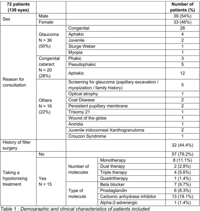
Dans cette étude, après analyse de 88 patients, 138 yeux (70 yeux droits et 68 yeux gauches) de 72 enfants ont été inclus.
16 patients ont été exclus par insuffisance de mesures.
Parmi les 72 patients inclus dans notre étude, 54% étaient de sexe masculin (Tableau 1). de 2.87 +/- 3.42 années (Tableau 2)
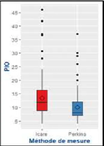
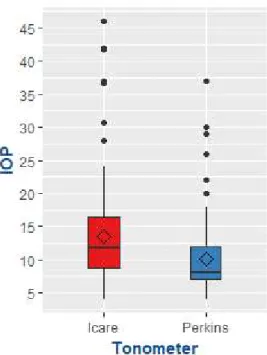
La moyenne de PIO mesurée avec la méthode Icare était de 13.43 +/- 7.43 mmHg [min 4 ; max 46] versus 10.06 +/- 5.20 mmHg [min 4 ; max 37] avec la méthode de Perkins (p < 0.001), soit une différence moyenne de 3.37 +/- 4.48 entre les deux tonomètres (Figure 1).
3.2.
Comparaison des deux méthodes de mesure de la PIO :
corrélation et concordance des 2 méthodes de mesure
, nous avons analysé la corrélation (test de Pearson) et la concordance (test de Bland-Altman) de leurs mesures respectives.
Le test de Pearson retrouvait une forte corrélation entre les deux méthodes de mesure (Figure 2) avec un coefficient de corrélation r = 0.8, p < 0.001
10 Cette corrélation était meilleure dans le sous-groupe des patients suivis pour glaucome (r = 0.84, p < 0.001) que dans le sous-groupe autres (r = 0.67, p < 0.001).
Il a ensuite été évalué la concordance entre les deux méthodes de mesures grâce au test de Bland-Altman (Figure 3)
Le couloir bleu en pointillés représente la concordance moyenne. Le couloir vert représente la limite de concordance maximale (moyenne + 2 déviations standard (DS)) tandis que le couloir orange représente la limite de concordance minimale (moyenne 2 DS). La droite bleue représente la corrélation entre la différence des PIO et la PIO moyenne. Cette corrélation est de r = 0.5, p < 0.001. Pour la totalité de notre échantillon de patients, la différence moyenne des mesures était de 3,37 mmHg (SD +/- 4,48) et les limites de concordance montraient que 95% des mesures se situaient entre -5.41 et +12.15 mmHg.
Figure 2
PIO mesurée au Perkins selon le test de Pearson dans la population étudiée.
Figure 1 : Comparaison de la PIO mesurée à
11
Figure 3
de Bland-Altman dans la population étudiée : différence de PIO (PIO Icare PIO Perkins) en fonction de la PIO moyenne ; Une différence proche de zéro montre une bonne entente entre les deux mesures. Le couloir bleu en pointillés représente la concordance moyenne. Le couloir vert représente la limite de concordance maximale (moyenne + 2 déviations standard (DS)) tandis que le couloir orange représente la limite de concordance minimale (moyenne 2 DS). La droite bleue représente la corrélation entre la différence des PIO et la PIO moyenne (r = 0.5, p<0.001)
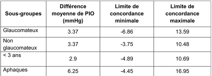
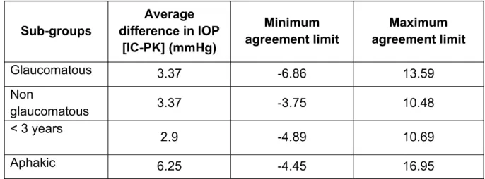
Concernant les analyses en sous-groupes, les résultats du test de Bland-Altman sont reportés dans le tableau 3. Les limites de concordance étaient variables en fonction des sous-groupes.
En effet, certains sous-groupes présentaient des limites de concordance plus étroites. Ceci
les deux tonomètres dans ces populations notamment le cas pour les patients de moins de 3 ans [-4.89 ;10.69] et les non glaucomateux [-3.75 ;10.48]. A inverse, les aphaques présentaient des limites de concordance beaucoup plus larges [-4.45 ;16.95], donc une moins bonne concordance.
12 Sous-groupes Différence moyenne de PIO (mmHg) Limite de concordance minimale Limite de concordance maximale Glaucomateux 3.37 -6.86 13.59 Non glaucomateux 3.37 -3.75 10.48 < 3 ans 2.9 -4.89 10.69 Aphaques 6.25 -4.45 16.95
Tableau 3 : Bland-Altman pour les différents sous-groupes de patients étudiés
3.3.
Recherche de facteurs explicatifs de la différence de PIO
entre les deux méthodes de mesure
es différences de mesure de PIO des analyses univariées puis multivariées ont été réalisées.
Il a donc été testé de manière indépendante plusieurs variables telles que patient, la longueur axiale, la pachymétrie, la PIO moyenne,
glaucome, le type de glaucome, le nombre de traitement hypotonisants, .
Parmi ces variables, seules la pachymétrie (r = 0.38 ; p = 0.0007) et la PIO moyenne (r = 0.52 ; p = 0.006) étaient faiblement mais significativement corrélées à la différence de mesure [Icare-Perkins]. Ces résultats montraient donc que plus la PIO moyenne et la pachymétrie augmentaient, plus la différence de PIO
13
4. DISCUSSION
Le gold standard actuel pour mesurer la PIO chez les enfants est le tonomètre à
population pédiatrique, imposant souvent une anesthésie générale.
Récemment, de nouveaux appareils de mesure pressionnelle ont été développés , intérêt, à terme, serait de réduire le no développement de ces nouveaux appareils de mesure.
L cette étude était de comparer les mesures des deux tonomètres : le Perkins et lcare Ic200 une population pédiatrique examinée sous anesthésie générale.
Dans la cohorte, les deux méthodes de mesures étaient bien corrélées (r = 0.8, p < 0.001). Cette corrélation était meilleure dans le sous-groupe des patients suivis pour glaucome (r = 0.84, p<0.001) que dans le sous-groupe autres (r = 0.67, p < 0.001). Cette différence pourrait être expliquée par une plus grande hétérogénéité des pathologies rencontrées au sein de ce dernier sous-groupe.
ont retrouvé des résultats similaires au sein de différentes populations pédiatriques: en consultation (11), avec un antécédent de glaucome congénital sans (14) ou sous AG (17), présentant une aphaquie (15) ; mais également chez des adultes (16)
tonomètres chez des enfants, toutes pathologies confondues, examinés sous AG. Ce travail montre pour la première fois la corrélation des deux méthodes dans une population pédiatrique de bas âge, diversifiée, et examinée sous AG.
Concernant la concordance des mesures entre les deux appareils, nos résultats montraient une concordance modérée des mesures [-5.41 ; +12.15 mmHg] avec une surestimation de la PIO mesurée à Icare par rapport au Perkins. Dans notre cohorte, la moyenne de la différence de PIO entre les deux tonomètres était de 3.37 SD +/- 4.48.
s ont également constaté cette surestimation de PIO par Icare mais celle-ci est un peu plus faible dans la littérature, aux alentours de 2 mmHg. Elle est
14 de 2.2 (+/- 3.4mmhg, p < 0.0001) dans étude de Mendez-Hernandez et al., chez des enfants glaucomateux en bas âge (17) e Serafino retrouve une différence moyenne de PIO de 1.97mmhg (+/- 1.23, p < 0.05) chez des enfants aphaques ou strabiques (7). Esmael A et al. (14) retrouvent une différence de moyenne des PIO bien moindre avec une valeur de 0.59 (+/- 2.59 mmHg, p = 0.001).
Ces différences de surestimation de la PIO pourraien e modèles différents entre la cohorte et les différentes études précédemment citées. Dans la
Mendez-Hernandez (17) et Serafino (7) (14) utilisait le tonomètre IcTA01.
Pour expliquer la différence de mesures entre les deux appareils, les résultats ont retrouvé deux variables faiblement mais significativement corrélées à cette différence : la PIO moyenne (r = 0.52 ; p = 0.006) et la pachymétrie (r = 0.38 ; p = 0.0007).
Ces résultats étaient concordants avec ceux retrouvés dans la littérature (9) ; la différence de PIO est encore plus importante lorsque les valeurs de PIO dépassent 20 mmHg (10) voire 23 mmHg (18).
De la même façon, plus la pachymétrie était épaisse, plus la différence de PIO était importante entre les deux tonomètres (r = 0.38 ; p = 0.0007). Li Y. et al. retrouvent le même effet avec un faible coefficient de corrélation ( r = 0.408 p < 0.001) (15).
Aucune autre variable expl cette étude. En effet, selon
les résultats, .
différence de PIO statistiquement significative.
Daxer et al. (23) âge sur la différence de PIO. Ils ont montré dans une étude post mortem que le vieillissement modifie
conséquences sur la mesure de la PIO.
15 Mendez-Hernandez et al. (17) retrouvent un effet de la LA sur la différence de PIO dans la plupart des cas. Certains auteurs (22) ne retrouvent, quant à eux, une relation positive avec la profondeur de la chambre antérieure, paramètre qui pas été évalué dans notre étude.
Différentes hypothèses ont été avancées dans la littérature
Muttulevu (19) montre en effet que les mesures prises au centre de la cornée par le tonomètre Icare sont significativement supérieures de 3 à 4 mmHg par rapport aux
sonde de 10°.
différence de PIO (20).
tonomètre de Perkins utilise une compression cornéenne constante qui pourrait entrainer une
mesure prise. A , qui ne touche que superficiellement et plus brièvement la cornée,
à une PIO
sous--Pardo (11), Prabhakar (21) et Borrego Sanz (22). Cependant, nous avons également retrouvé une surestimation de e mesure était prise après le tonomètre de Perkins,
Au final, plusieurs éléments font la force de la présente étude.
nsécutive des patients, ce qui limite le biais de sélection.
De plus, cet échantillon, recruté
16 représentative de la population cible à laquelle nous souhaitons extrapoler nos résultats.
En ce qui concerne les faiblesses de étude, on peut noter que le taux de patients exclus est de 18% (16 patients exclus sur 88 sélectionnés) ce qui est relativement
par insuffisance de mesures.
Par ailleurs, un seul opérateur était disponible par session
avons décidé de mesurer systématiquement la PIO dans le même ordre :
anation de Perkins puis avec le tonomètre à rebond Icare,
lors de sa mesure subjective au Perkins. Nous aurions également pu choisir de
par Serafino (7) ou celle de Molero et al. (8).
, il est admis (24) que les anesthésiques utilisés en pratique courante induisent une baisse significative de la PIO chez les enfants. De plus, cette baisse pressionnelle est variable selon les temps anesthésiques. Pour limiter ce biais , nous
profonde.
Enfin, q
relevés. Certaines études (8,25) ont en effet excluent les patients présentant un astigmatisme supérieur à 2 voire 3 dioptries car cette déformation cornéenne pouvait engendrer un biais dans la mesure de la PIO.
Malgré une concordance modérée des deux appareils de mesure, retrouvant une
hypert
les PIO permettra de ne pas sous-diagnostiquer cette pathologie glaucomateuse. Nous recommanderions de confirmer les PIO au Perkins pour les PIO plus élevées.
17
5. CONCLUSION
En conclusion, notre étude a montré que les
tonomètres à aplanation de Perkins et à rebond Icare Ic200 sont bien corrélées. à surestimer la PIO, en particulier pour les valeurs de PIO hautes. Cependant, aucune sous- avec cet appareil. En ce sens, le tonomètre portable Icare Ic200 reste un outil facile à utiliser et particulièrement intéressant pour dépister des cas de glaucome, y compris chez les enfants.
18 1. Cécité et déficience visuelle [Internet]. [cité 28 sept 2020]. Disponible sur:
https://www.who.int/fr/news-room/fact-sheets/detail/blindness-and-visual-impairment
2. OMS | Causes de la cécité et des déficiences visuelles [Internet]. WHO. World Health Organization; [cité 22 sept 2020]. Disponible sur: http://www.who.int/blindness/causes/fr/
3. Hamard P. Que faire si la mesure de la pression intraoculaire semble peu fiable ? J Fr Ophtalmol. 1 avr 2010;33(4):279 84.
4. Js B, Tm G, Je T. A comparison of Perkins and Goldmann applanation tonometry [Internet]. Vol. 57, Journal of the American Optometric Association. J Am Optom Assoc; 1986 [cité 21 sept 2020]. Disponible sur: https://pubmed.ncbi.nlm.nih.gov/3782712/
5. Gk K, Wk W. Goldmann applanation versus hand-applanation and .schiötz indentation tonometry [Internet]. Vol. 194, Albrecht von Graefes Archiv fur klinische und experimentelle Ophthalm
clinical and experimental ophthalmology. Albrecht Von Graefes Arch Klin Exp Ophthalmol; 1975 [cité 21 sept 2020]. Disponible sur: https://pubmed.ncbi.nlm.nih.gov/1079117/
6. Tonomètre portable à rebond ICARE [Internet]. Abioz Technologies. [cité 28 sept 2020]. Disponible sur: https://abioz.com/tonometre/
7. Serafino M, Villani E, Lembo A, Rabbiolo G, Specchia C, Trivedi RH, et al. A comparison of Icare PRO and Perkins tonometers in anesthetized children. Int Ophthalmol. 16 juill 2019;
8. Molero-Senosiaín M, Morales-Fernández L, Saenz-Francés F, García-Feijoo J, Martínez-de-la-Casa JM. Analysis of reproducibility, evaluation, and preference of the new iC100 rebound tonometer versus iCare PRO and Perkins portable applanation tonometry. Eur J Ophthalmol. 30 sept 2019;1120672119878017. 9. Diaz A, Yebra-Pimentel E, Resua CG, Gilino J, Giraldez MJ. Accuracy of the
ICare rebound tonometer in glaucomatous eyes with topical ocular hypotensive medication. Ophthalmic Physiol Opt J Br Coll Ophthalmic Opt Optom. janv 2008;28(1):29 34.
10. García-Resúa C, González-Meijome JM, Gilino J, Yebra-Pimentel E. Accuracy of the new ICare rebound tonometer vs. other portable tonometers in healthy eyes. Optom Vis Sci Off Publ Am Acad Optom. févr 2006;83(2):102 7.
11. Arribas-Pardo P, Mendez-Hernández C, Valls-Ferran I, Puertas-Bordallo D. Icare-Pro Rebound Tonometer Versus Hand-held Applanation Tonometer for Pediatric Screening. J Pediatr Ophthalmol Strabismus. 19 nov 2018;55(6):382 6.
19 12. Ting SL, Lim LT, Ooi CY, Rahman MM. Comparison of Icare Rebound
Tonometer and Perkins Applanation Tonometer in Community Eye Screening. Asia-Pac J Ophthalmol Phila Pa. juin 2019;8(3):229 32.
13. Wasielica-Poslednik J, Butsch C, Lampe C, Elflein H, Lamparter J, Weyer V, et al. Comparison of Rebound Tonometry, Perkins Applanation Tonometry and Ocular Response Analyser in Mucopolysaccharidosis Patients. PloS One. 2015;10(8):e0133586.
14. Esmael A, Ismail YM, Elhusseiny AM, Fayed AE, Elhilali HM. Agreement profiles for rebound and applanation tonometry in normal and glaucomatous children. Eur J Ophthalmol. juill 2019;29(4):379 85.
15. Li Y, Tang L, Xiao M, Jia S, Liu P, Zhou Y, et al. Comparison of the Icare tonometer and the hand-held goldmann applanation tonometer in pediatric aphakia. J Glaucoma. sept 2013;22(7):550 4.
16. Jablonski KS, Rosentreter A, Gaki S, Lappas A, Dietlein TS. Clinical use of a new position-independent rebound tonometer. J Glaucoma. déc 2013;22(9):763 7.
17. Mendez-Hernandez C, Arribas-Pardo P, Sanchez Jean R, Garcia-Feljoo J. Influence of Axial Length on Intraocular Pressure Measurement With Three Tonometers in Childhood Glaucoma. J Pediatr Ophthalmol Strabismus. 1 janv 2020;57(1):27 32.
18. Schild AM, Rosentreter A, Hermann MM, Muether PS, Schroeter SI, Lappas A, et al. [Comparison of Rebound tonometry versus Perkins tonometry in the supine glaucoma patient]. Klin Monatsbl Augenheilkd. févr 2011;228(2):125 9.
19. Muttuvelu DV, Baggesen K, Ehlers N. Precision and accuracy of the ICare tonometer - Peripheral and central IOP measurements by rebound tonometry. Acta Ophthalmol (Copenh). juin 2012;90(4):322 6.
20. Jorge JMM, González-Méijome JM, Queirós A, Fernandes P, Parafita MA. Correlations between corneal biomechanical properties measured with the ocular response analyzer and ICare rebound tonometry. J Glaucoma. sept 2008;17(6):442 8.
21. Prabhakar SK, Mahesh BS, Shanthamallappa M. A comparative study of intraocular pressure measurement by three tonometers in normal subjects. Nepal J Ophthalmol Biannu Peer-Rev Acad J Nepal Ophthalmic Soc NEPJOPH. déc 2013;5(2):201 6.
22. Borrego Sanz L, Morales-Fernandez L, Martínez de-la-Casa JM, Sáenz-Francés F, Fuentes M, García-Feijóo J. The Icare-Pro Rebound Tonometer Versus the Hand-held Applanation Tonometer in Congenital Glaucoma. J Glaucoma. févr 2016;25(2):149 54.
20 23. Daxer A, Misof K, Grabner B, Ettl A, Fratzl P. Collagen fibrils in the human
corneal stroma: structure and aging. Invest Ophthalmol Vis Sci. mars 1998;39(3):644 8.
24. Termühlen J, Gottschalk A, Eter N, Hoffmann EM, Van Aken H, Grenzebach U, et al. Does general anesthesia have a clinical impact on intraocular pressure in children? Paediatr Anaesth. sept 2016;26(9):936 41.
25.
intraocular pressure measurements in different tonometry methods. Int J Ophthalmol. 2019;12(12):1853 8.
- AG : Anesthésie générale
- CHU : Centre hospitalo-universitaire - LA : Longueur axiale
21 Comparison of the Icare rebound tonometer and the Perkins applanation tonometer in anesthetized children
Dr Chagny M. (1), Dr Stolowy N. (1), Pr Denis D. (1), Dr Sauvan L. (1) (1) Universitary Hospital Center Nord in Marseille, France
Objective
The aim of the study was to compare Perkins flattening tonometry and Icare rebound tonometry, evaluating their correlation and agreement, in a diverse paediatric population examined under general anaesthesia.
Materials and methods
138 eyes of 72 children examined under GA were included consecutively. The average age was 2.87 +/- 3.42 years. Intraocular pressure was successively measured using the Perkins flattening tonometer and then the Icare Ic200 rebound tonometer. Ultrasonic central pachymetry and axial length measurements were also taken.
Results
IOP measurements obtained with the two tonometers were statistically and significantly highly correlated (r = 0.8, p < 0.001) although the Icare overestimates IOP by an average of 3.37 mmHg (SD+/-4.48). Bland-Altman plot showed that the agreement between the two tonometers was moderate; the 95% agreement limits ranged from -5.41 to +12.15 mmHg (r = 0.5, p < 0.001). The difference in IOP between the two tonometers was weakly but significantly correlated with mean IOP (r = 0.52; p = 0.006) and pachymetry (r = 0.38; p = 0.0007). On the other hand, no correlation was found with axial length.
Conclusion
In this study, the IOP values obtained using the Perkins flattening tonometers and the Icare Ic200 rebound tonometer were well correlated. The Icare tended to overestimate the IOP, especially for high IOP values. However, no underestimation of IOP has been found with this device, hence its interest in screening for glaucoma in children.
22
1. INTRODUCTION
Glaucoma is one of the main causes of visual impairment in children (1,2). It is vital to be able to diagnose it early enough to treat it effectively. For this, measuring intraocular pressure (IOP) is essential. However, in children, due to poor cooperation during the ophthalmological examination, this IOP measurement is not always easy and often justifies the use of an examination under sedation or general anaesthesia (GA) to make a diagnosis of glaucoma.
Currently, the gold standard is based on Goldman's flattened tonometry (3). In supine position, the Perkins flat tonometer (Perkins; Clement-Clarke, Haag-Streit, UK) is classically used. This is a portable derivative of the Goldman tonometer and their reliability is comparable (4.5).
New technologies in tonometry, easier to achieve, are now available on the market. Among them, the Icare rebound tonometer (Icare, Tiolat Oy, Helsinki, Finland) has several advantages. Painless, it requires no instillation of anaesthetic eye drops. Portable and easy to use, it does not require any particular cooperation from the patient (6). Its use could therefore be particularly interesting in the screening of glaucoma in children because it could make it possible to limit the number of examinations under GA if the IOP was easily measurable in consultation.
Recent studies (7,8) indeed show promising results with a good correlation between IOP values measured with the Icare and those measured with the Perkins tonometer. However, most of these studies have been carried out in a small number of patients (9), in healthy patients (10,11), adults (8,12) or only in certain ophthalmopediatric pathologies (7,13).
The aim of the study is therefore to compare Perkins flattening tonometry and Icare rebound tonometry by evaluating their correlation and agreement in children examined under GA in different paediatric populations encountered in current practice.
23
2. MATERIALS AND METHODS
2.1.
Experimental design
This monocentric, analytical cohort prospective study was conducted at the Centre Hospitalo-Universitaire (CHU) Nord in Marseille, France, between November 2019 and March 2020. A total of 138 eyes from 72 children were included.
2.2.
Objective
The aim of this study was to compare and evaluate the correlation between intraocular pressure values measured with the Icare Ic200 rebound tonometer (Icare, Tiolat Oy, Helsinki, Finland) versus the Perkins flattening tonometer (Perkins; Clement-Clarke, Haag-Streit, UK) in a paediatric population examined under GA. In the long term, if the IOP values measured with the Icare were comparable to those obtained with the Perkins, it could reduce the number of GA examinations and thus limit the impact of repeated anaesthesia in children.
2.3.
Population
2.3.1. Criteria for inclusion
All consecutive patients between the ages of 0 and 18 years who were to be examined under general anaesthesia during the relevant period and had their IOP measured using both Perkins and Icare Ic200 measuring devices were included.
2.3.2. Exclusion criteria
Patients over 18 years of age and children for whom there was insufficient data on intraocular pressure measurement were excluded.
2.4.
Protocol
The patients analysed were all fasting. The anaesthetic protocol consisted of inhalation of sevoflurane only. After anaesthetic induction, children undergoing GA examination alone were ventilated using a laryngeal mask, while patients requiring surgery were intubated. All measurements in our study were performed under deep anaesthesia, i.e. during the mechanical ventilation phase.
24 The measurements were carried out by 8 different operators during the study.
For each patient, eye pressure was successively measured using the Perkins flat tonometer (Perkins; Clement-Clarke, Haag-Streit, UK) and then the Icare Ic200 rebound tonometer (Icare, Tiolat Oy, Helsinki, Finland). This order of measurement was chosen so that when subjectively measuring IOP with Perkins, the operator is not influenced by the IOP value obtained automatically with the Icare. The IOP value selected for the Icare was the average of the 6 measurements taken automatically.
The Perkins flattening tonometer (Perkins; Clement-Clarke, Haag-Streit, UK), which can be used in particular in the supine position, is currently the standard gold in portable tonometry. Its operating principle is based on Imbert and Fick's law. To use it, a drop of anaesthetic eye drops and fluorescein must first be instilled and then a knob must be turned manually so that the two semi-circles of fluorescein (or hemi-mires) face each other symmetrically at their ends.
The Icare Ic200 (Icare, Tiolat Oy, Helsinki, Finland) is a rebound tonometer that requires no contact anaesthesia. Its operating principle is based on the measurement of the return speed of a thin metal rod after an impact on the cornea. The higher the IOP, the faster the speed of rebound will be.
The following measurements were also carried out: ultrasonic central pachymetry, axial length (LA) with A-mode ultrasound, vertical and horizontal corneal diameters and cup/disc ratio measurement.
In addition, each patient's ophthalmological history was taken. In the case of a history of glaucoma, the following were specified: the type of glaucoma, a history of surgery and its delay in relation to our examination, the taking of a hypotonising treatment (drug classes and number of molecules administered). In the case of a congenital cataract, the following were specified: a previous history of surgery and therefore the crystalline status (phakic / pseudophakic / aphakic) and the delay of this surgery in relation to the examination.
Informed consent was obtained from all legal patient representatives and the study was conducted in accordance with the ethical principles of the Declaration of Helsinki.
25
2.5.
Primary outcome measure
Primary outcome measure was a comparison of the pressure measurements obtained with the Icare Ic200 tonometer and the Perkins tonometer in children under GA. The difference of IOP measured between the Icare and the Perkins tonometer was defined by the following formula: [IC-PK].
2.6.
Secondary outcome measure
Secondary outcome measure was to assess the correlation between the two methods of measuring IOP in different subgroups (glaucomatous or not, aphakic or not, age less than or greater than 3 years).
2.7.
Statistical analysis
The statistical analyses were carried out using the R software using the ggplot2, tidyr, dplyr, Rmisc, RColorBrewer and psych functions.
The Kruskal-Wallis test was used to compare continuous quantitative variables between more than 3 subgroups.
The strength of the correlation between the two measurement methods was assessed using the Pearson test.
The agreement between the two methods of measuring IOP was analysed using Bland-Altman plot.
Different univariate and multivariate analyses were carried out to try to explain the difference in results between the IOP measurements obtained at Perkins and Icare. Relevant variables were included in a multiple linear regression model and selected in a stepwise procedure.
26
3. RESULTS
3.1.
Clinical and demographic characteristics of the study
population
In this study, after analysis of 88 patients, 138 eyes (70 right eyes and 68 left eyes) from 72 children were included.
16 patients were excluded due to insufficient measures.
Of the 72 patients included in our study, 54% were male (Table 1). The average age was 2.87 +/- 3.42 years (Table 2).
The mean IOP measured with the Icare method was 13.43 +/- 7.43 mmHg [min 4 ; max 46] versus 10.06 +/- 5.20 mmHg [min 4 ; max 37] with the Perkins method (p < 0.001), i.e. a mean difference of 3.37 +/- 4.48 between the two tonometers (Figure 1).
3.2.
Comparison of the two IOP measurement methods:
correlation and agreement of the two measurement methods
In order to assess the agreement of the IOP measurements made by the two devices, we analysed the correlation (Pearson test) and the agreement (Bland-Altman test) of their respective measurements.
The Pearson test found a strong correlation between the two measurement methods (Figure 2) with a correlation coefficient r = 0.8, p < 0.001
This correlation was better in the subgroup of patients followed for glaucoma (r = 0.84, p < 0.001) than in the other subgroup (r = 0.67, p < 0.001).
Then, the agreement between the two measurement methods using the Bland-Altman test was assessed (Figure 3).
The dotted blue corridor represents the average agreement. The green corridor represents the maximum agreement limit (average + 2 standard deviations (SD)) while the orange corridor represents the minimum agreement limit (average - 2 SD). The blue line represents the correlation between the IOP difference and the mean IOP. This correlation is r = 0.5, p < 0.001. For our entire sample of patients, the mean
27 difference of the measurements [IC-PK] was 3.37 mmHg (SD +/- 4.48) and the agreement limits showed that 95% of the measurements were between -5.41 and +12.15 mmHg.
For the subgroup analyses, the results of the Bland-Altman test are reported in Table 3. The limits of agreement varied according to the subgroups.
Indeed, some subgroups had narrower boundaries of agreement. This was in favour of a better agreement of IOP values between the two tonometers in these populations. This was particularly the case for patients under 3 years of age [-4.89 ; 10.69] and non-glaucoma patients [-3.75 ; 10.48]. Conversely, aphakic patients had much wider agreement limits [-4.45 ; 16.95], and therefore worse agreement.
3.3.
Searching for factors explaining the difference in IOP
between the two measurement methods
To try to explain the differences in IOP measurement between Icare and Perkins [IC-PK], univariate and then multivariate analyses were performed.
Therefore, several variables were independently tested such as: patient age, axial length, pachymetry, mean IOP, existence or not of glaucoma, type of glaucoma, number of hypotonic treatments, aphakia.
Of these variables, only pachymetry (r = 0.38; p = 0.0007) and mean IOP (r = 0.52; p = 0.006) were weakly but significantly correlated with [IC-PK]. Therefore, these results showed that as mean IOP and pachymetry increased, [IC-PK] also increased.
28
4. DISCUSSION
The current gold standard for measuring IOP in children is the Perkins flat tonometer. It is a reliable but difficult test to perform in a paediatric population, often requiring general anaesthesia.
Recently, new pressure measuring devices have been developed, including the Icare Ic200, which is easier to use. The long-term interest would be to reduce the number of examinations under GA thanks to the development of these new measuring devices.
The aim of this study was to compare the measurements of the two tonometers: the Perkins and the lcare Ic200 in a paediatric population examined under general anaesthesia.
In the cohort, the two measurement methods were well correlated (r = 0.8, p < 0.001). This correlation was better in the subgroup of patients followed for glaucoma (r = 0.84, p<0.001) than in the other subgroup (r = 0.67, p < 0.001). This difference could be explained by a greater heterogeneity of disorders encountered within the latter subgroup.
Other studies have found similar results in different paediatric populations: in consultation (11), with a history of congenital glaucoma without (14) or under GA (17), with aphakia (15); but also in adults (16). However, no study has compared the two tonometers in children, with any conditions, examined under GA. This work shows for the first time the correlation of the two methods in a young, diverse paediatric population examined under GA.
Moreover, this study showed a moderate agreement of the measurements between the two devices [-5.41; +12.15 mmHg] with an overestimation of the IOP measured at the Icare compared to the Perkins. In our cohort, the mean difference in IOP between the two tonometers was [IC-PK] = 3.37 SD +/- 4.48.
Other authors have also noted this overestimation of IOP by Icare, but it is slightly lower in the literature, at around 2 mmHg. It is 2.2 (+/- 3.4mmhg, p < 0.0001) in the study by Mendez-Hernandez et al. in young glaucomatous children (17). The Serafino team found a mean difference in IOP of 1.97mmhg (+/- 1.23, p < 0.05) in aphakic or
29 strabic children (7). In ophthalmopediatric consultations, Esmael A et al (14) found a much smaller mean difference in IOP with a value of 0.59 (+/- 2.59 mmHg, p = 0.001).
These differences in the overestimation of IOP could be explained by using different Icare models between the cohort and the various studies previously mentioned. In the cohort, the model used was the Icare Ic200 whereas the studies by Mendez-Hernandez (17) and Serafino (7) used the Icare PRO and the team by Esmael et al (14) used the IcTA01 tonometer.
To explain the difference in measurements between the two devices, the results found two variables weakly but significantly correlated with this difference: mean IOP (r = 0.52; p = 0.006) and pachymetry (r = 0.38; p = 0.0007). The higher the mean IOP, the higher the difference in IOP. These results were consistent with those found in the literature (9); the difference in IOP is even greater when IOP values exceed 20 mmHg (10) or even 23 mmHg (18).
Similarly, the thicker the pachymetry, the greater the difference in IOP between the two tonometers (r = 0.38; p = 0.0007). Li Y. et al. found the same effect with a low correlation coefficient ( r = 0.408 p < 0.001) (15).
No other explanatory variables were found in our study. According to our results, there did not seem to be any effect of age or the time elapsed since surgery, or any learning effect. No correlation was found with corneal diameter and axial length was not associated with a statistically significant difference in IOP.
Daxer et al (23), for their part, mentioned a possible role of age on IOP difference. They showed in a post-mortem study that ageing modifies the organisation of collagen fibres in the corneal stroma, which could have consequences on the measurement of IOP.
Other articles (15) do not find any effect of the axial length on the difference in IOP between tonometers either. Conversely, Mendez-Hernandez et al (17) find an effect of axial length on the IOP difference in most cases. Some authors (22) only find a positive relationship with the depth of the anterior chamber, a parameter which did not evaluate in this study.
Various hypotheses have been put forward in the literature in an attempt to explain this overestimation of IOP by Icare.
30 Indeed, Muttulevu (19) shows that the measurements taken in the centre of the cornea by the Icare tonometer are significantly higher by 3 to 4 mmHg compared to measurements taken at the periphery, or when there is a horizontal angulation of the probe of 10°.
Other authors also mention the role of corneal hysteresis in this difference in IOP (20).
This overestimation of IOP values by the Icare could also be explained by the measurement method which differs between Perkins and Icare. Indeed, the Perkins tonometer uses constant corneal compression which, by flushing out the aqueous humor, could lower the IOP. On the other hand, Icare only touches the cornea superficially and more briefly. So, we could have an underestimation of the IOP with the Icare if this measure was taken after Perkins tonometer as it was done in the studies conducted by Arribas-Pardo (11), Prabhakar (21) and Borrego Sanz (22). However, we also found an overestimation of the IOP with Icare even if this measure was taken after Perkins tonometer. Therefore, this hypothesis does not explain this difference in values.
To sum up, several elements make up the strength of the study.
First of all, its prospective nature, with the consequent inclusion of patients, which limits selection bias.
The size of the sample is interesting as it includes 138 eyes from 72 children.
Moreover, this sample, recruited from patients in a university hospital, is made up of many patients suffering from serious disorders found in ophthalmopediatrics (congenital glaucoma, congenital cataracts...). This population is therefore representative of the target population to which we wish to extrapolate our results.
With regard to the weaknesses of the study, it can be noted that the rate of excluded patients is 18% (16 excluded patients out of 88 selected) which is relatively high. Most of the patients excluded from the study were excluded due to insufficient measurements.
In addition, only one operator was available per session, so there was no double blind for data collection. To limit this information bias, we decided to systematically measure IOP in the same order: first with the Perkins flattening tonometer and then with the
31 Icare rebound tonometer, so that the operator would not be influenced by the IOP value given by the Icare during his subjective Perkins measurement. We could also have chosen to randomise the order of IOP measurement as was done in the study by Serafino (7) or that of Molero et al (8).
On the other hand, it is accepted (24) that the anaesthetics used in current practice, notably sevoflurane, as is the case in our study, induce a significant drop in IOP in children. Moreover, this drop in pressure is variable according to the anaesthetic times. In order to limit this information bias, we decided to take all IOP measurements during the deep anaesthesia phase.
Finally, some confounding factors such as corneal astigmatism were not identified. Indeed, some studies (8,25) have excluded patients with astigmatism > 2 or even > 3 dioptres because this corneal deformation could lead to a bias in the measurement of IOP.
Despite a moderate agreement between the two measuring devices, finding an overestimation of the IOPs measured at Icare, the interest of this device is to detect ocular hypertonia and consequently glaucoma. The fact that the Icare overestimates the IOP will make it possible not to under-diagnose this glaucomatous pathology. We would recommend confirming IOPs with Perkins for higher IOPs.
32
5. CONCLUSION
As a conclusion, our study showed that the IOP values obtained using the Perkins flattening tonometers and the Icare Ic200 rebound tonometers are well correlated. The Icare tends to overestimate the IOP, especially for high IOP values. However, no underestimation of IOP has been found with this device. In this sense, the Icare Ic200 portable tonometer remains an easy to use tool and is particularly interesting for detecting cases of glaucoma, including in children.
33 1. Blindness and visual impairment [Internet]. cited 28 Sept 2020]. Available at:
https://www.who.int/fr/news-room/fact-sheets/detail/blindness-and-visual-impairment
2. WHO | Causes of blindness and visual impairment [Internet]. WHO. World Health Organization; [cited 22 Sept 2020]. Available at: http://www.who.int/blindness/causes/fr/
3. Hamard P. What to do if the intraocular pressure measurement seems unreliable? J Fr Ophtalmol. 1 Apr 2010;33(4):279-84.
4. Js B, Tm G, Je T. A comparison of Perkins and Goldmann applanation tonometry [Internet]. Vol. 57, Journal of the American Optometric Association. J Am Optom Assoc; 1986 [cité 21 sept 2020]. Disponible sur: https://pubmed.ncbi.nlm.nih.gov/3782712/
5. Gk K, Wk W. Goldmann applanation versus hand-applanation and . schiötz indentation tonometry [Internet]. Vol. 194, Albrecht von Graefe's Archive for Clinical and Experimental Ophthalmology. Albrecht von Graefe's archive for clinical and experimental ophthalmology. Albrecht von Graefe's Arch Clin Exp Ophthalmol; 1975 [cité 21 sept 2020]. Available on: https://pubmed.ncbi.nlm.nih.gov/1079117/ 6. ICARE portable rebound tonometer [Internet]. Abioz Technologies. cited 28 Sept
2020]. Available at: https://abioz.com/tonometre/
7. Serafino M, Villani E, Lembo A, Rabbiolo G, Specchia C, Trivedi RH, et al. A comparison of Icare PRO and Perkins tonometers in anesthetized children. Int Ophthalmol. 16 juill 2019;
8. Molero-Senosiaín M, Morales-Fernández L, Saenz-Francés F, García-Feijoo J, Martínez-de-la-Casa JM. Analysis of reproducibility, evaluation, and preference of the new iC100 rebound tonometer versus iCare PRO and Perkins portable applanation tonometry. Eur J Ophthalmol. 30 sept 2019;1120672119878017. 9. Diaz A, Yebra-Pimentel E, Resua CG, Gilino J, Giraldez MJ. Accuracy of the ICare
rebound tonometer in glaucomatous eyes with topical ocular hypotensive medication. Ophthalmic Physiol Opt J Br Coll Ophthalmic Opt Optom. janv 2008;28(1):29-34.
10. García-Resúa C, González-Meijome JM, Gilino J, Yebra-Pimentel E. Accuracy of the new ICare rebound tonometer vs. other portable tonometers in healthy eyes. Optom Vis Sci Off Publ Am Acad Optom. févr 2006;83(2):102-7.
11. Arribas-Pardo P, Mendez-Hernández C, Valls-Ferran I, Puertas-Bordallo D. Icare-Pro Rebound Tonometer Versus Hand-held Applanation Tonometer for Pediatric Screening. J Pediatr Ophthalmol Strabismus. 19 Nov 2018;55(6):382-6.
34 12. Ting SL, Lim LT, Ooi CY, Rahman MM. Comparison of Icare Rebound
Tonometer and Perkins Applanation Tonometer in Community Eye Screening. Asia-Pac J Ophthalmol Phila Pa. juin 2019;8(3):229-32.
13. Wasielica-Poslednik J, Butsch C, Lampe C, Elflein H, Lamparter J, Weyer V, et al. Comparison of Rebound Tonometry, Perkins Applanation Tonometry and Ocular Response Analyser in Mucopolysaccharidosis Patients. PloS One. 2015;10(8):e0133586.
14. Esmael A, Ismail YM, Elhusseiny AM, Fayed AE, Elhilali HM. Agreement profiles for rebound and applanation tonometry in normal and glaucomatous children. Eur J Ophthalmol. juill 2019;29(4):379-85.
15. Li Y, Tang L, Xiao M, Jia S, Liu P, Zhou Y, et al. Comparison of the Icare tonometer and the hand-held goldmann applanation tonometer in pediatric aphakia. J Glaucoma. sept 2013;22(7):550-4.
16. Jablonski KS, Rosentreter A, Gaki S, Lappas A, Dietlein TS. Clinical use of a new position-independent rebound tonometer. J Glaucoma. déc 2013;22(9):763-7. 17. Mendez-Hernandez C, Arribas-Pardo P, Sanchez Jean R, Garcia-Feljoo J. Influence of Axial Length on Intraocular Pressure Measurement With Three Tonometers in Childhood Glaucoma. J Pediatr Ophthalmol Strabismus. 1 janv 2020;57(1):27-32.
18. Schild AM, Rosentreter A, Hermann MM, Muether PS, Schroeter SI, Lappas A, et al. [Comparison of Rebound tonometry versus Perkins tonometry in the supine glaucoma patient]. Klin Monatsbl Augenheilkd. févr 2011;228(2):125-9.
19. Muttuvelu DV, Baggesen K, Ehlers N. Precision and accuracy of the ICare tonometer - Peripheral and central IOP measurements by rebound tonometry. Acta Ophthalmol (Copenh). juin 2012;90(4):322-6.
20. Jorge JMM, González-Méijome JM, Queirós A, Fernandes P, Parafita MA. Correlations between corneal biomechanical properties measured with the ocular response analyzer and ICare rebound tonometry. J Glaucoma. sept 2008;17(6):442-8.
21. Prabhakar SK, Mahesh BS, Shanthamallappa M. A comparative study of intraocular pressure measurement by three tonometers in normal subjects. Nepal J Ophthalmol Biannu Peer-Rev Acad J Nepal Ophthalmic Soc NEPJOPH. déc 2013;5(2):201-6.
22. Borrego Sanz L, Morales-Fernandez L, Martínez de-la-Casa JM, Sáenz-Francés F, Fuentes M, García-Feijóo J. The Icare-Pro Rebound Tonometer Versus the Hand-held Applanation Tonometer in Congenital Glaucoma. J Glaucoma. Feb 2016;25(2):149-54.
23. Daxer A, Misof K, Grabner B, Ettl A, Fratzl P. Collagen fibrils in the human corneal stroma: structure and aging. Invest Ophthalmol Vis Sci. mars 1998;39(3):644-8.
35 24. Termühlen J, Gottschalk A, Eter N, Hoffmann EM, Van Aken H, Grenzebach U,
et al. Does general anesthesia have a clinical impact on intraocular pressure in children? Paediatr Anaesth. sept 2016;26(9):936-41.
25.
intraocular pressure measurements in different tonometry methods. Int J Ophthalmol. 2019;12(12):1853 8.
- GA: General anaesthesia
- CHU: University Hospital Centre - LA: Axial length
36
Figure 1 : Comparison of IOP measured with Icare and Perkins
Figure 2 : Relationship between IOP measured with Icare and IOP measured with Perkins using the Pearson test in the study population
37
Figure 3 : Assessment of agreement between the two methods of measuring IOP according to the Bland-Altman test in the study population: difference in IOP [IC-PK] as a function of
38 72 patients (138 eyes) Number of patients (%) Sex Male 39 (54%) Female 33 (46%) Reason for consultation Glaucoma N = 36 (50%) Congenital 28 Aphakic 4 Juvenile 2 Sturge Weber 1 Myopia 1 Congenital cataract N = 20 (28%) Phakic 3 Pseudophakic 5 Aphakic 12 Others N = 16 (22%)
Screening for glaucoma (papillary excavation /
myopization / family history) 5
Optical atrophy 1
Coat Disease 2
Persistent pupillary membrane 2
Trisomy 21 1
Wound of the globe 1
Aniridia 1
Juvenile iridocorneal Xanthogranuloma 2
Crouzon Syndrome 1 History of filter surgery 32 (44.4%) Taking a hypotonising treatment No 57 (79.2%) Yes N = 15 Number of molecules Monotherapy 8 (11.1%) Dual therapy 2 (2.8%) Triple therapy 4 (5.6%) Quadritherapy 1 (1.4%) Type of molecule Beta blocker 7 (9.7%) Prostaglandin 6 (8.3%) Carbonic anhydrase inhibitor 13 (18.1%) Alpha-2-adrenergic 1 (1.4%)
39 Parameters (unit of measurement) Mean +/- standard deviation (minimum
; maximum)
Age (years) 2.87 +/- 3.42 (0.15 ;17.4)
Pachymetry (um) 550.71 +/- 74.08 (423 ; 800)
Axial length (mm) 21.46 +/- 2.01 (16.14 ; 28.5)
Average corneal diameter (mm) 11.56 +/- 1.01 (8.25 ; 15)
IOP with Perkins tonometer (mmHg) 10.06 +/- 5.20 (4 ; 37)
IOP with the Icare tonometer (mmHg) 13.43 +/- 7.43 (4 ; 46)
Table 2 : Clinical characteristics of patients included
Sub-groups Average difference in IOP [IC-PK] (mmHg) Minimum agreement limit Maximum agreement limit Glaucomatous 3.37 -6.86 13.59 Non glaucomatous 3.37 -3.75 10.48 < 3 years 2.9 -4.89 10.69 Aphakic 6.25 -4.45 16.95