HAL Id: dumas-01446308
https://dumas.ccsd.cnrs.fr/dumas-01446308
Submitted on 25 Jan 2017
HAL is a multi-disciplinary open access archive for the deposit and dissemination of sci-entific research documents, whether they are pub-lished or not. The documents may come from teaching and research institutions in France or abroad, or from public or private research centers.
L’archive ouverte pluridisciplinaire HAL, est destinée au dépôt et à la diffusion de documents scientifiques de niveau recherche, publiés ou non, émanant des établissements d’enseignement et de recherche français ou étrangers, des laboratoires publics ou privés.
Évolution du trouble ventilatoire obstructif chez les
patients infectes par le VIH : un impact inquiétant du
maintien du tabagisme
Céline Michelangeli
To cite this version:
Céline Michelangeli. Évolution du trouble ventilatoire obstructif chez les patients infectes par le VIH : un impact inquiétant du maintien du tabagisme. Médecine humaine et pathologie. 2016. �dumas-01446308�
UNIVERSITE DE NICE SOPHIA-ANTIPOLIS
FACULTE DE MEDECINE DE NICE
ANNEE 2015 – 2016
THESE DE MEDECINE
EVOLUTION DU TROUBLE VENTILATOIRE OBSTRUCTIF CHEZ LES PATIENTS INFECTES PAR
LE VIH : UN IMPACT INQUIETANT DU MAINTIEN DU TABAGISME
Céline MICHELANGELI Le Lundi 7 novembre 2016
UNIVERSITE DE NICE SOPHIA-ANTIPOLIS FACULTE DE MEDECINE DE NICE
EVOLUTION DU TROUBLE VENTILATOIRE OBSTRUCTIF CHEZ LES PATIENTS INFECTES PAR LE VIH : UN IMPACT INQUIETANT DU MAINTIEN DU TABAGISME
THESE DE MEDECINE
Par Céline MICHELANGELI Née le 12 Avril 1987 à NICE Présentée et soutenue publiquement
Le Lundi 7 Novembre 2016
En vue de l’obtention du titre de Docteur en Médecine
Membres du Jury
Président du Jury Monsieur le Professeur Pierre Marie ROGER Assesseurs Monsieur le Professeur Christian PRADIER
Monsieur le Professeur Charles-Hugo MARQUETTE
Madame le Docteur Francine DE SALVADOR-GUILLOUET
UNIVERSITÉ NICE-SOPHIA ANTIPOLIS
FACULTÉ DE MÉDECINE
Liste des professeurs au 1er septembre 2015 à la Faculté de Médecine de Nice
Doyen M. BAQUÉ Patrick
Vice-Doyen M. BOILEAU Pascal
Assesseurs M. ESNAULT Vincent
M. CARLES Michel Mme BREUIL Véronique M. MARTY Pierre
Conservateur de la bibliothèque Mme DE LEMOS Annelyse
Directrice administrative des services Mme CALLEA Isabelle
Doyens Honoraires M. AYRAUD Noël M. RAMPAL Patrick M. BENCHIMOL Daniel Professeurs Honoraires M. BALAS Daniel M. BATT Michel M. BLAIVE Bruno M. BOQUET Patrice M. BOURGEON André M. BOUTTÉ Patrick M. BRUNETON Jean-Noël Mme BUSSIERE Françoise M. CAMOUS Jean-Pierre M. CHATEL Marcel M. COUSSEMENT Alain M. DARCOURT Guy M. DELLAMONICA Pierre M. DELMONT Jean M. DEMARD François M. DOLISI Claude M . FRANCO Alain M. FREYCHET Pierre M. GÉRARD Jean-Pierre M. GILLET Jean-Yves M. GRELLIER Patrick M. HARTER Michel M. INGLESAKIS Jean-André M. LALANNE Claude-Michel M. LAMBERT Jean-Claude M. LAZDUNSKI Michel M. LEFEBVRE Jean-Claude M. LE BAS Pierre
M. LE FICHOUX Yves Mme LEBRETON Elisabeth M. LOUBIERE Robert M. MARIANI Roger M. MASSEYEFF René M. MATTEI Mathieu M. MOUIEL Jean Mme MYQUEL Martine M. OLLIER Amédée M. ORTONNE Jean-Paul M. SAUTRON Jean Baptiste M. SCHNEIDER Maurice M. SERRES Jean-Jacques M. TOUBOL Jacques M. TRAN Dinh Khiem
M VAN OBBERGHEN Emmanuel M. ZIEGLER Gérard
M.C.A. Honoraire Mlle ALLINE Madeleine
M.C.U. Honoraires M. ARNOLD Jacques
M. BASTERIS Bernard
Mlle CHICHMANIAN Rose-Marie Mme DONZEAU Michèle M. EMILIOZZI Roméo M. FRANKEN Philippe M. GASTAUD Marcel M.GIRARD-PIPAU Fernand M. GIUDICELLI Jean M. MAGNÉ Jacques Mme MEMRAN Nadine M. MENGUAL Raymond M. POIRÉE Jean-Claude Mme ROURE Marie-Claire
PROFESSEURS CLASSE EXCEPTIONNELLE
M. AMIEL Jean Urologie (52.04)
M. BENCHIMOL Daniel Chirurgie Générale (53.02)
M. BOILEAU Pascal Chirurgie Orthopédique et Traumatologique (50.02) M. DARCOURT Jacques Biophysique et Médecine Nucléaire (43.01)
M. DESNUELLE Claude Biologie Cellulaire (44.03) Mme EULLER-ZIEGLER Liana Rhumatologie (50.01)
M. FENICHEL Patrick Biologie du Développement et de la Reproduction (54.05) M. FUZIBET Jean-Gabriel Médecine Interne (53.01)
M. GASTAUD Pierre Ophtalmologie (55.02)
M. GILSON Éric Biologie Cellulaire (44.03)
M. GRIMAUD Dominique Anesthésiologie et Réanimation Chirurgicale (48.01) M. HASSEN KHODJA Reda Chirurgie Vasculaire (51.04)
M. HÉBUTERNE Xavier Nutrition (44.04)
M. HOFMAN Paul Anatomie et Cytologie Pathologiques (42.03) M. LACOUR Jean-Philippe Dermato-Vénéréologie (50.03)
M. MARTY Pierre Parasitologie et Mycologie (45.02)
M. MICHIELS Jean-François Anatomie et Cytologie Pathologiques (42.03) M. MOUROUX Jérôme Chirurgie Thoracique et Cardiovasculaire (51.03)
M. PAQUIS Philippe Neurochirurgie (49.02)
M. PRINGUEY Dominique Psychiatrie d'Adultes (49.03)
M. QUATREHOMME Gérald Médecine Légale et Droit de la Santé (46.03) M. M.ROBERT Philippe Psychiatrie d’Adultes (49.03)
M. SANTINI Joseph O.R.L. (55.01)
PROFESSEURS PREMIERE CLASSE
PROFESSEURS DEUXIEME CLASSE
M. ALBERTINI Marc Pédiatrie (54.01)
Mme BAILLIF Stéphanie Ophtalmologie (55.02) M. BAHADORAN Philippe Cytologie et Histologie (42.02) M. BARRANGER Emmanuel Gynécologie Obstétrique (54.03) M. BENIZRI Emmanuel Chirurgie Générale (53.02)
M. BENOIT Michel Psychiatrie (49.03)
Mme BLANC-PEDEUTOUR Florence Cancérologie – Génétique (47.02)
M. BREAUD Jean Chirurgie Infantile (54-02)
Mlle BREUIL Véronique Rhumatologie (50.01)
M. CANIVET Bertrand Médecine Interne (53.01)
M. CARLES Michel Anesthésiologie Réanimation (48.01)
M. CASSUTO Jill-Patrice Hématologie et Transfusion (47.01) M. CHEVALLIER Patrick Radiologie et Imagerie Médicale (43.02) Mme CHINETTI Giulia Biochimie-Biologie Moléculaire (44.01) M. DELOTTE Jérôme Gynécologie-obstétrique (54.03) M. DUMONTIER Christian Chirurgie plastique
M. FONTAINE Denys Neurochirurgie (49.02)
M. FOURNIER Jean-Paul Thérapeutique (48-04)
M. FREDENRICH Alexandre Endocrinologie, Diabète et Maladies métaboliques Mlle GIORDANENGO Valérie Bactériologie-Virologie (45.01)
M. GUÉRIN Olivier Gériatrie (48.04)
M. HANNOUN-LEVI Jean-Michel Cancérologie ; Radiothérapie (47.02) Mme ASKENAZY-GITTARD Florence Pédopsychiatrie (49.04)
M. BAQUÉ Patrick Anatomie - Chirurgie Générale (42.01)
M. BÉRARD Étienne Pédiatrie (54.01)
M. BERNARDIN Gilles Réanimation Médicale (48.02)
M. BONGAIN André Gynécologie-Obstétrique (54.03)
M. CASTILLO Laurent O.R.L. (55.01)
Mme CRENESSE Dominique Physiologie (44.02)
M. DE PERETTI Fernand Anatomie-Chirurgie Orthopédique (42.01) M. DRICI Milou-Daniel Pharmacologie Clinique (48.03)
M. ESNAULT Vincent Néphrologie (52-03)
M. FERRARI Émile Cardiologie (51.02)
M. FERRERO Jean-Marc Cancérologie ; Radiothérapie (47.02)
M. GIBELIN Pierre Cardiologie (51.02)
M. GUGENHEIM Jean Chirurgie Digestive (52.02)
Mme ICHAI Carole Anesthésiologie et Réanimation Chirurgicale (48.01)
M. LONJON Michel Neurochirurgie (49.02)
M. MARQUETTE Charles-Hugo Pneumologie (51.01)
M. MOUNIER Nicolas Cancérologie, Radiothérapie (47.02) M. PADOVANI Bernard Radiologie et Imagerie Médicale (43.02)
Mme PAQUIS Véronique Génétique (47.04)
M. PRADIER Christian Épidémiologie, Économie de la Santé et Prévention (46.01) M. RAUCOULES-AIMÉ Marc Anesthésie et Réanimation Chirurgicale (48.01)
Mme RAYNAUD Dominique Hématologie (47.01)
M. ROSENTHAL Éric Médecine Interne (53.01)
M. SCHNEIDER Stéphane Nutrition (44.04)
M. STACCINI Pascal Biostatistiques et Informatique Médicale (46.04)
M. THOMAS Pierre Neurologie (49.01)
PROFESSEURS DEUXIEME CLASSE (suite) M. IANNELLI Antonio Chirurgie Digestive (52.02) M JEAN BAPTISTE Elixène Chirurgie vasculaire (51.04)
M. JOURDAN Jacques Chirurgie Thoracique et Cardiovasculaire (51.03) M. LEVRAUT Jacques Anesthésiologie et Réanimation Chirurgicale (48.01) M. PASSERON Thierry Dermato-Vénéréologie (50-03)
M. PICHE Thierry Gastro-entérologie (52.01)
M. ROGER Pierre-Marie Maladies Infectieuses ; Maladies Tropicales (45.03)
M. ROHRLICH Pierre Pédiatrie (54.01)
M. RUIMY Raymond Bactériologie-virologie (45.01)
Mme SACCONI Sabrina Neurologie (49.01)
M. SADOUL Jean-Louis Endocrinologie, Diabète et Maladies Métaboliques (54.04) M. TROJANI Christophe Chirurgie Orthopédique et Traumatologique (50.02) M. VENISSAC Nicolas Chirurgie Thoracique et Cardiovasculaire (51.03)
PROFESSEUR DES UNIVERSITÉS
M. HOFLIGER Philippe Médecine Générale
PROFESSEURS AGRÉGÉS
Mme LANDI Rebecca Anglais
Mme ROSE Patricia Anglais
MAITRES DE CONFÉRENCES DES UNIVERSITÉS - PRATICIENS HOSPITALIERS Mme ALUNNI Véronique Médecine Légale et Droit de la Santé (46.03) M. AMBROSETTI Damien Cytologie et Histologie (42.02)
Mme BANNWARTH Sylvie Génétique (47.04)
M. BENOLIEL José Biophysique et Médecine Nucléaire (43.01) Mme BERNARD-POMIER Ghislaine Immunologie (47.03)
Mme BUREL-VANDENBOS Fanny Anatomie et Cytologie pathologiques (42.03)
M. DOGLIO Alain Bactériologie-Virologie (45.01)
M DOYEN Jérôme Radiothérapie (47.02)
M FAVRE Guillaume Néphrologie (52.03)
M. FOSSE Thierry Bactériologie-Virologie-Hygiène (45.01) M. GARRAFFO Rodolphe Pharmacologie Fondamentale (48.03) Mme GIOVANNINI-CHAMI Lisa Pédiatrie (54.01)
Mme HINAULT Charlotte Biochimie et biologie moléculaire (44.01) Mme LEGROS Laurence Hématologie et Transfusion (47.01) Mme MAGNIÉ Marie-Noëlle Physiologie (44.02)
Mme MOCERI Pamela Cardiologie (51.02)
Mme MUSSO-LASSALLE Sandra Anatomie et Cytologie pathologiques (42.03) M. NAÏMI Mourad Biochimie et Biologie moléculaire (44.01) M. PHILIP Patrick Cytologie et Histologie (42.02)
Mme POMARES Christelle Parasitologie et mycologie (45.02)
M. ROUX Christian Rhumatologie (50.01)
M. TESTA Jean Épidémiologie Économie de la Santé et Prévention (46.01) M. TOULON Pierre Hématologie et Transfusion (47.01)
PROFESSEURS ASSOCIÉS M M. COYNE John GARDON Gilles Anatomie et Cytologie (42.03) Médecine Générale
Mme PACZESNY Sophie Hématologie (47.01)
Mme POURRAT Isabelle Médecine Générale
MAITRES DE CONFÉRENCES ASSOCIÉS
M BALDIN Jean-Luc Médecine Générale
M. DARMON David Médecine Générale
Mme MONNIER Brigitte Médecine Générale
M. PAPA Michel Médecine Générale
PROFESSEURS CONVENTIONNÉS DE L’UNIVERSITÉ
M. BERTRAND François Médecine Interne
M. BROCKER Patrice Médecine Interne Option Gériatrie
M. CHEVALLIER Daniel Urologie
Mme FOURNIER-MEHOUAS Manuella Médecine Physique et Réadaptation
M. JAMBOU Patrick Coordination prélèvements d’organes
REMERCIEMENTS
A Monsieur le président du Jury Monsieur le Professeur Pierre-Marie Roger
C’est un grand honneur de vous avoir pour président de ma thèse.
Je vous remercie également de m’avoir acceptée en DESC de pathologies Infectieuses, de m’avoir donné l’opportunité d’apprendre autant de si belles choses qui me passionnent depuis mon externat. Merci pour votre enseignement et votre soutien durant mon internat. Et je ne vous remercierai jamais assez pour votre implication dans mon avenir en tant qu’infectiologue. J’espère être à la hauteur de vos attentes.
Aux membres du Jury
Monsieur le Professeur Christian Pradier
Je suis honorée de vous compter parmi les membres de mon Jury. Je vous remercie pour ces précieux conseils que vous avez apportés alors que ma thèse n’était encore qu’un projet. Vous m’avez permis de réaliser un travail soigné et bien ficelé. Je vous adresse toutes mes reconnaissances et mes plus sincères remerciements.
Monsieur le Professeur Charles-Hugo Marquette
Je suis enchantée que vous ayez accepté de participer à mon Jury de Thèse. Depuis un échange d’e-mail d’un bout à l’autre de la planète, en passant par vos enseignements si précieux au cours de mon externat, je vous remercie du fond du cœur pour tout ce que vous avez fait pour moi à cette époque. Je vous prie de recevoir toute ma gratitude et mon profond respect.
Madame le Docteur Francine De Salvador-Guillouet
Mille mercis d’avoir accepté de faire partie de mon Jury de thèse. Merci également pour votre aide à la réalisation de ce travail, mais aussi pour vos enseignements et votre gentillesse au sein de cette belle famille qu’est le service d’infectiologie.
A ma directrice de thèse Madame le Docteur Karine Risso
Je ne saurais jamais comment te remercier à la hauteur de tout ce que tu m’as apporté depuis que tu as accepté de t’engager dans ce travail de longue haleine. Je ne saurais jamais te remercier suffisamment pour tout ce que tu m’as appris, pour tout ce que tu as fait pour moi, pour m’avoir accompagné tel que tu l’as fait. C’était loin d’être simple, c’était même parfois déprimant, mais nous avons fini par y arriver, en grande partie grace à ta patience, à ton sens de l’organisation et à ton optimisme. Tu es une personne si talentueuse et passionnée, une source d’inspiration pour moi. Je te souhaite tellement de bonheur, tellement de réussite. Merci, merci encore et à jamais.
Au service d’infectiologie
Merci à toute l’équipe de m’avoir accueillie lors de mon externat initialement, puis lors de mes 2 semestres d’internat. Je pense que vous vous souviendrez tous de cette externe aux cheveux roses, puis de cette interne en short en jean en plein hiver puis et qui a ensuite adopté des leggings aux motifs divers et variés, plus étranges les uns que les autres. Ce que je me souviendrai, c’est surtout d’une atmosphère très agréable, où tous ceux qui y travaillent sont plus fous les uns que les autres. De cette petite ruche où le travail d’équipe arrive malgré tout à être impeccablement bien organisé, sous l’œil bienveillant de Marinette et Danièle, les meilleurs cadres du monde, il faut le dire, du savoir-faire et de la patience de toute l’équipe d’infirmiers et d’aides-soignants qui réalisent au quotidien un travail formidable dans l’intérêt des patients, et avec l’aide précieuse de nos et secrétaires préférées. Plein souvenirs positifs évidemment, mais comme toujours dans la vie, parfois des peines et des pleurs également. Une chose est sûre c’est que je ne vous oublierai jamais.
Merci à tous mes chefs pour votre enseignement, votre patience, vos conseils, votre soutien. Apprendre à devenir docteur à vos côtés était un honneur, je trouve en chacun de vous une source inépuisable d’inspiration qui alimente mon désir d’apprendre et de devenir un meilleur médecin.
Un grand merci à Colette et Audrey de l’équipe de l’unité d’exploration sans qui cette thèse n’aurait jamais pu se faire. Merci pour votre aide, pour le travail que vous avez fourni, pour votre coopération et votre patience.
A mes amis
Tout d’abord à toi Aurélie, car tu es l’amie la plus importante à mes yeux, une amie de longue date (l’année prochaine on arrête de compter…), avec qui j’ai tant partagé et vécu tant d’aventures. Des fous rires et des bons moments, ça c’est certain, mais aussi des peurs, des doutes, des peines, des réflexions, des secrets, des chagrins et des larmes, des discussions interminables en voiture, des quotes, des wayne’s, des wei, des soirées médecine, des cocktails, du fromage, des voyages… Je t’aime si fort!!!
A Kevin C, merci pour… être présent à ma thèse. Rien que ça, ça en dit long sur notre amitié. Vivement que tu reviennes dans le sud pasqu’on s’ennuie à mourir sans toi. Tu es mon binôme pour embêter mon frère, sans toi c’est beaucoup plus difficile et ô combien moins drôle!!
A mes amis de la promo que j’ai quittée, depuis le tutorat (que de souvenirs!!) jusqu’au début – catastrophique – de mon externat et mon exile en Australie, vous êtes tous formidables, je suis chanceuse de vous avoir comme amis.
To my friends from the other side of the planet, Earth isn’t wide enough to get in the way of our friendship. Cian, Missy, Brianna and Tom, even if we don’t see each other that often, when I’m back if feels like I’ve never left. You are my second family. A part of my heart lies with you, and thanks to you all, I always feel I’m home when I come to Sydney. To Ben, my tattoo artist and dear friend, I trust you enough to give you my skin, so you can illustrate with great skills and passion my love for life, and all the crazy things I like. I love you guys, I miss you heaps.
To my dear friend Lachlan, you have the worst taste in music – except for Nirvana – but mate you sure know how to party. Thanks for putting a smile on my face every time I see you, thanks for being such a good friend, thanks for being awesome, thanks for being in my life. I wish you all the best in Barcelona, and I can’t wait to meet you there!!
A mes amis de la promo qui m’a chaleureusement accueillie après mon aventure australe :
Fillot, mon âme sœur, la pièce manquante de mon puzzle, à tes côtés je me sens moi-même, normale dans ma folie et dans ma bizarrerie, encouragée, belle, géniale et aimée. Tu es une personne tellement adorable, tellement sensible, et tu occupes une place tellement importante dans ma vie. That’s why I’ve got you under my skin, so we can be together for ever and ever.
Philou, le co-externe en or par définition, qui m’a aidée à surmonter ce qui me semblait à l’époque impossible. Tu as bien plus grandi que moi depuis, je t’admire tellement, j’espère un jour arriver à t’égaler.
A mes co-internes
Benja, le petit Boussac, avec Kevin on a formé un trio qui a surgit de l’enfer et j’en suis vraiment fière. Nos débuts dans la cour des grands ne pouvait pas mieux se passer. Cette compatibilité, cette complicité entre nous 3, c’était vraiment magique. We’ve been welcomed to the machine and all in all it was just another brick in the wall.
Les co-internes de Fréjus, quelle expérience inoubliable!! Ce semestre varois était vraiment très agréable, bon certes, c’est en grande partie dû au fait qu’on a rien glandé la majeure partie du temps mais c’est justement ça qui était cool!! Comme quoi, on se sent à la maison vraiment n’importe où tant qu’on est bien entourés. Et il y avait deux petits lapins à l’internat!!
Aux co-internes de mes 2 semestres en infectio, et tout particulièrement :
A Nico W, mon modèle, tu m’as appris énormément de choses, tu m’as donné des conseils vraiment précieux et tu m’as encouragée à me lancer dans cette voie si compliquée mais si passionnante qu’est l’infectiologie. Mais surtout on s’est vachement bien marré pendant ce semestre!! Je n’oublierai jamais tes bassines de café et tes pauses clopes à la 42 bis.
A Adrien, tout simplement toi, c’est suffisant pour te décrire. Je n’ai jamais autant ri au travail qu’avec toi. C’est tellement un plaisir d’être avec toi. Tes jeux de mots débiles, qui sont si débiles qui me font quand même éclater de rire, tes imitations, ton tact légendaire, ton naturel. Tu es vraiment talentueux. On pourra refaire des tours de parc la prochaine fois qu’on retourne à Londres, c’est promis!!!
A David, tu es tellement apaisant, tellement intelligent, tellement intéressant. Je t’admire, et pas seulement parce que tu es un peu british. Merci d’être dans ma vie, et surtout merci pour ton aide si précieuse à la relecture de ma thèse, je te dois tellement!!!
Aux co-internes de ce dernier semestre aux urgences, mon pet peeve…
Déjà, vous avez fait en sorte que je survive à ce semestre et rien que ça, c’était un énorme challenge. Ensuite, vous avez transformé cet horriiiiiiible semestre en un semestre non seulement surmontable mais surtout vraiment fabuleux, riche en émotions. Cette complicité, ce travail d’équipe remarquable, alimenté de bons sentiments, de désirs de faire en sorte que notre travail soit plus aisé et plaisant, même avec les épreuves que nous avons dues tous plus ou moins affronter, toutes ces qualités que vous avez chacun su apporter pour faire en sorte que notre quotidien soit plus facile à vivre, ça m’a profondément touchée. Vous m’avez été d’un soutien tout particulièrement fort pour cette dernière ligne droite, que ce fut pour traverser ce stage difficile mais aussi pour ma thèse et mes problèmes de post internat. Je vous remercie tous, Diane, Axel, Lyson, Jerem, Wil, Alexiane, Marie, Tiphaine, Pierre, Sam, charlotte, Toto et grand Toto, Jennifer, PM, merci infiniment!!!
Et en tant que vieille, je peux vous dire à quel point vous êtes tous vraiment géniaux et que je suis vraiment très fière de vous!!
A mes co-internes de GEASP : Alexia, Fabien, Yohan, Adil, Lucile, Deborah, Amandine, François, Thomas, grâce à vous les GEASP c’était pas de la torture (c’est vrai, vous étiez le seul côté positif de ces GEASP). Et c’est enfin fini!! On s’est quand même bien marré, surtout la dernière année. C’était super de grandir avec vous. Et c’est bon de savoir qu’on ne va pas se quitter!!
Aux plus petits, aux nouveaux venus, externes, internes, néo-internes, et autres :
Et tout particulièrement à ceux qui me ressemblent le plus finalement, qui m’ont acceptée les bras et le cœur grand ouverts, et avec qui je passe des moments magiques, des supers nouvel an, à ceux qui acceptent de se faire kidnapper à Londres ou à la montagne, et peut-être même au Canada si on insiste un peu. Vous êtes trop nombreux pour tous vous citer, vous vous reconnaîtrez. John, peut-être qu’un jour je jouerai à LOL, continue d’insister, je finirai par céder à un moment donné. Camille, on se fait un verre de Jazz quand tu veux!!
A ma famille
Merci tout particulièrement à Guinness et Beurre de Cacahuète, à qui il ne manque que la parole. Vous êtes ma joie de vivre au quotidien. Cette thèse ne se serait jamais écrite sans votre aide. Vous êtes des petits lapins exceptionnels!!
Grand frère, tu es toujours là pour moi, même pour me casser les pieds, ce qui fait donc de toi le grand frère parfait. Tu as fait de moi la battante que je suis tout comme tu as entretenu mon arachnophobie. Tu es mon confident des bons et des mauvais jours, tu es une des rares personnes à vraiment me comprendre et tu m’accompagnes à construire ma vie. Je suis enfin à nouveau disponible pour refaire le monde avec toi quand tu veux, ou tout simplement pour manger des sushi ou boire un verre et surtout aller skier!!!!
A mon père, dont j’ai hérité le sang corse et le mauvais caractère, à qui je dois avant tout ma passion pour le rock des années 70, mon addiction pour la conduite, mes capacités à savoir tant de choses car tu as toujours réussi à alimenter ma curiosité et ma soif d’apprentissage, sans te poser de question, tu m’as permis de tout affronter dans la vie et de ne jamais me laisser faire. M’aider à concrétiser mes rêves et mes ambitions a certainement dû être difficile pour toi, surtout lorsque je t’ai annoncé mon départ pour l’Australie. En y réfléchissant, ça ne doit pas être tous les jours facile de m’avoir pour fille, mais c’est un défi que tu relèves parfaitement bien.
A ma mère, tu es ma première fan je pense, et la fierté que tu as pour moi me rend heureuse d’être telle que je suis. Tu es tellement dévouée et attentionnée à mon égard, tu es vraiment la maman parfaite, idéale. Tu rends mon quotidien bien plus simple, tu es toujours là pour moi, et peu importe comment tu peux m’aider, tu le fais. Et je te suis éternellement reconnaissante pour tout ce que tu fais pour moi.
Et enfin, à toi, mon amour. Sans toi, je n’aurais jamais réussi à aller au bout de ces études, sans toi, je n’aurais pas eu le courage de me lancer dans le DESC. Sans toi, je ne serais certainement pas là où j’en suis aujourd’hui. Sans toi, je ne serais pas celle que je suis. Tu acceptes mes délires et mes impulsivités avec humour et entrain, tu les entretiens et tu en apportes d’autres. Tu me permets d’avoir la tête dans les nuages tout en veillant à ce que je garde quand même au moins un pied sur terre. Tu m’harmonises, tu m’équilibres, tu m’apportes la paix intérieure. Tu me rends tellement, tellement heureuse. Je t’aime. Et je te remercie de m’aimer.
1
Evolution of the Obstructive Lung Disease in HIV-infected patients: a worrying impact of
smoking maintenance
Céline Michelangeli1a, David Chirio1a, Johan Courjon1a, Francine de Salvador-Guillouet1, Pascal Pugliese1, Christian Pradier2a, Pierre-Marie Roger1a, Karine Risso1
1Infectious Diseases Department, Archet Hospital, Centre Hospitalier Universitaire de Nice, 151, route de Saint Antoine de Ginestière, BP 3079, 06202 Nice Cedex
3, France.
2Department of Public Health, Centre Hospitalier Universitaire de Nice, Nice, France. aUniversité de Nice-Sophia-Antipolis, 28 avenue de Valombrose, 06103 Nice, France.
Abstract:
Aims: HIV infected patients are more frequently impacted by COPD. We analyzed the evolution of the respiratory function and its determinants in our cohort of HIV-infected patients diagnosed with an obstructive lung disease (OLD) 4 years ago.
Methods: This monocentric study conducted during 1 year included the 64 HIV-infected patients diagnosed with an OLD in the Infectious Disease department of Nice University Hospital in 2012. A questionnaire, plus our electronic medical record NADIS® and a new spirometry permitted to analyze the evolution of respiratory function and factors correlated (smoking, body mass index, age, markers of HIV infection).
Results: 31 patients with 74% of men and a median age of 58 years were included. Viral load was undetectable for 93% and median CD4 cell count was 656 cells/mm3. The mortality rate was 7.8% in 4 years. Current smokers have a decline of their forced expiratory volume in 1 second (FEV1) of 300mL over 4 years, whereas former smokers had a gain of 40mL (P = 0.0008). Smoking maintenance was the only parameter correlated with an unexpected worsening of FEV1 and an aggravation of obstruction, in multivariate analysis, respective values: OR 18.5 CI [1.95 – 166.6], P = 0.01; OR 13.5 CI [2.19 – 83.3], P = 0.005, respectively. Smoking maintenance was correlated with cannabis use (P = 0.03).
Conclusion: Smoking maintenance appears to be the main risk factor of OLD aggravation in HIV-infected patients who show an immune and virologic control of their infection. Clinicians must be more aware of the benefit of smoking cessation which probably requires a specific care.
Introduction
Nowadays, with the use of antiretroviral therapies (ART), the life expectancy of HIV-infected patients has improved greatly (1). However, this ageing population is faced with an increase of non-AIDS comorbities, compared to the general population, with more cardiovascular, metabolic, osteoarticular, nephrological and neurological concerns (2, 3). Immunodeficiency and continuous immune activation seem implicated in these comorbidities via an accelerated ageing along with other factors such as environmental exposure (4-11).
Chronic obstructive pulmonary disease (COPD) is a largely underdiagnosed disease in the general population and is directly responsible of an increased mortality (12, 13). This disease is projected as the third cause of mortality in the general population by 2030 (14). COPD is also a comorbid condition more prevalent in HIV-infected people than in the general population (2, 15-18). This condition is in relation with cigarette and marijuana smoking, which are more frequent in the HIV-infected population (19, 20). In a study conducted 4 years ago in the Infectious Disease Department of Nice University Hospital, we identified that a low CD4 cell count was also independently correlated with COPD (article submitted to publication). This finding confirmed previous suspicions described in the literature (21-24).
We aimed to analyze the evolution of the respiratory function in this cohort of patients with obstructive lung disease (OLD) diagnosed in 2012. Our objective was to characterize factors correlated with evolution of respiratory function and more specifically to analyze the influence of CD4 cell count and other markers of HIV disease.
Abstract:
Aims: HIV infected patients are more frequently impacted by COPD. We analyzed the evolution of the respiratory function and its determinants in our cohort of HIV-infected patients diagnosed with an obstructive lung disease (OLD) 4 years ago.
Methods: This monocentric study conducted during 1 year included the 64 HIV-infected patients diagnosed with an OLD in the Infectious Disease department of Nice University Hospital in 2012. A questionnaire, plus our electronic medical record NADIS® and a new spirometry permitted to analyze the evolution of respiratory function and factors correlated (smoking, body mass index, age, markers of HIV infection).
Results: 31 patients with 74% of men and a median age of 58 years were included. Viral load was undetectable for 93% and median CD4 cell count was 656 cells/mm3. The mortality rate was 7.8% in 4 years. Current smokers have a decline of their forced expiratory volume in 1 second (FEV1) of 300mL over 4 years, whereas former smokers had a gain of 40mL (P = 0.0008). Smoking maintenance was the only parameter correlated with an unexpected worsening of FEV1 and an aggravation of obstruction, in multivariate analysis, respective values: OR 18.5 CI [1.95 – 166.6], P = 0.01; OR 13.5 CI [2.19 – 83.3], P = 0.005, respectively. Smoking maintenance was correlated with cannabis use (P = 0.03).
Conclusion: Smoking maintenance appears to be the main risk factor of OLD aggravation in HIV-infected patients who show an immune and virologic control of their infection. Clinicians must be more aware of the benefit of smoking cessation which probably requires a specific care.
2 Patients and Methods
1. Population and study design
This prospective monocentric study took place in the Infectious Disease outpatient clinic department of the Nice University Hospital in France. We included the 64 patients diagnosed in 2012 with an OLD during a study performed in a randomly selected section of 623 HIV-infected patients from our entire cohort of 2453 HIV-infected patients (article submitted to publication). We used spirometric definition of OLD and COPD recommended by American Thoracic Society (ATS) / European Respiratory Society (ERS) guidelines (25, 26). Obstructive lung disease was defined as a Forced Expiratory Volume in one second (FEV1) / Forced Vital Capacity (FVC) < 70% prior bronchodilator test and COPD was defined as a FEV1/FVC < 70% after bronchodilator test [27]. In 2016 as in 2012, patients with a recent lower respiratory tract infection (LRTI) < 2 months or with mental or physical incapacity to perform a spirometry were excluded.
Guidelines recommend annual control of spirometry for COPD HIV-infected patients (27, 28). During a follow-up consultation, we performed a conventional spirometry, and the patients filled a questionnaire with the help of one of the two investigators (K.R, C.M). This questionnaire re-evaluated the same parameters than 4 years ago, with the same questions (respiratory symptoms, smoking history, marijuana use, previous LRTI or hospitalizations for a respiratory related conditions, occupational respiratory exposure), (Appendix).
Data concerning HIV-infection (date of contamination, CD4 nadir, CDC staging, HIV Viral Load, CD4 count within the last 4 years, and history of opportunistic infections) and comorbidities were collected from our electronic medical record (NADIS®) (29).
2. Ethics
All subjects provided their written informed consent to participate to this study. This study was realized in accordance with the ethical committee of our institution
3. Statistical analysis
In our study, we defined as an unexpected worsening of the FEV1, an annual loss of FEV1 over 50mL. This value corresponds in the literature to the mean annual loss of FEV1 in patients diagnosed with COPD still currently smoking. It is therefore a stringent value to define an unexpected aggravation of FEV1 (30-33). An aggravation of the airway obstruction was defined by a loss in the variation rate of the FEV1/FVC ratio.
We defined as former smokers, patients who have been abstinent for at least one year.
First, we compared epidemiological, respiratory exposure, respiratory symptoms, spirometric parameters and HIV markers between 2012 and 2016. Then, we searched for parameters correlated with the evolution of respiratory function. Our population including only patients with OLD, we compared patients who showed an annual loss of their FEV1 over 50mL, defined as an unexpected aggravation, with the other patients. Because FVC significantly decreased during 4 years, in order to target more specifically the obstructive disease, we compared patients who had an aggravation of their airway obstruction, based on a decreased of FEV1/FVC ratio, with the other patients.
3
Tobacco smoking remaining highly correlated with respiratory lung function degradation, in order to distinguish a correlation between markers of HIV-infection and respiratory function evolution, we separately compared the evolution of FEV1 and airway obstruction with marker of HIV-infection, in the population of former smokers and current smokers. These groups were compared in univariate analysis using Fisher’s exact test and Mann-Whitney U test. Then, we performed two multivariate analyses, using a logistic regression model testing including all factors identified with a tendency (p <0.1) in previous univariates analyses, firstly to search for a correlation with an unexpected aggravation of FEV1 and secondly to search for a correlation with an aggravation of airway obstruction.
Continuous variables are presented as median [minimum, maximum]. Wilcoxon signed rank test was used to compare continuous variables and Chi-squared and Fisher’s exact tests were used for discrete variables. Statistical significance was considered at P < 0.05. Statview version 5.0 software was used for statistical analysis.
Results
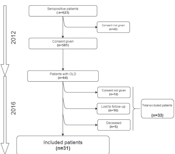
The study’s flowchart is presented in Figure 1. Over the 64 patients at baseline, 31 patients (48.4%) agreed to participate to the study, 16 patients (25%) were lost to follow-up, 12 patients (18.8%) refused to participate to the study and 5 patients (7.8%) had died.
Evolution of our population from baseline to 2016
Patients’ characteristics at baseline and in 2016 are detailed in Table 1. Seventy-four percent were men and median age at baseline was 54 years [43, 73]. At baseline, the median HIV-infection duration was 23 years [4, 29], with a median CD4 nadir cell of 171 cells/mm3 [4, 711]. All of the patients had an undetectable viral load (VL) at baseline, with 100% of patients under ART versus an undetectable HIV VL for 29 patients (93.5%) 4 years later (P = 0.1). Median CD4 cell count increased from 574 cells/mm3 [144 – 2632] at baseline to 656 [106, 2300] in 2016 (P = 0.04).
The proportion of current smokers decreased from 25 patients (77.4%) to 20 patients (64.5%) (P = 0.0013) with a median pack-year history at baseline of 68 [1, 135]. Respiratory symptoms were present in the same proportion of patients in 2012 and 2016 (19 patients (61.3%) vs. 21 patients (67.7%), respectively), (P = 0.6). Dyspnea was the most reported pulmonary condition, followed by chronic bronchitis and recurrent acute exacerbation (Table 1).
The mortality during 4 years in our OLD cohort and in our entire population followed-up from 2012 in our Infectious Disease consultation department was 7.8 % (5/64 patients) and 3.9% (90/2516 patients) respectively. In our OLD cohort, two patients died of a cardiac arrest, 1 patient of a pulmonary embolism, 1 patient of an acute respiratory failure due to a community acquired bacteria pneumonia, and 1 patient committed suicide.
Evolution of respiratory function
In 4 years, all the functional respiratory parameters tested decreased with a median of 2320mL [970, 3340] to 2190mL [670, 3210] and 3880mL [2020, 5190] to 3620mL [1910, 4840] for FEV1 and FVC respectively (P = 0.0006 and P < 0.0001). Airway obstruction, expressed by FEV1/FVC ratio, did not differ in 2012 and in 2016. The median value of the decrease
4
of FEV1 over 4 years was of 130mL [-1060, 220], 260 mL [-1060, 120] and 300mL [ -1060, 220] for the entire OLD cohort (31 patients), COPD patient and current smokers, respectively. The median value of FEV1 increased by 40 mL [-260, 180] in former smokers (P = 0.0008). Fourteen patients (45%) showed an unexpected worsening of their FEV1 and 17 patients (54.8%) an aggravation of their FEV1/FVC ratio.
Factors correlated with respiratory function degradation
The results are summarized in table 2. The unexpected worsening of the FEV1 was only correlated with the maintenance of tobacco smoking (92.8% current smokers in unexpected worsening vs. 41.2%, P = 0.008). This result remained significant even when smoking cessation was of 1 year (data not shown). The aggravation of the FEV1/FVC ratio was correlated with the smoking status in 2016 (88.2%current smokers vs. 35.7%, p = 0.007) and with the variation rate of CD8 cell count between 2012 and 2016 (delta CD8) (+21.9 % [-8.6, 185.7] P = 0.03). Smoking exposure being intensely correlated with worsening of respiratory function, we compared current smokers with former smokers to analyse the benefit of smoking cessation (table 3). Tobacco smoking cessation was not significantly correlated with a modification of respiratory symptoms but was correlated with a gain of 40 ml of FEV1 in 4 years [-260, 180] vs. a loss of 300mL [-1060, 220], (P = 0.0008) in patients who had continued smoking. Tobacco smoking maintenance was also correlated with cannabis use (P = 0.03).
Then, we analyzed separately our population of current and former smokers. We did not find any correlation between the variation of FEV1, obstruction or an unexpected worsening of FEV1 with CD4 cell count, variation rate of CD4 cell count, or CD8 cell count, variation rate of CD8 cell count, CD4/CD8 or its variation rate, or with HIV viral load (data not shown).
In multivariate analysis, only tobacco smoking remained correlated with an unexpected worsening of FEV1 (OR 18.5 CI [1.95 – 166.6], P = 0,01). The maintenance of tobacco smoking was also correlated with an aggravation of the FEV1/FVC ratio (OR 13.5 CI [2.19 – 83.3], P = 0.005), in multivariate analysis.
Analysis of the care and treatment of OLD
Results are summarized in table 4. At baseline, only 4 patients (12.9%) were diagnosed with COPD prior to the previous study. Twenty-five patients (80.6%) were current smokers, 24 (96%) received advice related to smoking cessation, with specialized help to smoking cessation proposed in 52% of cases. Three patients (11.5%) were receiving a pharmacologic treatment for their COPD. In 2016, 5 patients (20%) had quit smoking. Among the 20 patients who were still currently smoking in 2016, 11 patients (55%) stated having tried to quit smoking with nicotine substitutes, mainly electronic cigarette and nicotine patches, while 9 patients (45%) had reduced their consumption. Eleven patients (55%) were also cannabis users. Sixteen patients (51.6%) were informed in 2016 of the diagnosis of bronchopathy. Six patients (24%) received a pharmacologic therapy for COPD, 6 patients (24 %) had the recommended by guidelines follow-up, 7 patients (28 %) performed the recommended annual spirometry.
5 Discussion
The worsening of respiratory function was particularly important four years later for current smokers in our population of 31 HIV-infected patients with an OLD. This worsening seemed higher than what is observed in HIV-uninfected COPD individuals, even if we did not find any independent correlation between HIV parameters and respiratory function decline to explain these findings. On the other hand, we highlighted that smoking cessation was highly profitable. Our study also showed a concerning mortality rate in our OLD HIV-infected patients, and we pointed out a lack of COPD management from the clinicians despite a diagnosis established in a previous study.
The annual FEV1 decline is known in the general population of COPD individuals. It presents a high inter-individual variation and is markedly influenced by the maintenance of tobacco smoking and the severity of COPD (34-42). The FEV1 decline in our total cohort did not differ from what is expected in the general population (33mL/yr in general population
vs 32.5mL/yr [-265, 55] in our population) but it seemed globally worse when we focused on our patients diagnosed
with COPD and still currently smoking. Indeed, in HIV-uninfected individuals with COPD who maintain tobacco smoking, we expect an annual FEV1 loss of 50mL/yr, whereas in our study we observed a loss of 75mL/yr [-265, 55].
Then, given than data from previous studies seem to indicate an independent role-played by HIV-infection in the development and aggravation of COPD (2,17,21, 23, 24, 43, 44), we assessed the implication on the respiratory function evolution of diverse factors including HIV markers. With this goal and the suspicion that HIV could be a cofactor of COPD progression along with smoking maintenance, we arbitrarily chose to analyze patients with an unexpected decline of their FEV1 with the others patients. We did the same analysis with patients who presented an aggravation of their obstruction (decrease of FEV1/FVC ratio) with those who did not, to find correlated factors. The maintenance of tobacco smoking was the only factor correlated with an unexpected worsening of the FEV1, in univariate analysis (P = 0.008), and in multivariate analysis (OR 18.5 CI [1.95 – 166.6], P = 0,01).
Likewise, the maintenance of tobacco smoking was also the only factor correlated with an aggravation of the FEV1/FVC ratio, in univariate analysis (P = 0.007), and in multivariate analysis (OR 13.5 CI [2.19 – 83.3], P = 0.005), (table 2). Our findings on the consequences of the maintenance of tobacco smoking are in accordance with previous studies (34-42, 45).
While smoking maintenance is correlated with a higher worsening of lung function, smoking cessation appeared clearly beneficial for the evolution of FEV1 and airway obstruction. Actually, among the former smokers, we noted that 6 patients (54.5 %) showed a gain of their FEV1. (table 3).Seeing the impact of smoking, we chose separately to analyze in the sub-group of former smokers and of current smokers, the variation rate of FEV1, an unexpected worsening of FEV1 and an aggravation of FEV1/FVC ratio with markers of HIV-infection (such as HIV duration, the nadir CD4 cell count, the Viral Load, the CD4 cell count, the CD8 cell count and the CD4/CD8 ratio and their variation rates). We did not find any independent correlation between the unexpected worsening of FEV1 and markers of HIV-infection, nor with the aggravation of FEV1/FVC ratio and markers of HIV-infection, either in the sub-group of former smokers or the sub-group of continuing smokers (data not shown).
6
Considering our entire OLD population, we observed a high level of CD8 cells (928 cells/mm3 [357 – 2693]), P = 0.1) or an increase of CD8 cells count (21,9% [- 8.6 – 185.7], P = 0.03) in patients who showed an aggravation of airway obstruction, in univariate analysis but not in multivariate analysis. Increased CD8 cells had already been described as correlated with more inflammatory comorbidities especially when associated with a CD4/CD8 decrease (46). This association disappeared in multivariate analysis and was not correlated with the decrease of FEV1.
In recent studies, the results on the role played by HIV markers in the development and the decline of COPD are scarce and discordant. One study, performed in Italy, including 111 HIV-infected patients compared to HIV-uninfected age and sex-matched controls, found that HIV-infection was an independent risk factor of COPD (P = 0.008) but did not identify the parameters of HIV-infection implicated in this observation (17). The most suspected markers are mainly a low CD4 cell count (article submitted to publication, 2) or a high HIV viral load (43, 45). These appear only for extreme values, when HIV-infection is poorly controlled, with very low CD4 cell count and high viral load, which was not the case in our study. In our population, HIV-infection is highly controlled, as 93.5% have an undetectable VL, and the median CD4 cell count at baseline was 574 cells/mm3 (144, 2632) increasing in 2016 to 656 cells/mm3 (106, 2300), which did not allow us to test our hypothesis of an effect of these markers, especially CD4 cell count, in the progression of lung function and airway obstruction.
We observed the already known high mortality found in COPD patients, with a mortality rate of 7.8% in our study vs. 3.9% for our entire HIV-infected cohort [47, 48]. Furthermore, COPD as a cause of mortality in our out-patient active file may be under-estimated given the fact that we only considered the patients diagnosed with a COPD in our previous study in 2012 and we did not take into account the other outpatients who could have been diagnosed with COPD.
Early diagnosis and management have an impact on both the evolution and the prognostic of COPD (49, 50). Four years ago, patients and clinicians were informed of the diagnosis of OLD. We assessed their management (table 4). From this analysis, we noted that patients, in 2016, were under-informed about the diagnosis of OLD which was given to their clinician (51.6%) but smoking cessation was proposed to the major part of them (96%). A specific help including nicotine substitutes and consultations with the tabaccologist working in our department, were insufficiently offered (52%), when smoking cessation seems difficult in our population where 38.7% of our patients are current cannabis users. Actually, we noted that over half of the current smokers were also cannabis users (55%) vs 6 (54.5%) who managed to quit both tobacco smoking and cannabis use. Pharmacologic treatment was too largely under prescribed, with only 6 patients (24%) receiving a pharmacotherapy for their COPD. An annual spirometry was performed for 7 patients (28 %). We did not know the reasons for this lack of interest in COPD management by the clinicians in this population meticulously followed-up, with frequent consultations. Some studies show pharmacologic interactions between inhaled corticosteroids (ICS) and protease inhibitors (PI), which could be a reason why clinicians are reluctant to treat this population (51). Of course, while this pharmacologic interaction between ICS and PI must be taken into account, perhaps
7
it is important to inform clinicians that the indications of ICS in COPD management concern only GOLD 3 or 4 COPD patients with frequent exacerbations. In our study, only 1 patient required the use of ICS and did not have PI for the treatment of their HIV-infection. Nowadays, possibilities of switch PI to INI, which are particularly safe in this population, are numerous.
Our study has several limits. Considering our population, only 31 (48.4%) of OLD patients diagnosed in 2012 remained evaluable in 2016. Comparison with a HIV-negative controlled group with OLD would not have permitted to determine the effects of HIV-infection but would have been interesting in order to appreciate more precisely the evolution of respiratory function and airway obstruction in the absence of HIV-infection. With regard to the analysis of markers of HIV-infection, our population shows a very well-controlled infection. Consequently, we could not establish a suspected role of immune or viral markers on the evolution of respiratory function and airway obstruction. Finally, the size of our cohort also represents a limit to this study with only 48.4% of participation.
In conclusion, COPD in HIV-infected patients is frequent and presents a particularly serious evolution when with smoking maintenance. Smoking cessation, which is probably more difficult owing to the frequent association with cannabis use, seems to be particularly profitable and requires a particular concern including the use of specific tools. Our study did not allow us to conclude about the effects of markers of HIV-infection on the respiratory function evolution as all of our patients, as well as those usually followed-up in developed countries, present an excellent immune and virologic control of their HIV-infection.
8 Bibliography
1. Antiretroviral Therapy Cohort Collaboration al. Life expectancy of individuals on combination antiretroviral therapy in high-income countries: a collaborative analysis of 14 cohort studies. The Lancet, 2008, vol. 372, no 9635, p. 293-299.
2. CROTHERS, Kristina, BUTT, Adeel A., GIBERT, Cynthia L., et al. Increased COPD among HIV-positive compared to HIV-negative veterans. Chest Journal, 2006, vol. 130, no 5, p. 1326-1333.
3. LEONE, S., GREGIS, G., QUINZAN, G., et al.Causes of death and risk factors among HIV-infected persons in the HAART era: analysis of a large urban cohort. Infection, 2011, vol. 39, no 1, p. 13-20.
4. HELLMUTH, Joanna, MILANINI, Benedetta, et VALCOUR, Victor. Interactions between aging and NeuroAIDS. Current opinion in HIV and AIDS, 2014, vol. 9, no 6, p. 527.
5. MOORE, Richard D., GEBO, Kelly A., LUCAS, Gregory M., et al. Rate of comorbidities not related to HIV infection or AIDS among HIV-infected patients, by CD4 cell count and HAART use status.Clinical infectious diseases, 2008, vol. 47, no 8, p. 1102-1104.
6. DUFFAU, Pierre, WITTKOP, Linda, LAZARO, Estibaliz, et al. Association of immune-activation and senescence markers with non-AIDS-defining comorbidities in HIV-suppressed patients. AIDS, 2015, vol. 29, no 16, p. 2099-2108.
7. CASTILHO, Jessica L., SHEPHERD, Bryan E., KOETHE, John, et al. CD4+/CD8+ ratio, age, and risk of serious noncommunicable diseases in HIV-infected adults on antiretroviral therapy. AIDS, 2016, vol. 30, no 6, p. 899-908.
8. LAPADULA, Giuseppe, CHATENOUD, Liliane, GORI, Andrea, et al. Risk of Severe Non AIDS Events Is Increased among Patients Unable to Increase their CD4+ T-Cell Counts> 200+/μl Despite Effective HAART. PloS one, 2015, vol. 10, no 5, p. e0124741. 9. ABERG, J. A. Aging, inflammation, and HIV infection. Topics in antiviral medicine, 2011, vol. 20, no 3, p. 101-105.
10. APPAY, Victor et SAUCE, Delphine. Assessing Immune Aging in HIV-infected patients. Virulence, 2016, no just-accepted, p. 00-00.
11. NASI, M., DE BIASI, S., GIBELLINI, L., et al. Aging and inflammation in patients with HIV infection.Clinical & Experimental Immunology, 2016. 12. BERRY, Cristine E. et WISE, Robert A. Mortality in COPD: causes, risk factors, and prevention. COPD: Journal of Chronic Obstructive Pulmonary
Disease, 2010, vol. 7, no 5, p. 375-382.
13. BOUTOU, Afroditi K., SHRIKRISHNA, Dinesh, TANNER, Rebecca J., et al. Lung function indices for predicting mortality in COPD. European Respiratory Journal, 2013, vol. 42, no 3, p. 616-625.
14. WHO - Chronic obstructive pulmonary disease (COPD). Available at: www.who.int/respiratory/copd/en/. Date last update february 2013. Accessed 3 March 2013.
15. GINGO, Matthew R., MORRIS, Alison, et CROTHERS, Kristina. Human Immunodeficiency Virus–Associated Obstructive Lung Diseases.Clinics in chest medicine, 2013, vol. 34, no 2, p. 273-282.
16. DIAZ, Philip T., KING, Mark A., PACHT, Eric R., et al. Increased susceptibility to pulmonary emphysema among HIV-seropositive smokers.Annals of internal medicine, 2000, vol. 132, no 5, p. 369-372.
17. MADEDDU, G., FOIS, A. G., CALIA, G. M., et al.Chronic obstructive pulmonary disease: an emerging comorbidity in HIV-infected patients in the HAART era?. Infection, 2013, vol. 41, no 2, p. 347-353.
18. CROTHERS, Kristina, HUANG, Laurence, GOULET, Joseph L., et al. HIV infection and risk for incident pulmonary diseases in the combination antiretroviral therapy era. American journal of respiratory and critical care medicine, 2011, vol. 183, no 3, p. 388-395.
19. RAHMANIAN, Shiva, WEWERS, Mary Ellen, KOLETAR, Susan, et al. Cigarette smoking in the HIV-infected population. Proceedings of the American Thoracic Society, 2011, vol. 8, no 3, p. 313-319.
20. DIAZ, Philip T., WEWERS, Mark D., PACHT, Eric, et al. Respiratory symptoms among HIV-seropositive individuals. CHEST Journal, 2003, vol. 123, no 6, p. 1977-1982.
21. SHIRLEY, Daniel K., KANER, Robert J., et GLESBY, Marshall J. Screening for chronic obstructive pulmonary disease (COPD) in an urban HIV clinic: a pilot study. AIDS patient care and STDs, 2015, vol. 29, no 5, p. 232-239.
22. PETRACHE, Irina, DIAB, K., KNOX, Kenneth S., et al. HIV associated pulmonary emphysema: a review of the literature and inquiry into its mechanism.Thorax, 2008, vol. 63, no 5, p. 463-469.
9
23. RAYNAUD, Christine, ROCHE, Nicolas, et CHOUAID, Christos. Interactions between HIV infection and chronic obstructive pulmonary disease: Clinical and epidemiological aspects. Respiratory research, 2011, vol. 12, no 1, p. 1.
24. DRUMMOND, M. Bradley et KIRK, Gregory D. HIV-associated obstructive lung diseases: insights and implications for the clinician. The lancet Respiratory medicine, 2014, vol. 2, no 7, p. 583-592.
25. QASEEM, Amir, WILT, Timothy J., WEINBERGER, Steven E., et al. Diagnosis and management of stable chronic obstructive pulmonary disease: a clinical practice guideline update from the American College of Physicians, American College of Chest Physicians, American Thoracic Society, and European Respiratory Society. Annals of internal medicine, 2011, vol. 155, no 3, p. 179-191.
26. MILLER, Martin R., HANKINSON, J. A. T. S., BRUSASCO, V., et al. Standardisation of spirometry.European respiratory journal, 2005, vol. 26, no 2, p. 319-338.
27. 2015 Global Initiative for Chronic Obstructive Lung Disease
28. Prise en charge médicale des personnes infectées par le VIH. Rapport 2013. La Documentation Française.
29. Pugliese P, Cuzin L, Cabié A, Poizot-Martin I, Allavena C, Duvivier C et al. A large French prospective cohort of HIV-infected patients: the Nadis Cohort. HIV Med 2009; 10:504̻511.
30. CELLI, Bartolomé R., THOMAS, Nicola E., ANDERSON, Julie A., et al. Effect of pharmacotherapy on rate of decline of lung function in chronic obstructive pulmonary disease: results from the TORCH study. American journal of respiratory and critical care medicine, 2008, vol. 178, no 4, p. 332-338.
31. DECRAMER, Marc, RUTTEN-VAN MÖLKEN, Maureen, DEKHUIJZEN, PN Richard, et al. Effects of N-acetylcysteine on outcomes in chronic obstructive pulmonary disease (Bronchitis Randomized on NAC Cost-Utility Study, BRONCUS): a randomised placebo-controlled trial. The Lancet, 2005, vol. 365, no 9470, p. 1552-1560.
32. BURGE, P. Sherwood, CALVERLEY, P. M. A., JONES, Paul W., et al. Randomised, double blind, placebo controlled study of fluticasone propionate in patients with moderate to severe chronic obstructive pulmonary disease: the ISOLDE trial. Bmj, 2000, vol. 320, no 7245, p. 1297-1303. 33. PAUWELS, Romain A., LÖFDAHL, Claes-Göran, LAITINEN, Lauri A., et al. Long-term treatment with inhaled budesonide in persons with mild chronic
obstructive pulmonary disease who continue smoking. New England Journal of Medicine, 1999, vol. 340, no 25, p. 1948-1953.
34. ANTHONISEN, Nicholas R., CONNETT, John E., et MURRAY, Robert P. Smoking and lung function of Lung Health Study participants after 11 years. American journal of respiratory and critical care medicine, 2002, vol. 166, no 5, p. 675-679.
35. TANTUCCI, Claudio et MODINA, Denise. Lung function decline in COPD. Int J Chron Obstruct Pulmon Dis, 2012, vol. 7, p. 95-99. 36. FLETCHER, Charles et PETO, Richard. The natural history of chronic airflow obstruction. Br Med J, 1977, vol. 1, no 6077, p. 1645-1648.
37. KOHANSAL, Robab, MARTINEZ-CAMBLOR, Pablo, AGUSTÍ, Alvar, et al. The natural history of chronic airflow obstruction revisited: an analysis of the Framingham offspring cohort. American journal of respiratory and critical care medicine, 2009, vol. 180, no 1, p. 3-10.
38. SCANLON, Paul D., CONNETT, John E., WALLER, Lance A., et al. Smoking cessation and lung function in mild-to-moderate chronic obstructive pulmonary disease: the Lung Health Study. American Journal of Respiratory and Critical Care Medicine, 2000, vol. 161, no 2, p. 381-390.
39. XU, Xiping, DOCKERY, Douglas W., WARE, James H., et al. Effects of cigarette smoking on rate of loss of pulmonary function in adults: a longitudinal assessment. American Review of Respiratory Disease, 1992, vol. 146, no 5_pt_1, p. 1345-1348.
40. BURCHFIEL, Cecil M., MARCUS, Ellen B., CURB, J. David, et al. Effects of smoking and smoking cessation on longitudinal decline in pulmonary function. American journal of respiratory and critical care medicine, 1995, vol. 151, no 6, p. 1778-1785.
41. WILLEMSE, B. W. M., POSTMA, D. S., TIMENS, W., et al. The impact of smoking cessation on respiratory symptoms, lung function, airway hyperresponsiveness and inflammation. European Respiratory Journal, 2004, vol. 23, no 3, p. 464-476.
42. VESTBO, Jørgen, EDWARDS, Lisa D., SCANLON, Paul D., et al. Changes in forced expiratory volume in 1 second overtime in COPD. New England Journal of Medicine, 2011, vol. 365, no 13, p. 1184-1192
43. DRUMMOND, M. Bradley, KIRK, Gregory D., ASTEMBORSKI, Jacquie, et al. Association between obstructive lung disease and markers of HIV infection in a high-risk cohort. Thorax, 2012, vol. 67, no 4, p. 309-314.
10
44. CROTHERS, Kristina, MCGINNIS, Kathleen, KLEERUP, Eric, et al. HIV infection is associated with reduced pulmonary diffusing capacity. Journal of acquired immune deficiency syndromes (1999), 2013, vol. 64, no 3.
45. DRUMMOND, M. Bradley, MERLO, Christian A., ASTEMBORSKI, Jacquie, et al. The effect of HIV infection on longitudinal lung function decline among injection drug users: a prospective cohort. AIDS (London, England), 2013, vol. 27, no 8, p. 1303.
46. MUDD, Joseph C. et LEDERMAN, Michael M. CD8 T cell persistence in treated HIV infection. Current opinion in HIV and AIDS, 2014, vol. 9, no 5, p. 500
47. MANNINO, David M. et KIRI, Victor A. Changing the burden of COPD mortality. International Journal of COPD, 2006, vol. 1, no 3.
48. BERRY, Cristine E. et WISE, Robert A. Mortality in COPD: causes, risk factors, and prevention. COPD: Journal of Chronic Obstructive Pulmonary Disease, 2010, vol. 7, no 5, p. 375-382.
49. LOVRE, Vladimir et USTAMUJIC, Aida. The changes of pulmonary function in COPD during four-year period. Materia socio-medica, 2013, vol. 25, no 2, p. 88.
50. BERRY, Cristine E. et WISE, Robert A. Mortality in COPD: causes, risk factors, and prevention. COPD: Journal of Chronic Obstructive Pulmonary Disease, 2010, vol. 7, no 5, p. 375-382.
51. SABERI, Parya, PHENGRASAMY, Tony, et NGUYEN, Dong Phuong. Inhaled corticosteroid use in HIV-positive individuals taking protease inhibitors: a review of pharmacokinetics, case reports and clinical management. HIV medicine, 2013, vol. 14, no 9, p. 519-529.
Appendix : Questionnaire (translated in English)
QUESTIONNAIRE COPD STUDY
READ WITH PRECAUTION QUESTIONS WITCH CONCERN YOUR RESPIRATORY SYMPTOMS AND YOUR RISK FACTOR OF RESPIRATORY DISEASE AND TRY TO PRECISELY ANSWER.
Weight, size: You weight _ _ _ kilograms You are _ _ _meter
Symptoms
1- If we ask you “do you spit?” you will respond (ONLY ONE RESPONSE)
YES F I spit at last one time a day from 2 years No F I don’t spit, or very occasionally
2- If we ask you if you are breathless, you will say (ONLY ONE RESPONSE)
YES F I’m sometimes breathless if I climb the stairs, from 2 floors YES F I’m regularly breathless if I walk fast or if I walk a climb
YES F I’m regularly breathless if I walk on the flat with the same speed than somebody as old than me YES F When I walk, I usually have to stop to catch my breath after several minutes or 100 meters YES F I’m breathless for a lesser effort
NO F None of previous response, breathlessness is not a problem.
3- Have you do to take antibiotics or corticoids (solupred®, cortancyl®, prednisolone®, celestene®) for a bronchitis, and that at least 2 times during previous year?
YES F NO F
4- Have you already been hospitalized for respiratory problem? YES F NO F
Care of the OLD
5- Some doctor has already say you that you have a chronic bronchitis (or chronic bronchitis of smoker)?
YES F NO F
If yes, which year? ___________
6-Have you been taken in charge for your CPDO / Bronchopathy due to tobacco smoking since the diagnosis?
YES F NO F
Date of the last spirometry? ____________
Do you take a treatment for your Breath/ for your respiration*? YES F NO F
* exemple of treatment for respiratory disease: Sérétide®, Symbicort®, Ventoline®, Bécotide®, Béclojet®, Prolair®, Qvar®, Béclométasone®, Béclone®, Pulmicort®,
Flixotide®, Miflonil®, Bronchodual®, Combivent®, Atrovent, , Spiriva, Asmabec®, Bemedrex®, Miflasone®, Onbrez®, A Spir®, Nexxair®, Ecobec®, Timos®, Formoair®,
Breezhaler®, Atimos®, Formoair®, Tersigat®, Dilatrane®, Euphylline®, Theophylline®, Tédralan®, Xanthium®, Trentadil®, Singulair®
Toxic exposure
7- Tobacco smoking:
- Has a doctor ever explained to you the link between tobacco smoking and COPD? YES F NO F
- Have you ever received the advice to quit smoking? YES F NO F
- Have you ever received a help to quit smoking / a consultation with a tobaccologist? YES F NO F
- Have you reduced your consumption since the diagnosis of COPD? YES F NO F
If yes, from how many? ___________
- Have you quit smoking since the diagnosis of COPD? YES F NO F
8- Do you currently smoke? YES F NO F
If yes, have you tried to quit? YES F NO F
If yes, have you had recourse to a pharmacologic of professional help? YES F NO F Please, precise: _________________________________________________________
Why did you start again? __________________________________________________
9- If you smoke, or have ever smoked:
- When did you start: ______ years old
- On average, how much pack of cigarettes do you smoke (smoked) a day?
(only one answer)
F less than a half packet a day F between an half and one packet a day F between one and two packet a day F more than 2 packet a day
- Pack-year number: ______________ pack-year
10- If you stopped smoking: when did you stop? _______ years old 11- Cannabis
- Do you currently smoke cannabis (at least one joint a week) YES F NO F
If yes, on average: F between a joint a week and 1 joint a day F between 1 and 5 joints a day
F more than 5 joints a day
In the past, have you regularly (at least one joint a week) during a period, smoked cannabis? YES F NO F
12- Professional exposure