People's Democratic Republic of Algeria Series N°:…..
Higher Education Ministry And Scientific Research
Echahid Hamma Lakhdar El-Oued University
Faculty of Natural Sciences and Life
Department of Cellular and Molecular Biology
Master’s thesis
In order to obtain a diploma of an Academic Master
in biological sciences
Specialty: Applied Biochemistry
Theme
Presented by:
ATOUSSI Ouidad
CHETEHOUNA Sara
Examining Committee:
President
Mr. LAICHE Ammar Touhami
M.C.B.
El-Oued University
Examiner
Mm. AOUIMEUR Meriem
M.A.A.
El-Oued University
Supervisor
Mr. DEROUICHE Samir
M.C.A.
El-Oued University
2019/2020
Formulation and Effectiveness Evaluation of
U
30ACS as a New Therapeutic Approach to
ءادـــــــــــــــــــــــــــــــــــــــــــــــــــــــــــــــــــــــــــــها
ةظحل
ل
اعقاو شيعأ نأ ...ا ريثك اهترظتنا املاط
نأ وه و لاأ ... يسفن قامعأب اثبشتم...سملأاب اديعب املح تاب
دمحلا كلف...عساولا ملعلا رحب ي
ف ةعفان ةرط
ق َّب ُر اوفاضأ نمم نوكأ و ي تسارد ة ريسم جوتا
َ
" ي
ّبر"
ِ
ام لىع
لجأ نم يل ترخس و تابثلاو ريصلاب ي تتمركأ و يدحتلا اذه عفر لىع ي تتقفو نأ دمحلا كل و... ي تتعدوأ
ا اذه
...ما
يحلاا و ءانثلا و ركشلا تارابع لك مهمامأ
تزجع نم... ي تبحأ ليبسل
ْ
ةصلاخلا ي
تايحت
فرأ و مىسأ ُجزا اذهل
... يمىهو يحرف تاظحل لك تل
َّمحتو تلم َح و ، يلىقع ملاظ تءاضأ ي تلا سمشلا لا
" يمأ "نانحلا عبن
ي
ف ريعلا نا و ةاجنم قدصلا ناب ي تملعي لازلا و ناك يذلا لا
...تاياهنلا
" ي ّبأ "تباثلا يدنس
... ي
تايح ء ي
ضي يذلا رونلا و يسر تيب ، ي تمكح توص ،فورحلا اهفصت لا ي تلا ي تياكح لا
يصخش ةآرم
" ي تخأ"
... يمأ اهدلت مل ي تلا ي تخأو ي تقيفر ، ي
تدحو ةسنؤم ، ي
تايحل رخأ اقيرب اهدوجوب تفاضأ ي تلا لا
ي تيلاغ
" ي
خأ ةجوز"
... ي ت
كت ُمو
َ
تميلا يدي لا
ي
ب َوز ِعو ي تعفِر
"
" ّيوخأ
نامثأب ردقي لا و نا ريمب نزوي لا يذلا كاذ لا
ققحتي ليحتسملا نأ و ،راسرأ حاجنلل نأ ي تملع يذلا ،
...راصرلإاب
ي
باثلا ي ّبأ و يملعم ،يذاتسأ
"
يمس شيورد
"
ي تلا ،نامزلا اهديعي نل ي تلا لا
كسمأو يدحتلا اذه يعم تعفر نم ،ةظحل ي تع اهقيرب تفخي لا
و يديب ت
ايوس اومسنل
تع َس
ْ
...
ةمئادلا ي تس ِكاش
ُم
"يدادِو"
... ي تسارد راوشم ي
ف قوفتلل ي تعجشو ي تقفار نم لك لا و
لضافلأا " ي
بذتاسأ"
هراـــــــــــــــــــــــــــــــــس
ــــــــــــــــــــــــــــــــــــــــــــــها
ــــــ
ءادـــــــــــــــــــــــــــــــ
لـملأاب ٍةمـعف ُم ٍلمر
ةبـ َح ُجرحدتـ
ُ
ت
َ
..
، مللأا
ع
َ
دـص
ُ
ت اهـيف ُعفني أـل و حاـيرلا
َ
تاهاجتإ اـهــيرغ
ُ
ت اـل
ُ
، ي ِ نحرـف ِموي ىـلإ ي ِ نــل
د ،
ُ
كـيـلإ لاــص ِولا ليب
َ
َس لىإ
نـل
ي ِ
د
ُ
مكـيدهلأ ، ٍ يع ِةفر
طِب يِـتأي
ُ
دـغلا َتيلاـيأ
َ
، ي ِ نملـك
؟، ي ِ نـص
غ َموي ي ِ
َ
سفـن
تاـت
َ
ش َملـمل نم ي ِ نلأـسإ
ُ
.. ي ِ
تدح ِو َءاـنآ ي ِ فت
ك لىـع ُتبر
َ
ت ةنونح ٍدي ـِب
َ
كبيجأ
َ
َ
ةبيبـحلا يـمأ
؟،ي ِـتخر َـص
تَلـ َع موي ًادـنس يِـل ناـك ن َم ي ِ نلأــسإ
.. ي ِ نبيـ
خ موي هيلـع ئكتأ ٍرادجـِب ك ربـخأ
َ
يـلاـغلا يـبأ
ي ِ نلأـسإ
؟، ي ِ
ت بـ َح َموي يدي ـِب
ذخأي نم يـت رب ِخ ِةل ِق يـف
ي ِ
تاسـل ُد ِد ُ بـس ،يـنيـع ِةر
ق ـِب
ُ
ك ُبيـجأ
َ
..يـِتوخإ ..يـِتوخإ
؟اـهنيـملاـع نـع ي ِ نـتفطصإ ِبولـ
قلا ُّيأ يـ ِنلأـسإ
ُ
ٍلمأـتو ٍةماـستببإب
كبيـجأ
َ
نـيتيلاـغلا َّ ي نـخأ
،
ٍ
يـ َع َموي ي ِـحور َ ربصأ نم ي ِـنلأـسإ
؟،يـ ِتـ َعمد اـهتأـلم
،نلاذخلا ِجاج
ز لىع َّ ي
ُ
ض َملا ي نمل
َع و ي ِديب َذخأ نمِب َكبـيجأ
" ي
تاثلا يـبأ و
ملع ُم
ي ِ
بــمس شــيورد
"
؟،اـهعم ِةقشـ َملا َبر
د ُتلمكأ ن َم يـنلأـسإ
َ
" يره
د
َ
ةقيدص ، ي ِ
ُ
تسِّم و ي رسِّ
ِب
كبيـجأ
َ
ي ِ
تراـس
"
، مللأا
دافصأ عزنإ
َ
ارـيخأ و
ً
نلأ طقف ،اـي بـثولـيلإاب اـلهأ ٍلائاـق
كيدي د ُم و لمأـلاب ب ِحرو
َ
نكم ُم ٍء ي
ش ل
ك
ُ
!
دادو
Acknowledgement
The patience, the strength, the courage, the good health and the good people that we met is the best gift from Allah to us, our first thank and love is always to him.
We would like to express our honest gratitude to our supervisor Dr. DEROUICHE Samir for his constant confidence in us to be able to shed the new light in this miracle disease, and for
agreeing to supervise us, for his dynamism, his help, motivation and his precious advice, that allowed us to go further in our research, we also, so grateful to him for all
the experience that we gained it in our small carrier.
We thank Dr. LAICHE Ammar Touhami who has given us the honor to accept the presidency of this thesis. Respectful tribute.
We thank Mm. AOUIMEUR Meriem for having agreed to examine our thesis and the honor for her presence. Permit us to express to her our deep respect. We also thank the members of Kouinine laboratory, Bin Omar Jilani and Slimane Amirat Hospitals, Faculty of Natural and Life Sciences, Physic and Chemistry laboratory in Faculty of
Exact Sciences, Echahid Hamma Lakhdar El-Oued University, Central Research Test and Analysis Laboratory Application and Research Center is located in Ege Üniversitesi
Kampüsü, Bornova/İZMİR in Turky.
We are grateful to Dr. DEROUICHE Manel and Dr. MOKHTARI Madjda lecture
in Physic Department, Faculty of Exact Sciences in El-Oued University.
We do not forget to express our thanks to our big sister and member of our teem Ms GOBI Sana thanks for your encouragement and your honest work, with
Abstract
Our study aimed to investigate the impact of U30ACS that combines phytotherapy by purslane and bio-nanotherapy by zinc and copper as a new therapeutic approach to physiological and biochemical alterations induced by Alzheimer disease (AD) in rats.
For this purpose, thirty males albino Wistar rats were randomly divided into 6 groups (n=5); healthy rats (Control), untreated Alzheimer rats (Exp Alzheimer), Alzheimer rats treated with colloidal solution (U30ACS), Alzheimer rats treated with aqueous extract of P. oleracea (AEPo), Alzheimer rats treated with zinc nanoparticles (ZnNPs) and Alzheimer rats treated with copper nanoparticles (CuNPs). All types of treatments were given to rats orally for 21 days.
Alzheimer in rats was induced by oral administration of AlCl3 and D-galactose (200mg/kg) in each
for 76 days. Various parameters as neurological, hematological, biochemical and oxidative stress
markers were estimated. Histopathology of brain and liver tissues were observed.In in-vitro study
on plant and nanoparticles, some quantitative, qualitative and characterization parameters were analyzed using standard protocols.
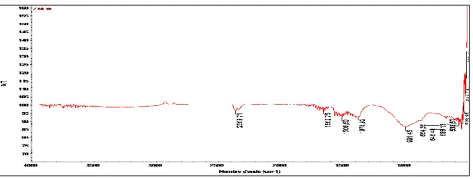
In vitro phytochemical test results revealed that purslane contains most of the principal active molecules, especially alkaloids of various kinds, such as trigonelline, sildenafil, indole and norharman. In vivo results, the Exp Alzheimer group showed an alteration in passive avoidance learning (PAL) and a significant increase in AChE activity (P<0.01) and protein levels (P<0.001). While, the antioxidant defend system in brain and liver was affected by increasing the MDA level and decreasing the GSH levels, GST and SOD activities compared to control. Furthermore, the hematological parameters revealed that Alzheimer disease induce an inflammation in rats by significant raise (P<0.01) of WBC, Monocyte, LYM and platelet (P<0.001) levels with a significant perturbation in lipid profile and biochemical parameters. In the other hand, histopathological analysis recorded an deep modification in brain and liver tissues of Alzheimer rats group compared to control. However, the treatment of Alzheimer rats by AEPo, ZnNPs and CuNPs ensured a partial amelioration and correction of the previous parameters. While the U30ACS approach was the best treatment that it give, improvement results in most of the studied parameters.
We conclude that the use of purslane and bio-nanoparticles of zinc and copper seems to be
the powerful limited of Alzheimer disease development, which opening the new horizons for the use of nanotherapy in medical approach. Where the new approach (U30ACS) relying on
phytotherapy and nanotechnology has proven highly effective against Alzheimer's symptoms, which shed a new lite and a new hopes for the possibility of treating Alzheimer’s patients.
صخلملا ىلإ انتسارد فدهت مييقت ريثأت جذومنلاب جلاعلا ( ACS 30 U ) عمجي يذلاو نيب جلاعلا يعيبطلا تابن ةطساوب ةلجرلا Portulaca oleracea تائيزجب جلاعلاو ونانلا ساحنلاو كنزلل ديدج يجلاع جذومنك ا دض ل فلا تاريغت زي ةيجولوي ةيئايميكويبلاو صملا حا ل ةب رمياهزلا ضرم دنع .نارئفلا نيثلاث ميسقت مت ،ضرغلا اذهل اذرج وشع راتسيو ونيبلأ ةليصف نم ا ًركذ ىلإ اًيئا 6 = ددعلا( تاعومجم 5 ) ؛ ناذرجلا ةميلسلا )ةدهاشلا( ، ب ةباصم ناذرج ةجلاعم ريغ رمياهزلا (Exp Alzheimer) ، ب ةباصم ناذرج ةجلاعم رمياهزلا جذومنلاب ) ACS 30 U ( ، ب ةباصم ناذرج رمياهزلا نم يئام صلختسمب ةجلاعم P. oleracea (AEPo) ، ب ةباصم ناذرج رمياهزلا ةيونانلا كنزلا تايئزجب ةجلاعم (ZnNPs) و ب ةباصم ناذرج رمياهزلا ةجلاعم ب ةيونانلا تاميسجلا ساحنلل .(CuNPs) تيطعأ تاجلاعلا عاونأ عيمج ناذرجلل ةدمل مفلا قيرط نع 21 زيفحت مت .اموي ضرم رمياهزلا ناذرجلا دنع يتدام ةطساوب ديرولك يثلاث موينمللأا و ةيموي ةعرجب زوتكلاجلا 200 ل غك/غلم امهنم لك مفلا قيرط نع ةدمل 76 دقو .اًموي ةيجولويبلا ريياعملا فلتخم ريدقت مت لثم ريياعم لقنلا ،مدلا تانوكم ،يبصعلا ريياعملا ةيئايميكويبلا يدسكاتلا داهجلااو . نم ةجسنلا ةيرهجملا ةظحلاملا تمت امك .دبكلاو خملا لل ةبسنلاب ةسارد ةيليلحتلا لل و ةتبنلا صلختسمل تاميسج ،ةيونانلا ضعب انمق لا حت ا ليل مادختساب ةيعونلاو ةيمكلا قرطلا ليلحتلا يف ةيدايتعلاا . جئاتن تفشك لاا رابتخ هنا ةلجرلا تابن صلختسمل ةيعونلاو ةيمكلا تا ، ةيسيئرلا ةطشنلا تائيزجلا مظعم ىلع يوتحي ةصاخو لا ليفانيدليس ،نيلينوغيرت لثم تاديولق .نامراهرونو لودنإ ، ىلع ةبرجتلا جئاتن اما ناف ناذرجلا جئاتن لا ةعومجم ةباصملا رمياهزلاب ترهظأ يف ا ًرييغت ةركاذلا رابتخا ( PAL ةدايزو ) ةيونعم ةريبك (P <0.01) طاشن يف زاريتسا نيلوك ليتيسلاا ميزنا و يف اضيا ( نيتوربلا تايوتسم P <0.001 نيح يف .) هذه تنيب للخ ةعومجملا يف دبكلاو غامدلا يف ةدسكلأل داضملا عافدلا ماظن ىوتسم ةدايزب نوهدلا ديسكوريب تايوتسم ضفخو نويثاتولجلا ةطشنأو ، نويثاتولجلا لقن تاميزنا نم لك (GST) و ميزنا ةدسكلاا يلاع زاتيمسيد (SOD) اذهو ةنراقم عم ةعومجم ةدهاشلا ناذرجلا كلذ ىلع ةولاع . تفشك ، تانوكم ريياعم مدلا نأ رمياهزلا ضرم ةيباهتلا تاباجتسا بحاصي دنع نارئفلا ب كلذو ( ةريبك ةدايز P <0.01 تايوتسمل ) و ءاضيبلا مدلا تايرك ايوافمللا ت تاديحو ايلاخلاو ةيلخلا ( ةيومدلا حئافصلاو P <0.001 يف ريبك بارطضا عم ) ضيا ريياعمو ةيئايميكويبلا ريياعملا ىرخأ ةيحان نم .نوهدلا يجيسنلا ليلحتلا لجس ، لالخ ةعومجمل دبكلاو خملا ةجسنأ يف اًقيمع ناذرجلا ةباصملا ب ةنراقم رمياهزلا ةعومجملاب ةدهاشلا كلذ عمو . نإف ، جئاتن جلاع ناذرج رمياهزلا ونانلا تائيزج وا ةلجرلا تابنل يئاملا صلختسملا لامعتساب ساحنلا وا كنزلل حيحصت ترهظا يئزج ريياعملا مظعم يف يجيسنلا ليلاحتلا و ةيجولويبلا ة ركذلا ةفلاسلا نأ نيح يف . جذومن ACS 30 U ناك لضفلاا لا ثيح نم جئاتن ةجلاعملا ناذرجلا ترهظا ثيح اهيلع لصحتملا انسحت هب ريبك ا فلتخم يف ضارعلاا ةبحاصملا ل ناذرجلا دنع رمياهزلا ضرم . مادختسا نأ جتنتسن و ةلجرلا تابن ةيويحلا ةيونانلا تاميسجلا روطت نم دحلا يف ةريبك ةيلاعف هل ساحنلا وا كنزلل ضرم رمياهزلا ، ام وهو و تاتابنلا لامعتسا ىلع عجشي تائيزج ونانلا تبثأ ثيح .يبطلا جهنلا يف نا لمعلا اذه جذومنلا ديدجلا ( ACS 30 U ىلع دمتعي يذلا ) لا يعيبطلا جلاعلا م معد تائيزجب تيلاعف ونانلا رمياهزلا ضرم ضارعأ دض ةيلاعلا ه ، حتفي ام وهو قافا .رمياهزلا ىضرم جلاع ةيناكمإ يف ةديدج لامآو :ةيسيئرلا تاملكلا ،يدسكأتلا داهجلإا ،رمياهزلا ضرم ةلجرلا تابن ، ACS 30 U ، كنز ونانلا ، ساحن ونانلا .
ABBREVIATIONS LIST
ACh: Acetylcholine
AChE: Acetyl cholinesterase
AChEI: Acetyl cholinesterase inhibitors AD: Alzheimer disease
AEPo: Aqueous extract Portulaca oleracea AGE: Advanced glycation end
ApoE: Apolipoprotein E
APP: Amyloid precursor protein
Aβ: Amyloid beta
BACE1: Beta- site APP cleaving enzyme 1 BASO: Basophile
BBB: Blood brain barrier
BHT: Butylated hydroxytoluene BPS: Phosphate buffer solution BSA: Bovine serum albumin bw: Body weight
CDNB: l-chloro-2,4-dinitrobenzene CNS: Central nerveuse system CuNPs : Copper nanoparticles
CuO NPs : Copper oxide nanoparticles d: Dilution factor
D-gal: D-galactose
DPPH: 2,2-diphenyl-1-picrylhydrazyl DTNB: 5,5'-dithiobis(2-nitrobenzoic acid)
DYRK1A: Dual specificity tyrosine‐phosphorylation‐regulated kinases activity
EDTA: Ethylenediaminetetra-acetic acid
EOAD: Early onset Alzheimer disease
Exp Alzheimer: Experimental Alzheimer FRAP assay: Ferric reducing antioxidant power FTIR: Fourier transform infrared spectroscopy GPT: Glutamate pyruvate transaminase
GPx: Glutathione peroxidase
GST: Glutathione-S-transferase h: Hemorrhage
HBG: Hemoglobin
HDL-C: High-density lipoprotein cholesterol i: Inflammation
IC50: Inhibition concentration 50
IL-1 β: Interleukin-1β IL-6: Interleukin-6
IP: Inhibition percentage
LDL-C: Low-density lipoprotein cholesterol
LOAD: Late-onset Alzheimer disease
LPO: Lipid peroxidation Lymph: Lymphocytes
mAChRs: Metabotropic muscarinic acetyl choline receptors MAO: Monoamine oxidases
MAP: Microtubules associated proteins
MDA: Malondialdehyde MNPs: Metallic nanoparticles Mono: monocytes
mRNA: Messenger ribonucleic acid n: Necrosis
nAChRs: Ionotropic nicotinic acetyl choline receptors NBT: Nitro-blue tetrazolium chloride
NFTs: Neurofibrillary tangles
NF-κB: Nuclear factor kappa-light-chain-enhancer of activated B cells NPs: Nanoparticles
OD: Optical density
PAL: Passive avoidance learning PLT: Platelet
PPAR-α: Peroxisome proliferator-activated receptor alpha Prot: Protein
PSEN1: Presenilin 1 PSEN2: Presenilin 2
RAGE: Advanced glycation end receptor RBC: Red blood cells
ROS: Reactive oxygen species SEM: Scanning electron microscope SEM: Standard error of the mean
SOD: Super oxide dismutase STL: Step-through latency TBA: Thiobarbituric acid
TBARS: Thiobarbituric acid reactive substances TBS: Tris buffer saline
TC: Total cholesterol TCA: Trichloroacetic acid
TDC: Time spent in the dark chamber TG: Triglyceride
TNB: 2-nitro-5-mercocapturic acid
TNF-α: Tumor necrosis factor-α
U30ACS: Ultrasonic technique for 30 min aqueous colloidal solution
UA: Uric acid
UHPLC: Ultra high performance liquid chromatography UV-VIS: Ultraviolet-visible spectroscopy
vc: Vacuolar cytoplasm
VLDL-C: Very low-density lipoprotein cholesterol WBC: White blood cell
XRD: X-ray powder diffraction ZnNPs : Zinc nanoparticles
FIGURES LIST
Number Title Page
Figure 01 Brain lobes fraction and function 5
Figure 02 Portulaca oleracea L. 10
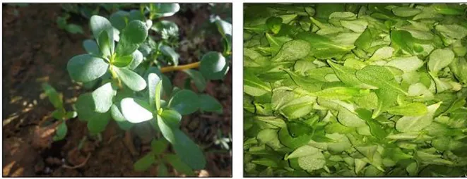
Figure 03 Portulaca oleracea leaves 17
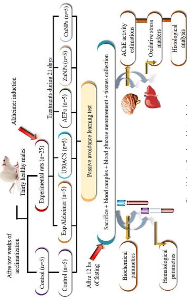
Figure 04 In vivo Experimental design of study 20
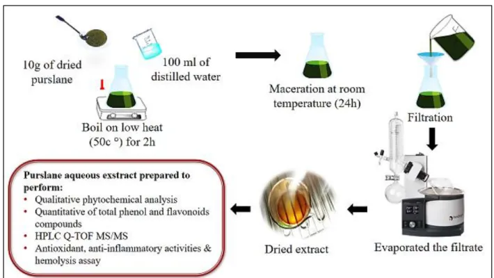
Figure 05 P.oleracea aqueous extract preparation 21
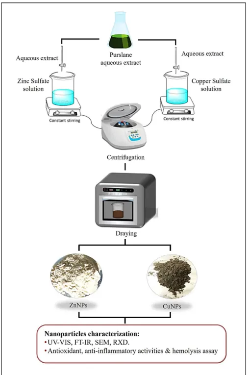
Figure 06 The green synthesis method of zinc and copper oxide nanoparticles 22
Figure 07 Step-through passive avoidance, Train (A) and Test (B) 28
Figure 08 chromatogram in positive ion (A) and negative ion (B) mode for tentative
assigned alkaloids compounds 36
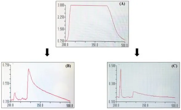
Figure 09 UV-Vis spectrums of purslane leaves aqueous extract (A), CuNPs (B) and
ZnNPs (C) 39
Figure 10 FT-IR spectrum of ZnNPs synthesized by P.oleracea aqueous extract 40
Figure 11 FTIR spectrum of powder CuNPs obtained from purslane aqueous extract 40
Figure 12 Scanning electron microscopy image of the biosynthesized Zn
nanoparticles from purslane aqueous extract 40
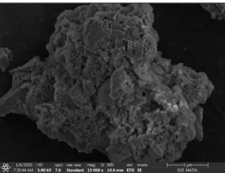
Figure 13 Scanning electron microscopy images of the biosynthesized CuNPs 41
Figure 14 Diffractograms of ZnNPs (A) and CuNPs (B) 41
Figure 15 IC50 levels of P.oleracea aqueous extract, ZnNPs, CuNPs and Vitamin C 42
Figure 16 IC50 levels of anti-inflammatory activity of P.oleracea aqueous extract,
ZnNPs, CuNPs and diclofenac 42
Figure 17 Effects of ZnNPs, P.oleracea aqueous extract and CuNPs on Red Blood
Cells (RBCs) hemolysis 43
Figure 18 Step-through latency in control and experimental groups 45
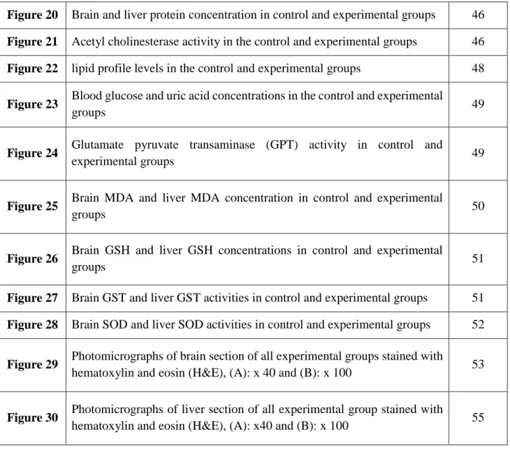
Figure 20 Brain and liver protein concentration in control and experimental groups 46
Figure 21 Acetyl cholinesterase activity in the control and experimental groups 46
Figure 22 lipid profile levels in the control and experimental groups 48
Figure 23 Blood glucose and uric acid concentrations in the control and experimental
groups 49
Figure 24 Glutamate pyruvate transaminase (GPT) activity in control and
experimental groups 49
Figure 25 Brain MDA and liver MDA concentration in control and experimental
groups 50
Figure 26 Brain GSH and liver GSH concentrations in control and experimental
groups 51
Figure 27 Brain GST and liver GST activities in control and experimental groups 51
Figure 28 Brain SOD and liver SOD activities in control and experimental groups 52
Figure 29 Photomicrographs of brain section of all experimental groups stained with
hematoxylin and eosin (H&E), (A): x 40 and (B): x 100 53
Figure 30 Photomicrographs of liver section of all experimental group stained with
TABLES LIST
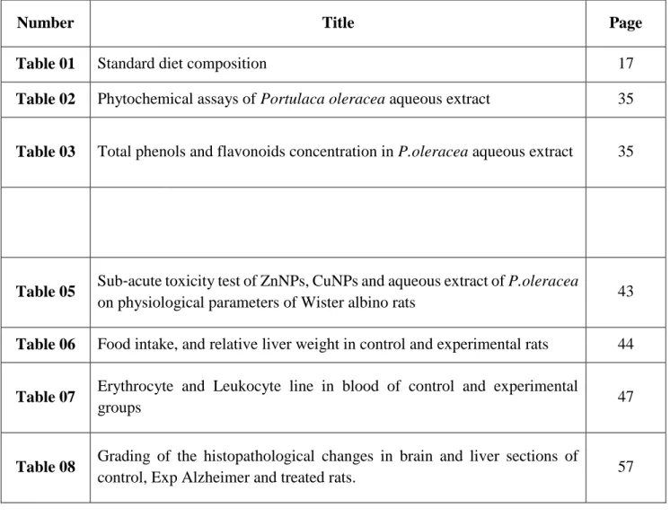
Number Title Page
Table 01 Standard diet composition 17
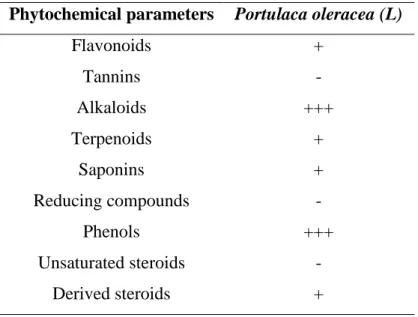
Table 02 Phytochemical assays of Portulaca oleracea aqueous extract 35
Table 03 Total phenols and flavonoids concentration in P.oleracea aqueous extract 35
Table 04 Different of positive and negative alkaloids existed in purslane aqueous
extract 36
Table 05 Sub-acute toxicity test of ZnNPs, CuNPs and aqueous extract of P.oleracea
on physiological parameters of Wister albino rats 43
Table 06 Food intake, and relative liver weightin control and experimental rats 44
Table 07 Erythrocyte and Leukocyte line in blood of control and experimental
groups 47
Table 08 Grading of the histopathological changes in brain and liver sections of
SUMMARY
Dedications Acknowledgement Abstract Abbreviations list Figures list Tables list IntroductionFirst part: Bibliographic synthesis
I. Alzheimer disease ……….. 5
I.1. Definition ……… 5
I.2. Pathophysiology ……….. 6
I.2.1. Genetic Mechanism ……….. 6
I.2.2. Amyloid beta (Aβ) hypothesis ………. 6
I.2.3. Protein Tau hypothesis ………. 7
I.2.4. Cholinergic hypothesis ………. 7
I.2.5. Oxidative stress ……… 7
I.2.6. Inflammatory mechanism ……… 8
I.3. Therapeutic approach for Alzheimer disease ……….. 8
I.3.1. Aβ- targeting therapies ………. 8
I.3.2. Tau-targeting therapies ………. 8
I.3.3. Acetylcholinesterase inhibitors ……… 9
I.3.4. Complementary treatments applied to AD ………. 9
II. Portulaca oleracea L. ………... 10
II. 1. Definition and localization ……… 10
II. 2. Botanical description and scientific classification ……….. 10
II. 2.1. Botanical description ……….. 10
II. 2.2. Taxonomy ……….. 10
II.3. Chemical composition ………... 11
II.4. Pharmacological propriety ………. 11
III. Nanoparticles ………... 13
III. 1. Definition ………. 13
III.2. Nanomedcine ……… 13
III.3. Metal nanoparticles ………... 13
III.4.1. Zinc oxide nanoparticles (ZnNPs) ………... 13
III.4.2. Copper oxide nanoparticles (CuNPs) ………... 14
III.5. Green synthesize of nanometals ………... 14
Second part: Experimental part Materials and methods I. Materials ………. 17
I.1. Plant material ………... 17
I.2. Animals ………... 17
I.2.1. Experimental Alzheimer induction ………... 18
I.2.2. Experimental design ………. 18
I.2.3. Passive avoidance learning (PAL) apparatus ………... 18
I.2.4. Sacrifice, Blood sampling and tissue collection ………... 19
I.3. Reagent and products ……….. 19
II. Methods ………. 21
II.1. Aqueous extract preparation ……….. 21
II.2. Green synthesis of nanoparticles ………... 21
II.2.1. Green synthesis of zinc oxide ZnO nanoparticles ………... 21
II.2.2. Green synthesis of copper oxide CuO nanoparticles ……….. 22
II.3. Phytochemical analysis ……….. 23
II.3.1. Phenols ……… 23
II.3.2. Flavonoids ………... 23
II.3.3. Alkaloids ………. 23
II.3.4. Tannins ……… 23
II.3.5. Terpenoids ………... 23
II.3.6. Reducing compound ……… 23
II.3.7. Saponins ……….. 23
II.3.8. Steroids ……… 24
II.4. Total phenols and flavonoids compounds ……….. 24
II.4.1. Total phenols ………... 24
II.4.2. Total flavonoids ……….. 24
II.5. HPLC-QTOF-MS/MS analysis ……….. 24
II.6. Characterization of ZnO and CuO nanoparticles ………... 25
II.7. Antioxidant activity (Ferric Reducing Antioxidant Power Assay "FRAP assay") ….... 25
II.8. Anti-inflammatory activity ……… 26
II.10 Sub-acute toxicity test ………... 27
II.11. Behavioural analysis ……… 27
II.11.1. Passive avoidance training ……… 27
II.11.2. Retention test ………. 28
II.12. Hematological parameters ………... 28
II.13. Biochemical parameters ………... 28
II.14. Homogenates preparation ……… 28
II.15. Determination of tissue proteins ……….. 29
II.16. Determination of Acetylcholinesterase (AChE) activity ………. 29
II.17. Oxidative stress parameters ………. 30
II.17.1. Determination of malondialdehyde (MDA) level ………. 30
II.17.2. Determination of reduced glutathione (GSH) level ……….. 30
II.17.3. Determination of Super Oxide Dismutase (SOD) activity ……… 31
II.17.4. Determination of Glutathione-S-transferase (GST) activity ………... 32
II.18. Histopathological study of the brain and liver tissues ………. 32
II.19. Statistical analysis ……… 33
Results I. In vitro assays ………. 35
I.1. Phytochemical analysis……… 35
I.1.1. Qualitative and quantitative phytochemical analysis of Portulaca oleracea L. …….. 35
I.1.1.1. Phytochemicals test ………... 35
I.1.1.2. Phenols and flavonoid compounds ……… 35
I.1.1.3. HPLC-QTOF-Mass Spectroscopy of P. oleracea aqueous extract ………... 36
I.2. Characterization of Zinc and Copper nanoparticles ……… 39
I.2.1. UV–Vis spectroscopy ………... 39
I.2.2. FT‑IR analysis ……….. 39
I.2.3. SEM analysis ……… 40
I.2.4. XRD analysis ……… 41
I.3. Biological Activities ……… 42
I. 3.1. Antioxidant activity “Ferric reducing antioxidant power (FRAP)” ………… 42
I.3.2. Anti-inflammatory activity ………... 42
I.3.3. Hemolysis assay ………... 43
II. In vivo assays ……… 43
II.1. Acute toxicity study ………... 43
II.3. Step through test ……… 44
II.3.1. Step-through latency (STL) ………. 44
II.3.2. Time spent in the dark compartment (TDC) ………... 45
II.4. Determination of protein level ………... 45
II.5. Determination of Acetyl cholinesterase activity ……… 46
II.6. Hematological parameters ………. 47
II.7. Lipid profile ………... 47
II.8. Biochemical parameters ………. 49
II.9. Oxidative stress parameters ………... 50
II.9.1. Malondialdehyde (MDA) level ………... 50
II.9.2. Reduced glutathione (GSH) level ………... 50
II.9.3. Glutathione-S-Transferase (GST) activity ……….. 51
II.9.4. Superoxide dismutase (SOD) activity ………. 52
II.10. Histological analysis ……… 52
Discussion I. In vitro study ……….. 59
I.1. Qualitative and quantitative phytochemical analysis of Portulaca oleracea L. ………. 59
I.2. Characterization of nanoparticles ……… 60
I.3. Biological activities ………. 60
II. In vivo discussion ………. 61
II.1. Acute toxicity ………. 62
II.2.Growth parameter ………... 62
II.3. Step through and acetyl cholinesterase activity ………. 63
II.4. Protein ……… 64
II.5. Hematological parameters ………. 66
II.6. Lipid profile ………... 67
II.7. Biochemical parameters ………. 67
II.8. Oxidative stress ……….. 68
II.9. Histological analysis ……….. 70
Conclusion ……… 74
Bibliographical references ……… 77
Introduction Neurodegeneration occurs in various diseases affecting the central nervous system (CNS), the loss of specific populations of neurons related to the functional neuronal networks determines the clinical presentation of acute and chronic neurodegenerative diseases including the Alzheimer disease (AD) (Sairazi & Sirajudeen, 2020). Alzheimer disease is an age-related neurodegenerative disorder, which progressive by nature, where the symptoms of dementia continue to increase and quality of life deteriorates gradually over a number of years (Chiroma et al., 2018). The amyloid hypothesis proposes β-amyloid (Aβ) among the major pathological hallmark of AD of the disease and suggests that misfolding of the extracellular Aβ protein accumulated in senile plaques and the intracellular deposition of misfolded tau protein in neurofibrillary tangles cause memory loss, confusion and result in personality and cognitive decline over time (Chen et al., 2017). The disordered functioning of neurotransmitters in brain, including acetylcholine (Ach), also belongs to AD pathological (Zhang et al., 2016).
Many of recent landmarks in scientific research have shown that in human beings, oxidative stress is an important factor causing physiological alterations and various diseases in the body (Derouiche et al., 2019). The oxidative stress may be a crucial link in AD initiation and development. The imbalance between the generation and elimination of reactive oxygen species (ROS) can cause extensive and lasting damage in the central nervous system (CNS), which finally accelerates the occurrence and development of AD (Wei et al., 2017).
Alzheimer disease is a devastating neurodegenerative disorder with no cure and limited treatment solutions that are unable to target any of the suspected causes (Robinson et al., 2015). Current treatment strategies are aimed to reducing the symptoms and to slow down the progression of the disease (Cavalli et al., 2020).
The use of medicinal plants for the treatment of AD as an alternative therapy, which is a vital focus of researchers. The medicinal plants have been reported to enhance the memory and learning process that normally decline with AD. The phytochemicals have been clinically proven the significant potentials anti-AD by different mechanism of action (Ovais et al., 2018). Purslane (Portulaca oleracea) listed in the World Health Organization as one of the most used medicinal plants (Lim & Quah, 2007). It possesses a wide spectrum of pharmacological properties such as neuroprotective, antioxidant, anti-inflammatory, and anticancer activities (Zhou et al., 2015). These effects have been demonstrated to result from various active components.
The emergence of nano-medicine has enriched the knowledge and strategies of treating diseases, and especially some incurable diseases, such as neurodegenerative diseases. The application of nanoparticles in medicine is in the core of nano-medicine (Leng et al., 2018). The
Introduction nano-form offer to pass the blood brain barrier (BBB) more than the bulk one and that there is an inverse relationship between particle size and the ability to penetrate the BBB (Simko & Mattsson, 2014). Metallic nanoparticles (MNPs) have considerable interest owing to unique properties of nanoparticles (NPs) such as small size and the greater surface area to volume ratio (Yao et al., 2019). Zinc and copper known as potential metal involved in metabolic organism regulation, where the zinc oxide nanoparticle (ZnNPs) is considered as one of the most promising and novel magic materials because of its unique catalytic and antimicrobial properties as well as its low cost and extensive applications in diverse areas (Erjun et al., 2006). Also the copper nanoparticles (CuNPs), from their unique physical and chemical properties and the low cost of preparation, have been of great interest recently (Honary et al., 2012) copper nanoparticles have potential applications in diverse fields including biomedicine (Rubilar et al., 2013).
Due to the complexity of Alzheimer disease and inability researchers to obtain an appropriate and effective treatment, this allows us to try new treatment strategy including bio-nanotechnology.
In light of these data, the present study was carried out to investigate the two following complementary aspects:
The first part: based on the in-vitro study; extraction of plant extract, preparation of zinc and copper nanoparticles, quantitative and qualitative characterization of these compounds and evaluation of their biological property.
The second part: based on the in-vivo study for evaluation of the therapeutic efficiency of
P.oleracea leave aqueous extract, ZnNPs, CuNPs and a novel approach (U30ACS) against
metabolic, physiological and histological alteration induced by experimental Alzheimer disease in rats.
First part
Bibliographic synthesis
Bibliographic synthesis Alzheimer disease
5
I. Alzheimer disease I.1. Definition
Alzheimer disease (AD) is the most common form of neurodegenerative dementia in the elderly. It refers to the neurodegenerative brain disorder regardless of clinical status (El Kadmiri et al., 2018). AD characterized by loss of neurons and synapses in the cerebral cortex and certain subcortical regions. This loss results in gross atrophy of the affected regions, including degeneration in the temporal lobe and parietal lobe, and parts of the frontal cortex and cingulate
gyrus (Rani et al., 2016).Wherethe temporal lobe including hippocampus and adjacent amygdala
is the starting point of the neurodegenerative process in this neurodegenerative disease (Coupé et al., 2019).In Alzheimer disease, the hippocampus is one of the first regions of the brain to suffer damage and therefore memory loss and disorientation are included among the early symptoms
(Rani et al., 2016).The second structure of temporal lobe region is the amygdala; the atrophy of
this structure with a rate of change less important than or similar to the hippocampal one (Coupé et al., 2019). Thus, this make the AD as CNS disorder by progressive deterioration of neurons resulting in loss of cognitive behavior, memory impairment, and disturbance in a daily routine activity (Agrawal et al., 2018).
Bibliographic synthesis Alzheimer disease
6
I.2. Pathophysiology
Over the last twenty years, neuroimaging has increasingly contributed to major advances in Alzheimer’s disease (AD), especially for the clinical diagnosis and to improve the understanding of the pathophysiological mechanisms of the disease (Perry et al., 2018).
At the macroscopic level, there is the atrophy of the cerebral cortex, hippocampus and amygdala, which in AD appears more sharply due to age. Microscopically it is possible to observe the formation of amyloid plaques, or senile plaques, accumulation of hyperphosphorylated Tau protein, which implies the formation of neurofibrillary tangles, and extensive neuronal loss (Singh et al., 2016).
Recent research showed other mechanisms are related to the formation of these AD markers, ranging from genetic imprint factors as family heritage, and mechanisms that involve apolipoprotein E, the mechanism of oxidation processes culminating in the neurodegeneration process (Dos Santos Picanço et al., 2018).
I.2.1. Genetic Mechanism
Molecular genetic investigation of multiple generations affected by a rare early onset form of Alzheimer disease (EOAD, <65 years) resulted in the identification of the amyloid precursor protein (APP), presenilin 1 (PSEN1) and presenilin 2 (PSEN2) genes as disease genes in AD. Pathogenic mutations in these genes converge to a general mechanism of increased amyloid beta (Aβ) accumulation. While a pathogenic mutation in APP, PSEN1 or PSEN2 is infrequently identified in late-onset Alzheimer disease (LOAD) patients, LOAD is typically considered multifactorial, with a strong polygenic component and an estimated heritability of up to 80%. The most well-known genetic risk factor for AD is the allele ε4 of apolipoprotein E (ApoE) (Verheijen & Sleegers, 2018). The mechanisms that correlate the presence of ApoE with AD are not well understood yet, however, it is being suggested that in such cases there is a decrease in clearance of Aβ in the brain (Dos Santos Picançoa et al., 2018).
I.2.2. Amyloid beta (Aβ) hypothesis
There are several theories, which have been proposed to explain the progression of Alzheimer disease with the amyloid cascade hypothesis remaining the most widely accepted. This hypothesis states that the primary neurologic insult is caused by toxic and soluble Aβ
oligomers (Robinson et al., 2015). The transmembrane amyloid precursor protein (APP) is an
integral membrane protein expressed in many tissues, especially in the synapses of neurons, which cleaved by β and γ secretases to produce Aβ monomers (Chen et al., 2017), which misfold to form toxic β-sheet oligomers and eventually form larger fibrils and plaques that known as senile
Bibliographic synthesis Alzheimer disease
7
plaque. After the enzymatic processing of APP, Aβ monomers are free to misfold back onto themselves through side chain interactions producing a hairpin structure with residues in the central region and N-terminus forming intermolecular hydrogen bonds with other monomers. If amyloid burden can be reduced, it is speculated that it may be possible to slow the progression of Alzheimer’s disease (Robinson et al., 2015).
I.2.3. Protein Tau hypothesis
Microtubules are essential in morphogenesis, cell division, and intracellular trafficking of organelles. Microtubule formation is stabilised by a protein family called Microtubules Associated Proteins (MAP). A major member of the MAP family is tau protein (Al-Hilaly et al., 2017). Hyperphosphorylation of Tau occurs during the course of AD, and is known to underlie the missorting of tau from a primarily axonal to a somatodendritic location in neurons. This alteration is thought to contribute not only to the deregulation of microtubule dynamics, but also to tau
polymerization, aggregation and the formation of neurofibrillary tangles (NFTs) are a fundamental
neuropathological hallmark of AD (Metaxas & Kempf, 2016).
I.2.4. Cholinergic hypothesis
Acetylcholine (ACh) is the neurotransmitter used by all cholinergic neurons, which exerts its physiological functions by activating either ionotropic nicotinic ACh receptors (nAChRs) or metabotropic muscarinic ACh receptors (mAChRs) (Jiang et al., 2014). Thus, it has a very important role in the peripheral and central nervous systems that are widely distributed. The cholinergic system is pointed as an important factor in many forms of dementia including AD, by its involving in critical physiological processes, such as attention, learning, memory, and sensory information (Ferreira-Vieira et al., 2016). The third important hallmark of A is cholinergic hypofunction that is closely linked to amyloid β and tau pathologies. In AD brains there are reduced choline acetyltransferase levels accompanied by decreased ACh synthesis, significant loss of cholinergic neurons, reduction in the numbers of postsynaptic neurons accessible to Ach, cholinergic neuronal and axonal abnormalities and reduction in nAChR levels (Jiang et al., 2014).
I.2.5. Oxidative stress
Oxidative stress, is a process increased in the brain with aging, is induced by an imbalance in the redox state, involving the generation of excess reactive oxygen species (ROS) or the dysfunction of the antioxidant system. Consequently, oxidative stress on nervous tissue may seriously damage the brain via several interacting mechanisms. The dysfunction mitochondrial mechanisms constitutes a risk for developing Alzheimer's disease (AD), when no efficient antioxidant system is available leads to neuron degeneration in AD are believed to be associated
Bibliographic synthesis Alzheimer disease
8 with ROS generation, activation of mitochondrial permeability transition, excitotoxicity, impaired production of adenosine triphosphate and altered calcium homeostasis (Huang et al., 2016).
I.2.6. Inflammatory mechanism
Inflammation has been strongly implicated in the pathogenesis of Alzheimer disease, and associated with increased populations of activated microglia and astrocytes. Accordingly, multiple inflammatory stimuli can trigger the activation of microglia and astrocytes; these stimuli can be peripheral (e.g., systemic infections and peripheral chronic inflammation) or local (e.g., brain injury and the presence of various forms of Aβ and tau proteins). Activated microglia and astrocytes release neurotoxic proinflammatory cytokines such as interleukin-1β, tumor necrosis factor-α (TNF-α), and interleukin-6 (Ng et al., 2015). Production of these molecules can perpetuate a cycle of neuroinflammatory processes including amyloidosis, neuronal death, cortical thinning, reduced brain volume, infarcts and neurodegeneration (McGrattan et al., 2019).
I.3. Therapeutic approach for Alzheimer disease
There is no cure for AD; however, drug treatments are available to help relieve symptoms in several aspects of the disease, and researchers around the world are focusing on finding better treatments, preventive strategies, and ultimately cure. A variety of cellular mechanisms can lead to the generation of Alzheimer’s disease (Singh et al., 2016).
I.3.1. Aβ- targeting therapies
Amyloid precursor protein (APP) is a transmembrane protein that has both cytoplasmic
and extracellular cleavage products that are relevant in Alzheimer disease, APP is initially cleaved via α-secretase or β-secretase, which leads to normal or pathological products, respectively (Panek et al., 2017). Therefore, therapeutic targeting of this protein processing is an attractive target. Thus, both α-secretase agonists and β-secretase antagonists could act as Alzheimer therapeutics. Following α or β cleavage, γ-secretase cleavage yields extracellular p3 or Aβ peptides respectively, therefore inhibitors of β and γ secreatas could be one of the main targets for the development of therapeutics (Arbor et al., 2016).
I.3.2. Tau-targeting therapies
Although most drug development efforts so far have focused on Aβ, interest in tau as a drug target is currently growing. Several approaches are being employed which target various aspects of tau pathogenesis, including modulation of tau gene expression, modulation of posttranslational modifications, immunotherapy, and microtubule stabilization (Wu et al.,
Bibliographic synthesis Alzheimer disease
9 2017). In addition, the block tau spreading, tau anti-aggregates, tau kinase inhibitors, and gene therapy approaches that inhibit tau production are also in development (Grundman, 2019).
I.3.3. Acetylcholinesterase inhibitors
The competitive inhibitors of the acetyl cholinesterase (AChE) that cleaved the Ach, which is a neurotransmitter molecule that associated with memory function, is the mostly wide used drugs to treat Alzheimer’s disease (AD) patients (Ferreira-Vieira et al., 2016). The acetylcholinesterase inhibitors (AChEI) were developed as an effect of the cholinergic assumption of cognitive reduction. AChEI compounds are demonstrated for the symptomatic treatment of mild-to-moderate AD. They inhibit AChE, which is accountable for the separation of acetylcholine (ACh) (Turkan et al., 2019).
I.3.4. Complementary treatments applied to AD
The anti-inflammatory drugs act by reducing the inflammatory response in the brain tissue. It inhibits the activation of astrocytes in brain tissue and several other signaling cascades related to inflammation and oxidative stress. Thus, these activities favor the inhibition of the inflammatory processes associated with the physiology of AD and reduce the deleterious consequences of this disease (Dos Santos Picanço et al., 2018).
The aggregation of Aβ amyloid plaques and, consequently, the formation of reactive oxygen species (ROS), that are typical, for instance, of the first stages of AD (Piemontese, 2017). The ROS-mediated pathway is related to AD. It has been suggested that ROS levels are increased and oxidative stress occurs. For this reason, targeting ROS or ROS-related enzymes has been a therapeutic strategy in AD (Chun& Lee, 2018).
Bibliographic synthesis Portulaca oleracea L.
10
II. Portulaca oleracea L.
II. 1. Definition and localisation
Purslane (Portulaca oleracea L.) is an annual herbaceous plant belongs to the family Portulacaceae, it is cosmopolitan in distribution and occurring particularly in tropical and subtropical areas (Iranshahy et al., 2017). Purslane used as a potherb in Asian, central European and Mediterranean countries, which is utilized as one of medicinal plants and has been given term “Global Panacea”, it also is popular as a traditional medicine in Algeria for the treatment of various diseases (Derouiche et al., 2017). P. oleracea is known to contain many active substances are also considered as sources of many dietary supplement (Derouiche et al., 2019). It has several pharmacological effects including; neuroprotective, hepatoprotective, antioxidant,
anti-inflammatory and immunomodulatory effects (Khazdair et al., 2019).
II. 2. Botanical description and scientific classification
II. 2.1. Botanical description
Portulaca oleracea L. is considered invasive weed, which may reach 40 cm in
hight (Chowdhary et al., 2013). Leaves are alternate, fleshy, obovate or spathulate with a cuneate base and obtuse apex, smooth and waxy on upper surface, margins are sometimes purple; sessile or indistinctly petiolate, 1–3 cm long, 0.5–1.5 cm wide. Flowers are solitary or clustered axillary or terminal, surrounded by 2 glabrous bracts; 2 unequal sepals, 5 glabrous yellow petals, stamens 6–15. Fruit are brown rounded capsule, 6–10 mm long, opening at top with lid. Seeds are numerous, small, 0.8 mm broad, reniform, and black in color (Alam et al., 2014).
II. 2.2. Taxonomy (Chugh et al., 2019)
Kingdom: Plantae Subkingdom: Tracheobionta Superdivision: Spermatophyta Division: Magnoliophyta Class: Magnoliopsida Subclass: Caryophyllidae Order: Caryophyllales Family: Portulacaceae Genus: Portulaca L.
Species: Portulaca oleracea L. Figure 02: Portulaca oleracea L.
Bibliographic synthesis Portulaca oleracea L.
11
II.3. Chemical composition
Purslane presents a variable chemical constituents mainly belong to flavonoids, alkaloids, terpenoids and organic acid and other classes of natural compounds including fatty acids, vitamins, and minerals (Sadhana & Deepali, 2017).
Flavonoids are one of the main active ingredients of purslane such as quercetin, kaempferol, myricetin, luteolin, and apigenin, (Andarwulan et al., 2015). Oleraceins A, Oleraceins B, Oleraceins C, Oleraceins D and Oleraceins E are exist in P. oleracea. Other alkaloids like dopa, dopamine and noradrenaline are also reported which are higher in leaves then in stem and seeds (Syed et al., 2016). Portulaca oleracea contains monoterpenes such as portulosides A and B, diterpenes as portulene, and 𝛽amyrin type triterpenoids (Zhou et al., 2015). It has high levels of tocopherols, vitamin C and some vitamins of complex-B (Petropoulos et al., 2016). Purslane has been demonstrated to be one of the major plant sources of omega-3 fatty acids, particularly α-linolenic acid (up to 30%) and other essential fatty acids such as palmitoleic, palmitic, linoleic, oleic, stearic eicosapentaenoic and docosahexaenoic acids (Iranshahy et al., 2017). Dietary minerals such as iron (Fe), zinc (Zn), potassium (K), boron (B), nitrogen (N), manganese (Mn), calcium (Ca), copper (Cu), magnesium (Mg) are the most abundant in purslane plant, whereas other minerals (i.e. phosphorus (P), sulfur (S), sodium (Na)) that exist in relative lower amounts, they also contribute to purslane valuable nutritional profile (Petropoulos et al., 2016).
II.4. Pharmacological propriety
Portulaca oleracea is considerable importance to the food industry and also possesses a
wide spectrum of pharmacological properties, which are associated with its diverse chemical constituents, including flavonoids, alkaloids, polysaccharides, fatty acids, terpenoids, sterols, proteins, vitamins and minerals (Zhou et al., 2015).
Purslane has a distinguished antioxidant capacity, which is attributed to high vitamin A content along with ascorbic acid and various flavonoids and polyphenolics. These provide direct free radical scavenging activity and enhance the activity of many enzymes such as glutathione reductase, glutathione peroxidase, superoxide dismutase, and catalase (Amin et al., 2020). The possible neuroprotection mechanism could be related to its antioxidant activity (Martins et al., 2016). When the alkaloid plant extract significantly inhibited acetyl cholinesterase (AChE) activity, where the use of AChE inhibitors has been a promising treatment strategy for Alzheimer’s disease (AD); therefore, purslane may be an effective agent for the prophylaxis and treatment of AD (Zhou et al., 2015).
Bibliographic synthesis Portulaca oleracea L.
12
P. oleracea has an anti-inflammatory activity, which inhibit the production of
inflammatory mediators such as NO and pro-inflammatory cytokines including the interleukins IL-1β and IL-6. The purslane extracts in a dose- dependent manner significantly inhibited tumor necrosis factor (TNF-α) that induce intracellular reactive oxygen species (ROS) production (Khazdair et al., 2019).The immunomodulating activity of purslane mainly is related to the polysaccharides. They activated phagocytes that are involved in the first mechanism of innate defense and the process of inflammation and, as a result, stimulate the activity of the immune system (Rahimi et al., 2019)
As the oxidative stress and free radical formation have, a key role in propagation of remote organ in jury the hepatoprotective activity of purslane was linked to their oxidant and anti-inflammatory activity (Askaripour et al., 2016). In addition, plant extract regulates the hepatic marker enzymes (Syed et al., 2016).
Bibliographic synthesis Nanoparticles
13
III. Nanoparticles III. 1. Definition
Nanotechnology is one of the most promising technologies of the 21st century. It is the ability to observe measure, manipulate, assemble, control, and manufacture matter at the nanometer scale generally from 1 to 100 nm (Bayda et al., 2019). Although this is the most used definition, a size limitation of nanotechnology to a below 100-nm range seems to exclude numerous materials and devices, particularly in the pharmaceutical area. For that reason, some experts disagree with a rigid definition based on a sub-100 nm size (Silvestre et al., 2015).
III.2. Nanomedcine
Medical applications of nanotechnology (commonly known as nanomedicine) are expected to significantly improve disease diagnostic and therapeutic modalities and subsequently reduce health care costs.The use of nanotechnology in medicine has the potential to significantly improve human health and wellbeing due to highly accurate and sensitive diagnostic tests (Satalkar et al., 2015).
III.3. Metal nanoparticles
The metal nanoparticles exhibit many properties that are very useful or can be manipulated for numerous new scientific studies and multitudes of technological applications. Recently, there are advances in the use of metal nanoparticles in the study of biomolecular interactions, developing bioassays and biomedical devices, and various other biomedical applications such as
immunodiagnostics, drug delivery, therapeutics, and gene transfer (Mei & Wu, 2017).
Nanoparticles have great interest due to their extremely small size and large surface to volume ratio, which lead to differ in their property such as the biological one, compared to bulk of the same chemical composition (Iravani, 2011).
III.4. Zinc and copper nanoparticles
Transition metal oxides nanoparticles (NPs) have received the most attention in a variety of industrial applications such as cosmetics and medicine. Zinc and copper oxides (ZnO and CuO) NPs are the most common and attractive metal oxide NPs in industries (Rafiei et al., 2014).
III.4.1. Zinc oxide nanoparticles (ZnNPs)
The ZnNPs, characterized by its small size facilitates the absorption of zinc by the body, which make it attractive in the biomedical application (Jiang et al., 2018). Where zinc (Zn) is a
Bibliographic synthesis Nanoparticles
14 well-known as essential trace element that is highly distributed and plentiful, it plays an important role in many cellular functions including neurotransmission, gene regulation, structural maintenance, enzymatic activity and protect the blood-brain barrier (BBB) from oxidative damage and prevent the progress of neurodegenerative diseases. Thus, ZnO NPs have drawn the attention of many biotechnologists as it has useful properties with potentially unlimited therapeutic applications (Hafez & Gad, 2018).
III.4.2. Copper oxide nanoparticles (CuNPs)
According to the distinctive properties of copper, the copper oxide (CuO) based nanomaterials are gaining importance in biomedical sciences that includes both, diagnosis and
therapy (Verma & Kumar, 2019). Thus, the research is increasingly carried out on replacement of standard Cu forms with nanoparticles (CuNPs), due to their very small size and specific surface area; it is assumed that, they may be differently utilized by the body than their macro counterparts. This suggests that they may be better absorbed in the body (Cholewińska et al., 2018). Moreover, copper is an essential trace element that is required as a cofactor for normal functioning of various metabolic enzymes. This property can be used to synthesize antitumor formulations that induce killing of tumor/diseased cells by altering the intracellular level of copper ions. Due to their nano size, they are easily accessible to the micrometer-sized human cellular entity and can readily interact with the biomolecules present on the cell surface and intracellularly (Verma & Kumar, 2019).
III.5. Green synthesize of nanometals
The green synthesis of nanoparticles is a novel way to synthesize nanoparticles by using biological sources. It is gaining attention and attracted the scientists from different fields due to its simplicity, and environmentally-friendly nature (Singha et al., 2014),especially in light of efforts to find greener methods of inorganic material synthesis (Philip, 2009), due to the growing need to develop environmentally benign technologies in material synthesis (Bar et al., 2009). The biosynthesis for obtaining nanoparticles using naturally occurring reagents such as vitamins, sugars, plant extracts, biodegradable polymers, and microorganisms as reductants and capping agents could be considered attractive for nanotechnology (Kharissova et al., 2013). Biosynthesis of nanoparticles is more unique and reliable not only because of its less toxicity as compared to some of physicochemical production methods but also because it can be used to produce large quantities of nanoparticles that have well shape and size. The biological synthesis approaches may actually produce nanoparticles of a better defined size and morphology as compared to some of other physicochemical methods of production (Khandel et al., 2018).
Second part
Experimental part
Materials and
Experimental part Materials and Methods
17
I. Materials
I.1. Plant material
The purslane (Portulaca oleracea L.) were collected in August from a village in Touggourt of Ouargla state, Algeria. The leaves were washed with distilled water, then dried at room temperature. The completely dried purslane leaves was powdered by using a mechanical grinder. The powder stored at room temperature in the airtight containers until the use.
Figure 03: Portulaca oleracea leaves (original photo) I.2. Animals
In this study, thirty males albino Wistar rats at the age of 8 weeks old, weighting 131.28 ± 5.58 g, obtained from the Institute Pasteur of Algiers, and were housed in plastic cages at animal room of molecular and cellular biology department, of nature and life sciences faculty, in Echahid Hamma Lakhdar-El-Oued university, Algeria. With a temperature of 25 ± 2°C, the rat were given a free access to their standard diet and tap water during the study. The animals were adapted to an inverse 12:12 h light/dark cycle. All experimental procedures employed, as well as rat care and handling, were in accordance with guidelines provided by the local ethics.
Tableau 01: Standard diet composition (Southon et al., 1984)
Raw materials Quantity (g/kg) Percentage (%)
Maize 326 32.6 Cellulose 326 32.6 Protein 168 16.8 Sacrose 60 6 Vitamin 40 4 Minerals 40 4 Oil 40 4
Experimental part Materials and Methods
18
I.2.1. Experimental Alzheimer induction
Alzheimer state was induced by oral administration 200 mg/kg b.w of both aluminum trichloride (AlCl3) and D-galactose in drinking water during 76 days (Zhang et al., 2016 ;
Debbache-Benaida et al., 2018). Therefore, treatment of AlCl3 + D-gal to normal rats is usually
considered as a model of Alzheimer. The co-administration of AlCl3 and D-gal for one week could
induce an ideal AD animal, which may produce neurodegenerative and neurobehavioral alterations similar to AD (Liaquat et al., 2017).
I.2.2. Experimental design
After two weeks of acclimatization, the animals were divided into six groups, 5 in each of, Alzheimer disease was induced by oral administration of 200 mg/kg b.w in drinking water for 76 days, the rats treated during 21 days as following:
Group 01 (Control): Healthy rats received distilled water.
Group 02 (Exp Alzheimer): Experimental Alzheimer rats received distilled water.
Group 03 (U30ACS): Experimental Alzheimer rats treated orally by colloidal solution which
contain the aqueous extract of P. oleracea (2g/Kg b.w/wk), ZnNPs (7mg/Kg b.w/wk) and CuNPs (3mg/kg b.w/wk).
Group 04 (AEPo): Experimental Alzheimer rats treated orally by aqueous extract of P.oleracea
(2g/Kg b.w/wk).
Group 05 (ZnNPs): Experimental Alzheimer rats treated orally by zinc oxide nanoparticles
(7mg/Kg b.w/wk).
Group 06 (CuNPs): Experimental Alzheimer rats treated orally by copper oxide nanoparticles
(3mg/Kg b.w/wk).
The colloidal solution obtained by ultrasonic technique for 30 min. The doses of P.oleracea according to (Derouiche et al., 2019). But for the ZnNPs and the CuNPs were chosen according to our experimental study.
Experimental part Materials and Methods
19
I.2.4. Sacrifice, Blood sampling and tissue collection
At the end of each treatment, and after 12 hours of fasting, animals were sacrified under slight anesthesia by Chloroform (94%) by inhalation; blood sampling was performed during the decapitation and blood samples were transferred into EDTA tubes to carried the hematological parameters and dry tubes previously labeled and numbered for each rat. The blood was separated by centrifugation for 3000 revolutions/min during 15 min, the obtained serum stored at -20°C until the use for biochemical analysis; fasting blood glucose level obtained by glucometer for each rat. Liver and brain were carefully removed, rinsed in NaCl 0.9% then weighed. Also, The organs stored in the freezer at -20°C until the preparation of homogenates for the determination of neurotransmitter and oxidative stress parameters.
I.3. Reagent and products
Hydrogen chloride (HCl), Methanol, Chloroform, Mayer reagent, Wagner reagent, Sulfuric acid, Fehling solution, Folin-Ciocalteu reagent (FCR), Magnesium (Mg), Sodium carbonate (Na2CO3), Aluminum trichloride (AlCl3), galactose (C6H12O6), Ferric chloride (FeCl3),
Potassium ferricyanide (K3Fe), Zinc sulfate (ZnSO4, 7H2O), Copper sulfate (CuSO4, 5H2O),
Bovine serum albumin (BSA), Gallic acid, Tris, Sodium chloride (NaCl) , Ammoniac, Nitric acid (HNO3), Salicylic acid, Coomassie brilliant blue, Trichloroacetic acid (TCA), Thiobarbituric acid
(TBA), Butylated hydroxytoluene (BHT), Phosphate buffer (Dipotassium phosphate K2HPO4 &
Potassium dihydrogen phosphate KH2PO4), 5,5'-dithiobis(2-nitrobenzoic acid) (DTNB), Reduced
glutathione (GSH), l-chloro-2,4-dinitrobenzene (CDNB), Sodium hydroxide (NAOH), Ethylenediaminetetra-acetic Acid (EDTA), Methionine, Nitro-blue tetrazolium chloride (NBT), Riboflavin, Acetylcholine (Ach) and Ethanol.
Experimental part Materials and Methods 20 F igu re 04 : In vivo Ex pe rim ental de sig n of stud y
Experimental part Materials and Methods
21
II. Methods
II.1. Aqueous extract preparation
The aqueous extract was prepared by adding 50 ml of distilled water to 5 g dry leaves powder of Portulaca oleracea L. at 50°C during 2 hours. After 24 h of maceration at room temperature, the mixture was filtered by Whatman paper then evaporated by using rotary evaporator (Derouiche et al., 2019), (figure 05).
Figure 05: P.oleracea aqueous extract preparation II.2. Green synthesis of nanoparticles
II.2.1. Green synthesis of zinc oxide ZnO nanoparticles
In order to prepare ZnNPs, as shown in figure 06, 200 ml of aqueous zinc sulfate was mixed with 20 ml of purslane leaves aqueous extract. The ions, which initiated the reaction, were afforded by the zinc sulfate in de-ionized water. The reaction mixture was incubated with constant stirring in the dark at 60 °C to avoid photo-catalysis. An observed off-white color marked the formation of ZnO NPs at the end of 24 h. The resultant product was further purified by centrifugation and washed in double-distilled water and ethanol, respectively, dried and kept in an amber-coloured sample bottle until use (Ezealisiji et al., 2019).
Experimental part Materials and Methods
22
II.2.2. Green synthesis of copper oxide CuO nanoparticles
In order to prepare CuNPs, 1g of the copper sulfate was added into the defined amount of the prepared walnut aqueous extract (10mL) and the reaction solutions were mixed using a heater-stirred, adjusted at 500 rpm and 70°C, for 15 min. Finally, the samples were put in an electric furnace memmert adjusted at 200°C for 2 h. The obtained powders washed in double-distilled water and ethanol respectively, as farmed CuO NPs, were then used for further studies (Asemani & Anarjan, 2019), (figure 06).
Experimental part Materials and Methods
23
II.3. Phytochemical analysis
The phytochemical analysis were carried out on the aqueous extracts prepared from the plant by qualitative characterization method according to (Evans, 2009 ; Harborne, 1998 ; Wadood et al., 2013 ; Harborne, 1973)
II.3.1. Phenols
Introduce 5 ml of extract in a test tube, drops a few of natural 5% ferric chloride solution. A dark green color indicates the presence of phenolic compounds
II.3.2. Flavonoids
In a test tube, introduce 5ml of extract, 5ml of diluted ammoniac and 1ml of H2SO4. The
appearance of a yellow color indicates the presence of flavonoids.
II.3.3. Alkaloids
1 ml of aqueous extract were treated with a few drops of hydrochloric acid then 1–3 drops of Wagner reagent were added. The appearance of brown precipitate reveals the presence of alkaloids in the sample.
II.3.4. Tannins
In a test tube, introduce 5 ml of extract and add 1 ml of a 2% aqueous solution of ferric chloride (FeCl3). The presence of tannins was indicated by a greenish or bluish-blackish
coloration.
II.3.5. Terpenoids
The formation of a reddish brown color indicates the presence of terpenoids, through the addition of chloroform (2ml) and concentrated sulfuric acid (3 ml) to 5 ml of plant extract.
II.3.6. Reducing compound
Add Fehling's liquor (1ml of reagent A and 1ml of reagent B) to the extract and incubate the whole in a boiling water bath, The appearance of a brick-red precipitate indicates the presence of reducing sugars.
II.3.7. Saponins
In a test tube, introduce 5ml of extract, mixed with 5ml of distilled water and with vigorous manual agitation. The formation of a steady foam indicates the presence of saponins.