protein complexes after gene duplication
Mémoire
Angel Fernando Cisneros Caballero
Maîtrise en biochimie - avec mémoire
Maître ès sciences (M. Sc.)
duplication
Master’s thesis
Angel Fernando Cisneros Caballero
Under the supervision of: Christian R. Landry
La duplication de gènes est l’un des plus importants mécanismes évolutifs pour la génération de diver-sité fonctionelle. Lorsqu’un gène est dupliqué, la nouvelle copie partage toutes ses fonctions avec la copie ancestrale car elles encodent pour des protéines identiques. Donc, les deux protéines, appelées paralogues, auront le même réseau d’interactions physiques protéine-protéine. Cependant, dans le cas de la duplication des gènes qui codent des protéines qui interagissent avec elles-mêmes (homomères), la nouvelle protéine interagira aussi avec la copie ancestrale, ce qui introduit une nouvelle interaction (heteromère) (Kaltenegger and Ober, 2015; Pereira-Leal et al., 2007). Puisque ces interactions peuvent avoir des différents motifs de rétention et de fonction (Ashenberg et al., 2011; Baker et al., 2013; Bon-coeur et al., 2012; Bridgham et al., 2008), il est important de mieux comprendre comment ces états sont atteints et quelles forces évolutives les favorisent. Dans ce memoire, je cible ces questions avec des simulations in silico de l’évolution des protéines suite à la duplication de gènes en travaillant avec des structures crystallographiques de haute qualité, provenant de la Protein Data Bank (Berman et al., 2000; Dey et al., 2018). Les simulations montrent que les sous-unités et interfaces partagées entraînent une forte corrélation entre les trajectoires évolutives de ces complexes. Ainsi, les simulations prédisent que la préservation de seulement les deux homomères ou seulement l’hétéromère ne devrait pas être fréquente. Toutefois, la simulation qui applique la sélection seulement sur un homomère montre que l’homomère neutre est destabilisé plus rapidement que l’hétéromère neutre. Nous avons comparé ces prédictions avec des résultats expérimentaux du réseau d’interactions protéine-protéine de la levure. Comme suggéré par les simulations, les patrons d’interactions les plus fréquents ont été la formation des trois complexes (deux homomères et un hétéromère) ou la formation de seulement un homomère. Les patrons correspondants à deux homomères sans hétéromères ou un hétéromère sans homomères sont rares. Nos résultats démontrent l’extension de l’hétéromérisation entre paralogues dans le réseau d’interactions physiques protéine-protéine de la levure, les mécanismes sous-jacents et ses implications.
Gene duplication is one of the most important evolutionary mechanisms for the generation of func-tional diversity. When a gene is duplicated, the new copy shares all of the ancestral copy’s functions because they encode identical proteins. Therefore, the two proteins, called paralogs, will have the same protein-protein interaction network. However, in the case of the duplication of genes encoding proteins that self-interact (homomers), the new protein will also interact with the ancestral copy, introducing a novel interaction (heteromer) (Kaltenegger and Ober, 2015; Pereira-Leal et al., 2007). As these interactions can have different retention and functional patterns (Ashenberg et al., 2011; Baker et al., 2013; Boncoeur et al., 2012; Bridgham et al., 2008), it is important to understand better how these states are reached and what evolutionary forces favor each of them. In this thesis, I approach these questions by means of in silico simulations of protein evolution after gene duplication by working with high-quality crystal structures from the Protein Data Bank (Berman et al., 2000; Dey et al., 2018). The simulations show that the shared subunits and interfaces lead to these complexes having highly correlated evolutionary trajectories. Thus, the simulations predict that the preservation of only the two homomers or only the heteromer is not likely to happen often. Nevertheless, simulating evolution with selection on only one homomer shows that the neutral homomer is destabilized faster than the neutral heteromer. We compared these predictions against experimental results from the yeast protein-protein interaction network. As suggested by the simulations, the most abundant interaction patterns were either the formation of all three complexes (two homomers and one heteromer) or the formation of only one homomer, with motifs corresponding to two homomers without a heteromer or a heteromer without homomers being rare. Our results highlight the extent of heteromerization between paralogs in the yeast protein-protein interaction network, the underlying mechanisms, and its implications.
Abstract . . . iii
List of Figures . . . v
List of Abbreviations and Acronyms . . . vi
Acknowledgements . . . viii Foreword. . . ix Introduction . . . 1 1 Chapter 1 . . . 8 Résumé . . . 8 Abstract . . . 9 Introduction . . . 10 Results . . . 12 Discussion . . . 22
Material and methods . . . 25
Author contributions . . . 36 Acknowledgements . . . 37 Competing interests . . . 37 2 Discussion . . . 38 Conclusion. . . 42 Bibliography . . . 43 Annex . . . 56 Supplementary text . . . 56
Comparison of PCA results with previous studies . . . 56
have pleiotropic effects on each other’s function due to their physical association . . . . 10
2 Homomers and heteromers of paralogs are frequent in the yeast protein interaction network 13
3 The loss of heteromerization between paralogs leads to greater functional divergence . . 16
4 Selection to maintain homomers also maintains heteromers. . . 18
5 Mutations have pleiotropic effects on homomers and heteromers. . . 19
• INDEL = insertion/deletion
• NMR = nuclear magnetic resonance • cryo-EM = cryo-electron microscopy • PDB = Protein Data Bank
• WGD = whole-genome duplication • SSD = small-scale duplication • GO = gene ontology
• HM = homomer • HET = heteromer
• HM&HET = homomer and heteromer
• PCA = Protein-fragment Complementation Assay • 2D = duplicated both by SSD and WGD
• DHFR = dihydrofolate reductase • NAT = nourseothricin
• HygB = hygromycin B
• EDTA = ethylenediaminetetraacetic acid • MTX = methotrexate
• DMSO = dimethylsulfoxide
• FRET = fluorescence resonance energy transfer • BiFC = bimolecular fluorescence complementation • PCR = polymerase chain reaction
• dNTP = deoxynucleotide triphosphate • YGOB = Yeast Gene Order Browser
made me the person I am today.
"If I have seen further than others, it is by standing upon the shoulders of giants"
Isaac Newton, 1675
This thesis completes a great adventure for me that started two years ago. It has been a very exciting journey that has taken me to discover a lot of new ideas, people, cultures, and places. Thus, this would be a good moment to thank everyone that has helped me through all this time.
It all starts with Carmen, Miguel, and Mauricio: my mom, my dad, and my younger brother. Their unconditional support has been a great source of motivation for me to keep moving forward. Through all these years, I have learned a lot of them, from my first words in Spanish and in English to how to live. Being away from them has been a challenge at times, but knowing they are there for me is a great source of inspiration.
I want to thank the rest of my family and my closest friends: Juan Pablo Terrazas, Rodrigo Ledesma, Gilberto Nava, Erick Cervantes, Patricia Hernández, Ricardo Morales, Fernando Ramírez, Daniel Rodríguez, Miguel Angel González, Salvador Paniagua, Rubén Pedrero, Jorge Peña, Rodrigo Delgado, Sergio Villicaña, Santiago Ceballos, David Lerner, Manuel Cortés, and Elisa Fernández. Though at different times, they have all been critical to me for their support on both a personal and a professional level.
New journeys bring new friends. I want to thank Carla Bautista, Andrés Carrillo, Natalia Sinuco, Rafael Ireta, Verônica Alves, Camilo Castañeda, Ulyses Caram, Daniela Pérez, Honey Jain, Johanna Benndorf, and Karolina Jackowska for making me feel at home far away from my country, even when on the other side of the Atlantic.
As for my scientific career, it is important for me to thank my teachers. Their hard work has also been an inspiration to me, and I am glad to say that I have learned from them. Special thanks to Jonathan Muñoz, Patricia Segura, Benito Antón, Susana Martiñón, Edgar Mixcoha, Ricardo Hernán-dez, Claudia Rangel, and Christian Landry. They all believed in me at different stages of my career and gave me platforms to work harder and reach further. In particular, working with Christian for the past two years has been an amazing experience in terms of the leadership, organization, communication, and scientific skills that he shows everyday.
Another great part of this work has been the contributions of my coworkers at the Landrylab and collaborators. Our laboratory has provided a great environment to discuss scientific ideas that gives us reasons to be proud about and push harder with our collective successes. Another great collaborator I want to thank is Joseph Marsh, who welcomed me in his laboratory for an internship. Similarly, I would like to thank my evaluators: Rong Shi and Patrick Lagüe, whose feedback helped me improve the quality of this thesis.
Last but not least, all of this is possible thanks to the funding organisms that support my research. I want to thank Mitacs; the Mexican Association for Cooperation and International Development (AMEXCID) and the Ministry of Education and Higher Education of Québec (MEES); Hydro-Québec and the Université Laval Foundation; the Québec Network for Research on Protein Function, Engi-neering, and Applications (PROTEO); and the Company of Biologists for their funding contributions.
This Master’s thesis contains the results of the main project I carried out at Christian Landry’s laboratory from September 2017 to April 2019. Such results were included in a journal article titled "The role of structural pleiotropy and regulatory evolution in the retention of heteromers of paralogs", which was initially submitted to eLife on March 26th, 2019. Revisions were received on April 26th, 2019, after which a revised version of the manuscript was submitted. Ultimately, it was accepted by
eLifeand published on August 27th, 2019. Axelle Marchant and I were co-first authors for this article,
and it also includes work done by Alexandre K. Dubé, Isabelle Gagnon-Arsenault, Diana Ascencio, Honey A. Jain, Simon Aubé, Chris Eberlein, Daniel Evans-Yamamoto, Nozomu Yachie, and Christian R. Landry. I am thankful to all the coauthors for accepting the use of this article in my Master’s thesis.
The version of the article I included here has a few differences with respect to the one that was published by eLife:
• I removed the references to the supplementary tables and the key resources table because they are too wide to be included properly in the annex of this thesis. The supplementary tables contain the dataset compiled from all the sources used, oligonucleotide sequences used, additional details on the RNA sequencing analysis, the list of PDB structures used for each analysis, and counts of substitutions accepted in each of the simulations. They are available online in the journal’s website.
• In the "Pleiotropy contributes to the maintenance of heteromers" section, I added a short com-ment on how Dey et al. (2018) determined high-quality structures in their analysis.
• In the "Identification of interfaces" section, I clarified that the definitions of contacting and nearby residues were taken from Keskin et al. (2005); Tsai et al. (1996). I also added a short comment on why nearby residues were defined using a cutoff of 6 Å.
• The supplementary text is included as part of the annex.
• Supplementary figures are labeled with an "S" and their number rather than the eLife system in which they are supplements of the main figures. They are shown in the same order as in the original article.
• The author contributions and acknowledgements are listed immediately after the materials and methods section.
As stated in the author contributions of the published article, I participated in the design of the computational part of the study, performed the in silico simulations of protein complex evolution and the analysis of protein structures, and analyzed the results. I wrote the manuscript together with Axelle Marchant and Christian R. Landry with input from all authors.
During my Master’s studies, I also performed other analyses together with Honey Ashok Jain, worked with Luciano Gama Braga from Sabine Elowe’s laboratory at the CHUL, and visited Joseph Marsh’s laboratory at the University of Edinburgh. This allowed me to explore other topics and ideas related to protein evolution. The collaboration with Luciano Gama Braga and Sabine Elowe resulted in a manuscript available as a preprint at https://www.biorxiv.org/content/10.1101/733378v1
Introduction
Protein complexes in biology
Proteins are the main workhorses of the cell. They are biomolecules formed from sequences of 20 different kinds of amino acids. They carry out a great diversity of functions, including the formation of biological structures, metabolic processes, and signaling functions (Boeckmann et al., 2005; Pandey et al., 2017; Scott and Pawson, 2009). Understanding how proteins get access to this wide diversity of functions has been one of the most important questions for modern biology because it has implications for diverse fields, such as medicine and evolutionary biology. Protein functions are conserved across evolution because they have an influence on traits. As such, knowing how they carry out specific tasks helps us understand better how new traits are acquired (Abrusán and Marsh, 2018; DePristo et al., 2005; Ferrada and Wagner, 2008; Kachroo et al., 2015; Payne and Wagner, 2019). As a result, the disruption of protein function could lead to alterations in such traits, and ultimately, diseases (Sahni et al., 2015; Schuster-Böckler and Bateman, 2008).
Research has pointed to two critical properties of proteins that drive their function: protein folding and the formation of protein complexes (Janin et al., 2008; Marsh and Teichmann, 2015; Pandey et al., 2017; Scott and Pawson, 2009; Vidal et al., 2011). Protein folding refers to the three-dimensional configuration of proteins in the cellular space. Current models of protein folding predict it to happen in an orderly fashion by means of local interactions between amino acids (Englander and Mayne, 2014; Maity et al., 2005). A protein’s fold then becomes important when studying how it interacts with its environment, most notably with other proteins. Protein complexes are physical associations of proteins, which can either be formed by identical or different chains and are found in a great diversity of stoichiometries (Ahnert et al., 2015). The formation of complexes has been pointed out to be critical for the function of proteins with respect to bringing components of metabolic pathways together and carrying out basic functions in the cell, such as the production of other proteins, because the formation of some active sites depends on complex assembly (Abrusán and Marsh, 2018; Korostelev et al., 2006). Whereas a protein’s fold determines how proteins interact with their environment, there are proteins, called chaperones, that contribute to the correct folding of other proteins by interacting with them (Mayer, 2010), so folding and interactions have an impact on each other.
Even though protein complexes are constrained by having to maintain their function, they also change throughout evolution. Consequences of the evolution of protein complexes are the gain of new subunits (Levy et al., 2008; Marsh and Teichmann, 2014), which leads to differences in the distribution of types of protein complexes in the different kingdoms of life (Lynch, 2012) and the appearance of new functions (Boncoeur et al., 2012; Bridgham et al., 2008). In the light of these observations, I am interested in modelling the evolution of protein complexes. However, in order to model it, we must take a closer look at where proteins come from, the processes through which they evolve, and their properties.
Where do proteins come from and how do they evolve?
Proteins are encoded by genes, that is, DNA sequences that organisms pass on to future generations. This process is carried out by two successive steps: transcription of the genetic information from DNA to RNA, and translation, in which proteins are produced based on the RNA sequence (Brenner et al., 1961; Gros et al., 1961; Jacob and Monod, 1961). Thus, protein sequences reflect the underlying DNA sequences of the genes that encode them, so studying genome evolution can yield insight into protein evolution.
Genomes evolve through different processes that lead to changes in the properties that allow proteins to perform their function, such as the efficiency of folding or binding to other molecules (DePristo et al., 2005). These effects ultimately have an effect on the fitness and the chances of survival of organisms, as they could lead to the appearance of a beneficial function or compromise a fundamental function. With time, natural selection will tend to reduce the frequency of genomic variants that are less fit, while the fitter variants will tend to increase their frequency in populations (Crow and Kimura, 1970; Orr, 2009). These processes include recombination, point mutations, and gene duplication. Recombination is a process by which homologous DNA molecules exchange genetic information, leading to a high variability among the individuals in a population (Kaniecki et al., 2018). Point mutations, on the other hand, are changes in a position in the DNA sequence that can have a wide variety of effects. They can be silent if they do not result in a change in the protein sequence due to the underlying architecture of the genetic code (Dufton, 1983; Firnberg and Ostermeier, 2013), but they can still be either beneficial or harmful (Parmley and Hurst, 2007). Other types of point mutations, termed INDELs because they insert or delete nucleotides from the DNA sequence, lead to shifts in the reading frame and result in changes to the protein sequence downstream of the mutation (Lin et al., 2017). Finally, gene duplication results in the birth of a new copy of a gene, which initially leads to an increase in the concentration of a protein but can lead to many different outcomes (Ohno, 1970).
Though these processes drive protein sequence evolution, there are several constraints that shape it. Mutation rates have been described as biased towards a higher genomic AT content, which, given the structure of the genetic code, would support the appearance of, on average, more hydrophobic residues (Bastolla et al., 2004; Dufton, 1983; Hershberg and Petrov, 2010). To some extent, these biases are reflected in the relative abundance of amino acids in the proteome, suggesting that evolution by neutral expectations can lead to proteomes with similar amino acid contents. Nevertheless, there are some deviations from those expectations, which would be imposed by natural selection (Dufton, 1983). This bias could happen due to the specific properties of different regions within the same protein chain. For example, proteins are exposed to a polar solvent with a largely crowded environment of different kinds of molecules with which they can interact. As such, hydrophobic residues play important roles for protein folding and in the binding interfaces of protein complexes, whereas more polar residues are exposed to the solvent (Levy et al., 2012). Still, contacts between protein chains must be restricted to very specific regions because the introduction of hydrophobic residues in other parts of the protein surface could lead to protein aggregation, which can compromise protein function (Garcia-Seisdedos
et al., 2017). Indeed, diseases such as sickle cell anemia and Alzheimer’s disease have been linked to protein aggregation (Aguzzi and O’Connor, 2010; Garcia-Seisdedos et al., 2017).
Gene duplication
I am particularly interested in the evolution of proteins after gene duplication because it is one of the main drivers of functional innovation. Sizable percentages of modern genomes arose through gene duplication, ranging from 17% in bacterial species to 65% in Arabidopsis thaliana (Zhang, 2003). In fact, whole-genome duplications (WGDs) have been documented several times across evolution. Some broadly described cases are that of Saccharomyces cerevisiae (Kellis et al., 2004; Marcet-Houben and Gabaldón, 2015; Wolfe, 2015), the two rounds of WGDs in the common ancestor of vertebrates (Dehal and Boore, 2005), and the several rounds of WGDs of the Arabidopsis (Bomblies and Madlung, 2014) and Paramecium lineages (Aury et al., 2006).
As gene duplication initially results in an increase of the production of a given protein, it can have effects on fitness. For example, the high copy numbers of ribosomal genes that arose from duplication have been conserved across evolution because their products are required at high levels (Sugino and Innan, 2006). Duplicate genes, or paralogs, can also show distinct retention patterns in different organisms due to the presence or absence of selective pressure on those functions. Such is the case of the reduction of the number of functional olfactory receptor genes in human with respect to mice, which has been attributed to a higher selective pressure on the sense of smell in mice (Rouquier et al., 2000). Other consequences of genome duplication could be the bridging of species barriers, leading to hybridization between species as a way to share genetic material and produce lineages that could have access to greater fitness because of the rapid production of genetic and phenotypic diversity (Arnold and Martin, 2010; Charron et al., 2019).
There are two classes of gene duplication events: whole-genome duplications (WGD) and small-scale duplications (SSD). WGDs occur due to errors in cell division, both in mitosis and meiosis. These events lead to daughter cells that have more copies of the whole set of chromosomes than the progenitor cells (Otto and Whitton, 2000). SSDs occur through unequal crossing over during recombination events and retrotransposition (Reams and Roth, 2015; Xing et al., 2006; Zhang, 2003). Unequal crossing over can happen when repetitive sequences flanking a gene engage in recombination with a slight shift, leading to the exchange of genetic material between two different loci such that one of the chromosomes gets two adjacent copies of the locus (Reams and Roth, 2015; Zhang, 2003). On the other hand, retrotransposons are DNA sequences that are transcribed to RNA molecules that are then used as templates for retrotranscription to produce more DNA copies of themselves that are later integrated elsewhere in the genome. These sequences are capable of carrying over other genetic elements with them, including genes, which has resulted in duplication events (Xing et al., 2006; Zhang, 2003).
Despite the positive effects on fitness of some gene duplications, others can have a negative effects. A mechanism for this negative effect is the dosage balance hypothesis, which proposes that proteins are expressed in levels required for their participation in protein complexes. In this sense, it is necessary to distinguish between WGDs and SSDs because they have different consequences on the balance of protein subunits (Hakes et al., 2007). WGD events imply an increase in the concentration of every protein, so they do not cause a change in the relative abundance of the subunits of protein complexes. SSD events, on the other hand, imply that only the concentration of one of the subunits increases, which results in an excess of protein chains that might not participate in complexes (Edger and Pires, 2009; Papp et al., 2003). The consequences of each type of duplication lead to differences in the types of genes that are retained after each of these and the similarity of the retained paralogs, as evidenced by sequence identity, gene ontology (GO) terms, and shared interactions (Guan et al., 2007; Hakes et al., 2007). As per lost duplicate genes, they often become pseudogenes, often described as "molecular fossils" in the sense that they once were genes but lost their expression or their function (Zhang, 2003). As some of the consequences of gene duplication can be explained through its effects on protein complexes, it is important to study them together. Following gene duplication, the new duplicate protein will share all of its interactions with the ancestral protein (Diss et al., 2017; Pereira-Leal et al., 2007). As there are now two genetic loci that produce the same protein, there is some degree of redun-dancy in their function, which results in the two paralogs buffering each other’s loss (DeLuna et al., 2008; Hsiao and Vitkup, 2008). A potential mechanism for compensation could be the upregulation of the maintained paralog (DeLuna et al., 2010) so that it can replace the lost one in protein complexes (Gagnon-Arsenault et al., 2013). However, the two paralogs diverge with time as they accumulate mutations. Potential results of this are the uncoupling of the ancestral protein’s functions as to make these functions independent from each other. Thus, the two paralogs might split the ancestral func-tions, a process called subfunctionalization (Baker et al., 2013; Force et al., 1999; Lynch and Force, 2000); or one of them might gain a new function, a process called neofunctionalization (Boncoeur et al., 2012; Bridgham et al., 2008). Some studies discuss the capability of proteins to accumulate neutral variants can prepare them later to acquire new functions, given that they maintain their proper fold, and how these new functions might increase their frequency in populations (Innan and Kondrashov, 2010; Rastogi and Liberles, 2005; Teufel et al., 2019, 2016). Others discuss that regulatory evolution could lead to differences in the expression patterns of paralogs, which has an impact on their retention (Braasch et al., 2016; Gout and Lynch, 2015; Lan and Pritchard, 2016; Lien et al., 2016; Sandve et al., 2018) and their involvement in tissue-selective diseases (Barshir et al., 2018).
A particular case that has been less studied is the duplication of self-interacting, or homomeric, proteins. In this specific case, the two duplicate copies will share the capability of self-interaction. Nevertheless, as they are undistinguishable at first, they will also interact with one another, leading to the formation of heteromers (Diss et al., 2017; Kaltenegger and Ober, 2015; Pereira-Leal et al., 2007). This phenomenon is called paralog interference because it results in a competition of the two proteins for their participation in protein complexes. Pereira-Leal et al. (2007) described the duplication of homomeric proteins as a way to introduce asymmetry, which they pointed out to promote the diversification of interaction partners. Notably, they also observed that heteromers of paralogs
are more often formed by contacts between paralogs than by contacts between identical subunits, suggesting that duplication influences the assembly of complexes. Since then, other studies have yielded insight into the variety of ways in which the function of protein complexes can evolve following paralog interference. Baker et al. (2013) identified that a pair of duplicated MADS-box transcription factors split the regulatory targets of the ancestral protein. In Kluyveromyces lactis, a species of yeast in which the transcription factor has not been duplicated, homodimers of the MADS-box transcription factors regulate the expression of genes involved in arginine metabolism and the Mat-α mating type. On the other hand, in Saccharomyces cerevisiae, in which the transcription factor has been duplicated, the homodimer of one of the duplicates regulates arginine metabolism while their heterodimer regulates the expression of Mat-α mating type genes. Other families of transcription factors also exhibit this pattern of different targets being regulated by homomers and heteromers (Amoutzias et al., 2008). Boncoeur et al. (2012) described how a pair of duplicates, patA and patB, evolved to provide a new function. The heteromer formed by these proteins is a bacterial multidrug transporter, but they are only capable of providing this function when they are expressed together and heteromerize. Ashenberg et al. (2011) showed that the duplicate histidine kinases EnvZ and RstB have evolved to form only homomers in Escherichia coli by means of competition assays. They suggest that such specificity might be achieved by the accumulation of neutral mutations and the later fixation of critical destabilizing mutations. Finally, they explain that the loss of the heterodimer could be attributed to potential deleterious effects of crosstalk between histidine kinases, as they tend to autophosphorylate upon homomerization and this could extend to phosphorylation upon heteromerization.
These results have prompted other studies on the extent and importance of heteromeric interactions between paralogs in protein-protein interaction networks. Diss et al. (2017) showed that when some proteins are deleted, their paralogs lose some of their interactions, which suggests that these paralogs might depend on each other to participate in protein complexes and carry out their functions. An observation that supports this model of paralog dependency is that the deletion of proteins is sometimes associated to the degradation of their paralogs (DeLuna et al., 2010). Interestingly, paralog dependency can extend directly to protein function, as in the case of the Fam20A and Fam20C pair of duplicates. Fam20A on its own is an inactive kinase, whereas Fam20C is an active kinase on its own. However, the heteromeric association of Fam20C with the Fam20A pseudokinase was shown to have an increased kinase activity with respect to the Fam20C homodimer (Cui et al., 2015). Generalizing this model of paralog dependency leads to the prediction that the deletion of proteins that interact with their paralogs should have more deleterious fitness effects than the deletion of non-interacting paralogs, which was confirmed recently by analyses of large-scale CRISPR-Cas9 deletion datasets (Dandage and Landry, 2019).
Considering the implications of heteromerization for the functional evolution of interfering paralogs, my objective is to model how sequence divergence affects the retention of protein interactions from a biophysical point of view. By means of this model, we would be able to study if there are evolutionary constraints for the loss of heteromers and the possibility of the development of specificity. As such, we must look at the biophysical properties of protein complexes and the available computational tools that will allow us to model evolution in silico.
Protein structure and biophysics
The field of protein biophysics has received considerable attention in recent years as a way to address the long-standing questions about protein folding and the assembly of protein complexes. Protein structures are usually solved by means of different experimental techniques, such as X-ray crystallog-raphy, nuclear magnetic resonance (NMR), and cryo-electron microscopy (cryo-EM), with each of these being favored for different objectives. While X-ray crystallography is the main workhorse of structural biology because of the atomic resolution of the structures it can generate (Shi, 2014), NMR can pro-vide useful information about protein dynamics (Kleckner and Foster, 2011), and cryo-EM can be used to look at larger protein complexes that are not easily crystallized (Costa et al., 2017), with recent developments pushing cryo-EM towards the range of atomic resolutions of X-ray crystallography (Shi, 2014). These techniques have contributed to the resolution of around 150 000 protein structures, which are deposited on databases such as the Protein Data Bank (PDB) (Berman et al., 2000), making them an invaluable resource for structural biology. However, care must be taken when selecting structures because the techniques used to solve them are not perfect and sometimes are subject to erroneous interpretations. Therefore, different surveys of quality of the structural models have been carried out, both based on the reinterpretation of results (Wlodawer et al., 2018) and the evolutionary conservation of assemblies, which would support the biological significance of the structures (Dey et al., 2018).
The availability of structural data has led to the development of increasingly complex models on protein structure. These include early amino acid contact potentials (Miyazawa and Jernigan, 1996); energy functions that look at the thermodynamic contributions of different kinds of interactions (hydrogen bonding, van der Waals forces, hydrophobic interactions, etc.), such as FoldX (Guerois et al., 2002) and Rosetta (Alford et al., 2017); and molecular dynamics simulations that integrate protein dynamics into the model (Hollingsworth and Dror, 2018). These models provide information about protein chain stability, that is, a measure of how favorable it is for a protein to fold the right way, and of the binding energy of complexes, which explains how favorable it is for proteins to assemble into complexes (Bastolla et al., 2017; DePristo et al., 2005; Liberles et al., 2012), on different levels. As such, the choice of energy function depends on the objective and the computational resources available. For example, molecular dynamics simulations are useful to address questions about protein dynamics and conformational changes with a higher level of detail but often require supercomputers due to the complexity of the calculations that must be carried out. In turn, other energy functions are more suitable for evolutionary models because they provide reasonable estimations of the energetic values in a shorter time frame, allowing a more extensive sampling of the sequence space.
Though based on prior knowledge of proteins, these models have led to the confirmation of hy-potheses through simulations and the proposal of new ideas on the properties of proteins. An early model suggested that symmetrical homomeric assemblies implied that any mutation in such complexes that creates an interaction between residues from different chains would create them at more than one spot. Thus, a prediction of this model was that these kinds of complexes would be more easily acquired than heteromeric assemblies and thus more abundant (Monod et al., 1965). This hypothesis
was confirmed later by means of simulations that showed that even random peptides have an inherent propensity to interact with other copies of themselves (Lukatsky et al., 2007, 2006). André et al. (2008) later observed that in simulations that selected for complex formation, symmetric complexes became dominant over time, even if they started out as a very small fraction of the total complexes. As a result, they concluded that the most favorable assemblies of homomers out of random samples were symmetric, which is in line with analyses of quaternary structures (Ahnert et al., 2015; Bergendahl and Marsh, 2017).
Other models have used fitness functions based on energetic terms to describe the evolution of protein complexes. Kachroo et al. (2015) showed that some proteins that have similar functions in different species, termed orthologs, are capable of complementing each other when the native one is deleted and replaced with the one from the other species. Using simulations, they explained that com-plementarity between complexes is maintained because the mutations that distinguish the orthologous proteins have much lower effects on the binding energy and stability of the protein chains than ran-dom mutations. As such, those would be the product of neutral variation that preserves the protein’s properties and function. Teufel et al. (2019) used a similar evolutionary model to study the evolution of a heterodimeric complex after the gene that encodes one of its subunits is duplicated. They con-cluded that, depending on the selection scenario, the evolutionary trajectories of the simulations can lead to quite different outcomes. In particular, when selection acts to preserve binding to one of the duplicates but acts against the other, the model predicts that the two resulting complexes could be quickly destabilized at first with subsequent recovery of the favored interface. This model highlights how a heteromeric interface could become specific to only one of the two duplicates.
As per my interests, these last two models represent powerful tools to model the evolution of duplicate genes. While Teufel et al. (2019) looked at the evolution of paralogs after gene duplication, this model is most suitable for heteromeric interactions that are not subject to paralog interference. Therefore, the model I am interested in working with is a model with paralog interference, in which the ancestral protein forms a homomer and is then duplicated. As discussed above, this gives birth to a second homomer and a heteromer of the duplicates. Considering that selection on these complexes would have to distinguish between a protein chain binding to itself and binding to a highly similar copy of itself, the evolution of these complexes might be subject to different evolutionary constraints to those modelled by Teufel et al. (2019). By working with different scenarios, I will be able to evaluate whether selection can favor or not a particular kind of complex over the other. As such, my working hypothesis will be that different selection scenarios will lead to different outcomes, with selection on one of the complexes maintaining it while destabilizing the others.
1
Chapter 1
Marchant, A.*, Cisneros, A. F.*, Dubé, A., Gagnon-Arsenault, I., Ascencio, D., Jain, H., Aubé, S., Eberlein, C., Evans-Yamamoto, D., Yachie, N., Landry, C. R., (2019). The role of structural pleiotropy and regulatory evolution in the retention of heteromers of paralogs. eLife, 8, 46754.
* Denotes equal contributions
Résumé
La duplication des gènes favorise l’évolution de nouvelles fonctions. La duplication des gènes codant des protéines homomériques entraîne la formation d’homomères et d’hétéromères de paralogues, ce qui crée de nouveaux complexes suite à un seul événement de duplication. La perte de ces hétéromères peut être requise pour que les deux paralogues évoluent des fonctions indépendantes. En utilisant la levure comme modèle, nous démontrons que l’hétéromérisation est fréquente entre les homomères dupliqués et est corrélée avec la similarité fonctionnelle entre paralogues. En utilisant, l’évolution in
silico, nous démontrons que pour les homomères et les hétéromères qui partagent des interfaces de
liaison, les mutations chez un paralogue peuvent avoir des effets structuraux pléiotropiques sur les deux interactions, ce qui entraîne des réponses fortement corrélées à la séléction. En conséquence, l’hétéromérsiation pourrait être préservé indirectement à cause de la sélection sur la rétention des homomères, ce qui ralentit la divergence fonctionnelle entre paralogues. Nous suggérons que les par-alogues peuvent supérer l’obstacle de la pléiotropie structurelle grâce à l’évolution régulatrice aux niveaux transcriptionnel et post-traductionnel.
Abstract
Gene duplication is a driver of the evolution of new functions. The duplication of genes encoding homomeric proteins leads to the formation of homomers and heteromers of paralogs, creating new complexes after a single duplication event. The loss of these heteromers may be required for the two paralogs to evolve independent functions. Using yeast as a model, we find that heteromerization is frequent among duplicated homomers and correlates with functional similarity between paralogs. Using
in silicoevolution, we show that for homomers and heteromers sharing binding interfaces, mutations in
one paralog can have structural pleiotropic effects on both interactions, resulting in highly correlated responses of the complexes to selection. Therefore, heteromerization could be preserved indirectly due to selection for the maintenance of homomers, thus slowing down functional divergence between paralogs. We suggest that paralogs can overcome the obstacle of structural pleiotropy by regulatory evolution at the transcriptional and post-translational levels.
Introduction
Proteins assemble into molecular complexes that perform and regulate structural, metabolic and signal-ing functions (Janin et al., 2008; Marsh and Teichmann, 2015; Pandey et al., 2017; Scott and Pawson, 2009; Vidal et al., 2011; Wan et al., 2015). The assembly of complexes is necessary for protein function and thus constrains the sequence space available for protein evolution. One direct consequence of protein-protein interactions (PPIs) is that a mutation in a given gene can have pleiotropic effects on other genes’ functions through physical associations. Therefore, to understand how genes and cellular systems evolve, we need to consider physical interactions as part of the environmental factors shaping a gene’s evolutionary trajectory (Landry et al., 2013; Levy et al., 2012).
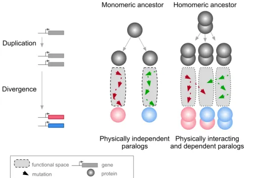
A context in which PPIs and pleiotropy may be particularly important is during the evolution of new genes after duplication events (Amoutzias et al., 2008; Baker et al., 2013; Diss et al., 2017; Kaltenegger and Ober, 2015). The molecular environment of a protein in this context includes its paralog if the duplicates derived from an ancestral gene encoding a self-interacting protein (homomer) (Figure 1). In this case, mutations in one paralog could have functional consequences for the other copy because the duplication of a homomeric protein leads not only to the formation of two homomers but also to a new heteromer (Figure 1) (Pereira-Leal et al., 2007; Wagner, 2003). We refer to these complexes as homomers (HMs) and heteromers of paralogs (HETs).
Duplication
Divergence
Monomeric ancestor Homomeric ancestor
Physically independent
paralogs and dependent paralogs Physically interacting functional space
mutation
gene protein
Figure 1: Mutations in paralogous proteins originating from an ancestral homomer are likely to have pleiotropic effects on each other’s function due to their physical association.
Gene duplication leads to physically interacting paralogs when they derive from an ancestral homomeric protein. The evolutionary fates of the physically associated paralogs tend to be interdependent because mutations in one gene can impact on the function of the other copy through heteromerization.
evo-lution can lead to the maintenance or the loss of these HETs. Consequently, paralogs that maintained the ability to form HETs have often evolved new functional relationships (Amoutzias et al., 2008; Baker et al., 2013; Kaltenegger and Ober, 2015). Examples include a paralog degenerating and becoming a repressor of the other copy (Bridgham et al., 2008), pairs of paralogs that split the functions of the ancestral HM between one of the HMs and the HET (Baker et al., 2013), that cross-stabilize and that thus need each other to perform their function (Diss et al., 2017), or that evolved a new function to-gether as a HET (Boncoeur et al., 2012). However, there are also paralogs that do form HMs but that have lost the ability to form HETs through evolution. Among these are duplicated histidine kinases (Ashenberg et al., 2011) and many heat-shock proteins (Hochberg et al., 2018). For the majority of HETs, we do not know what novel functions, if any, contribute to their maintenance.
Therefore, one important question to examine is: what are the evolutionary forces at work for the maintenance or the disruption of HETs arising from HMs? Previous studies suggest that if a paralog pair maintains its ability to form HMs, it is very likely to maintain the HET complex as well (Pereira-Leal et al., 2007). For instance, Lukatsky et al. (2007) showed that proteins tend to intrinsically interact with themselves and that negative selection may be needed to disrupt HMs. Since nascent paralogs are identical just after duplication, they would tend to maintain a high propensity to assemble with each other. Hence, the two paralogs would form both HMs and HETs until the emergence of mutations that specifically destabilize one or the other (Ashenberg et al., 2011; Hochberg et al., 2018). In addition, the rate at which the HET is lost may depend on epistasis since it may cause mutations to be more or less disruptive together for the HET than they are individually for the HMs (Diss and Lehner, 2018; Starr and Thornton, 2016). Here, we hypothesize that the association of paralogs forming HETs acts as a constraint that may slow down the functional divergence of paralogs by making mutations on one paralog affect the function of the other.
Previous studies have shown that HMs are enriched in eukaryotic PPI networks (Lynch, 2012; Pereira-Leal et al., 2007). However, the extent to which paralogs interact with each other has not been comprehensively quantified in any species. We therefore analyze the physical assembly of HETs exhaustively in a eukaryotic interactome by integrating data from the literature and by performing a large-scale PPI screening experiment. Then, using functional data analysis, we examine the conse-quences of losing HET formation for paralogs forming HMs. We perform in silico evolution experiments to study whether the molecular pleiotropy of mutations, caused by shared binding interfaces between HM and HET complexes, could contribute to maintain interactions between paralogs originating from ancestral HMs. We show that selection to maintain HMs alone may be sufficient to prevent the loss of HETs. Finally, we find that regulatory evolution, either at the level of gene transcription or protein localization, may relieve the pleiotropic constraints maintaining the interaction of paralogous proteins.
Results
Homomers among singletons and paralogs in the yeast PPI network
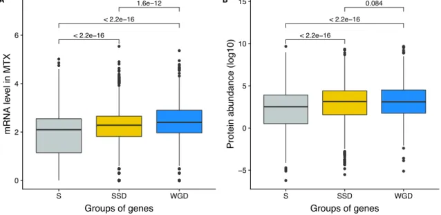
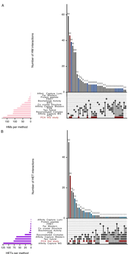
We first examined the extent of homomerization across the yeast proteome (see dataset in Mate-rials and methods and the supplementary text) for two classes of paralogs, those that are small-scale duplicates (SSDs) and those that are whole-genome duplicates (WGDs). We considered these two sets separately because they may have been retained through different mechanisms (see below). The dataset for this analysis, which includes previously reported PPIs and novel DHFR Protein-fragment Complementation Assay experiments (referred to as PCA, see Materials and methods and supple-mentary text), covers 2521 singletons, 2547 SSDs, 866 WGDs and 136 genes that are both SSDs and WGDs (henceforth referred to as 2D). We find that among the 6070 tested yeast proteins, 1944 (32%) form HMs, which agrees with previous estimates from crystal structures (Lynch, 2012). The propor-tion of HMs among singletons (n = 630, 25%) is lower than for all duplicates: SSDs (n = 980, 38%, p-value<2.0e-16), WGDs (n = 283, 33%, p-value=1.6e-05) and 2D (n = 51, 38%, p-value=1.7e-03) (Figure 2A).
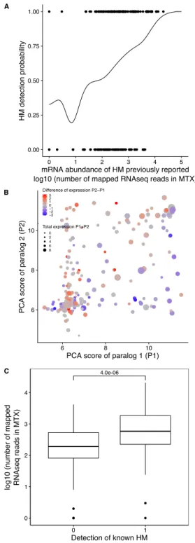
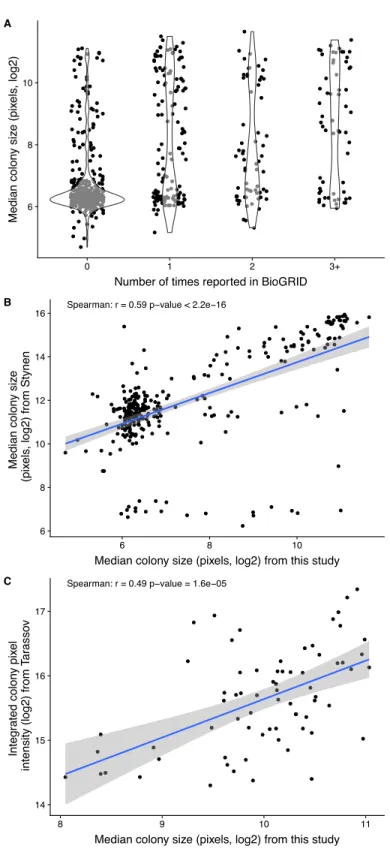
Although a large number of PPIs have been previously reported in Saccharomyces cerevisiae, it is possible that the frequency of HMs is slightly underestimated because they were not systematically and comprehensively tested (see Materials and methods). Another reason could be that some interactions were not detected due to low expression levels. We measured mRNA abundance in cells grown in PCA conditions and used available yeast protein abundance data (Wang et al., 2012) to test this possibility. As previously observed (Celaj et al., 2017; Freschi et al., 2013), we found a correlation between PCA signal and expression level, both at the level of mRNA and protein abundance (Spearman’s r = 0.33, p-value = 3.5e-13 and Spearman’s r = 0.46, p-value < 2.2e-16 respectively). When focusing only on previously reported HMs, we also observed both correlations (Spearman’s r = 0.37, p-value = 3.9e-08 and Spearman’s r = 0.38, p-value = 6.0e-08 respectively). The association between PCA signal and expression translates into a roughly two-fold increase in the probability of HM detection when mRNA levels change by one order of magnitude (Figure S1A). We also generally detected stronger PCA signal for the HM of the most expressed paralog of a pair, confirming the effect of expression on our ability to detect PPIs (Figure S1B). Finally, we found that HMs reported in the literature but not detected by PCA have on average lower expression levels (Figure S1B-C). We therefore conclude that some HMs (and also HETs) remain undetected because of low expression levels.
The overrepresentation of HMs among duplicates was initially observed for human paralogs (Pérez-Bercoff et al., 2010). One potential mechanism to explain this finding is that homomeric proteins are more likely to be maintained as pairs after duplication because they might become dependent on each other for their stability that is enhanced through the formation of HET (Diss et al., 2017). Another explanation is that proteins forming HMs could be expressed at higher levels and thus more easily detected, as shown above. High expression levels are also associated with a greater long term probability of genes to persist after duplication (Gout et al., 2010; Gout and Lynch, 2015). We indeed
Interaction motifs Percentag e (%) SSD WGD Duplication E Homeologs True ohnologs Origin of WGDs A B C D Total S SSD WGD 2D Groups of genes P ercentage of homomers (%) 0 10 20 30 40 50 D 0 4 8 12 z−score Scer (P2) Scer (P1) Lkluy Zrou Lkluy Scer (P2) Scer (P1) Zrou Tom70 (P1) / Tom71 (P2) Scer (P2)Zrou Scer (P1)Lkluy Scer (P2) Zrou Scer (P1) Lkluy Tal1 (P1) / Nqm1 (P2) 0.073 32 25 38 33 38 1.7e-03 1.6e-05 <2.0e-16 40 30 20 10 0 0.084 0.025 HM&HET HET HM NI 1HM 2HM 1HM&HET 2HM&HET HM HM&HET HM HM&HET 0.017 25 Pair wise ami no acid sequ ence iden tity (%) Protein region
Full sequence Interface Full sequence Interface
8.6e-05 2.0e-04 50 75 100 HM HM&HET HM HM&HET Interaction motif Relative con servation score 0.01 0.5 1.0 1.5 G H SSD WGD Interaction motifs Pairwise ami no acid se qu ence ident ity (%) F 100 75 50 25 0 0.065 1.3e-04 HM HM&HET HM HM&HET
Figure 2: Homomers and heteromers of paralogs are frequent in the yeast protein interaction network. (A)The percentage of homomeric proteins in S. cerevisiae varies among singletons (S, n = 2521 tested), small-scale duplicates (SSDs, n = 2547 tested), whole-genome duplicates (WGDs, n = 866 tested) and genes duplicated by the two types of duplication (2D, n = 136 tested) (global Chi-square test: p-value<2.2e-16). Each category is compared with the singletons using a Fisher’s exact test. P-values are reported on the graph. (B and C) Interactions between S. cerevisiae paralogs and pre-whole-genome duplication orthologs using DHFR PCA. The gray tone shows the PCA signal intensity converted to z-scores. Experiments were performed in S. cerevisiae. Interactions are tested among: (B) S. cerevisiae (Scer) paralogs Tom70 (P1) and Tom71 (P2) and their orthologs in Lachancea kluyveri (Lkluy, SAKL0E10956g) and in Zygosaccharomyces rouxii (Zrou, ZYRO0G06512g) and (C) S. cerevisiae paralogs Tal1 (P1) and Nqm1 (P2) and their orthologs in L. kluyveri (Lkluy, SAKL0B04642g) and in Z. rouxii (Zrou, ZYRO0A12914g). (D) Paralogs show six interaction motifs that we grouped in four categories according to their patterns. HET pairs show heteromers only. HM pairs show at least one homomer (one for 1HM or two for 2HM). HM&HET pairs show at least one homomer (one for 1HM&HET or two for 2HM&HET) and the heteromer. NI (non-interacting) pairs show no interaction. We focused our analysis on pairs derived from an ancestral HM, which we assume are pairs showing the HM and HM&HET motifs. (E)Percentage of HM and HM&HET among SSDs (202 pairs considered, yellow) and WGDs (260 pairs considered, blue) (left panel), homeologs that originated from inter-species hybridization (47 pairs annotated and considered, dark blue) (right panel) and true ohnologs from the whole-genome duplication (82 pairs annotated and considered, light blue). P-values are from Fisher’s exact tests. (F) Percentage of pairwise amino acid sequence identity between paralogs for HM and HM&HET motifs for SSDs and WGDs. P-values are from Wilcoxon tests. (G) Pairwise amino acid sequence identity for the full sequences of paralogs and their binding interfaces for the two motifs HM and HM&HET. P-values are from paired Wilcoxon tests. (H) Relative conservation scores for the two motifs of paralogs. Conservation scores are the percentage of sequence identity at the binding interface divided by the percentage of sequence identity outside the interface. Data shown include 30 interfaces for the HM group and 28 interfaces for the HM&HET group (22 homomers and 3 heterodimers of paralogs). P-value is from a Wilcoxon test.
observed that both SSDs and WGDs are more expressed than singletons at the mRNA and protein levels, with WGDs being more expressed than SSDs at the mRNA level (Figure S2A-B). However, expression level (and thus PPI detectability) does not explain completely the enrichment of HMs among duplicated proteins. Both factors, expression and duplication, have significant effects on the probability of proteins to form HMs. It is therefore likely that the overrepresentation of HMs among paralogs is linked to their higher expression along with other factors.
Paralogs that form heteromers tend to have higher sequence identity
The model presented in Figure 1 assumes that the ancestral protein leading to HET formed a HM before duplication. Under the principle of parsimony, we can assume that when at least one paralog forms a HM, the ancestral protein was also a HM. This was shown to be true in general by Diss et al. (2017), who compared yeast WGDs to their orthologs from Schizosaccharomyces pombe. To further support this observation, we used PCA to test for HM formation for orthologs from species that diverged prior to the whole-genome duplication event (Lachancea kluyveri and Zygosaccharomyces
rouxii). We looked at paralogs of the mitochondrial translocon complex and the transaldolase, which
show HETs according to previous studies (see Materials and methods). We confirm that when one HM was observed in S. cerevisiae, at least one ortholog from pre-whole-genome duplication species formed a HM (Figure 2B-C). We also detected interactions between orthologs, suggesting that the ability to interact has been preserved despite the millions of years of evolution separating these species. The absence of interactions for some of these orthologous proteins may be due to the incompatibility of their expression in S. cerevisiae or the use of a non-endogenous promoter for these experiments.
We focused on HMs and HETs for 202 pairs of SSDs and 260 pairs of WGDs. This is a reduced dataset compared to the previous section because we needed to consider only pairs for which there was no missing PPI data (see Materials and methods). We combined public data with our own PCA experimental data on 86 SSDs and 149 WGDs (see supplementary text, Figures S3-S4). Overall, the data represents a total of 462 pairs of paralogs (202 SSDs and 260 WGDs) covering 53% of the SSDs and 50% of the WGDs. This dataset encompasses 493 binary interactions of paralogs with themselves (HMs) and 214 interactions with their sister copy (HET).
We classified paralogous pairs into four classes according to whether they show only the HET (HET, 10%), at least one HM but no HET (HM, 39%), at least one of the HM and the HET (HM&HET, 37%) or no interaction (NI, 15%) (Figure 2D, supplementary text). Overall, most pairs forming HETs also form at least one HM (79%). For the rest of the study, we focused our analysis and comparisons on HM and HM&HET pairs because they most likely derive from an ancestral HM. Previous observations showed that paralogs are enriched in protein complexes comprising more than two distinct subunits, partly because these complexes evolved by the initial establishment of self-interactions followed by the duplication of the homomeric proteins (Musso et al., 2007; Pereira-Leal et al., 2007). However, we find that the majority of HM&HET pairs could be simple oligomers of paralogs that do not involve other proteins and are thus not part of large complexes. Only 70 (41%) of the 169 cases of HM&HET are in
complexes with more than two distinct subunits among a set of 5535 complexes reported in databases (see Materials and methods).
We observed that the correlation between HM and HET formation is affected by whether paralogs are SSDs or WGDs (Figure 2E). WGDs tend to form HETs more often when they form at least one HM, resulting in a larger proportion of HM&HET motifs than SSDs. We hypothesize that since SSDs have appeared at different evolutionary times, many of them could be older than WGDs, which could be accompanied by a loss of interactions between paralogs. Indeed, we observed that the distribu-tion of sequence divergence shows lower identity for SSDs than for WGDs, suggesting the presence of ancient duplicates that predate the whole-genome duplication (Figure S5A). Higher protein sequence divergence could lead to the loss of HET complexes because it increases the probability of divergence at the binding interface. We indeed found that among SSDs, those forming HM&HET tend to show a marginally higher overall sequence identity (p=0.065, Figure 2F, Figure S5B-C). We also observed a significantly higher sequence identity for WGD pairs forming HM&HET, albeit with a wider distribu-tion (Figure 2F, Figure S5C). This wider distribudistribu-tion derives at least partly from the mixed origin of WGDs (Figure S5D-E). A recent study (Marcet-Houben and Gabaldón, 2015; Wolfe, 2015) showed that WGDs likely have two distinct origins: actual duplication (generating true ohnologs) and hybridization between species (generating homeologs). For pairs whose ancestral state was a HM, we observed that true ohnologs have a tendency to form HET more frequently than homeologs (Figure 2E). Because homeologs had already diverged before the hybridization event, they are older than ohnologs, as shown by their lower pairwise sequence identity (Figure S5D). This observation supports the fact that younger paralogs derived from HMs are more likely to form HETs than older ones.
Amino acid sequence conservation could also have a direct effect on the retention of HETs, inde-pendently of the age of the duplication. For instance, among WGDs (either within true ohnologs or homeologs), which all have the same age in their own category, HM&HET pairs have higher sequence identity than HM pairs (Figure S5B, C, E). This is also apparent for pairs of paralogs whose HM or HET structures have been solved by crystallography (n = 58 interfaces). Indeed, we found that pair-wise amino acid sequence identity was higher for HM&HET than for HM pairs for both entire proteins and for their binding interfaces (Figure 2G). Furthermore, the conservation ratio of the binding inter-face to the non-interinter-face regions within the available structures is higher for those forming HM&HET, suggesting a causal link between sequence identity at the interface and assembly of HM and HETs (Figure 2H). We extended these analyses to a dataset of human paralogs (Lan and Pritchard, 2016; Singh et al., 2015) to evaluate if these trends can be generalized. Whereas interfaces within PDB struc-tures (n = 65 interfaces) are more conserved than the full sequence for both HM and HM&HET motifs (Figure S6A), we did not observe differences in the ratio of conservation of interfaces to non-interfaces (Figure S6B). The reasons for this difference between yeast and humans remain to be explored but it could be caused by mechanisms that do not depend on interfaces to separate paralogous proteins in humans, for instance tissue-specific expression.
Considering that stable interactions are often mediated by protein domains, we looked at the domain composition of paralogs using the Protein Families Database (Pfam) (El-Gebali et al., 2019). We tested
if differences in domain composition could explain the frequency of different interaction motifs. We found that 367 of 448 pairs of paralogs (82%) shared all their domain annotations. Additionally, HM&HET paralogs tend to have more domains in common but the differences are non-significant and appear to be caused by overall sequence divergence (Figure S9A-B). Domain gains and losses are therefore unlikely to contribute to the loss of HET complexes following the duplication of homomers.
Heteromer formation correlates with functional conservation
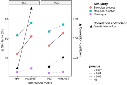
To test if the retention of HETs correlates with the functional similarity of HM and HM&HET paralogs, we used the similarity of Gene Ontology (GO) terms, reported growth phenotypes of loss-of-function mutants and patterns of genome-wide genetic interactions. These features represent the relationship of genes with cell growth and the gene-gene relationships underlying cell growth. The use of GO terms could bias the analysis because they are often predicted based on sequence features. However, phenotypes and genetic interactions are derived from unbiased experiments because interactions are tested without a priori consideration of a protein’s functions (Costanzo et al., 2016). We found that HM&HET pairs are more similar than HM for SSDs (Figure 3 and Figure S10). We observed the same trends for WGDs, although some of the comparisons are either marginally significant or non-significant (Figure 3, comparison between true ohnologs and homeologs in Figure S11). The higher functional similarity observed for HM&HET pairs could be the result of the higher sequence identity described above. However, for a similar level of sequence identity, HM&HET pairs have higher correlation of genetic interaction profiles, higher GO molecular function (for SSDs) and higher GO biological process similarity (for both SSDs and WGDs) than HM pairs (Figure S12). Overall, the retention of HETs after the duplication of HMs appears to correlate with functional similarity, independently from sequence conservation. HM HM&HET HM HM&HET Interaction motifs p-value Biological process Molecular function Phenotype Similarity Genetic interaction Correlation coefficient < 0.001 < 0.01 < 0.05 NS Similar ity (%) Corr elation coe fficie nt 30 60 90 0.03 0.06 0.09 SSD WGD
Figure 3: Maintenance of heteromerization between paralogs leads to greater functional similarity. The similarity score is the average proportion of shared terms (100% * Jaccard’s index) across pairs of paralogs for GO molecular functions, GO biological processes and gene deletion phenotypes. The mean values of similarity scores and of the correlation of genetic interaction profiles are compared between HM and HM&HET pairs for SSDs and WGDs. P-values are from Wilcoxon tests.
Pleiotropy contributes to the maintenance of heteromers
Since molecular interactions between paralogs predate their functional divergence, it is likely that physical association by itself affects the retention of functional similarity among paralogs. Any feature of paralogs that contributes to the maintenance of the HET state could therefore have a strong impact on the fate of new genes emerging from the duplication of HMs. A large fraction of HMs and HETs use the same binding interface (Bergendahl and Marsh, 2017), so mutations at the interface may have pleiotropic effects on both HMs and HETs (Figure 1), which would lead to correlated responses to selection. If we assume that HMs need to self-interact in order to perform their function, it is expected that natural selection would favor the maintenance of self-assembly. Negative selection on HM interfaces would act on their pleiotropic residues and thus also preserve HET interfaces, preventing the loss of HETs as a correlated response.
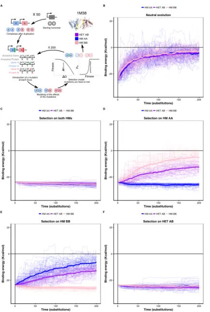
We tested this correlated selection model using in silico evolution of HM and HET protein com-plexes (Figure 4A). We used a set of six representative structures of HMs defined as high-quality based on a consensus of different quality measures including the evolutionary conservation of their quater-nary structures and thermodynamic models (Dey et al., 2018). We evolved these HM complexes by duplicating them and following the binding energies of the resulting two HMs and HET. We let muta-tions occur at the binding interface 1) in the absence of selection (neutral model), 2) in the presence of negative selection maintaining only one HM, and 3) with negative selection retaining both HMs. In these three cases, we applied no selection on binding energy of the HET. In the fourth scenario, we applied selection on the HET but not on the HMs to examine if selection maintaining the HET could also favor the retention of HMs. Mutations that have deleterious effects on the complex under selection were lost or allowed to fix with exponentially decaying probability depending on the fitness effect (see Materials and methods) (Figure 4A).
We find that neutral evolution leads to the destabilization of all complexes derived from the sim-ulated duplication of a HM (PDB: 1M38) (Figure 4B), as is expected given that there are more destabilizing mutations than stabilizing ones (Brender and Zhang, 2015; Guerois et al., 2002). Selec-tion to maintain one HM or both HMs significantly slows down the loss of the HET with respect to the neutral scenario (Figure 4C-E). Interestingly, the HET is being destabilized more slowly than the second HM when only one HM is under negative selection. The difficulty of losing the HET in the simulations could explain why for some paralog pairs, only one HM and the HET are preserved, as well as why there are few pairs of paralogs that specifically lose the HET (Figure S13). The reciprocal situation is also true, i.e. negative selection on HET significantly decelerates the loss of stability of both HMs (Figure 4F). These observations hold when simulating the evolution of duplication of five other structures (Figure S14) and when simulating evolution under different combinations of the parameters that control the efficiency of selection and the length of the simulations (Figure S15). By examining the effects that single mutants (only one of the loci gets a nonsynonymous mutation) have on HMs and HET, we find that, as expected, their effects are strongly correlated and thus highly pleiotropic (Pearson’s r between 0.64 and 0.9 (Figure S16)). We observe strong pleiotropic effects of mutations for the six structures tested, which explains the correlated responses to selection in the in silico evolution.
A Neutral evolution 0 50 100 150 200 -20 0 20 Time (substitutions)
Binding energy (Kcal/mol)
HM AA HET AB HM BB B Selection on both HMs 0 50 100 150 200 -20 0 20 Time (substitutions)
Binding energy (Kcal/mol)
HM AA HET AB HM BB C Selection on HM AA 0 50 100 150 200 -20 0 20 Time (substitutions)
Binding energy (Kcal/mol)
HM AA HET AB HM BB D Selection on HM BB 0 50 100 150 200 -20 0 20 Time (substitutions)
Binding energy (Kcal/mol)
HM AA HET AB HM BB E Selection on HET AB 0 50 100 150 200 -20 0 20 Time (substitutions)
Binding energy (Kcal/mol)
HM AA HET AB HM BB F Introduction of a mutation at each locus a b a b
Complexes after duplication
AA AB BB
BB
AB
AA
Modeling of the effects of the mutations Starting homomer BB A B AA Selection model: mutations are fixed or lost
X 200 X 50 HET AB HM AA HM BB G Fitness threshol d G Fitness P fix previo us fitness 1M38 Ancestral Gene Ancestral Protein Gene A Protein A Gene B Protein B A GCC A GCC A GCC H CAT T CAA H CAT N AAT N AAT H CAT
Figure 4: Negative selection to
main-tain homomers also maintains
het-eromers.
(A)The duplication of a gene encoding a ho-momeric protein and the evolution of the com-plexes is simulated by applying mutations to the corresponding subunits A and B. Only mu-tations that would require a single nucleotide change are allowed. Stop codons are disal-lowed. After introducing mutations, the se-lection model is applied to complexes and mu-tations are fixed or lost. (B to F) The bind-ing energy of the HMs and the HET resultbind-ing from the duplication of a HM (PDB: 1M38) is followed through time under different selec-tion regimes applied on protein stability and binding energy. More positive values indicate less favorable binding and more negative val-ues indicate more favorable binding. (B) Ac-cumulation and neutral fixation of mutations. (C) Selection on both HMs while the HET evolves neutrally. (D) Selection on HM AA or (E) HM BB: selection maintains one HM while the HET and the other HM evolve neu-trally. (F) Selection on HET while the HMs evolve neutrally. (E) Selection on HM AA or (F) HM BB: selection maintains one HM while the HET and the other HM evolve neu-trally. Mean binding energies among repli-cates are shown in thick lines and the indi-vidual replicates are shown with thin lines. Fifty replicate populations are monitored in each case and followed for 200 substitutions. PDB structure 1M38 was visualized with Py-MOL (Schrödinger LLC, 2015).
Additionally, mutations tend to have greater effects on the HM than on the HET (Figure S16, Figure S17), which agrees with observations on HMs having a greater variance of binding energies than HETs (André et al., 2008; Lukatsky et al., 2007, 2006). As a consequence, HMs that are not under selection in our simulations show higher variability in their binding energy than HETs.
We examined the effects of double mutants (the two loci get a non-synonymous mutation at the interface) on HET formation to study how epistasis may influence the maintenance or loss of HET and HMs when the former or the latter are under selection. We defined epistatic effects as deviations between the observed and the expected effects of mutations on binding energy. Expected effects on HETs were calculated as the average of the effects on the HMs, which each have two subunits with the same mutation. We defined positive epistasis as cases where the observed binding is stronger than expected (more negative ∆∆G) and negative epistasis when it is weaker (more positive ∆∆G). In terms of evolutionary responses, positive epistasis would contribute to the retention of the HET and
negative epistasis to its loss.
Regardless of the selection scenario, the mutations sampled are slightly enriched for positive epis-tasis, since the slope values of regression models are smaller than one (0.91 and 0.89 under selection on HMs and HET respectively). When the HMs are maintained by selection, this slightly positive epistasis is also visible in the mutations that are fixed because the epistatic effects are not selected upon. This results in a similar slope for the selected mutations as for the rejected ones. Positive epistasis may therefore contribute to the maintenance of the HET (Figure 5A). On the other hand, selection on the HET results in a further enrichment of mutations with positive epistasis (slope = 0.51, Figure 5B). In this case, mutations tolerated in the HETs and thus fixed are more destabilizing to the HMs. This is also visible in the higher number of fixed substitutions when selection acts on the HET than when it acts on both HMs, particularly for mutations having opposite effects on the HMs (Figure S18). This is also manifested in significantly stronger positive epistasis among fixed pairs of mutations when the HET is under negative selection (t-test, p-value = 0.009). These observations suggest that epistasis may make HETs more robust to mutations than HMs with respect to protein complex assembly, con-tributing to their maintenance when the HMs are under selection and concon-tributing to the loss of HMs when the HET is under negative selection. This effect is visible in our simulations since selection on the HET results in a slow destabilization of the two HMs (Figure 4, Figure S14), especially when more mutations are attempted (Figure S15), and is observed for all six structures tested (Figure S19).
y = 0.03 + 0.92 ⋅ x , R2 = 0.431 y = 0.03 + 0.91 ⋅ x , R2 = 0.797 Selection on both HMs -5 0 5 10 15 20 -5 0 5 10 15 20
Expected HET ΔΔG (kcal/mol)
Obser ved HET ΔΔ G (kcal/mol) Lost Fixed A y = -0.064 + 0.51 ⋅ x , Ry = 0.091 + 0.89 ⋅ x , R2 = 0.383 2 = 0.747 Selection on HET AB -5 0 5 10 15 20 -5 0 5 10 15 20
Expected HET ΔΔG (kcal/mol)
Obser ved HET ΔΔ G (kcal/mol) Lost Fixed B
Figure 5: Epistasis favors the maintenance of HETs and the loss of HMs.
(A and B)Observed effects of double mutants on HET (y-axis) are compared to their expected effects (x-axis) based on the average of their effects on the HMs when selection is applied on both HMs (n = 6777 pairs of mutations) (A) or on the HET (n = 6760 pairs of mutations) (B). Dashed lines indicate the diagonal for perfect agreement between observations and expectations (no epistasis), black regression lines indicate the best fit for the lost mutants, and red regression lines indicate the best fit for the fixed mutants. Data were obtained from simulations with PDB structure 1M38. The regression coefficients, intercepts and R2values are indicated on the figure for fixed and lost mutations. A regression coefficient lower than one means that pairs of mutations have a less destabilizing effects on the HET than expected based on their average effects on the HMs.