University of Montreal
Role of the Protein Tyrosine Kinase 7 gene in human neural
tube defects
By Mingqin Wang
Program of Biomedical Sciences Faculty of Medicine
Thesis presented to Faculty of Medicine in order to obtain the degree of Masters
In Biomedical Sciences
June 2015
i
Abstract
Neural tube defects (NTDs) are among the most common congenital defects with a high incidence of 1-2 per 1000 births, causing a heavy burden to both the families and society. Various types of NTDs result from defects happening in the neurulation process during vertebrate embryonic development. In order to prevent the occurrence of NTDs, understanding the underlying mechanism is a prerequisite. The etiology of NTDs is complex involving environmental and genetic factors. Folic acid supplementation was proven to efficiently decrease the frequency of NTDs by 50-70% depending on the time point of this supplementation and demographic background. Gene identification studies in NTDs have adopted mainly a candidate gene approach investigating folate-related genes and genes derived from animal models. In particular, studies in mouse models have demonstrated a strong association between the non canonical Wnt/Planar Cell Polarity (PCP) pathway and NTDs. Protein Tyrosine Kinase 7 (PTK7) is a member of the PCP pathway and was shown to cause a very severe form of NTDs called craniorachischisis in a mouse model. Ptk7 genetically interacts with a core PCP member Vangl2 where double heterozygotes suffer from spina bifida. These data make PTK7 a strong candidate for NTDs in humans. We sequenced the coding region and the exon-intron junctions of PTK7 in a cohort of 473 patients affected with various forms of open and closed NTDs. Novel and rare variants (<1%) were genotyped in a cohort of 473 individuals. Their pathogenic effect was predicted in silico and functionally in an overexpression assay in a well established zebrafish model. We identified in our cohort 6 novel rare mutations, 3 of which are absent in all public databases, in 1.1% of our NTD cohort. One variant, p.Gly348Ser, acted as a hypermorph when overexpressed in the zebrafish model. Our findings implicate mutation of PTK7 as a risk factor for NTDs and provide additional evidence for a pathogenic role of PCP signaling in these malformations.
Keywords: Protein Tyrosine Kinase 7, neural tube defects, planar cell polarity, convergent
ii
Résumé
Les anomalies du tube neural (ATN) sont des anomalies développementales où le tube neural reste ouvert (1-2/1000 naissances). Afin de prévenir cette maladie, une connaissance accrue des processus moléculaires est nécessaire. L’étiologie des ATN est complexe et implique des facteurs génétiques et environnementaux. La supplémentation en acide folique est reconnue pour diminuer les risques de développer une ATN de 50-70% et cette diminution varie en fonction du début de la supplémentation et de l’origine démographique. Les gènes impliqués dans les ATN sont largement inconnus. Les études génétiques sur les ATN chez l’humain se sont concentrées sur les gènes de la voie métabolique des folates du à leur rôle protecteur dans les ATN et les gènes candidats inférés des souris modèles. Ces derniers ont montré une forte association entre la voie non-canonique Wnt/polarité cellulaire planaire (PCP) et les ATN. Le gène Protein Tyrosine Kinase 7 est un membre de cette voie qui cause l’ATN sévère de la craniorachischisis chez les souris mutantes. Ptk7 interagit génétiquement avec
Vangl2 (un autre gène de la voie PCP), où les doubles hétérozygotes montrent une spina bifida.
Ces données font de PTK7 comme un excellent candidat pour les ATN chez l’humain. Nous avons re-séquencé la région codante et les jonctions intron-exon de ce gène dans une cohorte de 473 patients atteints de plusieurs types d’ATN. Nous avons identifié 6 mutations rares (fréquence allélique <1%) faux-sens présentes chez 1.1% de notre cohorte, dont 3 sont absentes dans les bases de données publiques. Une variante, p.Gly348Ser, a agi comme un allèle hypermorphique lorsqu'elle est surexprimée dans le modèle de poisson zèbre. Nos résultats impliquent la mutation de PTK7 comme un facteur de risque pour les ATN et supporte l'idée d'un rôle pathogène de la signalisation PCP dans ces malformations.
Mots clefs: Protéine Tyrosine Kinase 7, anomalies du tube neural, polarité cellulaire planaire,
iii
Table of contents
1 Introduction………1
1.1Neural tube defects………1
1.1.1 Classification and clinical symptoms………1
1.1.2 The epidemiology ………3
1.1.3 Aetiology of NTDs………3
1.1.3.1 Folic acid ………3
1.1.3.2 Genetic factors in NTDs ……… 5
1.2 Neural Tube Formation ………8
1.2.1 Neurulation in general………8
1.2.2 Primary Neurulation ………8
1.2.3 Secondary Neurulation ………12
1.2.4 Neurulation in Zebrafish ………15
1.3 Wnt signaling pathways ………15
1.3.1 Canonical/ β-catenin Wnt pathway………16
1.3.2 Non-canonical Wnt pathways……..………17
1.3.2.1 The planar cell polarity (PCP) pathway……….…17
1.3.2.1.1 PCP members………..17
1.3.2.1.2 PCP and convergent extension……….…..18
1.3.2.1.3 PCP signaling and ciliogenesis……….………….…...18
1.3.2.2 Wnt/Ca2+pathway...19
1.4 Protein Tyrosine Kinase 7………21
1.4.1 The structure of Protein Tyrosine Kinase 7………21
1.4.2 The expression pattern of Ptk7 during embryonic development………22
1.4.3 PTK7 and NTDs………23
1.4.4 PTK7 and the Wnt pathways………24
1.4.5 PTK7 and other related genes………26
1.5 The Zebrafish Model for studies of convergent extension……..………27
1.5.1 Advantages of the zebrafish model…...………28
iv
1.5.2.1 PCP genes and convergent extension in zebrafish ………31
1.5.2.2 Other genes mediating convergent extension in zebrafish………33
1.5.2.3 PTK7 and CE in zebrafish………..………33
2 Research Project………34
2.1 Rationale and significance………34
2.2 Hypothesis and objectives………34
2.3 Material and Methods ………35
2.3.1 Patients and controls………35
2.3.2 Re-sequencing of PTK7………37
2.3.3 Genetic validation of rare PTK7 mutations ………37
2.3.4 Pathogenic Effect Prediction using bioinformatics ………37
2.3.5 Expression constructs………38
2.3.6 In vitro transcription………39
2.3.7 Zebrafish Maintenance, mRNA and Morpholino Injection ………40
2.3.8 Statistical analyses………40
2.4 Results and Discussion………42
2.4.1 Results………...………42
2.4.2 Discussion……….………47
2.4.3 Conclusions………49
2.4.4 Future directions….…………..………50
2.4.4.1 Functional validation of PTK7 variants identified in NTDs...50
v
List of the tables
Table 1: Characteristics of the 473 NTDs patients………...………36
Table 2: Primers for sequencing of hPTK7………...………38
Table 3: Primers for cloning and mutagenesis ………...…40
vi
List of figures
Figure1: Representative NTDs and their defective sites in neural tube closure………2
Figure2: Transverse sections for neural tube formation………10
Figure3: Schematic diagram of cytoskeleton rearrangements in neural tube formation……12
Figure4: Schematic diagram of secondary neurulation………14
Figure5: Schematic diagram of neurulation in zebrafish………16
Figure6: Schematic model of Wnt pathways………20
Figure7: Schematic diagram for convergent extension………21
Figure8: The structures of the PTK7 gene and its encoded protein………22
Figure9: Diagram for convergent extension of zebrafish during gastrulation………31
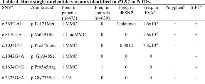
Figure10: Diagram for distribution of the six rare variants identified in PTK7 in NTDs and partial alignments of Ptk7 orthologues among varies species……….43
vii
List of abbreviations
APC Adenomatous polyposis coli
BMP Bone morphogenetic proteins
CE Convergent extension CK1 Casein kinase 1 Crc Circle tail Dgo Diego Dkk1 Dickkopf1 DSH (Dvl) Dishevelled
EMT Epithelial-mesenchymal transition
FGF Fibroblast growth factor
Fmi Flamingo
GSK3 Glycogen synthase kinase 3
Kny Knypek
Lp Loop tail
LRP6 Low density lipoprotein receptor-related protein 6
MDCK Madin-Darby canine kidney
MET Mesenchymal–epithelial transition
MO Morpholino oligomers
MT1-MMP Membrane type 1-matrix metalloproteinase 1
NTDs Neural tube defects
viii
pMLC Phospho-Myosin Light Chain
pk prickle
ppt pipe tail
PTK7 Protein tyrosine kinase 7
rMLC Regulatory myosin light chain
ROCK Rho-associated protein kinase
slb silberblick
SHH Sonic hedgehog
TCF/LEF T-cell factor-1/ lymphoid enhancing factor-1
TGF Transforming growth factor
ix
AcknowledgementsI would like to give my great appreciation to my advisor Zoha Kibar for her kind and patient help and direction.
I thank all participants in the study and appreciate the help of our colleagues for collecting the samples.
I thank my lab mates for helping me a lot and making research so much fun together. I would also like to thank the team of Dr. Pierre Drapeau for training me in zebrafish experiments.
1
1. Introduction
1.1Neural tube defects
1.1.1 Classification and clinical symptoms
Neural tube defects (NTDs) are among the most common congenital defects of the
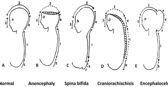
central nervous system, affecting 1-2/1000 births. They are caused by complete or partial failure of the closure of the neural tube during neurulation [1]. NTDs comprise a wide array of morphologically distinct malformations. There are two types of NTDs according to the morphological appearance at birth. Open NTDs of which the brain and/or spinal cord are exposed and include anencephaly, craniorachischisis, encephalocele, anencephaly and spina bifida. In closed NTDs, the spinal defects are covered by skin and include lipomeningocele and tethered spinal cord. Within these distinct morphologically classified malformations, the clinical syndromes vary from mild to severe such as stillborn or immediate death after birth [2, 3]. The following paragraph will discuss mainly the most common NTDs with their corresponding clinical symptoms. NTD happens following failure of neural tube closure. In both humans and mouse models, there exist multiple neural closure sites. The number and sites of neural tube closure in humans are not as clear as in mouse models [4].
Anencephaly is a severe NTD resulting from failure of complete closure of the rostral
end of neural tube during the third to fourth week of conception. It is characterized by absence of telencephalon and the exposure of the residues of brain tissue without the cover of skin and skull (Fig. 1B). The affected babies are unconscious due to the absence of the telencephalon which is responsible for cognition and they cannot survive for long after birth. It is estimated to have an occurence about 1 in 10000 births in the United States. Due to the fact that many of these pregnancies result in miscarriage, this number might be underestimated [5]. Females babies are reported to be more affected than males [6].
2
Myelomeningocele also known as spina bifida affects the posterior region of the
spinal cord (Fig.1 C). It is characterised by the opening of spinal cord mainly in the lumbar and sacral regions to varying extents. The clinical symptoms vary and include leg weakness and paralysis, orthopedic abnormalities and bladder and bowel control problems [8].
Craniorachischisis is characterized by an open neural tube in both cranial and spinal
regions all along the body axis (Fig.1 D). It is the most severe type of NTDs with a relative low prevalence leading to invariable death at birth [9].
Encephalocele is characterized by sac-like protrusions of the brain and the membranes
that cover it through openings in the skull (Fig.1 E). The protrusion can happen in different regions along the cranial and forehead line. The clinical symptoms may include neurological
Fig. 1. Representative NTDs and their defective sites in neural tube closure. A: Normal neural tube formation marked with 5 closure initiating sites. B: Anencephaly due to the failure of closure site 2. C: Spina bifida due to the failure of closure site 5. D: Craniorachischisis due to the failure of closure site 1. E: Encephalocele due to the failure of closure site 4 (it also can be due to defect of the closure site 2 or 3 and have this sac-like protrusion at different region). Adapted from Van allen et al, 1993 [7].
3
problems, hydrocephalus, spastic quadriplegia, microcephaly, ataxia, developmental delay, vision problems, mental and growth retardation, and seizures.
1.1.2 The epidemiology of NTDs
NTDs have a world average prevalence of 1-2 per 1000 live births. The prevalence varies between different populations and different geographic regions. For example, the NTD prevalence is as high as 138.7 per 10,000 births in some areas in Shanxi province located in North China [10]. In England and Wales, the NTD prevalence declined from 5-6 per 1000 live birth in the 1950s to less than 1 per 1000 live birth in the 1990s due mainly to the increase in dietary folate intake and to peri-conceptional supplementation with folic acid [11]. The same declining trend was also demonstrated in North America [11]. Recurrence risks among women who have had an NTD-affected pregnancy are markedly higher (5%) than that in the first affected pregnancy in the general population (0.2%). And the risk would go up to 10% for the third pregnancy and 28% for the fourth pregnancy [12]. Concordance rates of monozygotic twins (7.7%) for NTDs exceeds that of dizygotic twins (4.0%) suggesting a genetic contribution [12].
1.1.3 Aetiology of NTDS
The etiology of NTDs is complex involving environmental and genetic factors as suggested by epidemiological studies and genetic studies mainly conducted in animal models. Environmental factors associated with NTDs include folic acid insufficiency during the pregnancy, maternal obesity, maternal hyperinsulinemia and hot water exposure in the first trimester. Previous pregnancy wastage and family history of NTDs are also considered as risk factors for NTDs [11]. Here, I will mainly discuss the role of folic acid in protection against NTDs and the genes associated with NTDs to date.
1.1.3.1 Folic acid
Folic acid (folate) is a form of the water-soluble vitamin B9. After absorption, it acts as an acceptor or donor of one-carbon units in a variety of reactions involved in amino acid and nucleotide metabolism, which is important for methylation, DNA synthesis and cell division.
4
A severe folic acid deficiency causes the macrocytic megaloblastic anemia characterized by many large immature and dysfunctional red blood cells in the bone marrow [13].
The relation of folic acid to NTDs was revealed by epidemiological studies where it was confirmed that peri-conceptional intake of folic acid decreases both the recurrence rate among pregnancies following an NTD-affected pregnancy and the occurence rate in the general population [14]. It has been estimated that around 70% of human NTDs are preventable by adequate folate intake [15, 16]. Daily intake of 0.8 mg of folic acid as a vitamin supplement by pregnant women shows a significantly decreased occurrence of NTDs as compared to the control population (NTD cases in the number of pregnancies: 0 in 2420 versus 6 in 2333 with and without folate supplementation respectively in Caucasians [14]).
The protective effect of folic acid against NTDs was confirmed by many subsequent observations or/and interventions conducted in distinct regions in the world. A study conducted in 1999 in China confirmed this preventive effect and at the same time showed that this effect is more marked in the region with high prevalence of NTDs than in the region with lower prevalence [17]. Food fortification is the practice of adding essential vitamins and minerals to staple foods to improve their nutritional content.A study conducted in Canada also showed that the prevalence of spina bifida decreased from 0.86/1,000 in the pre-fortification to 0.40 in the full fortification period [18].
Despite a well established protective effect of folate against NTDs, the underlying mechanisms remain unclear. Metabolic abnormalities of folic acid were suggested as one of the possible mechanisms as studies demonstrated a link between levels of maternal plasma and/or red cell folate level, plasma Vitamin B12, plasma Vitamin C (both are involved in the metabolism of folic acid), homocysteine and the risk of having fetuses affected with NTDs [19-24]. A study conducted on 81 women carrying NTD-affected fetuses during their pregnancies and 247 women carrying normal fetuses showed that both plasma folate and B12 levels were significantly lower in women who were carrying affected fetuses than in controls [20]. The prevalence decreased from 6.6 per 1000 births in women whose red cell folate levels
5
were below 150 ng/ml (340 nmol/l) to 0.8 per 1000 births in women whose red cell folates were over 399 ng/ml (906 nmol/l) [21]. Homocysteine can be transformed to methionine with the help of Vitamin B12 while the 5-Methyl tetrahydrofolic acid (5-MTHF) is transformed to tetrahydrofolic acid (THF) in the same reaction. The relation of homocysteine with NTDs was first indicated where mothers of NTDs-affected children tended to have higher total plasma homocysteine as compared to controls with no NTD-affected children in the methionine loading test [22]. Higher amniotic fluid total homocysteine was also detected in the NTD case mothers and not in controls [23]. These results were further confirmed in pregnant woman carrying NTDs affected children and controls [24].
All these studies provide strong evidence for defects in the metabolism of folic acid in NTDs. Consequently, folate-related genes were investigated for involvement in NTDs by direct re-sequencing and association studies. The variant c.677C→T detected in 5,10 methylenetetrahydrofolate reductase (MTHFR) that catalyzes the essentially irreversible conversion of 5,10-methyleneTHF to 5-methylTHF was the first folate-related genetic factor reported to be associated with NTDs. It was shown that homozygous TT individuals carry a mildly higher plasma homocysteine concentration [25]. The T variant results in an enzyme that binds its cofactor flavin adenine dinucleotide (FAD) with lower affinity than the C variant. This affinity reduction can be compensated by addition of the cofactor FAD or by addition of folate, which explains why the elevated homocysteine levels seen in individuals homozygous for the TT variant can be partially reversed by riboflavin intervention [26-28]. Many genetic studies were conducted to find candidate variants in folate related genes, but the results were not as exciting as the original ones with many conflicts between different reports [29]. Clearly, additional studies are needed to better understand the relationship of folic acid metabolism and the occurrence of NTDs.
1.1.3.2 Genetic factors in NTDs
The evidence from epidemiological studies suggests a strong genetic contribution to the etiology of human NTDs [12]. Concordance rates of monozygotic twins (7.7%) for NTDs exceeds that of dizygotic twins (4.0%) [30].
6
The genetic contribution is also supported by the fact that NTDs are a feature (or symptom) of known genetic syndromes, such as trisomy 13, trisomy 18, and Meckel-Gruber syndrome [31]. Despite the strong evidence for a genetic component in NTDs, the major associated gene(s) or pathways have not been determined yet, mainly due to the complexity of the NTDs’ etiology. At the same time, with the dramatic improvement in ENU (N-ethyl-N-nitrosourea) mutagenesis and other transgenic techniques, many NTDs-related genes were identified from genetic studies conducted in mouse models [4]. These genes are involved in regulation of actin dynamics, cell adhesion, electron transport, DNA damage repair, and other processes [32, 33]. I will present a few examples of NTD genes identified in mouse models focusing mainly on planar cell polarity genes and their possible upstream and downstream effectors.
The Loop-tail mouse is a well established mouse model for NTDs and is characterized by a looped tail phenotype in heterozygous mice, and craniorachischisis in homozygous embryos. The gene defective in Lp was identified as Vangl2 and is a mammalian ortholog of the Drosophila gene stbm/vang that forms part of the planar cell polarity (PCP) pathway controlling the convergent extension (CE) process in vertebrates which will be discussed later in section
1.3.2.2.
Lp was the first mouse model to implicate PCP signalling in the pathogenesis of NTDs [34]. Since this study, many other mouse NTD mutants with PCP genes including Frizzled1, Frizzled3, Celsr1, Scribble1, Dishevelled1, Dishevelled3, and Proteintyrosine kinase7, were identified and showed various degrees of genetic interaction among
them in the incidence of NTDs , confirming an important role of PCP signalling in development of NTDs [35-38]. Other potential downstream effectors of PCP signalling were also shown to cause NTDs in mice like Rho [39].
Canonical Wnt/β-catenin pathway and the non-canonical Wnt/ PCP pathway together regulate a lot of developmental processes including convergent extension. Dishevelled (Dsh),
axin, and glycogen synthase kinase 3(GSK3) are involved in both pathways. They are very
7
The human orthologues of PCP genes were investigated for their role in human NTDs by re-sequencing analyses of their coding regions. The first study that aimed at screening
VANGL1 in human NTDs identified 3 novel rare missense mutations in this gene in 3 NTD
patients: p. Val239Ile and p.Arg274Gln in 2 familial cases of NTDs and p.Met328Thr in a sporadic NTD case. Subsequent functional validation of these mutations using the yeast two hybrid system demonstrated that p.Val239Ile abolished interaction of Vangl2 with Dishevelled proteins and hence is most likely pathogenic [41]. The two variants p.Val239Ile and p.Met328Thr were also proven to be damaging in a zebrafish model where they failed to rescue a defective CE phenotype in zebrafish embryos injected with a morpholino oligonucleotide directed against Vangl2 [42]. Following these initial studies, larger studies conducted in larger cohorts of open and closed forms of NTDs have identified additional novel rare missense mutations in VANGL1 or VANGL2 associated with NTDs thereby confirming their role in these malformations. For VANGL1, 7 patients were heterozygous for 5 novel missense mutations, two of which, p.Ser83Leu and p.Arg181Gln, occurred in familial settings. Four mutations, p.Phe153Ser, p.Arg181Gln, p.Leu202Phe and p.Ala404Ser, were “private” and one mutation, p.Ser83Leu, was recurrent in 3 familial cases of tethered cord syndrome [43]. For VANGL2, six novel rare missense mutations were identified in 7 patients which are absent in the controls, five of which were detected in closed spinal NTDs [44].
A study of another PCP gene, PRICKLE1, in the same cohort used in the above mentioned studies identified 7 rare missense heterozygous mutations that were absent from controls. Functional validation in zebrafish has demonstrated that one variant; p.Arg682Cys antagonized the CE phenotype induced by the wild-type zebrafish prickle1a in a dominant fashion [45]. Other PCP genes including CELSR1, FRIZZLED 6, DVL1 and DVL3 were also demonstrated to be associated with NTDs in a small fraction of patients. All these studies taken together confirm an important contribution of PCP genes to the occurrence of human NTDs.
8
CE is a process which is governed by many signalling pathways including PCP. The detailed morphological or molecular changes that occur in this process will be discussed later in section 1.3.2.2. To better understand the association of PCP genes with NTDs, I will describe the normal neural tube formation in the following section.
1.2 Neural Tube Formation
1.2.1 Neurulation in general
Neurulation is an embryonic process by which the neural tube is transformed into the primitive structures that will later develop into the central nervous system. Neurulation in mammalian embryos occurs in two phases: primary and secondary neurulation [46]. These two phases occur in distinct areas along the rostro-caudal axis of the embryo. Primary neurulation happens on most of the axis from rostral end to the tail bud while the secondary neurulation is limited to the tail bud, which lies beyond the caudal neuropore. Primary neurulation occurs during the third and fourth weeks of pregnancy. It is the process by which the neural plate folds into the cylindrical neural tube leading to the formation of the brain and most of the spinal cord. Secondary neurulation occurs during the fifth and sixth weeks of pregnancy. It is the process by which mesenchymal cells in the dorsal part of the tail bud undergo condensation and epithelialisation (MET, mesenchymal–epithelial transition) to form the secondary neural tube which in turn forms the lowest portion of the spinal cord including most of the sacral and all the coccygeal regions [47]. The lumen of the secondary neurulation is continuous with that of the primary neural tube.
1.2.2 Primary Neurulation
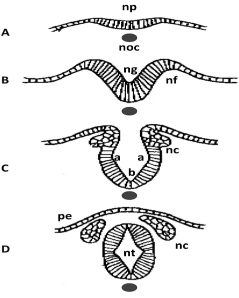
Primary neurulation can be subdivided into several stages: induction and formation of the neural plate, formation and elevation of the neural folds and closure of the neural groove (Figure 2) [48]. In each of these stages, specific tissue changes and specific molecules or genes are involved. Induction of the neural plate is mediated by some molecules secreted by primitive node that include Noggin, Chordin, Cerberus, Xnr3, Follistatin, Frzb, and eFGF [49]. These molecules suppress the default epidermal fate of the naive neural ectoderm by inhibiting
9
the BMPs and Wnts signals. After the induction, the formation of the neural plate is autonomous without the signalling from the surrounding epidermal ectoderm [50]. Then the neural plate conducts a CE morphogenetic process to narrow and elongate the neural axis in the mediolateral and rostro-caudal axes. This process will be discussed in detail in Section 1.3.2.2. At the same time the neural progenitor cells in the neural plate undergo apical constriction that would lead to changes in cell shape from cuboidal to columnar and finally to wedge- or bottle-like while the cell volume keeps constant [51, 52]. Combining with the extrinsic forces generated by the CE movement in non-neural ectoderm, the intrinsic forces generated from the shape change initiate the bending of the neural plate [53, 54]. A groove is formed during this bending and finally develops the lumen of the neural tube after its closure. After its closure, the neural tube is separated from the overlaying epidermal ectoderm, which will further develop in to the skin of the back of the embryo. At this stage, another important cell population called neural crest cells and located between the neural tube and the epidermal ectoderm starts to migrate and finally develops into various tissues in different structures [48].
Closure of the neural tube groove is initiated at some specific sites and then elongates uni-directionally or bi-directionally to meet with the elongation from another site leading to complete closure of the whole neural tube. It is well established that there are multiple initiation sites for neural tube closure in both humans and mouse models, but the exact number of these sites is still undefined [4, 7]. For example in mouse models, 3 main distinct initiation sites along the neural axis are thought to function in a temporal and spatial-specific pattern to complete neural tube closure while in humans there are probably 5 sites [7]. Defects happening in these different initiation sites or in the elongation region of these sites will result in various NTD types [1, 7]. The representative NTDs and the corresponding closure defects are shown in Fig1.
10
Fig. 2. Transverse sections for neural tube formation. Neural plate is induced by notochord under it (A). Neural folds form and elevate to create the neural groove (B). Bending or hinge points form at two sites: the dorsolateral hinge points (a) at the lateral extremes of the neural plate and the median hinge point (MHP) (b) overlying the notochord. With continued bending, the neural tube is closed and separates from the presumptive epidermis while the neural crest form and migrate to lateral sides. noc, notochord; np, neural plate; nf, neural fold; ng, neural groove; nt, neural tube; nc, neural crest; pe, presumptive epidermis.
11
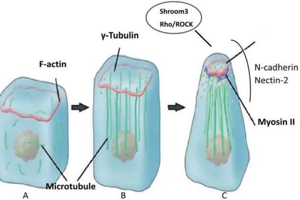
The major morphogenetic changes in cell shape or movement are caused by cytoskeletal rearrangements (Fig.3). Actin filaments (F-actin) constitute a major component of the cytoskeleton that is composed of monomeric actin (G-actin). It is present at the apical junction of the neural plate cells in the form of circular bands. During the neural tube closure process, the band thickens leading to apical constriction (Fig. 3C) [55, 56]. Another essential cytoskeletal component is myosin II that also contributes to the apical constriction [57]. Myosin II is composed of two heavy chains, two essential light chains and two regulatory light chains. The activity of myosin II is regulated through phosphorylation of its regulatory light chain (rMLC) mainly by Rho-associated kinase (ROCK), an effector of the small GTPase Rho. Myosin II is co-localized with F-actin in the apical side of neural plate cells (Fig. 3C) [58]. Either the disruption of F-actin or inhibition of Myosin II activity can cause the disappearance of the apical constriction and failure of neural tube closure in chick embryos [39, 56, 58]. This causative relationship was also observed in Xenopus in which the expression of myosin II was reduced by a morpholino oligonucleotide (MO) leading to failure of the F-actin accumulation and apical constriction [58]. Microtubules are involved in the cell elongation during the neurulation process. Consistent with the shape changes of the neural plate cells from cuboidal to columnar; the microtubules are reorganized from random distribution and disorientation throughout the cells to a parallel orientation to the axis of elongation (Fig. 3A, B, C) [56, 59]. It was shown that the neuroepithelial cells treated with microtubule polymerization inhibitors prior to the elongation process failed to elongate in chick embryo. In shroom3 mutants and MO-injected embryos, apical constriction is significantly inhibited, associated with a severe reduction in apical accumulation of F-actin and phosphorylated MLC (pMLC) [60]. In addition to intracellular molecules, cell adhesion molecules such as N-cadherin and nectin-2 also play crucial roles in cellular morphogenesis during neural tube closure (Fig. 3C). All these studies demonstrate that cytoskeletal rearrangements generate the driving force for shaping the neural plate cells and closure of the neural tube. The molecular mechanisms involved in these rearrangements still remain poorly defined.
12
Fig. 3. Schematic diagram of cytoskeleton rearrangements in neural tube formation. In cuboidal neuroepithelial cells prior to neurulation (A), F-actin exists in a thin circular band (red) at apical junction, while microtubules (green) are diffusively distributed in cytoplasm. During cell elongation, non-centrosomal γ-Tubulin particles (yellow) are distributed apically while microtubules polymerize and assemble parallel to the apicobasal axis (B). During apical constriction, actin filament bands constrict and become thickened. Non-muscle myosin II (green) actively slides and generates contractile force along apical actin filaments. The molecules that are accumulated at the apical junctions are shown in the circle while the adhesion molecules crucial for this constriction are marked besides. Adapted from Suzuki M et al, 2012 [60].
1.2.3 Secondary Neurulation
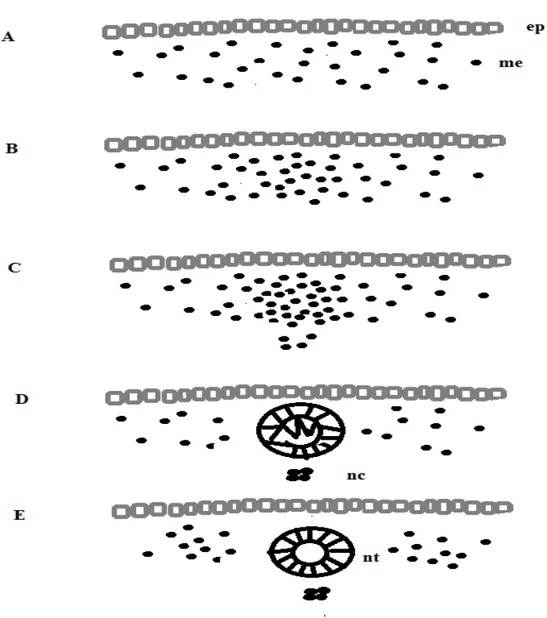
Secondary neurulation is initiated immediately after completion of the primary neurulation in the most posterior part of the caudal axis. Distinct from the primary neurulation, there is no neural plate formation or neural folds elevation. As indicated in Fig. 4, the bilateral
13
mesenchymal cells under the epithelial ectoderm undergo a mesenchymal-to-epithelial transformation (MET) characterized by a series of changes in the cell surface oligosaccharide complement of the differentiating cells. These cells together with the undifferentiated central mesenchymal cells migrate at the same time to form the cell mass located in the midline [61]. This is followed by formation of small cavities at the boundary between the two cells’ populations. These cavities enlarge while the central cells intercalate with the peripheral cells, and merge with each other resulting in a single lumen which becomes continuous with the neurocoele of the primary neural tube [61]. Developmental defects in this process are associated with closed spina bifida where the developing neural tube fails to separate from other tissues of the tail bud [62].
MET is the reverse process of epithelial–mesenchymal transition (EMT). Unlike epithelial cells which are stationary and characterized by an apical-basal polarity, tight junctions, and expression of cell-cell adhesion markers, mesenchymal cells do not have these mature cell-cell contacts and can invade the extracellular matrix. This transformation is very well studied in kidney ontogenesis, somitogenesis and cancer. The mechanisms by which the mesenchymal cells conduct this transformation are unknown. Some changes in gene expression patterns were reported during this transformation [63]. For example, Sox2 expression is not detected in the mesenchymal cell mass at the beginning of secondary neurulation and become detectable at later stages in a cell cluster in the medullary cord which is the precursor of the neural tube. A similar pattern was observed for other epithelial apical-basal polarity markers such as laminin and fibronectin (apical-basal markers), N-cadherin and atypical protein kinase C (aPKC) (apical markers). At the same time, it was shown that these markers also propagate in dorsal to ventral orientation [63]. Activity levels of Rho family GTPases Rac1 and Cdc42 were also shown to be critical for the MET during secondary neurulation.
14
Fig. 4. Schematic diagram of secondary neurulation. The bilateral mesenchymal cells under the epithelial ectoderm (A) proliferate, migrate and finally condensate at the midline (B, C). The condensed mesenchymal cells undergo MET and transformed to be neural epithelial cells (D). At the same time, small cavities starts to form inside the cell mass (D). Finally the neural tube lumen formed with the complete neural epithelial cells transformation (E). me, mesenchymal cells. ep, epithelial cells. nt, neural tube. nc, notochord. Adapted from: http://scienceblogs.com/pharyngula/2008/01/02/neurulation-in-zebrafish/.
15
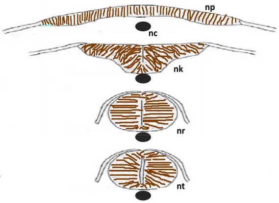
1.2.4 Neurulation in Zebrafish
As zebrafish (Danio rerio) will be used in our study as an animal model, I will point out some specific characteristics of the neurulation process in zebrafish. Neurulation in zebrafish tail is conducted through secondary neurulation as in other vertebrates. Whether the neurulation in the head and trunk (Fig. 5) goes through primary or secondary neurulation is still in dispute. The process of this neurulation does not start from the condensation of mesenchymal cells in the midline as in traditional secondary neurulation. Instead, it starts from the columnarization of neural plate cells. A solid neural keel is formed by the accumulation of these columnarized neural plate cells. A solid neural tube (neural rod) will be formed following the separation of the neural keel from the originating neural plate. The lumen will appear after the solid neural tube goes through the cavitation process [64].
1.3 Wnt signalling pathways
The Wnt members are secreted glycoproteins which can bind to different membrane- anchored receptors to activate multiple important pathways including canonical and non-canonical signalling pathways. The determinant of which pathway will be activated has been recently suggested to be the different Wnts and different co-receptor crossbinding [65]. Wnt signalling functions in cell proliferation, stem cell maintenance and cell fate decisions, as well as organized cell movements and the establishment of tissue polarity in one layer. It is also known to be related to human cancers and degenerative diseases, mainly by activation of canonical and non-canonical Wnt pathways [65].
The NTD phenotypes were observed in mouse mutants of genes involved in canonical
(Wnt3 and Lrp6) as well as non-canonical Wnt signalling (Vangl2 and Ptk7) [66]. This
suggests that the balance between these two pathways is very critical to the neurulation process.
16
1.3.1 Canonical/
β-catenin Wnt pathway
The core member in the canonical Wnt pathway is β-catenin, which is constantly synthesized and then degraded through phosphorylation and ubiquitination by a so-called destruction complex consisting of the scaffolding protein Axin, APC (adenomatous polyposis coli), GSK3 (Glycogen synthase kinase-3) and CK1 (casein kinase 1) (Fig. 6) [67]. Without the stimulation by Wnts, β-catenin is constantly degraded by this destruction complex. Once
Fig. 5. Schematic diagram of neurulation in zebrafish. The neural plate folds inward at the midline (A,B) and form the neural keel, the keel progressively rounds up, forming the neural rod (C) without a lumen inside while the cells from lateral fuse to form the epidermal layer . Cavitation starts from the ventral side of the neural tube to the dorsal side finally forming the neural tube (D). np, neural plate; nc, notochord; nk, neural keel; nr, neural rod, nt, neural tube. Adapted from: http://scienceblogs.com/pharyngula/2008/01/02/neurulation-in-zebrafish/.
17
the appropriate Wnt like Wnt3 or Wnt8 binds to the right Frizzled receptor, with the presence of other co-receptors like Lrp5/6 (low density lipoprotein receptor 5 or 6), Dsh is recruited to the membrane followed by its phosphorylation and this leads to disassembly of the destruction complex. β-catenin is then released from the complex, accumulates in the cytoplasm and then translocates into the nucleus. By relieving TCF/LEF (T-cell factor/Lymphoid enhancer-binding factor ) from enhancer-binding with transcription repressor Groucho, Wnt functions as a transcriptional activator and targets gene expression [65]. Canonical Wnt/β-catenin pathway is proven to function in cell proliferation, stem cell maintenance and cell fate decisions [68]. Defects in this pathway are responsible for tumorigenesis and developmental defects. β-catenin was found to be mutated and causing hyperactivation of Wnt/β-catenin signalling in virtually all intestinal cancers and other malignancies. In Drosophila, the mutation of Wnt1 homolog Wingless causes a segment polarity defect [ 66, 69].
There are disputed findings about the direct stream of Wnt, β-catenin, TCF/LEF, target gene activation pathway [70]. For example, TGF-β can also regulate the nucleus translocation of β-catenin independent of Wnts. Blocking the transcription activity of TCF could not block all the phenotypes induced by canonical Wnt ligands indicating another branch of downstream effectors besides TCF/LEF [70]. However, another group show that a dominate negative form of TCF4 abrogates DNA recruiting by any of the known transcription factors [71].
1.3.2 Non-canonical Wnt pathway
1.3.2.1 The planar cell polarity (PCP) pathway
1.3.2.1.1 PCP members
The PCP pathway was first demonstrated in Drosophila and is highly conserved in vertebrates [72, 73]. It shares some common members with canonical/β-catenin Wnt pathway like Fz and Dsh (Fig. 6). Genetic studies of a wide range of mutants affecting some highly organized structures in the fly, like the distal orientation of wing hairs, the posterior orientation of the abdominal bristles and the more complex organization of the ommatidia (eye units) in the adult eye, have identified a group of so-called core PCP genes required for PCP signalling in all tissues and which include: Frizzled (Fz), Dishevelled (Dsh/Dvl),
18
Strabismus/Van Gogh (Stbm/Vang), Flamingo (Fmi), Prickle (Pk) and Diego (Dgo) [73-75]. In planar cells, PCP members are asymmetrically localized at the cell membrane: Fmi, Fz, Dsh, and Dgo localize to the distal region of the cell, whereas other PCP members Fmi, Vang and Pk localize to the proximal region. After Dsh recruitment to the membrane under the stimulation of some non-canonical Wnt activators (Wnt4, Wnt5a and Wnt11), PCP signalling through unknown mechanisms, lead to activation of the small GTPases RhoA and Rac, resulting in modification of the cytoskeleton like the contraction of F-actin and microtubule reorganization (Fig. 6). This modification ultimately results in the polarization of cells within epithelia and polarized cell migration [75, 76].
1.3.2.1.2 PCP and convergent extension
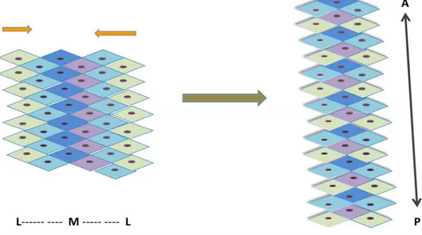
PCP signalling is essential to a morphogenetic pathway called CE during gastrulation and neurulation in vertebrates. CE is the process by which cells move from the lateral side to the midline and intercalate with each other giving rise to a longer and narrowed anterior – posterior axis (Fig. 7) [65]. CE is a major driving force for neural tube formation and closure. In Vangl2 mutant Loop tail mice, the NTD phenotype craniorachischisis was demonstrated to be the result of a defective CE process manifested by a shorter anterior-posterior axis and wider left-right axis [77]. These CE defects were also observed in other PCP mutant mice models like Crash (Celsr1), Dvl1/Dvl2 double knockouts, Fz3/ Fz6 double knockouts, Circle tail (Scribble1) and Chuzhoi (Ptk7) [32].
1.3.2.1.3 PCP signaling and ciliogenesis
Cilia are microtubule-based protrusions on the surface of most vertebrate cells. There are two types of cilia: motile cilia and non-motile cilia (primary cilium). It is well known that motile cilia beat in coordinated waves to sweep mucus out of the respiratory tract or move the ovum from the ovary to the uterus [78]. The function of primary cilium was first illustrated to be linked to patterning of the mouse embryo by transducing Hedgehog signaling [79]. The mouse mutants show phenotypes characteristic of Hedgehog signaling defects including an open neural tube on the head and preaxial polydactyly. Following this, additional studies confirmed the role of cilium in inducing rostral neural tube defects [80, 81]. In humans, genes
19
involved in ciliogenesis are proven to be responsible for some Meckel-Gruber syndrome that typically involves meningo-encephalocele, polycystic kidneys and postaxial polydactyly [82, 83]. In contrast to cilia mutants, PCP mutants are more affected with caudal neural tube defects [84]. Inversin, a homolog of the PCP member Diego in fruit fly, was more studied in the context of ciliogenesis and was responsible for some renal cystic diseases [85]. Reduced expression of either PCP effectors Inturned or Fuzzy generated rostral neural tube closure defects [86].
PCP was shown to regulate the orientation of cilium in both individual cells and at the tissue level of radial glial cells and ependyma to create a coordinated beating [87]. Additional studies are needed to better understand the relationship between PCP members and cilium polarity and relationship between PCP members and Hedgehog signaling.
1.3.2.2 Wnt/Ca
2+pathway
The Wnt/Ca2+ pathway is another major non-canonical Wnt pathway. There is crosstalk between the Wnt/Ca2+ pathway and the canonical β-catenin pathway or PCP pathway at different levels [88]. Wnt/Fz ligand receptor binding leads to a transient increase of certain intracellular signaling molecules, inositol 1,4,5-triphosphate (IP3), and 1,2 diacylglycerol (DAG). IP3 causes release of Ca2+ from ER (endoplasmic reticulum). Calcium ions in the presence of calmodulin or DAG can activate CaMKII (calcium calmodulin dependent protein kinase II) or protein kinase C (PKC). CaMKII and PKC can activate nuclear transcription factors (NFkB and CREB). Calcium can also activate phosphatase calcinurin (Cn) that can further activate nuclear factor associated with T cells (NFAT) (Fig. 6). These activated nuclear factors will regulate the expression of various genes related to development, cancer, and cellular responses as a consequence of inflammatory challenges. The Wnt/Ca2+ signaling pathway was proved to promote ventral cell fate through CaMKII during early embryogenesis in Xenopus [89].
20
Fig. 6. Schematic model of Wnt pathways. Canonical Wnt ligands bind to the membrane receptor Fzd forming a complex with some known and unknown co-receptors like Lrp5/6. The subsequent Dvl membrane recruitment and interaction with Axin induce the disassembly of the β-catenin degradation complex. The free
β-catenin accumulates in the cytoplasm and enters in the nucleus to activate the
transcription of canonical target genes via an association with the DNA binding factors LEF/TCF. Non–canonical Wnt ligands bind to Fz with the help of co-receptors and recruit Dvl to the membrane. Small GTPase family members Rac/ Rho are activated and regulate the cytoskeletal reorganization. The core PCP members Vangl, Diversin, Prickle, and Ceslr are all involved in the Dvl membrane localization. Diversin at the same time regulates the degradation of β-catenin. Wnt/Fz ligand receptor interaction with co-receptor Ror1/2 leads to the production of IP3 and DAG from phospholipid phosphatidyl inositol 4, 5-bisphosphate (PIP2). IP3 causes release of Ca2+ from ER; Cn and CamKII are activated which in turn
activate NFAT and NFkB. DAG is activated by released calcium from ER, which activates PKC. PKC activates NFkB and CREB which together with NFAT will regulate downstream gene expression. Dotted arrows show the pathway direction; Green lines show the genetic or physical interaction between two proteins. The red line shows the binding that can inhibit the non-canonical pathway.
21
1.4 Protein Tyrosine Kinase 7
1.4.1 The structure of Protein Tyrosine Kinase 7
The first study on Ptk7 involved the cDNA coding of KLG that is the homolog of Ptk7 in chicken [90]. Human PTK7 and mouse Ptk7 were subsequently cloned [91, 92]. The human
PTK7 gene is located to chromosone 6 and its coding region consists of 20 exons (NCBI Gene
ID: 5754; Ensembl ID: ENST00000230419). Among multiple splicing transcripts, the longest isoform encodes a protein composed by 1070 amino acids (Fig. 8, upper panel) [93]. Human PTK7 is a highly conserved single pass transmembrane protein that consists of one signal peptide which targets the protein to the membrane, seven extracellular Ig-like (immunoglobulin-like) domains and one cytoplasmic tyrosine kinase homolog domain which
Fig. 7. Schematic diagram for convergent extension. The polarized cells lining the lateral side (green and light blue) migrate to the midline. Cells intercalate to each other resulting in narrowing in the medial-later axis and extending in the anterior – posterior axis. M, midline; L, lateral line; A, anterior; P, posterior.
22
lacks kinase activity due to the absence of the DFG triplet which is essential for kinase activity (Fig. 8, lower panel ).
1.4.2 The expression pattern of Ptk7 during embryonic development
In mouse embryos, at E6.5, Ptk7 is expressed in the early primitive streak. At E7.5,
Ptk7 is broadly expressed throughout the embryo, with high levels in the primitive streak. At
E8.25-E8.75, Ptk7 is highly expressed in the anterior and posterior regions of the primitive streak which is consistent with a role for Ptk7 in neural tube closure [35]. In Xenopus, Xptk7 is expressed in the neuroectoderm from late gastrula through all neurulation stages [35].
In zebrafish, Ptk7 expression is ubiquitous at the shield stage and is pronounced in axial, paraxial and tailbud regions during late-gastrula stages. In the post-gastrulation stage,
Ptk7 is expressed in the head, neural tube and somites, with the highest level of expression Fig. 8. Schematic digrams of the structures of the PTK7 gene and its encoded protein. In the upper panel, each square stands for one exon where a different color is consistent with the domain that it codes for. On the lower panel, each oval stands for one domain whereas the TM domain is marked by a grey column. TM, transmembrane; Ig, immunoglobulin-like; Inactive PKD, inactive protein tyrosine kinase domain.
23
observed in the tailbud which persists until 24 hpf. The expression pattern of zebrafish ptk7 is similar to that observed in Xenopus and mouse [94].
1.4.3 PTK7 and NTDs
The role of Ptk7 in embryonic development ranges from axon guidance in Drosophila to the regulation of gastrulation, neural tube closure, neural crest migration, cardiac morphogenesis and epidermal wound repair in vertebrates [95]. Here I will mainly focus on its role in neural tube closure. Ptk7 was first identified as an NTD responsible gene in a gene trapping screen for transmembrane proteins important for neural development. Mutant mice that were homozygous for a truncating mutation in Ptk7 suffered from craniorachischisis that resulted from failure of neural tube closure initiation site 1. Besides the defects in the neural tube, the orientation of stereocilia bundles in the organ of Corti was also disrupted in these mice. Craniorachischisis and stereocilia orientation defects were also observed in Vangl2 and
mScrib1 mutants (Lp and Crc, respectively). A strong genetic interaction was detected
between Ptk7 and Vangl2 (Lp) where the frequencies of NTDs in the form of spina bifida increased dramatically from less than 10% (Lp/+) or zero (Ptk7/+) to 94% in double heterozygotes. No genetic interaction was observed between Crc and Ptk7. However, the NTD phenotype in Lp/+; Ptk7/+ double heterozygotes was not as severe as in Lp/+; Crc/+ double heterozygotes. The disruption in sensory hair cells orientation was also not observed in the
Lp/+; Ptk7/+ double heterozygotes.
Chuzhoi is an ENU-induced mutant which exhibits craniorachischisis and bundle
orientation disruption in the inner ear. Abnormal lung and heart development were also observed in chuzhoi mutants. Genetic mapping and subsequent cloning showed that the underlying mutation in chuzhoi is a nucleotide substitution in Ptk7 that created a new splice acceptor site leading to a 3 amino acid insertion in the protein, thereby resulting in disruption of Ptk7 protein expression in chuzhoi mutants. Chuzhoi was also shown to genetically interact with Vangl2/Lp and Celsr1/Crsh but not with Scrib/Crc [96].
24
In the frog model, reduced expression of Xptk7 by MOs (morpholino oligomers) also causes NTDs. This phenotype could be rescued by co-injection of full-length Xptk7 or a construct lacking the cytoplasmic domain, indicating an important role of the membrane anchored extracellular domain in neural tube formation [35].
1.4.4 PTK7 and the Wnt pathways
Recruitment of Dsh to the membrane is essential for the activation of both canonical and non-canonical Wnt pathways. The pseudokinase domain of Ptk7 is required for this recruitment. Loss of function of Fz7 does not affect Ptk7-mediated Dsh localization in animal caps. Interestingly, while overexpression of Fz7 leads to recruitment and phosphorylation of Dsh, overexpression of Ptk7 can recruit co-overexpressed Dsh to the membrane but no phosphorylated band was observed [97]. Rack1 is required for Ptk7-mediated Dsh membrane recruitment and the pseudo kinase domain of Ptk7 is necessary for Rack 1 translocation to the membrane. Furthermore, expression of a fusion construct consisting of a pseudo kinase domain-deleted mutant and Rack1 recruits Dsh to the membrane successfully suggesting that the pseudo kinase domain functions by recruiting Rack1 which in turn recruits Dsh independent of Fz [98]. These results seem to be inconsistent with the study of Lu et al. (2004) that demonstrated this domain to be dispensable in rescuing the NTD phenotype induced by injection of MOs against Xptk7 in n the frog model. This inconsistency could be due to the use of different functional assays: overexpression in cell lines versus a MO-knockdown in the frog. Alternatively, Ptk7 is a highly versatile protein implicated in many biological processes and hence recruitment of Dsh by its pseudo-kinase domain could reflect a functional aspect that is independent of PCP signaling.
Ptk7 co-precipitates with canonical Wnt3a and Wnt8 but not Wnt5a or Wnt11, indicating a role in canonical Wnt signalling [99], but Ptk7 inhibits rather than activates canonical Wnt activity. In Xenopus, Ptk7 inhibited secondary axis formation induced by Wnt8 or Wnt3a. The inhibition was also shown by siamois reporter assay which is readout of canonical Wnt pathway. The inhibition by Ptk7 is suggested to be upstream of β-catenin and Dsh. The non-canonical pathway was shown to be activated by Ptk7 through ATF2-based
25
luciferase reporter assay which is readout of the PCP pathway. For both the inhibitor and activator roles of Ptk7 in Wnt signalling, membrane-anchored extracellular part was shown to have the same activity as the full length one while the extracellular part alone or membrane-anchored intracellular part lost this inhibition or activation effect, indicating a crucial regulatory role of the membrane anchored extracellular domain of Ptk7 in canonical and non-canonical Wnt signalling. Ptk7 was suggested to act as a “molecular switch” that activates the Wnt/PCP non canonical pathway while inhibiting at the same time the Wnt/β-catenin canonical pathway [99]. One contradictory study showed that Ptk7 physically interacts with β-catenin in a yeast two hybrid system and can activate the canonical Wnt pathway stimulated by Wnt3a. In the same study, they also showed that Ptk7 functions upstream of the β-catenin destruction complex in the Wnt canonical signalling pathway probably by stabilizing β-catenin [100].
A recent study confirmed the role of ptk7 in zebrafish in both Wnt pathways and showed that this gene is also implicated in tailbud tissue differentiation. Overexpression of
ptk7 induces the expansion of dorsalization marker chd and the reduction of ventralization
marker vox and β-catenin target axin2. Co-injection of zebrafish ptk7 rescued the dorsoventral patterning defects induced by ectopic expression of wnt8. ptk7 genetically interacts with
Wnt5b in body length reduction caused by defective CE. Wnt11-induced cyclopia was also
shown to be potentiated by ptk7 overexpression [94]. In the same study, a mutant line made by a specifically engineered ZFN (zinc finger nuclease technology) showed that CE defects were observed in maternal-zygotic mutants, while this phenotype was absent in zygotic mutants demonstrating its important role in early embryo development [94]. Recently, another study by the same group described a skeletal defect observed in the zygotic mutant adult fish and demonstrated that this zebrafish model is an ideal model for idiopathic and congenital scoliosis [101]. One PTK7 mutation found in a scoliosis patient was shown to disrupt both PCP and Wnt/β-catenin signalling.
Lrp6 (Low-density lipoprotein receptor-related protein 6), a single-pass transmembrane protein, is required in Wnt/β-catenin signalling and acts as a co-receptor for Wnt [102].
26
Dickkopf-1 (Dkk1), a secreted protein, negatively modulates the Wnt/β-catenin pathway through its binding to Lrp6 [103]. The role of Dkk1 in modulating the canonical and non-canonical Wnt pathway was shown to be executed through the binding to Lrp6 or another membrane receptor Knypek (Kny) [104]. Lrp6 alone was also shown to be acting as a canonical and non-canonical Wnt pathway regulator with its different domains responsible for different functions [105]. Ptk7 was recently shown to modulate Wnt signalling activity via Lrp6 [64]. In HEK293 cells, PTK7 depletion by shRNA strongly inhibited LRP6 activation of β-catenin. Ptk7-depletion by MO caused a significant reduction of ectopically and endogenously expressed Lrp6 protein levels in Xenopus. Phenotypes of morphants of Lrp6 and
Ptk7 are very similar. Lrp6 is also similar to Ptk7 in rescuing the neural tube closure
phenotype caused by overexpressing Wnt5 or Wnt11. Ptk7 was also shown to physically interact with different Lrp6 constructs as long as they have the transmembrane domain which leads to the hypothesis that Ptk7 modulates Wnt signalling activity by binding and stabilizing Lrp6.
Combining all the studies above, Ptk7 was shown to be very important modulator of both canonical and non-canonical Wnt pathways and represents a strong candidate gene for NTDs in humans.
1.4.5 PTK7 and other related genes
Cdx genes encode transcription factors controlling anterior-posterior patterning via
integration of posteriorizing signals from retinoic acid and Wnt-canonical pathways [106]. They mediate neural tube closure through transcriptional regulation of Ptk7 [107]. Cdx1 and
Cdx2 double knock-out mice (DKO) showed the most severe NTD craniorachischisis. The
observed defects in axis elongation and in somite morphology in these mutants were similar to the Ptk7-null mutant but not the Vangl or Scribble mutants. This is further supported by the fact that Ptk7 expression was downregulated at E7.5 in Cdx1/2 DKO mutants. In addition, ChIP (chromatin immunoprecipitation) analysis revealed that the 5’ upstream promoter of
Ptk7 is responsive to the transcription factors Cdx1 and Cdx2. Combining all these data, Ptk7
27
Ptk7 was also demonstrated to be the primary cleavage target of the MT1-MMP (membrane type-matrix metalloproteinase) which cleaves ECM (extracellular matrix) proteins, initiates activation of soluble MMPs, and controls the functionality of cell adhesion and signalling receptors accompanied with other receptors including CD44. By reducing the expression of MT1-MMP in zebrafish using a MO, a body length reduction associated with PCP defects was observed and accumulation of full length Ptk7 in the 2-3 day old embryos was detected. A genetic interaction between Ptk7 and MT1-MMP was shown by co-injections of low, sub-threshold dosages of MT1-MMP and Ptk7 MO causing a synergistic effect on the CE phenotype [108].
In another cell migration and differentiation process which is called neural crest migration, Ptk7 precipitated with PlexinA at the gastrulation stage in Xenopus. Co–injection of
ptk7 aggravated the neural crest defects resulting from Plexin A injection alone [109]. Ptk7
was also shown to be needed for the facial branchiomotor (FBM) neuron migration. In E12.5
Ptk7 chz mutants, FBM neurons failed to migrate caudally [110].
Even though Ptk7 was implicated in the regulation of epithelial morphogenesis and PCP through modulation of the cytoskeleton, the underlying mechanism is not yet known. One possible pathway was raised and proven by the same group who first linked Ptk7 to NTDs [111]. The proto-oncogene tyrosine-protein kinase Src is traditionally considered to have an important role in cancer development. Endogenous Src and Ptk7 formed a complex in co-immunoprecipitation (coIP) experiments, and this interaction was dependent on the Ptk7 cytoplasmic domain. The glutathione S-transferase (GST) pull-down assay was further used to confirm this direct interaction. After testing this interaction with different domains of Src, Ptk7 was shown to directly interact with the SH3 domain of Src. The tyrosine kinase domain of Ptk7 is phosphorylated by active Src and then the interaction is fortified by direct interaction between this phosphorylated domain and the SH2 domain of Src. Reduced gene expression of Ptk7 using short hairpin RNAs (shRNAs) in MDCK (Madin-Darby canine kidney) cells causes changes in cell shape from high and small apical surface to short and an
28
increased apical surface. The distribution of ROCK2 and the cytoskeletal proteins myosin IIB and actin was also affected in Ptk7-depleted MDCK cells compared to controls. Src-EGFP rescues all those phenotypes caused by Ptk7 reduced expression. In conclusion, Ptk7 can regulate cell shape changes through Wnt signalling and/or interaction with Src in epithelial cells. Whether these two functional aspects of Ptk7 occur simultaneously or at different developmental stages or in different tissues remain unclear.
1.5 The Zebrafish Model for studies of convergent
extension
1.5.1 Advantages of the zebrafish model
Zebrafish is recognized to be a good animal model to research developmental disorders mainly because of the following characteristics:
a) It is evolutionally closer to humans than Drosophila and C. elegans.
b) The genome of zebrafish was sequenced and is available publically at http://zfin.org/. b) It has a relatively short new generation producing period. It only takes 3-4 months for the larvae to mature to produce a new generation.
c) More eggs from the same batch can be obtained compared with mice: around 200 eggs can be obtained from two fish every two weeks.
d) During all of the early stages of development, the embryo growing outside of mother body is transparent which allows us to observe the phenotype without interfering with the normal progress.
e) Availability of in vitro fertilization techniques and easier manipulation of microinjection compared to mouse.
f) The cost of keeping the strain and producing new strains is relatively low.
1.5.1 Zebrafish development
Thanks to its inherent properties like in vitro fertilization and transparency of eggs, zebrafish development has been well studied and described in detail. Seven broad periods of development are defined: zygote, cleavage, blastula, gastrula, segmentation, pharyngula and hatching periods [112].
29
The zygote period is the period from fertilization to the formation of one cell at the
animal pole. It occurs around 40 minutes post-fertilization in standard zebrafish incubation conditions. In this period, the chorion swells and lifts away from newly fertilized eggs and the non-yolky cytoplasm begins to stream toward the animal pole to form the first cell. In most cases, this is the best time for injection so that the injected content in yolk will diffuse to the cell.
The cleavage period is the period in which one cell will be divided six times via
meroblastic cytoplasmic division. It occurs around until two hours post-fertilization. The blastomeres remain interconnected by cytoplasmic bridges before the fourth cleavage. From the fourth to the sixth cleavage, more and more blastomeres will have complete boundaries isolating them from others. In this period, eventual axes of body symmetry cannot be predicted from the orientation of the cleavage.
The blastula period is a period when the blastodisc begins to look like a ball. It lasts
between 2.25 to 5.25 hours post-fertilization. Important processes occur during this blastula period: the embryo enters mid blastula transition (MBT), the yolk syncytial layer (YSL) forms, and epiboly begins.
The gastrula period is a period in which epiboly continues to finally cover all the yolk
and finally the tail bud forms. It occurs between 5.25 to 10 hours post fertilization. During this period, a series of morphogenetic cell movements occur, producing the primary germ layers and the embryonic axis. CE movements of cells starting from shield stage play an important role in the epiboly and axis formation. Epiboly is a cell movement characterized as being a thinning and spreading of cell layers to finally engulf the whole yolk. According to the percentage of yolk coverage in lateral view, different epiboly stages are named. For example, 50% percent epiboly means the blastmere margin is in the middle between animal pole and vegetal pole.
30
The segmentation period is the period during which the somites develop, the primary
organs start to become visible and the first body movements appear. It occurs around 24 hours post fertilization.
The pharyngula period is a period during which the pharyngeal arches develop
rapidly. It occurs around 2 days post-fertilization. The embryos are morphologically distinct from other vertebrates.
The hatching period refers to the period when the embryo will escape from the
chorion. It occurs around the end of the 3rd day of post-fertilization. During this period, rudiments of the pectoral fins, the jaws, and the gills develop rapidly.
1.5.2 CE in zebrafish
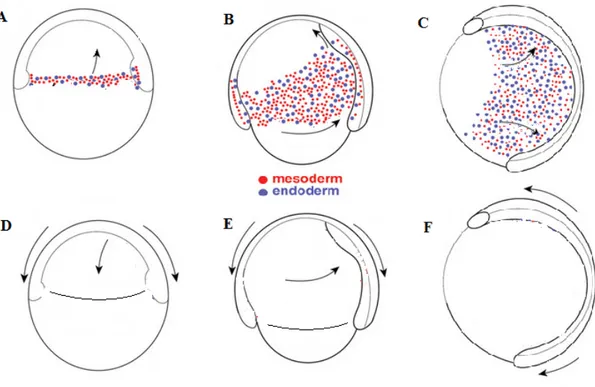
Neural tube formation in zebrafish implicates primary neurulation in the head and trunk and secondary neurulation in the tail as mentioned above in section 1.2.4. CE, as one of the very important processes for gastrulation and neurulation in zebrafish, happens after 50% epiboly when the cells surrounding the yolk migrate from ventral and lateral to dorsal regions. It consists of convergent and extension movements in different germ layers as demonstrated in
Fig. 9. At the early blastula stage, cells located in the animal pole start to migrate to the
vegetal pole. The cells at the blastoderm margin start to involute and form the prospective endoderm and mesoderm under the superficial layer of the prospective ectoderm. As endoderm and mesoderm originate from the same group of cells, the mechanisms involved in the separation of these two layers is not fully understood. These two layers will be discussed as combined in the following paragraphs.
31
1.5.2.1 PCP genes and convergent extension in zebrafish
Mutations in Wnt11 are responsible for the zebrafish mutant slb (silberblick) [114]. In zebrafish, Wnt11 expression is first detected in the germ ring at the shield stage and later in the paraxial head mesoderm and in the neuroectoderm at 90% epiboly. The main phenotype of slb
Fig. 9. Diagram for CE in zebrafish during gastrulation. At early gastrulation, mesendodermal cells form after involution from blastoderm margin cells and migrate mainly to the animal pole (A). Ectodermal cells basically migrate from the animal pole to the vegetal pole (D). At later stages, while migrating from the margin to the animal pole, ventro-lateral mesendodermal cells at the same time migrate to the dorsal midline (B). Overlying ectodermal cells continue the epiboly process and at the same time migrate from basal later to dorsal midline (E). At the end of gastrulation, mesendodermal cells continue migrating to the dorsal midline (C). Ectodermal cells elongate along the antero-posterior axis (F). All embryos were in lateral view with animal pole on the top and vegetal pole on the bottom. Modified from [113].