A mechanism for co-transcriptional recruitment of mRNA
localization factor on nascent mRNAs in budding yeast
par Zhi Fa Shen
Département de biochimie Programme de biochimie
Faculté de médecine
Thèse présentée à la Faculté des études supérieures en vue de l’obtention du grade de Philosophiae Doctor (Ph.D)
en biochimie
May 2010
Université de Montréal Faculté des études supérieures
Cette thèse intitulée :
A mechanism for co-transcriptional recruitment of mRNA
localization factor on nascent mRNAs in budding yeast
Présentée par : Zhi Fa Shen
a été évaluée par un jury composé des personnes suivantes :
Luc DesGroseillers Président-rapporteur Pascal Chartrand Directeur de recherche
Jacques Archambault Membre du jury François Bachand Examinateur externe
Résumé
Le transport et la localisation des ARN messagers permettent de réguler l’expression spatiale et temporelle de facteurs spécifiques impliqués dans la détermination du destin cellulaire, la plasticité synaptique, la polarité cellulaire et la division asymétrique des cellules. Chez S.cerevisiæ, plus de trente transcrits sont transportés activement vers le bourgeon cellulaire. Parmi ces transcrits, l’ARNm ASH1 (asymetric synthesis of HO) est localisé à l’extrémité du bourgeon pendant l’anaphase. Ce processus va entrainer une localisation asymétrique de la protéine Ash1p, qui sera importée uniquement dans le noyau de la cellule fille, où elle entraine le changement de type sexuel. La localisation asymétrique de l’ARNm ASH1, et donc de Ash1p, implique la présence de différents facteurs de localisation. Parmi ces facteurs, les protéines She (She1p/Myo4p, She2p et She3p) et les répresseurs traductionnels (Puf6p, Loc1p et Khd1p) participent à ce mécanisme. La protéine navette She2p est capable de lier l’ARNm ASH1 et va entrainer le ciblage de cet ARNm vers l’extrémité du bourgeon en recrutant le complexe She3p-Myo4p. Des répresseurs traductionnels régulent la traduction de cet ARNm et évitent l’expression ectopique de la protéine Ash1p pendant son transport. Alors que la fonction cytoplasmique de She2p sur la localisation des ARNm est connue, sa fonction nucléaire est encore inconnue.
Nous avons montré que She2p contient une séquence de localisation nucléaire non classique qui est essentielle à son import nucléaire médié par l’importine α (Srp1p). L’exclusion de She2p du noyau par mutation de son NLS empêche la liaison de Loc1p et Puf6p sur l’ARNm ASH1, entrainant un défaut de localisation de l’ARNm et de la protéine. Pour étudier plus en détail l’assemblage de la machinerie de localisation des ARNm dans le noyau, nous avons utilisé des techniques d’immunoprécipitation de chromatine afin de suivre le recrutement des facteurs de localisation et des répresseurs traductionnels sur les ARNm naissants. Nous avons montré que She2p est recruté sur le gène ASH1 pendant sa transcription, via son interaction avec l’ARNm ASH1 naissant. Puf6p est également recruté
nous avons détecté une interaction entre She2p et la plus grande sous-unité de l’ARN polymérase II (Rpb1p). Cette interaction est détectée avec la forme active en élongation de l’ARN polymérase II. Nous avons également démontré que She2p interagit avec le complexe d’élongation de la transcription Spt4p/Spt5p. Une délétion de SPT4 ou une mutation dans SPT5 (Ts spt5) à température restrictive empêche l’interaction entre She2p et Rpb1p, et diminue le recrutement de She2p au gène ASH1, entrainant un défaut de localisation de l’ARNm et un défaut de localisation asymétrique de la protéine Ash1p. De manière globale, nos résultats montrent que les facteurs impliqués dans la localisation cytoplasmique des ARNm et dans leur contrôle traductionnel sont recrutés de façon co-transcriptionnelle sur les ARNm naissants via leur interaction avec la machinerie de transcription, suggèrant un rôle important de la machinerie transcriptionelle dans la localisation des ARNm.
Mots-clés : localisation des ARNm, ARNm ASH1, séquence de localisation nucléaire
(NLS), She2p, recrutement co-transcriptionnel, ARN polymérase II, complexe Spt4-Spt5p, levure.
Abstract
Cytoplasmic transport and localization of messenger RNAs allows temporal and spatial expression of specific factors involved in cell fate determination, synaptic plasticity, cellular polarity or asymmetric cell division. In S. cerevisiae, over thirty transcripts are actively transported and localized to the bud tip of budding yeast. One of them, the ASH1 mRNA (for Asymmetric Synthesis of HO), is localized at the bud tip in late anaphase cells. This allows Ash1p, a transcriptional repressor of the HO endonuclease, to be sorted exclusively to the daughter cell nucleus, where it prevents mating type switching. Proper ASH1 mRNA localization and Ash1p asymmetric expression involve localization factors, which are part of the She-proteins (She1p/Myo4p, She2p and She3p), and translational repressors (the proteins Puf6, Loc1 and Khd1). The nucleo-cytoplasmic shuttling protein She2p binds the ASH1 mRNA and targets it for localization at the bud tip by recruiting the She3p-Myo4p complex. Translational repressors regulate the translation of ASH1 mRNA and avoid ectopic expression of the Ash1 protein during the transport of its transcript. While the cytoplasmic role of She2p in mRNA localization is known, its nuclear function is still unclear.
We now show that She2p contains a non-classical nuclear localization signal sequence (NLS) which is essential for its nuclear import via the importin α Srp1p. Exclusion of She2p from the nucleus by mutagenesis of its NLS disrupt the binding of Loc1p and Puf6p to the ASH1 mRNA, leading to defective mRNA localization and Ash1p sorting. To further investigate the assembly of the mRNA localization machinery in the nucleus, we used chromatin immunoprecipitation (ChIP) to follow the recruitment of localization factors and translational repressors on nascent localized mRNAs. We found that She2p is recruited on the ASH1 gene during transcription, via its interaction with the nascent ASH1 mRNA. Puf6p is also recruited on the ASH1 gene, but in a She2p-dependent manner. Interestingly, we detected an interaction between She2p and Rpb1p, the largest
subunit of RNA polymerase II in vivo. This interaction is independent of the RNA-binding properties of She2p, and involves the elongating form of the RNA polymerase II. We also found that She2p interacts with both members of the elongation factors Spt4p /Spt5p; Deletion of SPT4 or Ts spt5 mutants at restrictive temperature disrupted the interaction between She2p and Rpb1p, and then reduced the recruitment of She2p on the ASH1 gene, resulting in ASH1 mRNA delocalization and defective Ash1p sorting. Altogether, our results show that factors involved in cytoplasmic mRNA localization and translational control are recruited co-transcriptionally on nascent mRNAs via interation with the transcription machinery, pointing toward a role of the transcription machinery in the mRNA localization process.
Keywords: mRNA localization, ASH1 mRNA, nuclear localization signal (NLS), She2p,
Table of Contents
Résumé i Abstract iii Table of contents v List of tables x List of figures xi Abbreviations xivAmino acid codes xvii
Acknowledgments xviii
Dedication xix
Chapter I Introduction 1
1.1 . The intracellular transport and localization 2
of mRNAs in eukaryotes 1.1.1. Role of localized mRNAs in development 2
1.1.2. Functions of Localized mRNA in neurons 4
1.1.3. Localized mRNAs and cell motility 5
1.1.4. Localized mRNAs and asymmetric 7
divisions 1.1.5. Mechanisms of mRNA localization 8
1.1.6. Translational regulation and mRNA 10
localization 1.2. Yeast S. cerevisiae model organism and 10
localized mRNA. 1.2.1. The yeast life cycle 11
1.2.2. Mating type switching 12
1.2.3. Control of HO expression 15
1.2.5. Other bud localized mRNAs in S cerevisiae 18
1.3. Localized ASH1 mRNA as a model system to 20
study localization mechanism in eukaryotyes 1.3.1. The localization elements in ASH1 mRNA 20
1.3.2. The machinery of bud localized mRNA in yeast 21
1.3.3. She1/Myo4p- the yeast class V myosin motor 23
1.3.4. The adapter protein She3p 25
1.3.5. She2p- the mRNA binding protein 26
1.3.6. Translational regulators of ASH1 mRNA 31
1.3.6.1. Khd1p 32
1.3.6.2. Loc1p 33
1.3.6.3. Puf6p 34
1.4. Nucleo-cytoplasmic shuttling 35
1.4.1. Signals of nucleo-cytoplasmic shuttling and 36
transport factors 1.4.1.1. Nuclear localization signals and importin α 36
1.4.1.2. Nuclear localization signals and importins 37
1.4.1.3. Nuclear export signals and exportins. 38
1.4.2. Mechanism of receptor mediated nucleo- 40
cytoplamic shuttling 1.4.2.1. Nuclear pore complex (NPC) 39
1.4.2.2. RanGTP-RanGDP recycling 40
1.4.2.3. Model of nucleo-cytoplasmic shuttling 41
1.5. Coupling between transcription and mRNA maturation 43
1.5.1. Transcription and elongation factor Spt4-Spt5 complex 45
1.5.3. Coupling transcription with mRNA splicing 50
1.5.4. Coupling transcription with 3’end processing and 51
polyadenylation 1.5.5. Coupling transcription with mRNA export 52
1.5.6. Cytoplasmic mRNA Localization is initiated in the 54
Nucleus 1.6 Research objectives of this work 57
Chapter II Article: Nuclear shuttling of She2p couples ASH1 mRNA localization to its translation by recruiting Loc1p and Puf6p 2.1. Abstract 62
2.2. Introduction 63
2.3. Material and methods 65
2.3.1. Growth media and yeast strains 65
2.3.1. Immunoprecipitation and reverse transcription-PCR. 65
2.3.4. Fluorescence in situ hybridization and immunofluorescence 66
2.3.5. Protein expression and purification 67
2.3.6. GST pull-down assays 67
2.4. Results 68
2.4.1. Monomeric She2p interacts directly with the 68
importin α Srp1p in order to enter the 2.4.2. A non-classical NLS promotes the nuclear import of She2p 71
2.4.3. Nuclear import of She2p is required for proper localization 73
of the ASH1 mRNA at the bud tip and for the sorting of Ash1p. 2.4.4. Nuclear import of She2p is essential for the recruitment of 76
Loc1p and Puf6p to the ASH1 mRNA 2.5. Discussion 78
2.5.1. A non-classical NLS promotes the nuclear import of 79
She2p by binding importin α 2.5.2. Nuclear She2p couples mRNA localization and 80
translational repression 2.5.3. Roles of nuclear proteins in cytoplasmic mRNA localization 82
2.6. Acknowledgments 83
2.7. References 83
2.8. Figure legends 89
2.9. Supplementary data 103
2.9.1. Material and methods 103
2.9.1.1. Yeast three hybrid assay 103
2.9.1.2. Plasmid constructions 103
2.9.2. References 109
Chapter III Article: Cotranscriptional recruitment of She2p by RNA pol II elongation factor Spt4-Spt5 promotes mRNA localization to yeast bud 3.1. Abstract 113
3.2. Introduction 114
3.3. .Results 117
3.3.1. She2p is recruited cotranscriptionally at the 117
ASH1 and IST2 genes 3.3.2. She2p interacts with the elongating form of 119
RNA polymerase II in vivo 3.3.3. The interaction between She2p and RNA 121
polymerase II occurs via the transcription elongation factor Spt4-Spt5 3.3.4. Mutations in SPT4-SPT5 disrupt the cotranscriptional 122 recruitment of She2p to the ASH1
3.3.5. She2p is associated with genes coding for both 124
bud-localized and non-localized mRNAs 3.4. Discussion 126
3.5. Material and methods 129
3.5.1. Growth media and yeast strains 129
3.5.2. Chromatin immunoprecipitation 130
3.5.3. Fluorescence in situ hybridization (FISH) 132
3.6. Acknowledgments 133
3.7. References 134
3.8. Figure legends 141
3.9. Supplementary data 153
3.9.1. Material and methods 154
3.9.1.1. Co-Immunoprecipitation 154
3.9.1.2. Quantitative RT–PCR analysis 154
3.9.2. References 160
Chapter IV. Discussion 4.1. Nuclear factors are required for cytoplasmic 162
mRNA localization 4.2. Coupling transcription with ASH1 mRNA localization 169
4.2.1. She2p binds co-transcriptionally to nascent ASH1 mRNA 169
4.2.2. Association of She2p with Pol II via the Spt4-Spt5 174
complex is essential for the cytoplasmic fate of ASH1 mRNA Conclusion 180
List of tables
Chapter I
Table 1. The list of bud localized mRNA in yeast S. cerevisiae 19 Table 2. Trans-acting factors involved in ASH1 mRNA localization 25
Chapter II
Supplementary Table 1. Strains used in this study 105 Supplementary Table 2. Plasmid used in this study 106
Chapter III
Supplementary Table 1. Strains used in this study 156 Supplementary Table 2. Plasmid used in this study 158 Supplementary Table 3. PCR primers used for ChIP 159
List of figures
Chapter I
Figure 1. Examples of localized RNAs in different 7 organisms and cell types
Figure 2. Yeast life cycle 12 Figure 3. Mating type switching 14 Figure 4. Localization of ASH1 mRNA and Ash1p 17 to daughter cells of budding yeast.
Figure 5. Localizations elements of the ASH1 mRNA 22 and its second structure.
Figure 6. The ASH1 mRNA locasome 24 Figure 7. Model of ASH1 mRNA localization. 28 Figure 8. Schematic view of theShe2p homodimer with 31 each monomer in blue and grey.
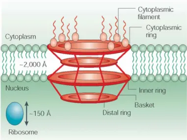
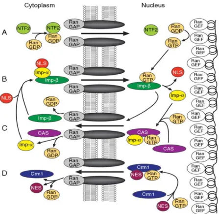
Figure 9. Schematic view of the nuclear pore complex. 40 Figure 10. Overview of some of the major nuclear 42 transport pathways in eukaryotic cells.
Figure 11. mRNA localization is a multistep process 44
Figure 12. Coupling between transcription and pre- 48 mRNA processing
Figure 13. Simplified model of mRNA export 54 Figure 14. oskar mRNA localization starts in the nucleus. 56
Chapter II
Figure 1. Monomeric She2p interacts directly with 95 the importin α Srp1p and is actively
imported into the nucleus.
Figure 2. Nuclear import of monomeric 96 She2p depends on Srp1p
Figure3. Identification of a NLS at the C-terminal 97 end of She2p
Figure 4. A non-classical NLS mediates nuclear 98 import of She2p.
Figure 5. Mutations in NLS of She2p impair interaction 99 with Srp1p and nuclear import of this factor.
Figure 6. The NLS-mutated She2 protein is as functional 100 as the wild-type She2p.
Figure 7. Nuclear import of She2p is required for proper 101 ASH1 mRNA localization and Ash1p sorting.
Figure 8. Nuclear She2p recruits Puf6p and Loc1p on 102 the ASH1 mRNA
Chapter III
Figure1. She2p -myc interacts cotranscriptionally 147 with bud-localized mRNAs.
Figure 2. She2p-myc interacts with the elongating 148 form of RNA polymerase II in vivo
Figure 3. She2p-myc interacts with RNA pol II via the 148 transcription elongation factor Spt4-Spt5
Figure 4. She2p-myc co-transcriptional interaction 149 with ASH1 depends on the transcription
Figure 5. Spt4-Spt5 is required for proper ASH1 150 mRNA localization and Ash1p sorting
Figure 6. She2p-myc interacts cotranscriptionally 151 with genes coding for both non-localized
and bud-localized mRNAs
Figure 7. Model for cotranscriptional recruitment 152 of localization factor Sh2p.
Supplementary data
Figure 1. Expression level of ASH1 mRNA in the 153 SPT4 and SPT5 mutant strains.
Chapter IV
Figure 1A. She2p is a phosphoprotein 165 Figure 1B. GST pull-down assay to detect interaction 165 between Srp1p and She2p-myc or
She2p-M2-myc from yeast extracts
Figure 2. Puf6p, Loc1p are recruited to ASH1 173 gene around the region of element E3.
Figure 3. Model for cotranscriptional recruitment 179 of ASH1mRNA trans-acting factors.
Abbreviations
Amp Ampicillin
ASH asymmetric synthesis of HO
ATP Adenosine triphosphate
BSA Bovine serum albumin
°C Grade Celsius
CTD Carboxy terminal domain
CTR Carboxy terminal region
Da Dalton
DAPI diamidino-2-phenylindol dihydrochloride
DEPC Diethylpyrocarbonate
DIC Differential interference contrast
DNA deoxyribonucleic acid
DNase Deoxyribonuclease
dNTPs Dideoxynucleotides
DTT Dithiotreitol
E. coli Escherichia coli
E1-3 ASH1 mRNA localization element
ECL Enhanced chemoluminiscence
EDTA Ethylenediaminotetraaceticacid
eIF Eukaryotic (translation-) initiation factor
ER endoplasmatic reticulum
et al. et alii (from Latin, “and others”)
FISH Fluorescence in situ hybridization
GDP Guanosine diphosphate GFP Green fluorescent protein GTP Guanosine triphosphate
HA Hemagglutinin
HEPES 4-(2-hydroxyethyl)-1-piperazineethanesulfonic acid hnRNP Heterogeneous nuclear ribonucleoprotein
HO endonuclease Homothallic switching endonuclease Ig Immunoglobulin kb Kilo bases l Litre LB-Medium Liquid-Broth-Medium M Molar mA Milliampere MDa Megadalton mg Milligramm μg Microgramm μl Mikroliter ml Millilitre mM Millimolar
mRNA Messenger ribonucleic acid NES Nuclear export signal
NLS Nuclear localization signal NP-40 Nonidet P-40 (Igepal-CA-630) NPC Nuclear pore complex
nt Nucleotide OD Optical density oligo Oligonucleotide ORF Open reading frame
PAGE Polyacrylamide gel electrophoresis
PBS Phosphate-buffered saline
PCR Polymerase chain reaction
pH Potential of Hydrogen Pol II Polymerase II
RBP RNA-binding domain
RNA Ribonucleic acid
RNP Ribonucleoprotein
rpm Rounds per minute
rRNA Ribosomal ribonucleic acid
RT-PCR Reverse transcriptase polymerase chain reaction
S Svedberg unit
S. cerevisiae Saccharomyces cerevisiae
SDS Sodium dodecyl sulfate
SHE Swi5p-dependent HO expression
TAE Tris-acetate-EDTA buffer
TAP Tandem affinity purification
TBS Tris-buffered saline
TEMED N, N, N’, N’-Tetramethylethylenediamine
Tris Trishydroxymethylaminomethane
tRNA Transfer ribonucleic acid
UTR Untranslated region
V Volt
wt Wild type
YEP Yeast Extract Peptone
Amino acid codes
Amino acid Three letter code One letter code
alanine ala A arginine arg R asparagine asn N aspartic acid asp D cysteine cys C glutamic acid glu E glutamine gln Q glycine gly G histidine his H isoleucine ile I leucine leu L lysine lys K methionine met M phenylalanine phe F proline pro P serine ser S threonine thr T tryptophan try W tyrosine tyr Y valine val V
Acknowledgments
I would like to express my sincere appreciation to my director Dr. Pascal Chartrand for providing me the opportunity to work in his laboratory and financial support, especially, for his essential guidance and inspirational encouragement in this project over the past five years. As well, I would like to thank my committee members Dr. Luc DesGroseillers, Dr. Jacques Archambault and Dr. François Bachand, for reading, correcting and evaluating this thesis.
I am grateful to Dr. Pascale Legault, Dr. Gertraud Burger, Dr. James G. Omichinski, Dr. Muriel Aubry, and Dr. Pierre Belhumeur, for their valuable discussions and suggestions. Special thanks go to my friend, Franck Gallardo for his French translation, thesis correction and, nice suggestions. I would like to thank Dr. Emmanuelle Querido for reading the manuscript.
I am also grateful to my colleagues at Dr. Pascal Chartrand’s laboratory: Dr. Anne-Laure Finoux, Amélie Forget, Anik St-Denis, Dr. Emilio Cusanelli and Faissal Ouenzar, for their collaboration, friendship, patience, and understanding. I also thank Dr. Nicolas Paquin and Catherine Olivier for their technical support during the first three years.
I want to thank Dr. Luc DesGroseillers, Dr. Gerardo Ferbeyre, Dr. Stephen Michnick, and all the members who have worked at their laboratory, for their help with equipments and reagents.
Finally, I want to thank my wife for everything she did to make my life easier. To my daughters, who never really know what I was doing in the lab, but their smile and healthy growing are enough to help me through my Ph.D.
To my mother
Chapter I
Following transcription in the nucleus, most RNAs are exported through the nuclear pores into the cytoplasm, where they are translated into a distinct set of proteins. For specific intracellular compartments and their localized distributions, most proteins are targeted to their destination on the basis of signals in the peptide sequence. In contrast to these post-translational event, messenger RNA localization has classically been considered to be a mechanism used to spatially and temporally restrict gene expression to specific regions within the cell or embryo. This mechanism has obviously different advantages. For instance, it can rapidly generate large amounts of proteins locally, offers the potential for a local regulation of protein expression and protects the rest of the cell from proteins that might be toxic or deleterious in other cellular compartments.
1.1 The intracellular transport and localization of mRNAs in eukaryotes
Twenty-seven years ago, beta-Actin mRNA that accumulates in the myoplasm of Ascidian eggs was discovered as the first localized mRNA (Jeffery et al. 1983). Since then, examples of localized transcripts dramatically increase year by year, encoding for either cytoplasmic, nuclear, secreted, membrane-associated or cytoskeletal proteins from yeasts to fruit flies, frogs, zebrafish and mammals (Bashirullah et al., 1998). The localization of specific mRNAs has been shown to play an important role during the development of multicelluar organisms and also in processes like synaptic transmission, cell motility and asymmetric cell division (Du et al., 2007; Kiebler et al., 2000; Kislaukis et al., 1997; Jan et al., 1998).1.1.1 Roles of localized mRNAs in development
The localization of specific mRNA has been shown to be essential for the establishment of the antero-posterior axis of Drosophila and animal-vegetal axis of Xenopus (Lasko 1999; Kloc et al., 2001). The disruption of their localization machineries results in gross abnormalities during the development of these organisms. In Drosophila, embryonic axis specification is determined by two localized mRNAs: bicoid, which is localized to the anterior pole, and nanos, which is localized to posterior pole (Figure 1A) (Van Eeden et al. 1999). These localized RNAs set up opposing protein gradients that define the anterior and posterior axes of the embryo.
In addition to bicoid and nanos mRNA, at least four other types of localized mRNA are required for the determination of the abdomen and the pole cells, the founders of germline lineage (St Johnston 1993). All of them are localized to the posterior region of the egg. The first mRNA to reach the posterior of the oocyte is oskar mRNA, which is sufficient to define the site of pole plasm formation. Unlike Bicoid and Nanos, however, Oskar protein does not seem to play a direct role in determining cell fates in the embryo and acts instead to nucleate the polar granules of the pole plasm by recruiting other mRNAs and proteins to the posterior pole (Breitweiser et al. 1996). One of the most important mRNA recruited to the posterior of the egg is nanos mRNA (Wang & Lehmann 1991). The second one is germ cell-less (gcl) mRNA that localizes to the posterior of the oocyte after stage 11 of oogenesis. The role of Gcl is unclear, but seems to function in formation of ectopic polar buds (Jongens et al. 1994). The third mRNA is the non-coding transcript Pgc, which localizes to the posterior at around stage 11 and is a component of the polar granules (Nakamura et al. 1996). The fourth class of localized RNAs specifically required for pole cell formation are the large and small ribosomal RNAs encoded by the mitochondrial genome (mt rRNA). In a recent striking study involving throughput and
high-resolution in situ hybridizations of over 3000 transcripts in Drosophila embryos, 71% were found to be expressed in spatially distinct patterns (Lecuyer et al., 2007), suggesting that the role of localization of mRNAs in cellular development may be much more prevalent than previously thought.
As in Drosophila, similar processes occur in oocytes of the frog Xenopus, where the mRNA encoding the T-box transcription factor VegT localizes to the vegetal pole and induces endodermal and mesodermal cell fates in the embryo (King et al., 2005). In addition, homologues of the localized maternal mRNAs that play a role in pole cell development in Drosophila have been implicated in the determination of the primordial germ cells (PGCs) in some other organisms. For instance, three Nanos homologues have been identified in the nematode Caenorhabditis elegans, and one of them has been shown to be a component of the P-granules, which segregate into primordial germ cells (PGCs), suggesting the existence of a conserved pathway for specifying the germline lineage (Subramaniam & Seydoux 1999, Schisa et al. 2001).
1.1.2 The localization and transport of mRNA in neurons
There is increasing evidence that an important aspect of gene expression in neurons involves the targeting of certain mRNAs to particular subcellular domains. It is now well known that localized mRNAs in dendrites encode proteins of different functional types. For example, the high molecular weight microtubule-associated protein (MAP2) mRNA and an activity-regulated cytoskeleton-associated protein (ARC) mRNA both encode certain cytoskeletal proteins; CaMKII α mRNA encode the α subunit of calcium/calmodulin-dependent protein kinase II that has a role in synaptic plasticity (Steward et al. 2001). During brain development, localization of mRNAs in axonal growth cones allows neurons to respond to local environmental cues as the distal axonal processes navigate toward their
synaptic partners (Lin and Holt, 2007). More recent studies indicate that hundreds of mRNAs are present in neuronal processes, where they encode diverse functions, suggesting that neurons may use the general mechanism of RNA targeting for different purposes at different times in their life time (Eberwine et al., 2002; Martin and Zukin, 2006).
Some evidences suggest that localized mRNAs contain a localization element in the 3’ untranslated region (UTR) required for sufficient localization in neurons. For example, MBP mRNA contains an A2RE localization element in its 3’ UTR, recognized by Heterogeneous nuclear ribonucleoprotein (hnRNP) A2 (Ainger et al., 1997). The A2RE has been shown to be sufficient to transport mRNAs into neuronal dendrites (Shan et al., 2003). The hnRNP E1, another RNA-binding protein that regulates translation of specific mRNAs, is recruited to A2RE RNA granules by binding to hnRNP A2. This recruitment inhibits translation of A2RE RNA during granule transport (Linda et al. 2006). Similar studies show that CamKIIα mRNA contains a 94 nucleotide-long element in its 3’UTR, required for dendritic localization in neurons. Deletion mutants of the 3’UTR of CaMKII mRNA drastically reduce the amount of transcript in mice dendrites (Mori et al. 2000; Ouyang et al. 1999). Interestingly, recent works show that microtubules play a critical role in the dendritic localization of RNA granules in neurons (Kiebler et al., 1999; Tang et al., 2001; Kanai et al., 2004). The purified large RNA granules from mouse brain that associate with the tail of the kinesin motor protein KIF5 contain localized mRNAs (like CamKIIa mRNA) and mRNA binding proteins (like Staufen and FMRP). This result further confirmed the importance of this mechanism in neurons (Kanai et al., 2004).
1.1.3 Localized mRNAs and cell motility
Localization of β-actin messenger RNA to sites of active actin polymerization modulates cell motility during embryogenesis and differentiation (Condeelis et al. 2005).
The localization of β-actin mRNA requires a conserved 54-nucleotide cis-acting element located in the 3’ UTR of the mRNA, called the “zipcode” (Condeelis et al. 2005). In chicken developing neurons, a protein of 68 kDa zipcode binding protein 1 (ZBP1) associates with the zipcode to regulate the localization of β-actin mRNA at the leading edge of fibroblasts and growth cones for enrichment of ß-actin protein and forward movement of growth cones (Figure 1B). Moreover, a mutated zipcode unable to localize was unable to bind ZBP1 (Zhang et al 2001; Condeelis et al. 2005). More recent evidence shows that β-actin translation can be regulated by the phosphorylation of ZBP1 through Src kinase (Huttelmaier et al. 2005). In Xenopus, the homolog of ZBP1, Vg1RBP (also known as Vera), is required for the motility of neural crest cells. This protein binds β-actin mRNA and controls its localization in retinal growth cones. During this process, Netrin-1 induces the movement of Vg1RBP granules into filopodia and plays an important role in the localization and translation of β-actin mRNAs in growth cones (Piper et al; Yisraeli et al. 2005; Leung et al. 2006). In cell movement, site-directed actin polymerization is induced at their surface, via the activation of the actin-related protein 2/3 (Arp2/3) complex by the WASp (Wiskott-Aldrich Syndrome protein) family proteins (Machesky et al. 1999). A recent study shows that the Arp2/3 complex required for actin polymerization is localized to the leading protrusions of migrating cells, further supporting that the Arp2/3 complex is targeted to its site of function by mRNA localization (Mingle et al. 2005).
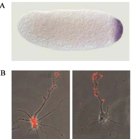
Figure 1. Examples of localized RNAs in different organisms and cell types
(A) The localization of maternal nanos mRNA is shown in purple at the posterior of an activated, unfertilized Drosophila egg (Lipshitz et al., 2000). (B) β-actin mRNA (Red) localization in the neurite and growth cone (left) and β-actin protein highly enriched in growth cone and filopodia (right) for forward movement of growth cones (Bassell et al., 2001).
1.1.4 Localized mRNAs and Asymmetric Cell Division
Asymmetric cell division gives rise to two daughter cells with different cell fates which are partly generated by active localization of the cell fate determinant mRNA at only one daughter cell during mitosis (Bernardoni et al. 1999). The examples of asymmetric segregation of mRNAs during cell division can be found in Drosophila, Yeast and other
higher eukaryotes. The asymmetric division of Drosophila neuroglioblasts provides a well characterized example where mRNA localization is responsible for determining different sibling cell fates (Akiyama-Oda et al. 1999, Bernardoni et al. 1999). In Drosophila embryos, Neuroblasts usually divide into an apical ganglion mother cell (GMC) and a new neuroblast daughter at the basal. One important cell fate determinant is prospero mRNA, which is localized to the cell cortex of the basal side of the GMC (Broadus et al., 1998). In the budding yeast Saccharomyces cerevisiae, specific mRNAs are localized to daughter cells to asymmetrically segregate cell-fate determinants during development (Paquin and Chartrand. 2008). Interestingly, in a mollusc embryo, IoEve mRNAs, encode a protein involved in anterior–posterior axis specification, associate with centrosomes in the cell and are also subsequently distributed asymmetrically to the pericentriolar matrix in a microtubule-dependent manner during division (Lambert and Nagy, 2002).
1.1.5 Mechanisms of mRNA localization
Studies in diverse organisms have demonstrated the existence of several potential mechanisms by which mRNAs can be localized, like the active directional transport of RNA along cytoskeletal elements or random cytoplasmic diffusion and trapping. However, the most common mechanism invoked for mRNA localization involves the active transport of the mRNA along the cytoskeleton (Bashirullah et al. 1998; St Johnston et al. 1995; Lipshitz et al. 2000). The observation that most localized mRNAs get delocalized in the presence of microtubule depolymerizing drugs supports the active transport model (St Johnston et al.1995). For some transcripts, actin filaments must remain intact in order to maintain a proper localization (Long et al. 1997; Sundell et al. 1991). Localized mRNAs are recognized by their localization machinery via specific cis-acting localization elements or “zipcode” present within the transcript sequence (Bashirullah et al. 1998). For most localized mRNA, these zipcode have been found in the 3’ UTR, while a few, like the yeast
ASH1 mRNA or the Drosophila gurken and bitesize mRNAs, also contain additional zipcodes in the coding sequence or 5’UTR, suggesting that localization elements may be quite variable between mRNAs, both in term of sequence and secondary structure (Chartrand et al. 1999; Gonzalez et al. 1999; Serano et al.2003; Thio et al. 2000).
During the transport process, localized mRNA is first packaged into a “locasome”, which contains the mRNA and several trans-acting factors, such as RNA-binding proteins and molecular motors (Oleynikov et al. 1998). Several localized transcripts have been shown to form large particles or granules when microinjected or expressed in vivo (Ainger et al. 1993; Ferrandon et al. 1994; Bertrand et al. 1998). While this observation has been frequently reported, the composition and function of these particles are still mostly unknown. Currently, a few RNA-binding protein have been reported to directly bind the zipcode region of a localized mRNA, such as ZBP1 binding to the chicken β-actin mRNA, Vera binding to the Xenopus Vg1 mRNA, hnRNP2 binding to MBP mRNA in oligodendrocytes, staufen and VPS36 binding to the Drosophila bicoid mRNA or She2p to the yeast ASH1 mRNA (Kelsey et al. 2009).
Recent evidences suggest that several molecular motors are involved in the transport of localized mRNAs in different organisms. The type V myosin Myo4p has been shown to actively transport the ASH1 mRNA along the actin cytoskeleton of budding yeast (Bertrand et al. 1998). More recently, microtubule-associated motors like dynein and kinesin were shown to be involved in the sorting of the Drosophila bicoid and oskar mRNA, respectively (Schnorrer et al. 1999; Brendza et al. 2000). Adaptor proteins which associate with both mRNA and molecular motor, like She3p in yeast and Swallow in Drosophila, have also been identified (Schnorrer et al. 1999; Takizawa et al. 2000).
1.1.6 Translational regulation and mRNA localization
Translational control is essential for the localization of zipcode-containing mRNAs and can be regulated by their localization machinery (Lipshitz et al. 2000). This mechanism prevents the untimely translation of mRNAs, which could have deleterious effects on cellular function. Several localized mRNAs are not translated unless they are properly localized. For instance, in Drosophila oocytes, the translation of unlocalized oskar mRNA is repressed by the binding of the Bruno protein on its 3’UTR (Kim-Ha et al. 1993). The unlocalized nanos mRNA is also translationally repressed by the binding of Smaug protein on its 3’UTR (Smibert et al. 1996; Crucs et al. 2000). In both case, the proper localization of these mRNAs results in the activation of their translation (Lipshitz et al. 2000). The mechanism by which some of these proteins repress translation has been recently identified. This mechanism reveals that eIF4E-binding proteins compete for the eIF4G binding site on mRNA, disrupting the eIF4E- eIF4G interaction that is essential for the recruitment of the 40S ribosomal subunit to the mRNA (Gingras et al. 1999). An example is the Drosophila Cup protein, an eIF4E-binding protein that blocks the interaction between eIF4E and eIF4G. Cup interacts with Smaug to repress the translation of unlocalized nanos mRNA, and with Bruno to repress the translation of the oskar mRNA (Nelson et al. 2004; Nakamura et al. 2004).
1.2 The yeast S. cerevisiae as model organism to study localized mRNA
Saccharomyces cerevisiae, the budding yeast, is the common yeast used in baking and brewing industry. Yeast was the first eukaryotic organism whose complete genomic sequence was established (Dujon 1996; Goffeau et al., 1996). With its 12.8 Mb, the yeast genome is divided up into 16 chromosomes ranging in size between 250 kb and >2500 kband about 200 times smaller than the human genome, but less than four times bigger than that of E.coli. The complete genome sequence now defines some 6000 open reading frames (ORFs), most of which are likely to encode specific proteins.
In biomedical science, the buding yeast is a popular "model" organism in laboratories because it is a unicellular eukaryote whose many essential cellular processes are conserved between yeast and other higher eukaryotes. Among all eukaryotic model organisms, S. cerevisiae combines several advantages, for instance, it is an unicellular organism and has a short generation time, so they can be easily cultivated and grown on defined media. In addition, its entire genome is known and it can be easily transformed
with genes from other sources. Importantly, in yeast, cell architecture and fundamental cellular mechanisms can be successfully investigated and many sophisticated genetic tools and biochemical approaches have been developped, making it a convenient and powerful model system to study eukaryotic cellular processes.
1.2.1 The yeast life cycle
Budding yeast can live with either two genomes (diploid) or one (haploid). In either case, it divides by forming buds, the production of a small outgrowth from the parent cell. In nature, and when nutrients are available, yeast reproduces asexually mainly in the diploid stage. Budding starts at late G1-phase. At the end of M-Phase, the emerged daughter bud has reached the size of the mother cell. The subsequent cell division results in two cells, termed “mother cell” and “daughter cell”. Under conditions of nutrient deprivation, diploid cells may undergo meiosis and revert to the haploid stage by sporulation. After meiosis, the formed tetrade consists of usually four ascospores, two of which with the mating type a and two with mating type α. When nutrients are available, the spores germinate and the resulting cells may either multiply asexually as haploids or may serve as a gamete (Figure 2). In yeast, this sexual process is termed “mating” and occurs
when two haploid cells with different mating types fuse to form a diploid (a/α) zygote. Cells from each haploid type produce a secreted mating-pheromone. These mating type-specific pheromones, termed a- and α-factor, act to synchronize the cell cycle of the mating partners and to prepare cells for mating (Herskowitz, 1988).
Figure 2: The yeast life cycle.
(A) The cell cycle of S. cerevisiae (Source: Lodish et al., 1999). Yeast cells multiply asexually by budding. At the end of G1, a bud emerges from the mother cell. Prior to cytokinesis, the daughter bud has reached size of the mother cell. After cell division, the resulting cells grow in G1 until reaching the appropriate size for bud formation. (B) Morphology of S. cerevisiae cells (Source: Herskowitz, et al., 1988). Upper panel shows an unbudded cell in G1 (a) and cells with different bud sizes S (b), G2 (c). Mating of a- and α-haploids leads to formation of a diploid (a/α) zygote (d). The zygote is able to produce diploid (a/α) daughter cells by budding. Bud emerges often at the neck (e).
1.2.2 Mating type switching
The cell mating type is determined by the MAT locus. After cytokinesis of a haploid cell, the interconversion between MATa and MATα is due to the protein product of the HO
gene, which is an endonuclease that promotes mating-type switching in S. cerevisiae (Herskowitz, 1988; Nasmyth, 1982). Mating type switching occurs only in mother cells but not in daughter cells during late G1.
Switching to the opposite mating type occurs by gene conversion at the active MAT locus. The template used for this conversion is issued from the silenced HMLa or HMRa locus of the opposite type. In haploid cells, expression of one of the two alleles leads to cells with either mating type a or α, whereas diploid cells express both alleles (Mating type a/α). MATα codes for two proteins termed α1p and α2p. The transcription factors Mcm1p and α1p are responsible for the activation of α-specific genes (Shore et al. 1995). In contrast, α2p and Mcm1p serve to repress a-specific genes (Wolberger, 1998). The MATa-locus codes for two proteins, of which only A1p is known to have a biological function. A1p and α2p form a heterodimer, which is required to repress haploid-specific genes (Li et al., 1995). Consequently, there is no expression of α-specific genes in a-cells because α1p and α2p are missing, whereas through the activation by Mcm1p, a-specific genes are expressed (Bruhn et al. 1994).
Mating type switching occurs when either HMLa or HMLα is recombined into the transcriptionally active MAT-locus by gene conversion (Hicks et al. 1977; Strathern et al.1982). Thus, the MAT-locus is replaced by the genetic information of the opposite mating type. This recombination event is initiated by a double-strand break, catalyzed by the haploid-specific Ho endonuclease. In diploid cells, binding of the heterodimer A1p/α2p inhibits HO expression (Herskowitz, 1992). Yeast strains used for biological studies in laboratories have lost their ability to change mating types due to a point mutation in the HO gene. These strains are called heterothallic and are more accessible to genetic manipulations because of a stable haploid phase.
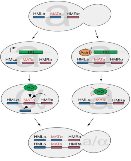
Figure 3: Mating type switching. After division of a haploid cell, only the mother but not the daughter cell can switch the mating type. Switching to the opposite mating type occurs by gene conversion at the active MATlocus. Ho endonuclease cuts the active MAT locus, initiating this replacement. In daughter cells, the transcriptional repressor Ash1p inhibits the expression of HO, thereby repressing mating type switching (Source: Darzacq et al., 2003).
1.2.3 Control of HO expression
The HO gene product of Saccharomyces cerevisiae is a site-specific endonuclease that initiates mating type interconversion during late G1 in mother cells. The transcription activation program of HO is cell cycle regulated. The expression occurs only transiently and starts during late mitosis, when Cdk1p is inactive and ends during late G1-phase, when Cdk1p is reactivated (Nasmyth, 1993). The HO promoter can be divided in two regions: a distant upstream region called URS1 (“Upstream Regulatory Sequence“), which regulates mother cell expression specificity, and a proximal region called URS2 that controls HO cell-cycle regulation (Nasmyth, 1993).The late-G1 specificity of HO transcription depends on a heteromeric factor, SBF, which is composed of the Swi4 and Swi6 proteins. Mother-cell specificity involves a second site-specific DNA-binding factor, Swi5, which is synthesized in the G2 and M phases and only enters the nucleus at the end of mitosis. Swi5 enters mother and daughter nuclei and binds to two sites of URS1 regions within the HO promoter in equal amounts. This event triggers the recruitment of the SWI/SNF chromatin-remodelling complex to URS1 and URS2, resulting in recruitment of RNA polymerase lI and the general factors TFIIB/TFIIH for transcription initiation (Bhoite et al., 2001; Cosma et al., 1999; Krebs et al., 1999). However, the highly concerted recruitment of all these transcription factors does not occur in daughter cell nuclei.
The ASH1 gene (Asymmetric synthesis of HO) was identified through the isolation of mutants in which the daughter cells were defective in HO repression and so were able to switch mating type. Ash1p is a 66-kDa zinc-finger transcriptional repressor that inhibits HO transcription through its asymmetric accumulation in the daughter nucleus in late anaphase (Bobola et al, 1996; Sil et al. 1996). Ash1p contains a domain which is highly homologous to the zinc-finger domain of the erythroid cell nuclear protein GATA-1 (Bobola et al., 1996; Sil et al. 1996). All GATA-like factors bind to GATA motifs, which leads to either transcriptional activation or repression. Recently, the Ash1-binding
consensus sequence, YTGAT, was identified within the HO promoter (Maxon et al. 2001). This consensus, which is related to the canonical (A/T)GATA(A/G) sequence bound by most GATA factors, is present at least 20 times within the upstream repression sequence 1 (URS1) region of the HO promoter (Maxon et al. 2001). Ash1p has two principal domains: the C-terminal DNA-binding domain, which binds to the YTGAT consensus within URS1 of HO; and the amino-terminal domain, which is devoted to the repression of HO transcription (Maxon et al. 2001). The asymmetric control of HO expression, which is caused by the sorting of Ash1p to daughter cells, explains why only haploid mother cells can undergo mating type switching. Moreover, a recent study show that Ash1p also involved in daughter cell size control using quantitative time-lapse microscopy (Di Talia et al., 2009).
1.2.4 ASH1 mRNA in yeast S. cerevisiae
The Ash1p localizes to the daughter cell nucleus in late anaphase where it prevents mating type switching. The asymmetric accumulation of this cell fate determinant in only daughter cell nuclei is mediated by the products of five genes, termed SHE1–SHE5 (Swi5p-dependent HO Expression), each of which has a specific function (Jansen et al, 1996). They were identified in a genetic screen for factors that are required for asymmetric HO-expression in yeast cells (Jansen et al., 1996). SHE mutants fail to restrict Ash1p to the daughter cells nucleus, therefore, HO transcription is repressed in both mother and daughter cells, resulting in none of cells switching. Interestingly, except for Ash1p sorting, SHE genes have no function in SWI regulation.
Some of the She proteins have been found to be localized asymmetrically to a crescent at the cortex of the daughter bud. Ash1p is unlikely to be directly targeted by She-proteins transport because Ash1p appears in nuclei of daughter cells at much later stages of the cell cycle, at a moment when the She-proteins are no longer localized (Chang et al. 1996). Thus, it soon became clear that asymmetric localization of Ash1p originates from
the asymmetric localization of the ASH1 mRNA (Figure 4) (Long et al., 1997). Each of the SHE gene products is essential for targeting the ASH1 mRNA to the bud tip (Long et al., 1997; Takizawa et al., 1997) (see section 1.3.2.).
The post-transcriptional level of regulation of gene transcription through the asymmetric localization of mRNA is well known in different organisms (Kloc et al, 2002). The key elements of ASH1 mRNA localization to the bud tip of daughter cell have been identified as follows: the cis-acting sequences within ASH1 mRNA; the secondary structure of the ASH1 mRNA; the cytoskeleton; a macromolecular complex that includes the She1–3 proteins, some factors that are required for the recognition of ASH1 mRNA in the nucleus and translational repression before proper bud tip localization (Paquin et al., 2008).
Figure 4: Localization of ASH1 mRNA and Ash1p to daughter cells of budding yeast. ASH1 mRNA (light yellow) is actively transported to the bud tip of daughter cells where it is translated into Ash1p (green), the repressor for HO expression. Ash1p is exclusively sorted to daughter cell nucleus to prevent mating type switching (Long et al., 1997).
1.2.5 Other bud-localized mRNAs in S. cerevisiae
In Saccharomyces cerevisiae, ASH1 mRNA is localized to bud tip of daughter cell where it is translated into Ash1p, a cell-specific transcription factor (Long et al. 1997; Takizawa et al. 1997). This represented the first description of RNA localization in a single-cell eukaryote. A second localized mRNA was also found, the IST2 mRNA, whose localization to the cortex of daughter cells creates a higher concentration of Ist2 protein in the bud, and this asymmetry is maintained by a septin-mediated membrane diffusion barrier at the mother-bud neck (Takizawa et al. 2000). A microarray-based screen identified a set of 22 additional mRNAs, all of which become localized to bud tip in a She-dependent manner (Shepard et al., 2003). These messages encode a wide variety of proteins, including several involved in stress responses, cell wall maintenance and membrane proteins. And recent studies show that over thirty mRNA are localized to the bud of daughter cells (Table 1) (Aronov et al. 2007; Oeffinger et al., 2007; Shepard et al. 2003). Among these
bud-localized mRNAs, including ASH1 mRNA, some were found to cofractionate with ER microsomes in a She2-dependent manner. Thus, asymmetric mRNA transport and cortical ER inheritance are connected processes in yeast (Aronov et al. 2007; Schmid et al. 2006). However, the biological significance of localizing these RNAs remains to be elucidated, since asymmetric distribution of several of these proteins also occurs in the absence of mRNA transport (Takizawa et al. 2000).
Table1: The list of bud localized mRNA in yeast S. cerevisiae
Gene Function
ASH1 M Transcription BRO1 Stress transduction CLB2 Cyclin B
CPS1 Carboxypeptidase DNM1 Mitochondrial fission EGT2 Cellulase
ERG2 Sterol isomerase IST2 Tranporter
MID2 Membrane receptor MMR1 Unknown
SRL1 Unknown
TPO1 Polyamine transport WSC2 Membrane receptor
TAM41 Import into the mitochondrial matrix TCB3 Lipid-binding protein
EAR1 Unknown
TCB2 Membrane trafficking
KSS1 Mitogen-activated protein kinase LCB1 Endoplasmic reticulum, lipid synthesis MET4 Transcription
MTL1 MID2-like
CDC42 Small rho-like GTPase RHO3 Cell polarity
YPT1 ER-to-Golgi step of the secretory pathway SEC1 Exocytose SEC3 Exocytose EXO84 Exocytose SRO7 Exocytose SRO77 Exocytose SEC4 Exocytose
BUD8 Bud-site selection
CIS3 Glycoprotein constituent of the cell wall
1.3. Localized ASH1 mRNA as a model system to study localization
mechanism in eukaryotyes
Data so far have shown that more than thirty mRNAs are localized to the bud of daughter cells in budding yeast Saccharomyces cerevisiae, among these localized mRNAs, the ASH1 mRNA is the best characterized example. Because mRNA transport and localization in yeast shares many features with RNA localization in higher eukaryotes, including cis-acting sequences within localized mRNA, formation of a large ribonucleoprotein (RNP) localization complex, cell cytoskeleton and molecular motors, some factors that are required for mRNA recognition in the nucleus, thus, the mechanism of ASH1 mRNA localization can be used as a paradigm to explore the molecular basis of this process.
1.3.1 The localization elements in ASH1 mRNA
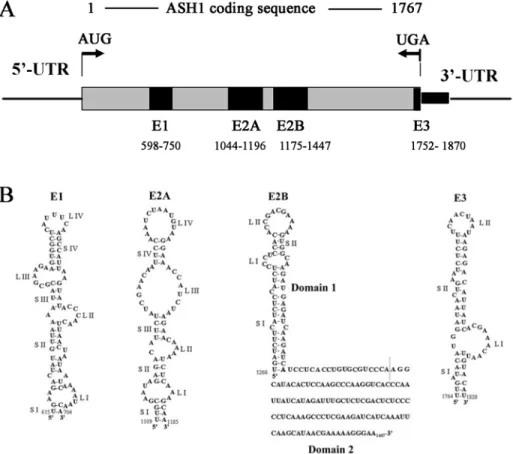
To address the cis-acting sequences required for the localization of the ASH1 mRNA, different fragments of ASH1 were inserted into a reporter mRNA and the cytoplasmic distribution of these chimeric mRNAs was determined by in situ hybridization (Chartrand et al., 1999; Gonzalez et al., 1999). These studies have shown that there are four minimal localization sequences within the ASH1 mRNA. Among these elements, three are located within the coding region of the ASH1 mRNA, and they have been named as E1 (spanning between nucleotides 598 and 750), E2A (between 1044 and 1196) and E2B (between 1175 and 1447), whereas the remaining element (E3), which is located primarily within the ASH1 3’UTR, spans seven nucleotides before and 67 after the stop codon (Figure 5A). Each single element is sufficient to localize mRNA at the bud of yeast cells (Chartrand, et al. 1999; Gonzalez, et al. 1999).
These localization elements are predicted to form RNA secondary structures containing stem-loops (Figure 5B) (Chartrand, et al. 1999; Chartrand, et al. 2001; Gonzalez, et al. 1999), and this secondary structure is crucial for ASH1 mRNA localization (Bertrand et al, 1998). For example, mutations in the stem loop of the E3 element can affect the localization of ASH1 mRNA (Chartrand et al, 1999). Recent work shows that the ASH1 mRNA contains a similar loop-stem-loop structure with a highly conserved CGA triplet in one loop and a single conserved cytosine in the other loop. Mutating these conserved nucleotides, or the stem separating them, resulted in the delocalization of a reporter mRNA (Olivier, et al. 2005). In addition, in an independent approach, a predicted single-stranded core CG dinucleotide appears to be an important component of the RNA-protein interface, although other nucleotides contribute in a context-dependent manner (Jambhekar et al., 2005).
1.3.2 The machinery of bud localized ASH1 mRNA
SHE1–SHE5, at beginning, were identified in a genetic screen for factors required for asymmetric HO-expression in yeast cells (Jansen et al., 1996). After that, it is soon clear that each of the SHE gene products was essential for targeting the ASH1 mRNA to the bud tip (Long et al., 1997; Takizawa et al., 1997). So far, several trans-acting factors involved in the localization of the ASH1 mRNA in yeast have been identified (Table 2). In budding yeast, ASH1 mRNA association with these trans-acting factors triggers the packaging into a locasome (Figure 6), which is actively transported to the bud in late anaphase, ensuring the exclusive translation of Ash1p in the daughter cell (Long et al, 1997; Takizawa et al, 1997). During this process, ASH1 mRNA localization is mediated by four cis-elements within the mRNA and each of them can be recognized by She2p, an RNA-binding protein (Long, et al. 2000; Bohl, et al. 2000). The link between the RNA-binding protein She2p and the motor She1p/Myo4p is mediated by She3p (Long, et al. 2000; Takizawa, et al. 2000). These studies lead to the development of a working model for this pathway (Figure 7) (Chartrand,
et al. 2001). In this model, the ASH1 mRNA is first recognized in the nucleus by proteins which act as “tags” for the localization machinery. Once in the cytoplasm, the localization machinery assembles on the tagged mRNA as a complex called “locasome”, the locasome is transported along the actin cytoskeleton to the bud tip where it becomes anchored. Once localized, the mRNA is translated and Ash1p appears at the bud tip and diffuses back to the daughter cell nucleus.
Figure 5: Localization elements of the ASH1 mRNA and its second structure.
(A) The position of localization elements in ASH1 mRNA. (B) Three of the localization elements, namely E1, E2A and E2B, are located within the ASH1 coding sequence. The E3 element spans the stop codon and the first 100 nucleotides of the 3’ untranslated region (UTR) (Source: Chartrand et al., 2002; Olivier et al., 2005).
1.3.3 She1/Myo4p- a yeast class V myosin motor
She1p, also called Myo4p, is the motor protein required for active transport of the ASH1 mRNA locasome along the actin cytoskeleton to the bud tip of daughter cell (Bertrand et al., 1998; Haarer et al., 1994; Jansen et al., 1996; Münchow et al., 1999). In budding yeast, there are two class V unconventional myosins, Myo4p and Myo2p (Titus, 1997). Both of them non-processively localize to the bud tip of daughter cells (Karpova et al., 2000; Lillie et al. 1994; Schott et al.1999). Myo2p is required to set up the orientation of the mitotic spindle (Yin et al., 2000), and has a role in the polarized transport of secretory vesicles (Govindan et al., 1995; Johnston et al., 1991; Lillie, et al. 1994; Pruyne et al., 1998; Schott et al., 1999), inheritance of the vacuole and the Golgi apparatus (Catlett et al., 2000; Catlett, et al, 1998; Rossanese et al., 2001). Myo4p, a single-headed and nonprocessive class V myosin in budding yeast, transports >20 different mRNAs asymmetrically to the bud (Long et al., 1997; Reck-Peterson et al., 2000). Since Myo4p does not contain intrinsic RNA-binding activity, accessory proteins are necessary to interface the myosin with ASH1 mRNA localization elements, some evidence show that Myo4p associates with ASH1 mRNA, which is dependent on She2p and She3p (Münchow et al. 1999, Takizawa et al. 2000). Furthermore, it was observed that Myo4p colocalizes with ASH1 mRNA-containing particles (Bertrand et al. 1998, Takizawa et al. 2000), and in living yeast cells, Myo4p directly transports ASH1 mRNA to daughter cells (Bertrand et al. 1998, Beach et al. 1999). While type V myosins are thought to require dimerization for processive movement, Myo4p is strictly monomeric at physiologic concentrations (Heuck et al., 2007). A recent study shows that the rod region of Myo4p contains the primary binding site for She3p and is essential for correct localization of ASH1 mRNA (Bookwalter et al., 2009). Interestingly, Myo4p is also involved in the inheritance of cortical ER (Estrada et al., 2003), and recent works suggested that both Myo4p-dependent processes (mRNA localization and cortical ER inheritance) are tightly coordinated (Aronov et al., 2007; Schmid et al., 2006).
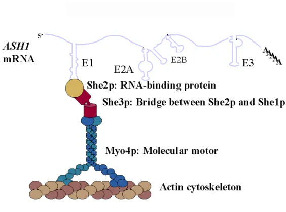
Figure 6: The ASH1 mRNA locasome
ASH1 mRNA contains four localization elements within its sequence and each of them can be recognized by She2p, an RNA-binding protein, containing a basic helical hairpin motif for RNA binding activity. The link between the RNA-binding protein She2p and the motor She1p/Myo4p is mediated by She3p (Chartrand et al., 2001).
1.3.4 The adapter protein She3p
She3p is a novel protein with no significant homology to any known proteins. Some studies have shown that She3p colocalizes with Myo4p and ASH1 mRNA at the bud tip and plays an important role in the association of Myo4 motor protein with ASH1 mRNA-She2p complex (Kruse et al., 2002; Münchow et al., 1999; Takizawa et al. 2000). Furthermore, by two-hybrid, three-hybrid, and coimmunoprecipitation experiments, the N-terminal half of the She3p was found to interact with the C-terminal tail of Myo4p (Münchow, et al. 1999; Böhl et al. 2000; Long et al. 2000). Moreover, sucrose density gradients demonstrated a cosedimentation of Myo4p together with She3p, suggesting a tight and permanent association of both proteins (Böhl et al., 2000). A recent study show that She3p binds to the rod of yeast Myosin V and prevents it from dimerizing, forming a single-headed motor
complex (Hodges et al., 2008). The C-terminus of She3p provides the binding to She2p. Thus, She3p serves as an adapter that docks the myosin motor onto an ASH1 mRNA– She2p ribonucleoprotein (RNP) complex. Interestingly, She3p might have an influence on ASH1 mRNA- She2p interaction, the evidence comes from binding of She2p to ASH1 mRNA zipcode was enhanced in the presence of She3p by a gel-shift assays (Böhl et al., 2000). She3p association with ASH1 mRNA was observed to be dependent on She2p, suggesting that the Myo4p-She3p complex interacts with the ASH1 mRNA cis-acting localization elements through She2p (Münchow, et al. 1999; Böhl et al. 2000; Long et al. 2000). In addition, She3p also possesses a novel activity required for ASH1 mRNA localization since some mutants of She3p are defective for ASH1 mRNA localization and retain the ability to associate with Myo4p and She2p (Landers et al., 2009). Importantly, this novel function for She3p could be negatively regulated by phosphorylation (Landers et al., 2009).
1.3.5 She2p- the mRNA binding protein
She2p is also a novel protein with no homology to known proteins in higher eukaryotes. As a small 28 kDa RNA binding protein, She2p is the key component in the assembly of the ASH1 mRNA locasome (Long et al, 1997; Takizawa et al, 1997).Yeast two-hybrid analysis and experiments using recombinant She2p and She3p demonstrated that She2p directly interacts with She3p through a domain in the C terminus of She3p (Böhl et al. 2000, Long et al. 2000), suggesting that She2p is required to link the Myo4p-She3p complex to ASH1 mRNA. Disruption of SHE2 abolishes Myo4p’s association with the mRNA (Jansen et al., 1996; Münchow et al., 1999). She2p is also required to mediate the association of She3p to ASH1 mRNA (Böhl et al., 2000). These results suggest that She2p is the factor acting directly on ASH1 mRNA, independent of She3p and Myo4p.
Although no classical RNA-binding motifs can be identified in the She2 protein, it directly and specifically interacts in vitro and in vivo with each of the ASH1 mRNA cis-acting elements, but with apparently weak affinity (Böhl et al., 2000; Darzacq et al., 2003; Long et al., 2000). This was well demonstrated by electrophoretic gel mobility shift assays (Böhl et al., 2000), and further confirmed using filter binding experiments (Niessing et al., 2004), where purified recombinant She2p displayed specific binding to ASH1 mRNA cis-acting localization elements.
Figure 7: Model of ASH1 mRNA localization.
(1) Synthesis of the ASH1 transcription in late anaphase, association of ASH1 mRNA with She2p, interaction with Loc1p in the nucleus. (2) Export of ASH1 mRNA-She2p RNP from nucleus. (3) Assembly of the locasome by association of the newly exported RNP with She3p and She1p/Myo4p. (4) Locasome atively transported to bud tip of the daughter cells along the actin cytoskeleton. (5) Dissociation of the locasome after proper localization at the bud. (6)- (7) Anchoring and translation. ASH1 mRNA was translated into Ash1p after it was accurately anchored at the bud, then Ash1p enters the daughter cell nucleus where prevent mating type switching (Source: Chartrand et al., 2001).
The first 70 amino acids residues of the She2p are apparently essential for RNA-binding activity, since a deletion of this region abolishes the ability of She2p to associate with ASH1 mRNA, resulting in an accumulation of this mutant in the nucleus (Kruse et al., 2002). This was the first evidence indicating that She2p’s nuclear export is dependent on RNA binding. A cluster of arginine residues within this region (R43, R44, R52 and R63) as well as asparagine residue 36 (N36) have been found to play a key role in She2p RNA-binding activity (Gonsalvez et al., 2003). Interestingly, classical RNA-RNA-binding motifs, like the RNA recoginition motif (RRM), the arginine rich motif (ARM), the RGG box and the double-stranded RNA-binding motif (dsRBD), all of them contain arginine residues critical for RNA binding activity. However, so far, no evidence shows that any of the arginine residues in She2p directly contact ASH1 mRNA or they play a more indirect role in mRNA association (Gonsalvez et al., 2003). Mutations of N36S, R43A, R44A, R52A, R52K, R63A, and R63K lead to a loss of RNA binding and consequently, to defective mRNA localization (Gonsalvez et al., 2003; Niessing et al., 2004). Moreover, block of mRNA export caused the accumulation of She2 in the nucleus as well (Kruse et al., 2002), and more recent evidence show that She2 accumulates in nucleoli during mRNA export block (Du, et al., 2008). This indicates that She2 can enter the nucleus for the binding of its RNA target and thus is able to shuttle between the nucleus and cytoplasm. However, other experiments using She2p mutants N36S and R63K, which are specifically defective for ASH1 mRNA association, do not support the assertion that nuclear export of She2p is dependent of both mRNA transport and the ability of She2 to bind mRNA (Gonsalvez et al., 2003). While She2p is not excluded from the nucleus and hence could bind localization substrates in the nucleus prior to export to the cytoplasm, resolution of these apparently contradictory results will require further investigation into the nuclear function of She2p nucleo-cytoplasmic shuttling.
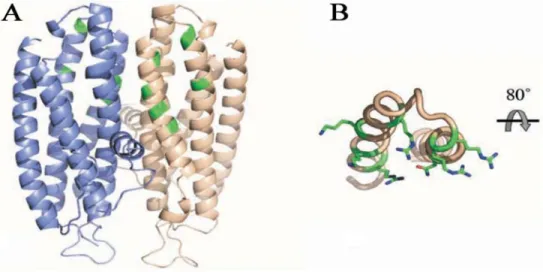
The crystal structure of She2p was recently determined and revealed that it is an almost exclusively α-helical protein unrelated to any previously described RNA-binding
protein (Niessing et al., 2004). Moreover, She2p forms symmetric homodimers that contains basic helical hairpin motif for RNA binding (Figure 8). Mutations in the motif result in loss of mRNA binding in vitro and defective mRNA transport in vivo (Niessing et al., 2004; Gonsalvez et al., 2003). A possible explanation for the need of She2p dimerization is that additional surface regions from both subunits are required for efficient RNA binding by She2p. However, these additional surface regions seem not enough for its RNA-binding functions. By using analytical ultracentrifugation, Müller and colleagues found that She2p adopts a tetrameric structure at physiological concentrations, which is required for RNA binding, efficient mRNP assembly, and mRNA localization in vivo (Müller, et al. 2009).
Indeed, besides ASH1 mRNA, She2p are also essential for some of other localized mRNAs to the bud of daughter cells (Takizawa, et al. 2000; Shepard et al., 2003; Oeffinger et al., 2007; Aronov et al., 2007). Meanwhile, recent evidence indicates that She2p is
required for the localized messenger ribonucleoprotein (mRNP) particles that comigrate with tubular endoplamic reticulum (ER) structures to the bud. Moreover, She2p associates with cortical ER independently of polysomes and She3p-Myo4p complex, suggesting that asymmetric mRNA transport and cortical ER inheritance are connected processes in yeast (Schmid et al., 2006; Aronov et al., 2007). However, it is still unknown how She2p asscociates with ER.
Figure 8: Schematic view of theShe2p homodimer with each monomer in blue and grey. (A) She2p forms a stable symmetric homodimer. Each monomer consists of five α helices. Amino acids and the basic helical hairpin RNA-binding motif are highlighted in green. (B) View of the basic helical hairpin motif of the gray monomer. Amino acids of the basic helical hairpin motif required for RNA binding are highlighted in green with their side chains shown (Source: Müller et al., 2007).
1.3.6 Translational regulators of ASH1 mRNA
The ASH1 mRNA needs to be translationally repressed to be efficiently localized to the bud tip of daughter cells, and this requires the presence of translational regulators. In the past years, several regulators have been identified and characterized, giving rise to the evidence that it requires more than just a functional motor complex to target a transcript effectively. Among them, Khd1p, Loc1p and Puf6p, which bind directly to the ASH1 mRNA, are clearly required to control ASH1 mRNA translation during its localization to the yeast bud tip (Irie et al., 2002; Long et al., 2001; Gu et al., 2004; Paquin et al., 2007; Komili et al., 2007).