by
Georges MATAR
MANUSCRIPT-BASED THESIS PRESENTED TO ÉCOLE DE
TECHNOLOGIE SUPÉRIEURE IN PARTIAL FULFILLMENT FOR THE
DEGREE OF DOCTOR OF PHILOSOPHY
Ph.D.
MONTREAL, SEPTEMBER 8, 2020
ÉCOLE DE TECHNOLOGIE SUPÉRIEURE
UNIVERSITÉ DU QUÉBEC
BY THE FOLLOWING BOARD OF EXAMINERS
Mr. Jean-Marc Lina, Thesis Supervisor
Department of Electrical Engineering, École de Technologie Supérieure
Mr. Georges Kaddoum, Co-supervisor
Department of Electrical Engineering, École de Technologie Supérieure
Mr. Luc Duong, President of the Board of Examiners
Department of Systems Engineering, École de Technologie Supérieure
Mrs. Rita Noumeir, Member of the Jury
Department of Electrical Engineering, École de Technologie Supérieure
Mrs. Carolina Bessega, External Independent Examiner Straidigi AI
THIS THESIS WAS PRESENTED AND DEFENDED
IN THE PRESENCE OF A BOARD OF EXAMINERS AND THE PUBLIC ON JUNE 4, 2020
co-director Professor Georges Kaddoum for the support of my Ph.D study and research. For their patience, motivation, and immense knowledge. The time and effort they have invested in my thesis have allowed me to successfully bear the challenges I have faced during my Ph.D journey. I thank Professor Lina for trusting my research intuition and ideas and supporting them while providing a leadership and scientific guidance to help me realize and get the results. I thank Professor Kaddoum for the motivation and support he provided, and for sharing his expertise and experience in research to help me face the challenges encountered during my PhD.
The data collection and part of my research work have been in the Hôpital du Sacré-Coeur de Montreal (HSCM) hospital. I would like to thank Professor Julie Carrier for motivation and support that she provided during my PhD. Her presence, remarks and words added a positive vibe and a smile to the team in the hospital. I express my sincere grattitude to Mrs. Sonia Frenette, who has never hesitated to provide full support regarding the experimental setup in the hospital. I thank everyone in the center of advanced research in sleep medicine (CÉAMS), including sleep professionals, technicians, researchers, and students, for making the workplace in the hospital friendly.
I would like to thank the Professors managing the Laboratoire d’analyse et de traitement d’information médicale (LATIS) laboratory, Professor Catherine Laporte and Professor Rita Noumeir, for their encouragement and their support through the Ph.D years.
I have a deep gratitude for my laboratory colleagues. They call me "Owner", for them, I am the owner of the laboratory as I have been always there, working. I am lucky to have such colleagues, specially Houssem, who supported me as a friend and colleague, and never hesitated to share his experience in research to help. I am grateful to have Nassim, Nicolas, Jihad, Jawad, Lei, Brieuc, Bo, Arnaud, Haythem, Rafic, and all LATIS students. They are ex-colleagues, and friends from now on. Their presence made the laboratory a pleasant place to work. Last but not least, I would like to thank my friends and family for their continuous support and love they
provided since childhood, and specially throughout the Ph.D years. It would have not been possible without your support and motivation, grateful to have each one of you!
RÉSUMÉ
Au moins 50% de la population mondiale des personnes âgées, dont le pourcentage est en croissance rapide, souffre d’un sommeil perturbé. Les études du sommeil sont une approche servant d’outil de diagnostic aux professionnels de la santé. Actuellement, la méthode conventionnelle d’étude du sommeil est la polysomnographie (PSG) enregistrée dans un laboratoire du sommeil. Cependant, la PSG est intrusive, nécessite des techniciens qualifiés, et nécessite une grande allocation des ressources matérielles et professionnelles. Avec l’introduction de technologies commerciales dans le domaine médical, des méthodes alternatives ont été conçues pour essayer de donner une estimation fiable des stades et de la qualité du sommeil. Cependant le manque de validation ou d’un consensus scientifique concernant la fiabilité de ces appareils demeure un défi pour les chercheurs et l’industrie. Ces outils peuvent désormais être utilisés à domicile plusieurs nuits. Les activités cardio-respiratoires et physiques sont les mesures physiologiques les plus prometteuses pour détecter les stades du sommeil sans PSG complète. Les impacts et les budgets liés aux troubles du sommeil sont phénoménaux, ce qui met l’accent sur le fait que le domaine nécessite davantage de recherche. Cette thèse vise à fournir au lecteur une perspective de recherche multidimensionnelle sur les études du sommeil et la surveillance des paramètres physiologiques pendant le sommeil à l’aide de techniques et d’appareils d’acquisition des données non-intrusifs d’acquisition de données. Dans cette optique, nous présentons une revue exhaustive de la littérature de recherche sur les développements en matière d’évaluation non-intrusive du sommeil. En outre, une catégorisation des approches actuelles est présentée en fonction de considérations méthodologiques, allant des protocoles d’acquisition de données et des mesures physiologiques au traitement de l’information. Nous examinons trois fonctions physiologiques principales qui pourraient potentiellement être explorées pour faire avancer les études du sommeil non intrusives basées sur des fonctions du système nerveux autonomes, principalement l’activité cardiaque, la respiratoire et le mouvement. Cette dernière revue nous a aidé à réaliser nos trois contributions. Premièrement, nous proposons une méthode autonome pour classifier les quatre postures du sommeil chez des adultes en bonne santé: dorsal, abdominal, latéral gauche et droit, sans capteurs ou câbles attachés sur le corps ni aucune contrainte imposée au sujet, en utilisant un matelas à capteur de pression. Contrairement à la majorité des travaux similaires antérieurs, les postures dorsale et ventrale ont été séparées avec succès dans la classification. Nous avons constaté que l’utilisation de l’information sur la répartition du poids corporel ainsi que sur la forme et les contours du corps contribue à une meilleure classification et, par conséquent, aide à séparer les postures dorsale et ventrale. Les résultats sont prometteurs pour une surveillance discrète de la posture de manière satisfaisante. La méthode peut être utilisée dans les études du sommeil, les procédures post-chirurgicales ou les applications nécessitant une identification de la posture sur le lit. Deuxièmement, nous tirons parti des résultats de la classification de la posture fiables pour mettre au point une surveillance non-intrusive de la fréquence respiratoire au lit adaptive à la posture à l’aide de
capteurs de pression. Tout au long de cette contribution, nous avons démontré qu’avec un traitement de signal approprié, le matelas à capteurs de pression pourrait être utilisé de manière interchangeable avec les ceintures respiratoires agréées pour l’usage médical par l’association Américaine de la médecine du sommeil (AASM), offrant ainsi une solution plus pratique et nécessitant moins de ressources pour les sujets et les professionnels de la santé. Et troisièmement, nous proposons et validons cliniquement une méthode de classification basée sur l’apprentissage machine profond pour identifier non-intrusivement les stades du sommeil pendant la nuit en utilisant un matelas à capteurs de pression. Bien que les résultats présentés ne donnent pas encore satisfaction pour prétendre l’adoption éventuelle de capteurs de pression de drap de lit en clinique du sommeil, nous estimons que le potentiel d’une telle application mérite d’être largement reconnu et approfondi. Nous soutenons que la méthode proposée pourrait constituer une étape vers des études du sommeil non-intrusives nécessitant moins de ressources. Par la suite, nous formulons des recommandations et des mesures pratiques pour les projets futurs cherchant à contribuer à des études non-intrusives de sommeil en utilisant des matelas à capteur de pression. Nous discutons des limites et des défis auxquels sont confrontées les solutions actuelles, et nous mettons en évidence des domaines de recherche ouverts, qui, nous l’espérons, ouvriraient la voie à de futurs efforts de recherche sur la question: comment évaluer les stades et la qualité du sommeil de manière moins intrusive et fiable?
Mots-clés: Surveillance non-intrusive du sommeil, matelas à capteurs de pression, respiration,
mouvements respiratoires, prévention des uclères/plaies, posture du corps humain, cartographie de la distribution du poids du corps, actigraphie, mouvements du corps, surveillance du patient, polysomnographie, activité cardiaque.
ABSTRACT
At least 50% of the world’s elderly population, whose range is fast growing, experience disturbed sleep. Sleep studies have become an extensive approach serving as a diagnostic tool for health-care professionals. Currently, the gold-standard is Polysomnography (PSG) recorded in a sleep laboratory. However, it is obtrusive, requires qualified technicians, is time consuming, and expensive. With the introduction of commercial off-the-shelf technologies in the medical field, alternatives to the conventional methods which may be now used at home on several nights have been conceived to ensure sleep stages and sleep quality detection. However, the lack of validation or scientific consensus regarding the reliability of these devices remains a challenge for researchers and the industry. Cardio-respiratory and physical activities remain the most promising physiological measurements to detect sleep stages without complete PSG. The statistically proven impacts and budgets related to sleep disorders are phenomenal, showing that the field needs more research. This thesis aims at providing the reader with a multidimensional research perspective on sleep studies and physiological parameters monitoring during sleep using unobtrusive data acquisition techniques and apparatus. In this vein, we present an exhaustive review of developments made in unobtrusive sleep assessment. Additionally, a categorization of current approaches is presented based on methodological considerations, from data acquisition frameworks and physiological measurements, to information processing. We discuss the three main physiological functions that could potentially be explored to advance unobtrusive sleep studies based on autonomous physiological functions, mainly cardiac, breathing, and movements activities. The latter review helped us achieve our three contributions. First we propose an autonomous method for classifying the four state-of-art human body lying postures (HBLP) in healthy adults subjects: supine, prone, left and right lateral, with no sensors or cables attached on the body and no constraints imposed on the subject, using a pressure sensor mattress. In contrast to the majority of previous similar works, prone and supine postures were successfully separated in the classification. We found that using the body weight distribution along with the shape and edges contributes to a better classification performance, and hence, helps separate supine and prone postures. The results are satisfactorily promising towards unobtrusively monitoring the posture for ulcer prevention. The method can be used in sleep studies, post-surgical procedures or applications requiring HBLP identification. Second, we leverage the reliable results of posture classification in order to develop an unobtrusive posture-adaptive in-bed breathing rate (BR) monitoring system using bed-sheet pressure sensors. Throughout this contribution, we demonstrated, that with proper signal processing, pressure sensor mattresses could be used interchangeably with respiratory belts, which have been approved for medical use by the American Association of Sleep Medicine (AASM), providing a more convenient solution for both subjects and health professionals. Third, we propose and clinically validate a deep learning based classification method for unobtrusive identification of sleep stages using bed-sheet pressure sensors. Although the results presented in this paper are primary and not yet satisfactory to claim an eventual adoption of bed-sheet pressure sensors in sleep clinics, we believe that the
potential of such applications is worth being recognized and further explored. We argue that the proposed method could be a step towards unobtrusive sleep studies that require less resources. Subsequently, we give recommendations and practical steps for future endeavors seeking to bring contributions to unobtrusive sleep studies using pressure sensor mattresses. We discuss limitations and challenges facing current solutions, and we highlight open research areas, which we hope would pave the way for future research endeavors addressing the question: how to assess sleep stages and sleep quality less intrusively, and reliably?
Keywords: Unobtrusive sleep monitoring, pressure sensor mattress, respiration, breathing
movements, bed pressure ulcer prevention, human body lying posture, body pressure mapping, actigraphy, body movements, patient monitoring, polysomnography, cardiac activity.
INTRODUCTION . . . .1
CHAPTER 1 LITERATURE REVIEW . . . 13
1.1 An overview of sleep stages, disorders, and monitoring techniques . . . 13
1.1.1 Sleep stages . . . 13
1.1.2 The burdensome impact of sleep disorders . . . 14
1.1.3 State-of-the-art sleep monitoring techniques . . . 15
1.2 Unobtrusively measurable physiological patterns during sleep . . . 18
1.2.1 Breathing activity patterns . . . 19
1.2.2 Body movement patterns . . . 21
1.2.3 Cardiac activity patterns . . . 22
1.3 Physical activity based unobtrusive sleep monitoring technologies . . . 24
1.3.1 Sleep actigraphy . . . 25
1.3.2 Sleep video monitoring . . . 25
1.3.2.1 Posture and movement monitoring . . . 26
1.3.2.2 Breathing activity monitoring . . . 27
1.4 Cardiac based unobtrusive sleep monitoring methods . . . 29
1.4.1 HRV Analysis in sleep studies . . . 29
CHAPTER 2 ARTIFICIAL NEURAL NETWORK FOR IN-BED POSTURE CLASSIFICATION USING BED-SHEET PRESSURE SENSORS . . . 33
2.1 Abstract . . . 33
2.2 Introduction . . . 34
2.3 Materials and methods . . . 38
2.3.1 System . . . 38
2.3.2 Data acquisition . . . 39
2.3.3 Feature extraction . . . 41
2.3.3.1 Histogram of oriented gradients . . . 42
2.3.3.2 Local Binary Patterns . . . 44
2.3.3.3 Principal Component Analysis . . . 45
2.3.4 Feed-Forward ANN (FFANN) . . . 46
2.3.4.1 Network Architecture . . . 47
2.3.4.2 Training and error back-propagation scheme . . . 48
2.3.5 Nested Cross-Validation . . . 48
2.4 Experimental Results . . . 50
2.5 Discussion . . . 52
CHAPTER 3 KALMAN FILTERING FOR POSTURE-ADAPTIVE IN-BED BREATHING RATE MONITORING USING BED-SHEET
PRESSURE SENSORS . . . 57 3.1 Abstract . . . 57 3.2 Introduction . . . 58 3.3 Methodology . . . 62 3.3.1 System hardware . . . 63 3.3.2 Experimental protocol . . . 64 3.3.3 Dataset . . . 65 3.3.4 Data pre-processing . . . 65
3.3.5 Breathing movements signal prediction model . . . 67
3.3.6 Parameters selection and initialization . . . 70
3.3.7 Validation methodology . . . 71
3.4 Experimental results and discussion . . . 73
3.4.1 Posture-adaptivity: . . . 78
3.4.2 Clinical validation: . . . 79
3.4.3 Inter-subject variance: . . . 79
3.4.4 Pressure sensor mattress choice: . . . 79
3.5 Conclusion and future perspectives . . . 80
CHAPTER 4 DEEP-LEARNING BASED UNOBTRUSIVE SLEEP STAGES CLASSIFICATION USING BED-SHEET PRESSURE SENSORS . . . 83
4.1 Abstract . . . 83
4.2 Introduction . . . 84
4.3 Materials and methods . . . 89
4.3.1 Subjects recruitment . . . 89
4.3.2 Materials and experimental procedure . . . 90
4.3.3 Data pre-processing . . . 94
4.3.4 Network architecture . . . 96
4.3.5 Model training . . . 99
4.4 Experimentation results and discussion . . . 101
4.5 Conclusion and future perspectives . . . .108
CONCLUSION AND RECOMMENDATIONS . . . 111
5.1 Recommendations and remarks for future works . . . .113
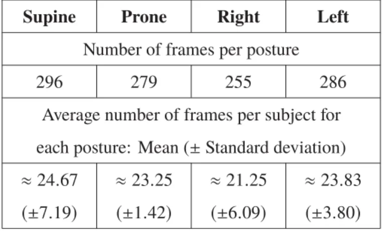
Table 2.1 Database structure table: number of frames for each posture, and the
average number of frames per subject per posture . . . 39 Table 2.2 An exhaustive comparison of the proposed method to the
state-of-the-art methods that use pressure sensor mattresses to classify body
posture unobtrusively . . . 53 Table 3.1 Testing confusion matrix of the ANN posture classifier . . . 72 Table 3.2 Posture-wise optimal q and r coefficients . . . 75 Table 3.3 Existing methods that focus on breathing monitoring during sleep
using bed pressure sensors . . . 76 Table 3.4 PCC values calculated on the BRs derived from the two measurement
methods for each of the four postures . . . 77 Table 4.1 Average age, height, weight, BMI, SL, TST, SE calculated for the 12
subjects . . . 94 Table 4.2 Detailed summary of the output shapes of the classification network’s
layers . . . 98 Table 4.3 Model training parameters and computational resources . . . 99 Table 4.4 Confusion matrix and results comparison . . . .102 Table 4.5 A comparison of the proposed method to the state-of-the-art methods
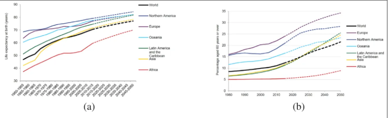
Figure 0.1 Population aging: (a) life expectancy at birth, and (b) percentage of people aged 60 years or older Adapted from United Nations, Department of Economic and Social Affairs, Population Division
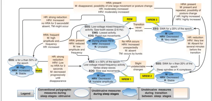
(2015). . . .1 Figure 1.1 Physiological changes during sleep stages in accordance with the
AASM . . . 14 Figure 1.2 Typical acquisition protocol and general workflow in a PSG
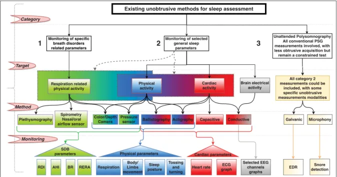
procedure. 22 sensors and wires, attached on the body . . . 16 Figure 1.3 Unobtrusive sleep monitoring methods . . . 18 Figure 1.4 HRV changes with sleep stages. R-R intervals [ms] plotted over
time [min] during: Awake, Light (Including stages 2 or 3), deep
(stages 3), and REM sleep. . . 23 Figure 1.5 Respiratory effort signal derived using a pressure sensor mattress
from a subject sleeping in a dorsal position . . . 29 Figure 2.1 A three steps general work-flow diagram showing bed pressure ulcer
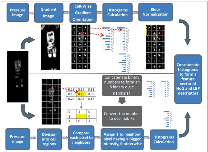
prevention using a pressure sensor mattress: from data acquisition
to analysis and action . . . 35 Figure 2.2 Feature extraction work-flow diagram showing HoG and LBP feature
vectors extraction to decode shape and patterns information in the
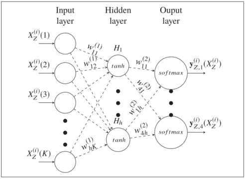
sample pressure images . . . 41 Figure 2.3 A diagram showing the FFANN architecture with weights, activation
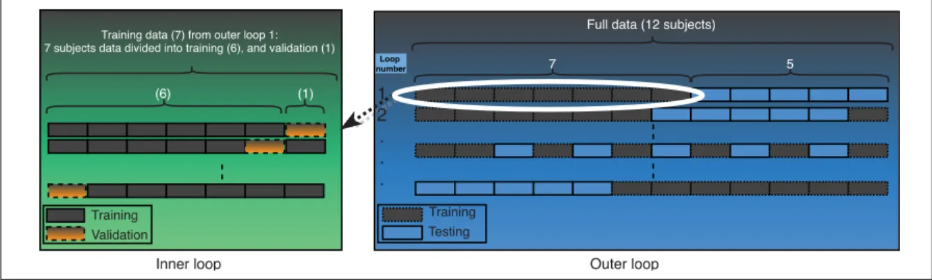
functions, and the input-output parameters and layers . . . 44 Figure 2.4 Nested validation scheme . . . 46 Figure 2.5 Body pressure distribution images of 4 postures: supine (a) to (d),
left (e) to (h), prone (i) to (l), and right (m) to (p) . . . 49 Figure 2.6 Training (a) and testing (b) confusion matrices of the selected
classifier model, showing the classification accuracy and confusion for each of the 4 classes in terms of percentage (%) and number of
Figure 2.7 Histogram of mean testing prediction accuracy performed in the outer loop of the nested validation, showing the number of models
that yield to corresponding classification accuracy . . . 52 Figure 3.1 Work-flow diagram of bed pressure ulcer prevention using a pressure
sensor mattress . . . 61 Figure 3.2 Body pressure distribution images of 4 postures: supine (a), prone
(b), left (c), and right (d) . . . 72 Figure 3.3 BA plots for the BRs calculated over 30 seconds windows for the 12
subjects (a color per subject) lying in each of the 4 postures . . . 74 Figure 4.1 Conventional PSG acquisition protocol and sensors placement on
the body . . . 86 Figure 4.2 Work-flow diagram of unobtrusive sleep stage classification using
bed-sheet pressure sensors. . . 89 Figure 4.3 Pressure images in the first row and differences of pressure images
in the second row for dorsal (a), prone (b), right (c), and left (d)
positions. Image dimensions are in pixels, and colorbar unit is PSI . . . 93 Figure 4.4 Classification network’s architecture: 3D-CNN + deep ANN layers . . . 97 Figure 4.5 Leave one subject out cross validation scheme . . . 101
ANN artificial neural network
AR auto-regressive
AUC area under curve
BA bland altman
BPM breaths per minute
BR breathing rate
CE cross-entropy
CÉAMS center for advanced research on sleep medicine
CGM conjugate gradient methods
CPAP continuous positive airway pressure
CPM counts per minute
DI digital integration
ECG electrocardiogram
EEG electroencephalogram
EMG electromyogram
EOG electroocculogram
FFANN feed-forward artificial neural netowrk
FP false positive
FSR force sensitive resistor
HF high frequency
HoGs histogram of oriented gradients
HRV heart rate variability
HSCM hôpital du sacré-coeur de Montréal
IR infrared
ICSD-3 international classification of sleep disorders-third edition LOA
LOE limit of agreement
LOE line of equality
LBPs local binary patterns
LF low frequency
MA moving-average
NIR near infrared
NREM non rapid eye movement
OSA nbstructive sleep apnea
OSAS obstructive sleep apnea syndrome
PCA principal component analysis
PCC pearson correlation coefficient
PSG polysomnography
PLM periodic limb movement
PLMD periodic limb movement disorder
PLMS periodic limb movement during sleep
PSD power spectral density
REM rapid eye movement
RGB red-green-blue
RBD rapid eye movement behavior disorder
R/K Rechtschaffen and Kales
RIP respiratory inductance plethysmography
RLS restless legs syndrome
RRV respiratory rate variability
SA sino-atrial node
SBD sleep breathing disorders
SBSM society of Behavioral Sleep Medicine
SCG scaled conjugate gradient
SD standard deviation
SDB sleep disordered breathing
SDNN standard deviation of N-N intervals
TN true negative
ToF time of flight
TP true positive
TST total sleep time
USB Universal Serial Bus
becoming phenomenal where at least half of the people over the age of 65 experience disturbed sleep (Monane, 1992). With the perpetual increase of the elderly population percentage, as shown in figure 0.1, this number is expected to fast grow with a tendency to continue, leading to an increase in sleep disorders around the world with increases in the demanding budgets and care (United Nations, Department of Economic and Social Affairs, Population Division, 2015). Researchers have shown the direct socioeconomic impact on the population and the public health, including work accidents, lack of productivity, isolation, depression and numerous more impacts were documented (Leger, 2000; Metlaine, Leger & Choudat, 2005; Sigurdson & Ayas, 2007).
(a) (b)
Figure 0.1 Population aging: (a) life expectancy at birth, and (b) percentage of people aged 60 years or older
Adapted from United Nations, Department of Economic and Social Affairs, Population Division (2015)
Moreover, sleep disorders are the result of physiological disturbances and an inducing factor to others, making sleep a very important consciousness state to maintain a healthy well-being and long-term physiological functions (Altevogt, Colten et al., 2006) and (Barnes & Drake, 2015).
Increased risk of depression, stroke, hypertension, heart attach, obesity, and diabetes, are some of the deleterious cumulative long-term effects induced by sleep disorders (Medic, Wille & Hemels,
2017). Over the last decades sleep research has revealed the widespread impacts of sleep alterations on human health. Altevogt et al. (2006) have reviewed the prevalence, manifestations, comorbodities, risk factors, and etiology of the most common sleep disorders, including restless legs syndrome, parasomnias, sleep-related psychiatric disorders, sleep-related neurological disorders, sleep loss, sleep-disordered breathing, insomnia, circadian rhythm sleep disorders, and narcolepsy.
Despite the fast spreading and impact of sleep disorders, the majority of people remain not willing to resorting to the current medical sleep evaluation (LeBlanc, Morin, Bélanger, Ivers & Coté, 2011). The reasons for that reduction include the burdensome physiological signal acquisition protocols and clinical conditions that alter both comfort and sleep quality of the subjects, the very high costs of sleep evaluations, and the long waiting lists before exam (Kingshott & Douglas, 2000).
Therefore the need for less constrained sleep evaluations in more ergonomic conditions has given rise to a prominent research line through which researchers have been trying to propose unobtrusive alternative solutions to the conventional methods. These alternatives mainly consist of significantly reducing the large number of sensors attached on the body and making the signal acquisition process more comfortable. The importance of such methods is that they allow to measure the ’real sleep’ of the individuals in their home, not the sleep in an hostile or unusual situation such as in sleep clinics. This is done by targeting unobtrusive physiological signals such as breathing, cardiac, and movement activities instead of obtrusive conventional measures such as Electroencephalogram (EEG), Electroocculogram (EOG), and Electromyogram (EMG) that are all subjected to direct contacts with the subject’s body. With the application of unobtrusive sleep studies, not only comfort, costs and waiting lists are bound to improve, but also 1) this gives the option to measure sleep in ecological conditions i.e., at home, through several nights and 2) it enables the reach of more people with sleep tests where the collected big data can
constitute an impactful step forward in sleep research. We spend the third of our lifetime sleeping at home. Subjects’ domicile could become a "laboratory" by assessing peoples general health through unobtrusive sleep evaluations. In this vein, several algorithms and hardware have been proposed, implemented, and some of them have succeeded to reach industrial gates, thus they can be classified in two groups: industrial and academic.
The concept behind these systems is to monitor certain physiological behaviours such as physical activity, HRV, and BR, and correlate their evolution with the occurrence of sleep stages as defined by the Polysomnography (PSG), or with general sleep parameters such as total sleep time (TST) or wake after sleep onset, which is the amount of wake time in minutes during the attempted sleeping period, after sleep onset has been achieved. However, due to the acquisition process induced challenges facing signal quality, none of the proposed unobtrusive methods has succeeded in joining the medico-industrial production who’s typical outcome is a validated, widely used, and class-defined medical device (Baig, Gholamhosseini & Connolly, 2013) and (Hao, Xing & Zhou, 2013).
Advancements in the last decade related to the design of alternative solutions for unobtrusive sleep assessments have shown that an interdisciplinary collaborative work is essential. Thus a substantial collaboration combining medicine with engineering to assess medical and technical constraints arising in the hardware integration and signal acquisition as well as in various levels of signal processing and data communication, is crucial. Previous works have been focusing on developing unobtrusive sensing devices and hardware (Zheng, Ding, Poon, Lo, Zhang, Zhou, Yang, Zhao & Zhang, 2014). Thus, several sensing approaches and sensor types have been conceived and regarded as potential hardware solutions to specific types of parameters or sleep monitoring like posture identification applications (Ni, Abdulrazak, Zhang & Wu, 2010) and sleep/wake measurement (Guerrero-Mora, Elvia, Bianchi, Kortelainen, Tenhunen, Himanen, Méndez, Arce-Santana & Gutiérrez-Navarro, 2012). Although the existing unobtrusive means
for sleep evaluation do not provide the necessary information for rigorous classification of sleep cycling and sleep stage scoring, yet, it can provide general and limited indications on certain important aspects of sleep such as the physical and cardiac activities during sleep.
Accordingly, the need for more advancement in this fields requires defining the challenges and opportunities paving this line of research.
In this thesis, we explore unobtrusive sleep monitoring techniques by presenting an exhaustive literature review. Moreover, we significantly contribute to the field by using a pressure sensor mattress placed under the subject’s bed-sheet in order to measure physiological data and validate the measurements with respect to clinical reference. More precisely, the following research questions are explored in this thesis:
1. From both physiological and technical points of view, how can we measure physiological signals to monitor sleep from autonomous nervous functions, consequently reducing the constraints imposed by conventional sleep measurements?
2. To what extent the techniques that have been proposed in the literature to unobtrusively monitor sleep can be trusted or generalized in order to gradually provide a reliable sleep stage estimation?
3. How can we provide a reliable unobtrusive sleep posture classification using a pressure sensor mattress placed under the subject’s bed-sheet, while separating supine and prone position?
4. How to unobtrusively monitor BR in all sleep postures using a pressure sensor mattress placed under the subject’s bed-sheet?
5. Is it possible to estimate sleep stages using the body pressure distribution on the mattress overnight?
6. What are the future research lines in unobtrusive sleep monitoring and what challenges are they facing?
In the light of the comprehensive review that we presented, we were able to identify and complete three contributions to the field.
We propose a set of features and algorithms to classify sleeping postures adaptively, with a state-of-the-art results while classifying prone and supine postures separately. In fact, sleep posture is an important factor to consider when studying the dynamic body pressure distribution on mattress. For illustration purposes, the breathing induced variation of the body pressure distribution on the mattress is different with posture changes as shown throughout the Chapter 3. Four main postures are considered: prone, dorsal, left, and right lateral. For instance, the breathing induced pressure variation tends to be ampler in prone position than dorsal one, and located in different areas on the pressure sensor mattress. Hence estimating the posture before monitoring the dynamic aspects such as breathing activity is crucial.
We propose an algorithm that, uses the estimated posture to reconstruct a noise-free and posture-adaptive breathing signal. We then compute the breathing-rate accordingly, achieving novel results with an exhaustive comparison to the literature.
We conducted experiments at the center for advanced research on sleep medicine (CÉAMS) of the hôpital du sacré-coeur de Montréal (HSCM). A certificate of ethics compliance was issued by the ethics committee of the HSCM. The experiments consisted of a simultaneous recording of the body pressure distribution on the mattress, and the conventional PSG test. A bed-sheet pressure sensor was placed under the subject’s bed-sheet during sleep overnight, while the PSG electrodes were attached on the body. A sleep professional labelled the collected data by giving a sleep stage label for each epoch (time segment) of 30 seconds. We removed the artefacts, anonymized, and stored the data for unobtrusive sleep monitoring.
Based on the collected data, we present a bed-sheet pressure based sleep stages classification method. We validate the method using the conventional PSG criteria and compare the obtained results with available methods.
The goal of this thesis is to develop an unobtrusive monitoring approach of physiological data during sleep. This approach could be leveraged to help reduce burdensome signal acquisition protocols during sleep and tends to optimize resources. The challenge is that using alternative unobtrusive techniques to measure physiological data is prone to noise and unwanted variations in the data. For instance, a breathing signal acquired using the body weight distribution on a pressure sensor mattress could highly vary depending on the sleeping posture, the body shape, and weight. The specific objectives are listed below:
- Investigate autonomous physiological parameters and monitoring techniques that could be used to provide reliable unobtrusive sleep monitoring. This also includes also exploring the physiological changes during sleep and their signification.
- Propose a method to identify the 4 aforementioned HBLP in healthy adults using bed-sheet pressure sensors. The challenge is to distinguish prone and abdominal postures as two separate classes while preserving a high accuracy and generalization performance, considering the importance of classifying the posture to adaptively detect the amplitude and position of breathing patterns on the mattress.
- Propose a method to unobtrusively monitor the BR using a bed-sheet containing elastic textile pressure sensors placed under the subject’s bed-sheets, while accounting for posture change during sleep. The realization of this objective proves that a pressure sensor mattress could be used interchangeably with a respiratory belt, which makes breathing monitoring less obtrusive.
- Create an original database of body pressure distribution on mattress acquired simultaneously with the conventional PSG signals with sleep stages labelled. The potential of such database lies in the design and training of a supervised deep learining classifier to build a sleep stages classification that is automatic and unobtrusive.
- Propose a method of unobtrusive sleep monitoring using bed-sheet pressure sensors by automatically estimating sleep stages using on-mattress body pressure distribution data.
This thesis is organized into four chapters. Chapter 1 is an exhaustive literature review in which we give an introduction on sleep physiology, disorders, and monitoring techniques. It presents a survey of the state-of-the-art techniques of sleep monitoring that constitute the central topic of this thesis. The following three chapters address each of the aforementioned objectives. The literature related to each specific topic is reviewed in the corresponding chapter.
In the following, we present the four main contributions we accomplished through this thesis, presented by chapters and peer-reviewed publications:
Contribution 1: exhaustive review of physiological patterns and measurement techniques explored in unobtrusive sleep monitoring.
The main objective of this contribution is to present a comprehensive review on the advancements in unobtrusively measuring autonomous physiological functions, i.e., cardiac activity, breathing and body movements activity. Technical considerations and challenges encountered in the acquisition and signal processing steps are discussed while comparing the proposed methods and algorithms.
The value of this work lies in what it adds to the existing literature in this field. For instance, previous works have reviewed only devices existing in the market. This work however, covers the behind-the-scenes researches that have not reached market yet and may have potential applications. In chapter 1, we present an overview of unobtrusively measurable physiological
patterns during sleep, and we present and discuss activity based unobtrusive sleep monitoring technologies in a technical way.
This work has been published in the following peer reviewed journal paper, which has been already cited 13 times as till 17 february 2020:
Matar, Georges, Jean-Marc Lina, Julie Carrier, and Georges Kaddoum. "Unobtrusive sleep monitoring using cardiac, breathing and movements activities: an exhaustive review." IEEE Access 6 (2018): 45129-45152.
Contribution 2: a feature extraction technique and algorithm for in-bed posture classifi-cation using bed-sheet pressure sensors
Video-surveillance, the conventional method of human body lying posture (HBLP) monitoring, suffers from various limitations, such as subject’s privacy and field-of-view obstruction. We propose an autonomous method for classifying four state-of-art HBLPs in healthy adults subjects, namely supine, prone, left and right lateral, with no sensors or cables attached on the body and no constraints imposed on the subject. In contrast to the majority of previous similar works, prone and supine postures were successfully separated in the classification. We found that a combination of body weight distribution, along with shape and edges features extracted from the pressure images, contributes to a better classification performance and enables the separation of supine and prone postures. The results are satisfactorily promising towards unobtrusively monitoring posture for ulcer prevention. The method can be used in sleep studies, post-surgical procedures, and applications requiring HBLP identification. We explain this contribution in chapter 2.
This work has been published in the following peer reviewed publications:
Georges Matar, Jean-Marc Lina, Julie Carrier, Anna Riley, and Georges Kaddoum. "Internet of Things in sleep monitoring: An application for posture recognition using supervised learn-ing." In 2016 IEEE 18th International Conference on e-Health Networking, Applications and Services (Healthcom), pp. 1-6. IEEE, 2016.
Georges Matar, Jean-Marc Lina, and Georges Kaddoum. "Artificial neural network for in-bed posture classification using bed-sheet pressure sensors." IEEE journal of biomedical and health informatics (2019).
Contribution 3: a method and algorithm for unobtrusively monitor BR using bed-sheet pressure sensors
We propose in this contribution an indirect and contactless method to adaptively monitor breathing movement using a pressure sensor mattress placed under the subject’s bed-sheet. In addition, we reconstruct the breathing movements signal, and compute the BR over 30 seconds time windows. A ten-sinusoidal model-based extended Kalman Filter was used to adaptively estimate the breathing movements signal from the body pressure distribution data. The model is posture-specific, i.e., the model’s parameters are optimized based on the detected posture. The artificial neural network (ANN) model developed and detailed in chapter 3, was used to detect four sleeping postures to perform the optimization step accordingly. Based on the classified posture, a kalman filter model is selected and the breathing signal is reconstructed to compute BR. We show and discuss the consistency of the proposed method and the potential usage in several medical applications requiring respiration monitoring during sleeping or in-bed patients. Finally, we show that pressure sensor mattresses could be used interchangeably with respiratory belts which have been approved for medical use by the AASM, providing a more convenient solution for both subjects and health professionals. We explain this contribution in chapter 4.
This work was submitted in the following peer reviewed journal paper (Under review):
Georges Matar, Jean-Marc Lina, Julie Carrier and Georges Kaddoum. "Kalman filtering for posture-adaptive in-bed BR monitoring using bed-sheet pressure sensors." Submitted to
IEEE journal of biomedical and health informatics journal in October 2019.
Contribution 4: a method and algorithm for an automatic and unobtrusive classification of sleep stages using bed-sheet pressure sensors
We propose a method to automatically classify sleep stages using data collected from bed-sheet pressure sensors. We propose an automatic feature extraction from pressure data divided into 30 seconds epochs using convolutional neural networks and feed the feature to an artificial deep neural network to classify sleep stages. We collected and labeled pressure and PSG data
and clinically train and validate the proposed classification model. We conclude that using the dynamic body pressure distribution on mattress it is possible to identify sleep stages. The advantage of the presented results lies in the potential application of pressure sensor mattresses, as an unobtrusive data acquisition method in the field of sleep stages classification.
This work was submitted in the following peer reviewed journal paper (under review):
Georges Matar, Jean-Marc Lina, Julie Carrier and Georges Kaddoum. "Deep learning based unobtrusive sleep stages classification using bed-sheet pressure sensors." Submitted to
1.1 An overview of sleep stages, disorders, and monitoring techniques
1.1.1 Sleep stages
Sleep is a physiological state defined by specific characteristics (Roehrs, 2000). Being periodic, naturally-occurring, reversible, recurring and involving suspension or reduction of alertness and muscular activity, it has always been a subject of interest for researchers, even regarding the question ’why humans need to sleep?’ a scientific consensus has not been reached yet (Cirelli & Tononi, 2008).
The sleep architecture is a sequence of sleep cycles, where each one is formed by different sleep stages. Each stage is characterized by specific physiological changes. There are 4 sleep stages: Rapid eye movement (REM) sleep, and three non-REM (NREM) stages: NREM1, NREM2, and NREM3 reflecting the progression from lighter (NREM1) to deep (NREM3) sleep. In 1968, Rechtschaffen and Kales (R/K) proposed the ”Manual of Standardized Terminology, Techniques, and Scoring System for Sleep Stages of Human Subjects” to score sleep stages based on pre-defined criteria of the physiological parameters measured during sleep (EA, 1969). The AASM issued the latest version to date (v.2.5.0) of the manual of sleep scoring and associated events in 2018 that is based on the R/K and researchers’ recent findings (Berry, Brooks, Gamaldo, Harding, Marcus & Vaughn, 2018). The manual is continuously upgraded. Moser, Anderer, Gruber, Parapatics, Loretz, Boeck, Kloesch, Heller, Schmidt & Danker-Hopfe (2009) compared the effects of both scoring systems on the derived scoring parameters and the overall scoring outcome. Fig. 1.1 shows a brief description of the physiological parameters changes with respect to each sleep stage, as well as the transition-specific physiological changes that are noted during transitions between sleep stages.
¬ Customer Service Central Management Service
EEG: α for ≥ than 50% of
the epoch
M: High HRV: Increased
R: Stable
EEG:¬α < 50% of the epoch
Low-voltage mixed-frequency activity Vertex sharp waves
EOG: Slow eye movements M: Low HRV: Decreased R: Instable amplitude Wake
EEG: SWA for < than 20% of the
epoch Sleep spindles or K-complexes
M: Low HRV: Decreased
R: Stable
EEG: SWA for ≥ 20% of the epoch. M: Low
HRV: Decreased R: Very stable EEG: Low-voltage mixed-frequency
activity. Saw-tooth waves (2-6 Hz).
EMG: Lowest activity. EOG: Rapid eye movements. M: During phasic REM
HRV: Increased R: Unstable NREM 2 REM HR: strong reduction HRV: Low no HRA for 3 seconds M: decreased progressively until disappearance HR: reduction HRV: Very low M: Absent for several minutes before the transition No HRA occurs HRV: Increased unexpectedly M: absent TM: occurs by bursts HRA: present
M: disappeared, possibility of one large movement or posture change HR: moderately increased HRV: moderately increased HRA: present M: present and repeated, possibility of posture change HR: highly increased HRV: highly increased Transition never occurs HR: strong reduction HRV: Increased no HRA for 3 secondsM: absent, TM might occur
HRA: present HRV: reduced M: low amplitude and frequency HRA: frequent M: high amplitude and frequency HR: increased Slight undetectable changes Legend NREM 3 NREM 1 Unobtrusive¬measures¬ during¬sleep¬stages¬ Unobtrusive¬¬measures¬ during¬¬transition¬ between¬¬sleep¬¬stages Conventional polygraphic¬ measures during ¬sleep stages: obtrusive
Figure 1.1 Physiological changes during sleep stages in accordance with the AASM
For instance, cardiac, breathing, and body movement activities that could be measured unobtru-sively are shown separately from the conventional polygraphic EEG, EOG, and EMG signals that are the gold standard used to score sleep stages, and derive the evolution of sleep stages over time, i.e., sleep hypnogram.
1.1.2 The burdensome impact of sleep disorders
There exist seven major categories of sleep disorders, according to the latest international classification of sleep disorders (ICSD-3) (Sateia, 2014). They can be classified as follows: 1) sleep-related breathing disorders, 2) insomnia disorders, 3) circadian rhythm sleep/wake disorders, 4) central disorders of hyper-somnolence, 5) parasomnias, 6) sleep-related movement disorders and 7) other sleep disorders. Numerous physiological dysfunctions underlaying sleep disorders have been described in the literature. Among these are neurological factors such as narcolepsy and periodic limb movement disorder (PLMD), or sleep breathing disorder (SBD) such as obstructive sleep apnea (OSA).
The negative physiological impacts of sleep disorders are serious. Accordingly, studies have shown that one night of sleep deprivation can cause impairments to insulin sensitivity equivalent
to a six months of a high-fat diet (Broussard & Brady, 2010). Moreover sleep disorders can alter pain tolerance in humans (Onen, Alloui, Gross, Eschallier & Dubray, 2001), can cause heart dysfunctions such us ischemic heart disease (Knutsson, Jonsson, Akerstedt & Orth-Gomer, 1986), and cause a wide range of health issues such as metabolism impairment and hormonal disturbances leading to severe physiological alterations and bad consequences (Naitoh, Kelly & Englund, 1990).
Sleep disorders are not only known to induce short and long-term health issues and have bad physiological consequences (Harvey Moldofsky, 2001; Manocchia, Keller & Ware, 2001), but they are also a consequence of physiological disturbances (Misra & Malow, 2008). In fact, occurrences of sleep disorders could also be a manifestation or a symptom displaying autonomous physiological function abnormalities that, if left untreated, could cause severe health conditions (Ribeiro, Hampton, Morgan, Deacon & Arendt, 1998). Beside health related problems, sleep disorders were shown to have a potential sociological, professional (Léger, Guilleminault, Bader, Lévy & Paillard, 2002), and economical (Hillman, Murphy, Antic & Pezzullo, 2006) impact on the world population. For instance, around 150 Million people are estimated to have sleep disorders. In the United States, 50-70 million adults have a sleep disorder, 48.0% report snoring, and 37.9% reported unintentionally falling asleep during the day at least once in the preceding month (Leger, 2000; Metlaine et al., 2005; Sigurdson & Ayas, 2007). Motivated by this major prob, numerous studies were designed to make the acquisition protocol during sleep studies less obtrusive, including home-based solutions, wearable textiles, and electronic gadgets. In the next sub section, we give a brief classification of the devices types based on their relative clinical significance, and based on the world-wide established medical devices classification (Cheng, 2003).
1.1.3 State-of-the-art sleep monitoring techniques
In this subsection, we classify the devices currently used in sleep studies in both research and clinical environments into four categories. Sleep studies are medical examinations performed to evaluate the sleep quality of people based on scoring schemes. The aim of sleep monitoring is
to explore a person’s abnormal sleeping state that is a result of some health issues, and a cause of others. Hence sleep monitoring techniques serve as a diagnostic tool and an identifier for several health problems. Depending on the application, sleep monitoring devices can be divided into four main types ranging from 1 to 4. In general terms they can be described as follows:
- Type 1: trust-worthiest among others for sleep diagnosis. Operate in attended sleep tests
that take place in clinical places, the most known among them is PSG (Fig. 1.2) (Douglas, Thomas & Jan, 1992b).
ECG EOG EEG Spirometry EMG RIP Oxymetry 92 % Text 0 Sleep interpretation by the physician Sleep report written containing results Sleep scoring based on the measured parameters and the derived Hypnogram
Figure 1.2 Typical acquisition protocol and general workflow in a PSG procedure. 22 sensors and wires, attached on the body
- Type 2: being able to carry out the full spectrum of PSG signals in an unattended signal
acquisition protocol, type 2 devices provide the advantage of longer term PSG recording, which makes them suitable to diagnose an important range of sleep disorders, with lower yet acceptable precision in the results (Fry, DiPhillipo, Curran, Goldberg & Baran, 1998; Iber, Redline, Gilpin, Quan, Zhang, Gottlieb, Rapoport, Resnick, Sanders & Smith, 2004).
- Type 3: unattended, physiological parameters specific, e.g, respiratory monitoring devices,
such as continuous positive airway pressure (CPAP) machines.
- Type 4: unattended, portable non-medical devices delivering highly unobtrusive
Several types of conventional sleep quality assessment methods exist. Each of them is prescribed depending on the person’s health status and the aim behind the study. Some are very specific and designed to measure somnolence and specific sleep characteritics. These include the multiple sleep latency test (Carskadon, 1986) and the maintenance of wakefulness test (Littner, Kushida, Wise, G. Davila, Morgenthaler, Lee-Chiong, Hirshkowitz, Loube, Bailey, Berry, Kapen & Kramer, 2005). Meanwhile, others are designed to measure sleep quality in general such as home-based portable monitor (Ajilore, Stickgold, Rittenhouse & Hobson, 1995) and the most widespread PSG test (Douglas et al., 1992b). In addition, more specialized sleep studies exist with a relatively narrow and focused application. Instead of aiming to classify sleep stages, such tests are only used to diagnose specific parameters such as breathing activity. The most common among them are the ones dedicated to analyze obstructive sleep apnea syndrome (OSAS) such as CPAP or CPAP Titration (Berkani, Lofaso, Chouaid, d’Ortho, Theret, Grillier-Lanoir, Harf & Housset, 1998), Bi-Level Titration (Kasai, Narui, Dohi, Ishiwata, Yoshimura, Nishiyama, Yamaguchi & Momomura, 2005), and Split Study for severe OSAS cases (Dernaika, Tawk, Nazir, Younis & Kinasewitz, 2007). Moreover, there are some modifications to the nocturnal PSG test (Robinson, Walsleben, Pollack & Lerner, 1998) such as the expanded EEG sleep recording test (Bonnet & Arand, 2000), where a recording of a full montage of EEG is required to analyze not only sleep disorders but also the existence of nocturnal seizures, and the nocturnal PSG Test with End Tidal CO2 (Vos, Folgering & Van Herwaarden, 1993).
Being the most accurate and trust-worthy among all other means to conduct sleep studies for general sleep disorders, the PSG abides, the decisive and far-reaching approach in many cases (McGregor, Weitzman & Pollak, 1978). Fig. 1.2 illustrates the sensors attached to the body and the general procedure followed in a PSG test. During a PSG procedure, the movement of the chest and abdominal wall, the blood O2 saturation, the brain and heart electrical activities, the eye movements, the respiration, the limb and the chin muscles’ activities are measured. Data is partitioned into 30 seconds epochs based on the criteria defined by the manual to score sleep stages (Berry et al., 2018). Afterwards, the scoring results are sent to the physician
for interpretation. However it remains a complex, high demanding and obtrusive procedure especially for some people having a low requirement for a sleep assessment. For example, for a suspicious diagnosis that need to be ensured, and mostly for adult and elderly people that for some reason need to be health-monitored during sleep, such as people having unjustified and frequent laziness, increased sleep propensity along day, or abnormal sleeping behaviour.
1.2 Unobtrusively measurable physiological patterns during sleep
During sleep monitoring, in order to detect abnormalities and irregularities, it is important to take into consideration the normal sleep patterns, or physiological changes that are supposed to occur during each stage. Monitoring the brain activities provides the most useful information about sleep regulation. In a PSG procedure, this is directly measured through EEG recording. The brain’s electrical activity measurements is in the range of a few hundreds of micro-volts, making it hard to measure using unobtrusive apparatus, i.e., sensors requiring a minimal contact with the subject and maintaining comfort during acquisitions. On the other hand,
Existing unobtrusive methods for sleep assessment
Unattended Polysomnography ¬All conventional PSG measurements involved, with less obtrusive acquisition but remain a constrained test Monitoring of selected general sleep parameters¬ Monitoring of specific breath disorders related parameters Brain electrical activity Physical activity Cardiac activity
Respiration Heart rate ECG
graph Body/ Limbs movement Sleep¬ posture Tossing and turning Selected EEG channels graphs Color/Depth Camera Pressure
sensor Ballistography Actigraphy Capacitive Conductive
Physical parameters¬ Cardiac parameters
All category 2 measurements could be
included, with some specific unobtrusive measurements modalities Galvanic EDR 1 2 3 Microphony Snore detection Respiration related physical activity Plethysmography Spirometry ¬Nasal/oral airflow sensor SDB parameters AHI BR RERA RDI Category Target Method Monitoring
since the autonomic nervous system is highly influenced by the activity of the central nervous one (Shannahoff-khalsa & Yates, 2000), autonomic physiological functions such as blood pressure, muscular activity, movements, and the cardiac activity are affected and altered by the central nervous system. In addition, these autonomic functions are displayed in ample variations (millivolts for ECG, movements, or breathing) compared to the EEG’s small amplitude variations, which makes them less sensitive to noise and more suitable for unobtrusive measurement apparatus that require less stable contact with the body. Hence, unobtrusive sleep monitoring consists of capturing physiological changes such as breathing and body activity, that are alternative to conventional PSG measures such as brain and muscles activities, but without interfering with the subject’s comfort during sleep. For instance, vagal activity related features have been showing promising results that could lead to a reliable estimation of sleep hypnogram using unobtrusively acquired physiological signals. The diagram depicted in Fig. 1.3 shows three categories of sleep monitoring methods that will be addressed in this chapter: monitoring of breath disorders related parameters, monitoring of selected general sleep parameters, and unattended PSG. For each monitoring method, the physiological behavior, the acquisition method and hardware, and the obtained physiological parameters are given. In this section, each of the three activity patterns and its relationship with sleep cycles and staging is described and discussed in details.
1.2.1 Breathing activity patterns
The respiratory effort has been used to analyze sleep in humans using several characteristics such as the spectral power features variations in respiratory rate and the respiratory effort’s signal regularity (Douglas, White, Pickett, Weil & Zwillich, 1982). Patterns of body movement are induced by breathing over time, and they depend on the posture of the body on the mattress (Oksenberg & Silverberg, 1998). Therefore, it is possible to continuously monitor the respiratory movement in order to calculate the respiration rate (BPM) and other characteristics such as regularity and depth of the signal. Several aspects make breathing activity a physiological activity to study during sleep:
1. sleep stages estimation: respiratory patterns have been shown to vary during different sleep stages which makes it possible to combine respiration with other parameters to improve the estimation of sleep stages and related hypnograms. Benarroch (2019)
2. SDB are one of the most widespread sleep disorders and they could be detected and identified by monitoring breathing.
3. BR is one of the five vital signs that provide measurements of the body’s most basic functions, thus an irregular BR may be a symptom of other medical conditions, which makes it a very interesting behavior to monitor during sleep to unveil abnormal conditions. Researchers have shown that respiratory rate variability (RRV) analysis during sleep could give potential insights on sleep stages. This latter is modelled in (Gutierrez, Williams, Alrehaili, McLean, Pirouz, Amdur, Jain, Ahari, Bawa & Kimbro, 2016) as follows:
RRV = (100 − H1
DC)%, (1.1)
whereH1andDC are the spectral amplitude of the first harmonic peak and the zero frequency
peak, respectively. For instance, as defined by (1.1), RRV is proven to have different values at each sleep stage, with the lowest occurring in NREM3, followed by NREM2, NREM1, REM, and wake that has a higher value than all the other sleep stages, including REM (Gutierrez et al., 2016).
During wake, breathing becomes irregular if the eyes are open and tends to be more regular with closed eyes. During NREM1. In NREM 2 and 3, breathing becomes regular with few disturbances or some variations in rate. In the REM phase, breathing becomes irregular with short breathing breaks.
It is worth noting that, as for the respiratory amplitude, the volume of inhaled air is more irregular with a smaller tidal volume during REM than NREM stages (Tabachnik, Muller, Bryan & Levison, 1981).
1.2.2 Body movement patterns
Body movements occur in specific stages of sleep with patterns, durations, and frequencies, that indicate the state of the person and can provide insights on further physiological changes. The movements can be analyzed and a variety of parameters can be used to monitor sleep or diagnose sleep disorders.
In this chapter, the term body’s physical condition during sleep is used to designate the absence or presence of physical activity of the body and limbs. Specific behaviors characterize a normal and abnormal body’s physical condition during sleep:
Normal physical conditions during sleep: random, periodic, and absence of movements that
occur naturally and are not associated with any disorder, including:
- Major body movements: can be defined as body movements and muscular activity that
are characterize of arousals and are used to discern wake periods from sleep. For instance, if they occur for more than 15 seconds during a 30 seconds epoch of sleep, the epoch is classified as wake state.
- Minor body movements: lighter body or limb movements that occur during sleep and do
not induce the identification of the corresponding epoch as a wake state. After deep sleep at NREM3, a transition to lighter sleep stages, NREM2 then NREM1, is accompanied by the occurrence of possible minor body movements.
- Periodic movements: periodic patterns of minor chest movements are induced by breathing
due to the change of the diaphragm’s volume during the recurring periodic inspiratory and expiratory phases. An increase, then a pause followed by a decrease of volume are induced by inspiration, inspiratory hold and expiration, respectively.
- Body paralysis: during REM sleep, the body and members undergo a muscular atonia with
occurring muscular twitches, also referred to as paralysis, which can be used as one of the biomarkers of the REM sleep state in both conventional and unobtrusive sleep monitoring techniques.
Abnormal physical conditions during sleep: includes all types of random and periodic
movements or movements that are not supposed to occur naturally. These movements are associated with well known disorders and physiological disturbances. For instance, the REM behavior disorder (RBD), where people physically act their dreams, could be potentially dangerous (Schenck & Mahowald, 2002). Another disorder is the periodic limb movement disorder (PLMS) and occurs commonly with ageing. It is one of the most widespread among sleep movement disorders and consists of repetitive movements of the limbs that occur sporadically, more often in the legs i.e., PLMS, than arms i.e., periodic arms movement disorder. These movements often involve an extension of the big toe accompanied by an occasional slight bend of the hip and knee, and a dorsiflexion of the ankle. They do not prevent the person from sleeping; however, they affect sleep quality. They can last between 0.5 to 5 seconds with a period of 20 to 40 seconds, where the process could be repeated for few minutes to an hour. Although they are not supposed to occur naturally, they are not considered as a disorder unless they severely affect sleep and daily life. In this case, then they are known as PLMD. REM SBD was shown to impact body movement patterns and muscle tone (Cygan, Oudiette, Leclair-Visonneau, Leu-Semenescu & Arnulf, 2010), such as punching, kicking, arm falling, or jumping from bed, in response to action-filled dreams. Several other sleep movement disorders exist such as hypnic jerks, bruxism, rhythmic movement disorder and nocturnal leg cramps (Merlino & Gigli, 2012).
1.2.3 Cardiac activity patterns
There exists a considerable number of works aiming to discuss and assess the relationship between HRV, or the physiological cardiac changes, and the evolution of sleep stages (Bonnet & Arand, 1997; Brandenberger, Buchheit, Ehrhart, Simon & Piquard, 2005; Hall, Vasko, Buysse, Ombao, Chen, Cashmere, Kupfer & Thayer, 2004; Lee, Chou, Lai, Liu & Chiu, 2005). Authors in (Jurysta, Van De Borne, Migeotte, Dumont, Lanquart, Degaute & Linkowski, 2003) and (Ako, Kawara, Uchida, Miyazaki, Nishihara, Mukai, Hirao, Ako & Okubo, 2003) have studied the correlation between EEG power spectrum and HRV. Jurysta et al. (2003) have shown that the HRV’s high frequency power normalized with respect to the total frequency power (low
frequency+high frequency) is linked to all EEG power bands, and that delta band changes in the EEG signal is preceded by a significant change in the cardiac vagal activity. In fact, the aforementioned electro-cardiac changes are correlated with cycling of sleep stages and could be measured and evaluated through a HRV analysis, which can help to a sleep quality assessment as it will be described in this section.
ECG, which has been used for decades as a partial measure and a part of the combination of signals needed to be acquired and analyzed. However, for non-diagnosis uses of sleep assessments, i.e., where a general insight on sleep and not detailed diagnosis of cardiac activity is required, ECG can be used as a standalone approach along with HRV analysis.
Figure 1.4 HRV changes with sleep stages. R-R intervals plotted over time during: Wake, Light (Including stages 2 or 3), deep (stages 3), and REM sleep Adapted from Togo & Yamamoto (2001)
Currently used conductive ECG electrodes requires a direct contact with the skin that should remain stable all night, which is a severe limitation for unobtrusive applications. Moreover, tosses involving posture changes are not allowed during conventional ECG acquisitions. Using unobtrusive means of acquiring ECG signals can overcome such limitations and should take into account unavoidable tosses that are a part of sleep. Precisely, unobtrusive acquisitions could take advantage and exploit, position changes, and tosses as an indicators that help the sleep assessment instead of altering it.
Sleep staging using HRV analysis
HRV analysis consists of assessing how much variability the time-duration between consecutive heart-beats can undergo over time (Saykrs, 1973). The time interval between two beats is referred to as R-R interval, where R is the peak of the QRS complex. Several methods have been used in the literature to show the strength of correlation between HRV and autonomic physiological functions (Laude et al, 2004) and (Carney et al, 2001). The first to observe beat-to-beat variability was Hon (1965). He noticed that R-R intervals were the only parametric variation to occur before a fetal distress. Since then, researchers have been trying to explore HRV by proposing hypothesizes to be tested, methods, and algorithms (Rajendra Acharya, Paul Joseph, Kannathal, Lim & Suri, 2006). Different types of HRV analysis methods exist. Time-frequency methods of HRV analysis consist of extracting time and/or spectral parameters from the ECG signal, such as time lapses between two consecutive R peaks. Time-frequency methods are the most widespread, whereas spectral methods yield the best results in sleep staging procedures. Several types of noise that should be taken into account can be found in unobtrusively acquired ECG signals. For instance, in applications where there is no direct contact between the electrodes and the person’s skin, e.g., capacitive coupling in which we have to acquire ECG signals from a subject that is not in a clinical room, a high input impedance for the system is induced due to the body-electrode poor contact interface (Stein & Pu, 2012).
1.3 Physical activity based unobtrusive sleep monitoring technologies
Acitivity based sleep monitoring methods have been existing since 1922 (Orthner, 1969), including systems that use slow motion cinematography (Cooper, 1965), motion induced ultrasonic interruption (Peacock & Williams, 1962), and many others.
Previous works have tried to compare between several acquisition methods such as video moni-toring and actigraphy (Borazio & Van Laerhoven, 2012) and (Sitnick, Goodlin-Jones & Anders, 2008). Here, only the widespread systems that are still used in modern research and sleep studies are covered and discussed, i.e., actigraphs, cameras and IR.
1.3.1 Sleep actigraphy
Sleep actigraphy consists of recording the body’s movements during sleep. Depending on the application, the recorded data can be used to predict some insights on the neurobehavioral state, infer sleep or discern wake periods. By counting the number of body movements and assessing their amplitudes, sleep parameters, such as quality, latency, duration, efficiency, and fragmentation, circadian rhythms, sleep and wake periods, and activity levels (Gorny & Spiro, 2001). Actigraphy is convenient in sleep studies because it is a low-cost unobtrusive method that could be used in both clinics and subjects’ homes. Hence, actigraphy can be used for acquiring sleep related data in situations where PSG is logistically impractical, or for long acquisition periods in the patients home.
The AASM indicates in it’s practice guidelines that actigraphy is reliable in measuring sleep for healthy adults (Morgenthaler, Alessi, Friedman, Owens, Kapur, Boehlecke, Brown, Chesson Jr, Coleman, Lee-Chiong et al., 2007). Moreover, actigraphy was shown to be sufficiently sensitive to be used in more specific applications of sleep studies. For instance, monitoring sleep changes following treatment for insomniac patients has been explored in (Brooks III, Friedman, Bliwise & Yesavage, 1993) and (Hauri & Wisbey, 1992). In 2015, the SBSM has published a guide to actigraphy monitoring, to assist clinicians and researchers using actigraphy, citing more than 150 actigraphy based works, including many on sleep applications (Ancoli-Israel, Martin, Blackwell, Buenaver, Liu, Meltzer, Sadeh, Spira & Taylor, 2015). Despite their usefulness in specific applications, sleep actigraphy systems have limitations such as inaccuracies in the number of activity accounts, and lack of clinical validation (Matar, Lina, Carrier & Kaddoum, 2018).
1.3.2 Sleep video monitoring
Several types of sensors have been used for video monitoring during sleep studies to monitor different physiological aspects such as posture, body and limb movements, breathing activity, and sleep/wake states (Yang, Cheung, Chan & Stankovic, 2014). Depending on the application,
the targeted measures and the clinical and environmental constraints, RGB, IR, or thermal cameras, are used as a standalone or in combination in the data acquisition steps (Falie, Ichim & David, 2008; Gade & Moeslund, 2014; Lee & Nevatia, 2007; Li, Yadollahi & Taati, 2017; Procházka, Schätz, Vyšata & Vališ, 2016; Shotton, Sharp, Kipman, Fitzgibbon, Finocchio, Blake, Cook & Moore, 2013; Yang et al., 2014; Yang, Cheung, Stankovic, Chan & Ono, 2017). Despite its limited clinical applications, sleep video monitoring offers several advantages over other conventional or unobtrusive methods, especially when used for specific applications such as periodic limb detection. Some of the main advantages of sleep video monitoring over conventional methods can be resumed as follows:
- Unobtrusive; requires no direct contact with the subject; does not induce skin discomfort caused by electrodes; does not limit or constraint movements; and can be adopted outside supervised clinical conditions.
- Apart from tibials, where EMG electrodes are applied (Berry, Brooks, Gamaldo, Harding, Marcus, Vaughn et al., 2012), some legs muscles activity are often missed by EMG. This information is preserved in 3D methods of video monitoring (Garn, Kohn, Dittrich, Wiesmeyr, Kloesch, Stepansky, Wimmer, Ipsiroglu, Grossegger, Kemethofer & Seidel, 2016).
- EMG signals are sensitive to tonic muscle contractions leading to an over detection of limb movements in most PSG procedures caused by signal deflections over time. Moreover, an alteration of skin-electrodes contact causes signal deflections and false movements annotations, which can affect sleep/wake detection and an overestimation of the PLM index (number of PLM per hour of sleep time). These problems can be avoided in video monitoring. Next, we will present the two widely used video-based monitoring methods.
1.3.2.1 Posture and movement monitoring
Researchers have been trying to develop and validate materials and methods to make video-surveillance a potential solution for posture and movement analysis and quantification during sleep. Image processing based approaches are essential to extract the human body from