The FASEB Journal
•
Research Communication
Soluble forms of VEGF receptor-1 and -2 promote
vascular maturation via mural cell recruitment
Sophie Lorquet,*
,†,1Sarah Berndt,*
,1Silvia Blacher,* Emily Gengoux,* Olivier Peulen,*
Erik Maquoi,* Agne`s Noe¨l,* Jean-Michel Foidart,*
,†,2Carine Munaut,*
and Christel Pe´queux*
*Laboratory of Tumor and Developmental Biology, GIGA-Cancer, University of Lie`ge, Institute of
Pathology, Lie`ge, Belgium; and
†Department of Gynecology and Obstetrics, University of Lie`ge,
CHR-Citadelle, Lie`ge, Belgium
ABSTRACT
Two soluble forms of vascular
endothe-lial growth factor (VEGF) receptors, sVEGFR-1 and
sVEGFR-2, are physiologically released and
overpro-duced in some pathologies. They are known to act as
anti-VEGF agents. Here we report that these soluble
receptors contribute to vessel maturation by mediating
a dialogue between endothelial cells (ECs) and mural
cells that leads to blood vessel stabilization. Through a
multidisciplinary approach, we provide evidence that
these soluble VEGF receptors promote mural cell
mi-gration through a paracrine mechanism involving
inter-play in ECs between VEGF/VEGFR-2 and
sphingosine-1-phosphate type-1 (S1P)/S1P1 pathways that leads to
endothelial nitric oxyde synthase (eNOS) activation.
This new paradigm is supported by the finding that
sVEGFR-1 and -2 perform the following actions:
1) induce an eNOS-dependent outgrowth of a mural
cell network in an ex vivo model of angiogenesis,
2) increase the mural cell coverage of neovessels in
vitro and in vivo, 3) promote mural cell migration
toward ECs, and 4) stimulate endothelial S1P1
over-production and eNOS activation that promote the
migration and the recruitment of neighboring mural
cells. These findings provide new insights into
mech-anisms regulating physiological and pathological
an-giogenesis and vessel stabilization.—Lorquet, S.,
Berndt, S., Blacher, S., Gengoux, E., Peulen, O.,
Maquoi, E., Noe¨l, A., Foidart, J.-M., Munaut, C.,
Pe´queux, C. Soluble forms of VEGF receptor-1 and
-2 promote vascular maturation via mural cell
recruit-ment. FASEB J. 24, 000 – 000 (2010). www.fasebj.org
Key Words: mural cell migration
䡠 vessel normalization
䡠 eNOS 䡠 NO 䡠 S1P1
In healthy tissues
,angiogenesis generates perfused
blood vessels and improves oxygenation (1). By
con-trast, tumor vasculature is abundant but disorganized,
immature, and poorly efficient. Tumor vessels are
tortuous and leaky and exhibit poor mural cell
cover-age (2). Tumor angiogenesis is therefore often “poorly
productive” because nonfunctional vessel abnormality
impairs oxygen supply (2). Tumor hypoxia, together
with hypoperfusion and increased interstitial pressure,
impedes the delivery and the efficacy of anticancer
drugs. Hypoxia also promotes invasion, metastasis, and
malignancy (3). Vessel normalization has therefore
gained interest as a therapeutic option to improve drug
delivery and anticancer treatment (4). In addition to
stabilizing vessels, the coverage of endothelial cells
(ECs) by mural cells also reduces tumor cell
intravasa-tion (5–7). Clinical observaintravasa-tions indicate that, besides
triggering vessel pruning, antiangiogenic treatments
targeting vascular endothelial growth factor (VEGF)
signaling induce tumor vessel normalization,
particu-larly by increasing vessel coverage by pericytes (PCs) (2,
4, 8 –10). However, their mechanisms of action remain
undefined.
The recruitment of mural cells, including PCs/
smooth muscle cells (SMCs), involves various factors
and their downstream molecular pathways, among
which the main ones are platelet-derived growth
factor (PDGF)-BB, basic fibroblast growth factor
(bFGF), sphingosine-1-phosphate (S1P),
angiopoi-etin-1 (Ang1), transforming growth factor (TGF)-
,
and nitric oxide (NO) (11–15). In particular,
grow-ing evidence identifies S1P and its G-protein-coupled
receptors as modulators of the cardiovascular system
physiology (15). Gene deletion experiments
demon-strate a key role of S1P type-1 receptor (S1P1) for the
coverage of vessels by mural cells (16, 17). Recently,
Mazzone et al. (6) reported that tumor vessels in
animals with reduced expression of the oxygen
sen-sor prolyl hydroxylase domain protein 2 (PHD2) are
less leaky, have a better PC coverage and have a more
consistent basement membrane, all hallmarks of
mature, quiescent vessels. Decreasing the
oxygen-sensing pathways of ECs led to the reshaping of
endothelial cells and the normalization of tumor
blood vessels. Normalization of these vessels
dramat-1These authors contributed equally to this work.
2Correspondence: University of Lie`ge, Laboratory of
Tumor and Developmental Biology, GIGA-Cancer, Insti-tute of Pathology, CHU-B23, B-4000 Lie`ge, Belgium. E-mail: jmfoidart@ulg.ac.be
doi: 10.1096/fj.09-149070
ically blocked tumor invasion and metastasis and
correlated with the up-regulation of vascular
endo-thelial (VE) cadherin and of both VEGF type 1
receptor (VEGFR-1) and its soluble isoform
sVEGFR-1. However, the molecular mechanisms
inte-grating these observations remain to be elucidated.
Soluble isoforms of VEGF receptors (VEGFR-1 and
-2) named sVEGFR-1 and sVEGFR-2 are detected in
blood circulation. These soluble receptors contain the
ectodomain of their corresponding full-length isoform
and are able to bind their ligands (18 –20), thereby
controlling their biodisponibility and inhibiting
tumor-or ischemia-induced angiogenesis (21–23). The plasma
levels of sVEGFR-1 become elevated in pregnant
women destined to become preeclamptic later in
ges-tation (24, 25), while those of sVEGFR-2 are increased
in leukemic patients and decreased in the presence of
an adrenocortical tumor and in systemic lupus
erythe-matus (26, 27). Moreover, sVEGFR-1 is crucial for
proper endothelial sprouting, migration, and
branch-ing (28 –30). Indeed, the VEGFR-1
⫺/⫺mutant exhibits
defects in sprouting and migration that impair vascular
branching. This phenotype could be rescued by
sVEGFR-1 transgene that also modulates VEGFR-2
sig-naling.
Altogether, these data suggest that the molecular
mechanisms by which sVEGFR-1 and -2 modulates
physiological and pathological angiogenesis is more
complex than a simple antiangiogenic effect. In this
context, the aim of this study is to understand and
characterize the potential role of sVEGFR-1 and
sVEGFR-2 in vascular maturation, by identifying their
implication in interactions between endothelial and
mural cells. We report that, during angiogenesis,
be-sides their role of VEGF inhibitors, sVEGFR-1 and
sVEGFR-2 are involved in a dialogue between ECs and
mural cells, leading to mural cell migration and
vascu-lar maturation. Our data provide new insights into
molecular mechanisms regulating physiological and
pathological angiogenesis and vessel normalization.
MATERIALS AND METHODS
Cell culture and animals
Human umbilical endothelial cells (HUVECs) were isolated as described previously (31). Clonetics human aortic smooth muscle cells (AoSMCs) were purchased from Lonza (Verviers, Belgium). Both primary cells were used from passages 3– 8. C57BL/6 and eNOS KO (eNOS⫺/⫺) mice (8 –12 wk old) were obtained from Charles River Laboratories (L’Arbresle, France). Housing and all animal studies were approved by the ethical committee for the care of experimental animals of the University of Lie`ge (Belgium).
Reagents
Recombinant VEGF was obtained from Peprotech Inc. (Lon-don, UK). Recombinant PDGF-BB, sVEGFR-1, sVEGFR-2, sVEGFR-3, Fc of IgG1, sVEGFR-2-specific antibody, and S1P1-specific antibody 1 (MAB2016) were purchased from R&D
Systems (Abingdon, UK). S1P1-specific antibody 2 (sc-25489) was obtained from Santa Cruz Biotechnology (Santa Cruz, CA, USA). S1P1 antagonist VPC 23019 was purchased from Avanti Polar Lipids (Alabaster, AL, USA), protein kinase C (PKC) inhibitor GF109203X from Biomol (Plymouth, PA, USA), and VEGFR-2 tyrosine kinase inhibitor ZM323881 from Tocris Bioscience (Ellisville, MI, USA). N-nitro-l-arginine-methyl-ester (L-NAME), N-nitro-d-arginine-N-nitro-l-arginine-methyl-ester (D-NAME), 1400W, and 12-myristate 13-acetate phorbol ester (PMA) were obtained from Sigma (St. Louis, MO, USA). Aortic ring assay, whole-mount immunostaining, and quantification
Mouse aortic rings were cultured in 3-D collagen gels as described previously (32). Effects of recombinant VEGF, sVEGFR-1 or -2, and ZM323881 were evaluated after 9 d of incubation on aortic rings. A modified assay consisted of incubated the rings with VEGF over 5 d, then media were supplemented or not with sVEGFR-1 or -2 for 1 d. Quantifi-cations of cellular network outgrowth were performed using image analysis algorithms with the software Aphelion 3.2 (Adcis, He´rouville Saint-Clair, France) (33). At the end of cultures, aortic fragments embedded in collagen gels fixed in 4% paraformalde´hyde and blocked with 1.5% milk were immunolabeled with primary lectins or antibodies: Griffonia
simplifolia isolectin-B4/Alexa Fluor 488 (IB4, 121411;
Invitro-gen Molecular Probes, Merelbeke, Belgium), rabbit anti-NG2 chondroitin sulfate proteoglycan (NG2, AB5320; Millipore-Chemicon, Brussels, Belgium), then with a secondary rabbit-IgG-specific biotinylated antibody (E432; DakoCytomation, Glostrup, Denmark), and finally mounted with Vectashield-DAPI mounting medium (H-1200; Vector Laboratories, Bur-lingame, CA, USA).
FITC staining and receptor binding assay in cell-free condition
Recombinant sVEGFR-1/Fc and sVEGFR-2/Fc were conju-gated with fluorescein isothiocyanate (FITC) using Fluo-rescein Protein Labeling Kit (1 386 093; Roche, Manheim, Germany) according to the manufacturer’s instructions. Then a 96-wells microplate was coated with VEGF (2 g/ml) or with vehicle (NaHCO3, pH 8.4), saturated with PBS containing 1% BSA, then incubated with various dilutions of FITC-conjugated sVEGFR-2. Bound sVEGFR-2 was evidenced using an anti-FITC-HRP linked antibody (16 848 17; Roche), and the reaction was revealed with TMB solution [0.42 mM TMB, 0.004% H2O (v/v), in 100 mM sodium acetate/citric acid, pH 4.9]. The reaction was stopped by addition of H2SO4 (0.9 M). Assay absorbency was measured using an automatic spectrophotometer (Mul-tiskan MS; Labsystems, Vantaa, Finland) at 450 vs. 620 nm. VEGF and VEGFR-2 screening
HUVECs and AoSMCs were cultured in serum-free media during 24 h. Then media were collected and cells were lysed in order to isolate either proteins or ARN. VEGF was quantified in media according to the manufacturer’s in-structions by Duoset ELISA (DY293B; R&D Systems). West-ern blot was performed using the following antibodies: VEGF-specific antibody (sc-152; Santa Cruz Biotechnology), VEGFR2-specific antibody (2479; Cell Signaling Technology, Danvers, MA, USA), and rabbit-IgG-specific HRP-linked anti-body (7074; Cell Signaling Technology). RT-PCRs were run with the following primers: VEGF (forward) 5
5⬘-CTCA-CCGCCTCGGCTTGTCACA-3⬘; VEGFR-2 (forward) 5⬘-TT-CCACGTGACCAGGGGTCCT-3⬘, (reverse) 5⬘-AGCTG-CCTGACCACGCAATGT-3⬘. RT-PCR products were quanti-fied by normalization with respect to 28S ribosomal RNA. Cell proliferation assay
The effect of VEGF, sVEGFR-1 or -2, used alone or mixed on HUVEC and AoSMC proliferation, was quantified after 24, 48, or 72 h by BrdU incorporation into DNA with a colori-metric cell proliferation ELISA kit (11 647 229 001, Roche) according to the manufacturer’s instructions. In some exper-iments, culture medium was replaced by HUVEC-conditioned medium. For HUVEC medium conditioning, see below. Modified Boyden chamber migration assay
For HUVEC and AoSMC migration assay, polycarbonate filters (8-m pore) of Transwell Permeable Support (Costar; Corning, Lowell, MA, USA) were treated overnight at room temperature with 0.005% gelatin. Cells were suspended in serum-free medium containing 0.1% BSA and placed on the upper compartment of the chamber (105 cells/filter). The lower compartment of the chamber was filled with medium containing 1% FCS and 1% BSA, supplemented with or without VEGF, sVEGFR-1, or -2. In some experiments, the lower compartment of the chamber was filled with HUVEC-conditioned medium prepared as described in the HUVEC medium conditioning section.
AoSMC migration was also evaluated in coculture with HUVECs. For this, HUVECs were seeded in the lower compartment of the Transwell coated with gelatin and filled with medium containing 1% FCS and 1% BSA, supplemented with the tested reagents. AoSMCs were seeded in the upper compartment of the chamber as described above. After an incubation at 37°C for periods of 12, 24, and 48 h, filters were fixed in methanol and media were collected, centrifuged (10,000 g, 15 min, 4°C), then stored at⫺20°C until ELISA was performed. Cells on filters were stained with 0.1% crystal violet solution (Sigma). Nonmigrated cells at the upper surface of the filters were wiped away with a cotton swab. Quantification of the migration assay was done by colorimetric measurement (⫽560 nm) resulting from cells having migrated at the lower surface of the filter. The following factors were measured, in media of the lower chamber, by ELISA, according to the manufacturer’s instructions: PDGF-BB (DY220; R&D Systems) Ang1 (DY923; R&D Systems), bFGF (DY233; R&D Systems), TGF-1 (DY240; R&D Systems), and S1P (K-1900; Echelon Biosciences, Salt Lake City, UT, USA). Cord-like structure formation assay and quantification in Matrigel
A cocultured 3-D model of cord-like formation in Matrigel was used to define mural cell organization around EC cord-like structures. Briefly, HUVECs stained with CellTracker-CMFDA dye (green, C2925; Invitrogen Molecular Probes, Merelbeke, Belgium) were seeded on Matrigel and allowed to form cord-like structures by 6 h of incubation. Then AoSMCs stained with CellTracker-CMRA dye (orange, C34551; Invitro-gen Molecular Probes) were seeded over EC cords simulta-neously with drugs. Analysis of the AoSMC distribution around the EC cord network was computer-assisted quanti-fied by implementing an algorithm using the image analysis toolbox of MATLAB 7.1 software (MathWorks, Natick, MA, USA). This method calculated the smallest distance present between neighboring green pixel corresponding to ECs and orange pixel corresponding to AoSMCs.
In vivo mouse Matrigel plug assay, immunostaining, and
quantification
The mouse Matrigel plug assay was performed as described previously (34). Briefly, Matrigel (500l) was injected subcu-taneously into both flanks of C57BL/6 mice. Matrigel con-taining heparin (10 U/ml) was supplemented or not with VEGF (250 ng/ml), sVEGFR-1, or sVEGFR-2 (2g/ml). After 10 d, plugs were collected either for hemoglobin (Hb) quantification, or for subsequent immunostaining. Hb quan-tification was performed as described previously (31). For immunostaining, fluorescein isothiocyanate (FITC)-conju-gated dextran was perfused for 5 min before mice were sacrificed. Matrigel plugs were collected, embedded in Tissue-Tek, and stored at⫺80°C. For immunostaining, thick sections (100 m) were cut, fixed for 10 min in 4% PFA, then immunolabeled with Cy3-conjugated anti-␣ smooth muscle actin (SMA) antibody (C6198; Sigma) and mounted with Vectashield-DAPI mounting medium. To evaluate the local-ization of mural cells, pictures were recorded using a Leica TCS SP2 confocal microscope (Leica Microsystems, Wetzlar, Germany). Analysis of the density of vessels covered by mural cells was quantified by implementing an algorithm using the image analysis toolbox of MATLAB7.1 software (MathWorks). For this analysis, 1– 4 optical fields/plug section (⫻10 or ⫻20) were randomly chosen and recorded using an Olympus AH-3 microscope (Olympus, Aarstelaar, Belgium). Images were registered in the RGB color space, and color images were split into their 3 components. Each green and red picture was binarized using an automatic threshold in order to determine the total area of vessel and mural cells, respec-tively. Then the area of intersected region, occupied by both vessels and mural cells, was determined. Colocalization den-sity was defined as the area of this intersected region divided by total area occupied by vessels.
HUVEC medium conditioning
HUVECs in stock culture were replated in 6-cm culture dishes and cultured in 2 ml of basal medium (culture medium supplemented with 1% FCS and 1% BSA) supple-mented or not with VEGF (50 ng/ml), sVEGFR-1 (250 ng/ml), or sVEGFR-2 (250 ng/ml). After 4, 24, or 48 h of incubation, media were collected, centrifuged (10,000 g, 15 min, 4°C) and stored at ⫺20°C until ELISA was per-formed or until they were used for BrdU uptake or Boyden chamber migration assays. The following factors were measured by ELISA, according to the manufacturer’s in-structions: PDGF-BB (DY220; R&D Systems) Ang1 (DY923; R&D Systems), bFGF (DY233; R&D Systems), TGF-1 (DY240; R&D Systems), S1P (K-1900; Echelon Bio-sciences).
Western blot and ERK1/2 phosphorylation analysis
For phosphorylated VEGFR-2 (phospho-VEGFR-2), phos-phorylated eNOS (phospho-eNOS) and S1P1 detection by Western blot 15g of whole-cell extracts were resolved on 8 or 10% SDS-PAGE after cells had been incubated with PDGF-BB, sVEGFR-1, sVEGFR-2, or vehicle (serum-free medium) for 10 min to 24 h. Protein loading was con-trolled by glyceraldehyde-3-phosphate dehydrogenase (GAPDH) immunodetection. Antibodies used at concen-tration recommended by the manufacturer were phospho-VEGFR-2 (Tyr1175)-specific antibody (2478; Cell Signaling Technology), phospho-eNOS (Ser-1177) -specific antibody (9571; Cell Signaling Technology), SP1-specific (EDG-1) antibody (MAB2016; R&D Systems), GAPDH-specific
anti-body (MAB374; Millipore-Chemicon), mouse-IgG-specific HRP-linked antibody (7076; Cell Signaling Technology), and rabbit-IgG-specific HRP-linked antibody (7074; Cell Signaling Technology). Immunocomplexes were visualized by chemiluminescence reaction on a luminescent image analyzer (LAS-4000; Fujifilm, Wavre, Belgium). Intensity of bands was quantified using Quantity-One software (Bio-Rad Laboratories, Nazareth Eke, Belgium) and normalized with respect to GAPDH expression.
Phosphorylation of ERK1/2 was quantified by Duoset IC ELISA DYC1018 (R&D Systems) according to the manufac-turer’s instructions.
Binding of sVEGFR-1/FITC and -2/FITC to EC
HUVECs were cultured in 24-well plates or on a coverslip with vehicle or with sVEGFR-1/FITC or sVEGFR-2/FITC mixed with VEGF (0.5 ng/ml) for 2 h at 4°C. After cross-linking, cells were either lysed for subsequent Western blot analysis, either fixed for 10 min in 4% PFA for subsequent immunocyto-chemistry analysis. Western blot was performed using a FITC-specific HRP-linked antibody (16 848 17, Roche). Protein loading was controlled by GAPDH immunodetection. Immu-nocomplexes were visualized by chemiluminescence reaction on a luminescent image analyzer (LAS-4000, Fujifilm, Wavre, Belgium). Immunocytochemistry was conducted with a FITC-specific alexa488-linked antibody (A11090; Invitrogen Molec-ular Probes), and cells were mounted with Vectashield-DAPI mounting medium. Photomicrographs were recorded using an Olympus AH-3 microscope.
S1P1 RT-PCR
HUVECs were preincubated or not for 15 min with the PKC inhibitor GF109203X. Then cells were incubated with vehicle or with PMA, VEGF, sVEGFR-1, or sVEGFR-2 for 60 min to 4 h before total RNA was extracted with the kit RNeasy (Qiagen, Valencia, CA, USA), according to the manufacturer’s proto-col. S1P1 mRNAs were measured with the following primers: S1P1 (forward) 5⬘-GCCCAGTGGTTTCTGCGGGAA-3⬘, (re-verse) 5⬘-ACCAAGGAGTAGATCCTGCAGTA-3⬘. S1P1 prod-ucts were quantified by normalization with respect to 28S ribosomal RNA.
Statistical analysis
All quantitation experiment data are expressed as means⫾ sd or means⫾ se. Statistical analyses were conducted with GraphPad Prism software (GraphPad, La Jolla, CA, USA) using 1-way ANOVA followed by Student-Newman-Keuls’s test or using Kruskal-Wallis followed by Dunn’s test, with regard to heterosedasticity. For computerized image anal-ysis, statistical analysis were performed with the statistics toolbox of MATLAB 7.1 (MathWorks) using Student’s t test or Wilcoxon test, with regard to heterosedasticity. Values of
Pⱕ 0.05 were considered as statistically significant.
RESULTS
sVEGFR-1 and -2 promote mural cell network
outgrowth in aortic ring assay
VEGF, but also, unexpectedly, the antiangiogenic
sVEGFR-1 or -2, stimulated the outgrowth of a cellular
network from the aortic ring (Fig. 1A) that was
quanti-fied by computer-assisted image analysis (Fig. 1B).
Im-munostaining of whole-mount aortic ring explants cultured
in the presence of VEGF identified vessel outgrowth as
composed of isolectin-B4 (IB4) -positive ECs (Fig. 1C).
However, in the absence of exogenous VEGF, addition of
sVEGFR-1 or -2 promoted the outgrowth of an IB4-negative,
but NG2 chondroitin sulfate proteoglycan (NG2) -positive
(identifying mural cells) cellular network. In this model,
isolated round IB4 positive cells were leukocytes. In addition,
when the aortic rings were first incubated with VEGF then
supplemented with sVEGFR-2 (Fig. 1D), a visible increase of
NG2-positive cells covering endothelial vessels was induced.
The VEGF-neutralizing effect of the chimeric
recombi-nant sVEGFR-1/Fc and sVEGFR-2/Fc, used in our
exper-imental conditions, was assessed with a receptor-binding
assay in cell-free conditions (Fig. 2A) and by measurement
of free VEGF in media supplemented with these soluble
receptor recombinant proteins (Fig. 3D). Both
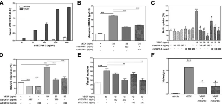
sVEGFR-1/Fc and -2/Fc inhibited VEGF-driven effect on
endothe-lial cell proliferation and migration (Fig. 2C, D), as well as
in the ex vivo aortic ring assay (Fig. 2E) and, finally, by the
in vivo matrigel plug assay (Fig. 2F). ERK1/2
phosphory-lation was also evaluated (Fig. 2B) in the presence of
VEGF, and a decrease of this VEGF-induced
phosphory-lation was observed when sVEGFR-1 or -2 was added.
Collectively, these results confirm that both recombinant
sVEGFRs/Fc possess the ability to bind VEGF and inhibit
VEGF-dependent endothelial cell angiogenesis. The soluble
VEGF receptors did not directly modulate EC
differentia-tion, proliferadifferentia-tion, or migradifferentia-tion, but primarily affected EC
activities by neutralizing VEGF. sVEGFR-1 and -2 modulated
angiogenesis not only by inhibiting VEGF action, but also by
promoting the outgrowth of mural cells.
sVEGFR-1 and -2 promote vessel stabilization in vitro
and in vivo
We evaluated the impact of sVEGFR-1 and -2 on the
codis-tribution of mural cells and ECs in a 3-D model of cord-like
formation in Matrigel (Fig. 4A). EC cords were preformed
before the addition of mural cells. Interestingly, perivascular
cell distribution calculated as a distance away from EC cords
showed that the density of mural cells closely opposed to the
EC cords (distance
ⱕ5 m) was 2-fold higher under
sVEGFR-1 or -2 treatments. Reciprocally, the density of
mural cells distant from EC cords by 75
m was 1.5-fold
lower on sVEGFRs treatment (Fig. 4B). The mean distance
between mural cells and EC cords was significantly reduced
by 40% in the presence of either sVEGFR. Moreover, in the
aortic ring assay, the addition of sVEGFR-1 or -2 after 5 d of
VEGF treatment increased the coverage by mural cells of the
endothelial cords (data not shown).
To investigate in vivo the impact of both sVEGFRs on
mural cell recruitment, we examined mural cell
cover-age of neovessels formed in Matrigel plugs
subcutane-ously injected to C57BL/6 mice. Perfused vessels were
visualized through intravenous injection of
FITC-con-jugated dextran. Mural cells were immunostained with
Cy3-conjugated
␣-SMA antibody. Staining with this
mu-ral cell marker revealed an increase of mumu-ral cell
coverage when the Matrigel plug was supplemented
with sVEGFR-1 (Fig. 4C). The quantification by
com-puter-assisted image analysis showed that the density of
vessels covered by mural cells was increased by 63 and
114% under treatment with sVEGFR-1 or PDGF-BB
(used as a positive control), respectively (Fig. 4D). For
each experimental condition, confocal microscopy
analysis attested that
␣-SMA positive cells surrounded
functional vessels (Fig. 4E). Similar data were obtained
with sVEGFR-2 (data not shown).
Induction of mural cell migration by sVEGFR-1 and -2
requires EC neighboring
The impact of sVEGFR-1 and -2 was evaluated on mural
cell migration (Fig. 5A) and proliferation (Fig. 5B) using
PDGF-BB as positive control. Neither VEGF nor sVEGFR
treatments modified aortic smooth muscle cell migration
or proliferation after 12, 24, 48, or 72 h exposure,
indicating that VEGF or sVEGFRs have no direct effect on
mural cell proliferation and migration.
We next evaluated the influence of these sVEGFRs on
AoSMC migration when cocultured with HUVECs in a
modified Boyden chamber assay. After 12, 24, or 48 h of
incubation, AoSMC migration was stimulated not only by
PDGF-BB treatment, but also in a dose-dependent way on
sVEGFR-1 or -2 exposure (Fig. 5C). The effect of
PDGF-BB persisted in the absence of HUVECs, showing a
direct effect of this cytokine on AoSMC migration. On the
other hand, the presence of HUVECs was required to
observe an increase in AoSMC migration in the presence
of sVEGFR-1 or -2. As the recombinant sVEGFR-1 and -2
used in this work were chimeric proteins fused to the Fc
region of human IgG1, we verified the specificity of their
effect, by demonstrating an absence of regulation of
AoSMC migration by the same Fc region of IgG1 alone
(2–250 ng/ml) and by another chimeric recombinant
protein of the VEGF family, sVEGFR-3, fused to the same
Fc region (Fig. 5C).
After 12 or 24 h of incubation, we observed that a
Figure 1.Effects of sVEGFR-1 and -2 on the aortic ring assay. A) Photomicro-graphs of mouse aortic rings incubated without specific treatment (vehicle) or with VEGF (10 ng/ml), sVEGFR-1, or sVEGFR-2 (250 ng/ml). Representative data ofⱖ3 experiments are shown. B) Quantification of the cellular network outgrowing from mouse aortic ring. Each curve is a mean of the cellular network distribution obtained by the average ofⱖ5 individual distributions generated for each experimental condition. Mouse aortic rings were incubated without specific treatment (vehicle) or with VEGF (10 ng/ml), sVEGFR-1, or sVEGFR-2 (250 ng/ml) treatments. *P⬍ 0.05. Representative data of ⱖ3 experiments are shown. C) Photomicrographs of mouse aortic ring immunostained with IB4-specific antibody (IB4, green) identifying ECs and with NG2-specific antibody (NG2, red) identifying PC/SMCs. Nuclei were stained with DAPI (blue). Aortic rings were incubated without specific treatment (vehicle) or with VEGF (10 ng/ml), sVEGFR-1, or sVEGFR-2 (250 ng/ml). Representative data ofⱖ3 experiments are shown. D) Photomicrographs of mouse aortic ring immunostained with IB4-specific antibody (IB4, green) identifying ECs and with NG2-specific antibody (NG2, red) identifying PC/SMCs. Nuclei were stained with DAPI (blue). Aortic rings were incubated without specific treatment (vehicle) or with VEGF (10 ng/ml) for 5 d, then media were supplemented or not with sVEGFR-2 (250 ng/ml). Scale bars⫽ 1 mm (A); 100 m (C, D).
neutralizing sVEGFR-2-specific antibody inhibited the
promigratory effect induced by sVEGFR-2 (Fig. 5D),
with a maximal neutralizing effect when used in a ratio
of 5:1 (anti-sVEGFR-2 vs. sVEGFR-2).
In our experimental conditions, VEGF is expressed at
the mRNA and protein levels by HUVECs. However,
the protein is not secreted at a detectable level in the
medium conditioned by HUVECs (Fig. 3). On the
other hand, the AoSMCs produce and secrete VEGF.
Note that the level of intracellular VEGF protein is
Figure 2.Anti-VEGF activity of sVEGFR-1/Fc and sVEGFR-2/Fc. A) Histogram of receptor binding assay to VEGF in cell-free conditions. Results are expressed as optical density (O.D.) of sVEGFR-2 bound to plate wells coated with VEGF or vehicle, means⫾ sd, n ⫽ 4. B) Histogram of ERK1/2 phosphorylation in HUVECs obtained after 10 min of incubation. For specific treatments see the figure. Results (means⫾se) show the concentration (pg/ml) of phosphorylated ERK1/2 of 6 independent experiments run in triplicate. ***P⬍ 0.001. C) Histogram of HUVEC growth after 48 h under treatment. For specific treatments see the figure. Results are expressed as percentage of BrdU uptake, means⫾ sd, n ⫽ 5. ***P ⬍ 0.001 vs. vehicle;#P⬍ 0.001
vs. VEGF treatment. Representative data of 3 independent experiments are shown. D) Histogram of HUVEC migration after
10 h under treatment. For specific treatments see the figure. Results (means⫾sd) show the percentage of HUVEC migration of 3 independent experiments run in triplicate. ***P⬍ 0.001 vs. vehicle. E) Histogram of vessel number outgrowing from mouse aortic rings. For specific treatments see the figure. Results are means⫾ se, n ⫽ 8. **P ⬍ 0.01; ***P ⬍ 0.001. F) Histogram of Hb matrigel plug content reported to plug weight. Matrigel was supplemented without specific treatment (vehicle) or with VEGF (250 ng/ml) and sVEGFR-1 or -2 (2g/ml). Results are means ⫾ se, n ⫽ 8. ***P ⬍ 0.001 vs. vehicle,#P⬍ 0.01 vs. VEGF.
Figure 3.Screening of VEGF and VEGFR-2 in HUVECs and AoSMCs.
A, B) mRNA levels of VEGF (A) and VEGFR-2 (B) in HUVECs and
AoSMCs were quantified by RT-PCR normalized to 28S rRNA and expressed as arbitrary units (A.U.) (means⫾sd), n ⫽ 4. C) VEGF (30 kDa) and VEGFR-2 (230 kDa) expression evaluated by Western blot on HUVEC and AoSMC protein extracts. Protein loading was controlled by GAPDH (36 kDa) immunolabeling. D) VEGF secretion measured by ELISA on HUVEC- and AoSMC-conditioned media supplemented or not with sVEGFR-1 or -2.
lower in AoSMCs than in HUVECs. These results reveal
that in HUVECs, the majority of VEGF produced
remains inside the cells, while VEGF produced by
AoSMCs is secreted in the extracellular milieu.
More-over, AoSMCs expressed VEGFR-2, albeit at a lesser
extent than in HUVECs.
sVEGFR-1 and -2 promote mural cell migration
through EC eNOS activation
To determine whether the indirect promigratory effect of
sVEGFR-1 and -2 could be mediated by a stable factor
secreted by ECs under sVEGFR-1 or -2 treatment, AoSMC
Figure 4.Effect of sVEGFR-1 and -2 on PC/SMC recruitment. A) Photomicrographs of 3-D cord-like structure obtained from HUVECs (green staining) cocultured with AoSMCs (orange staining) on Matrigel and incubated without specific treatment (vehicle) or with sVEGFR-2 (250 ng/ml). Arrows indicate area where AoSMCs are distant from or close to endothelial cord-like structure. Representative data ofⱖ3 experiments are shown. B) AoSMC distribution around a HUVEC cord network. Each curve is a mean of AoSMC distribution obtained by the average ofⱖ5 individual distributions generated for each experimental condition. Cocultures of HUVECs and AoSMCs were incubated without specific treatment (vehicle) or under sVEGFR-1 or sVEGFR-2 (250 ng/ml) treatments. **P⬍ 0.05. Representative data of ⱖ3 experiments are shown. C) Photomicrographs of vessels grown in Matrigel supplemented without specific treatment (control) or with sVEGFR-1 (2g/ml) or PDGF-BB (250 ng/ml). Vessels are visualized by FITC-conjugated dextran (green) perfusion, mural cells are stained by␣-SMA immunostaining (red). D) Quantification of mural cell coverage density. Results (means⫾se, n⫽12 animals/group) show the mural cell coverage density resulting from the ratio between the area of vessel covered by mural cells and the total vessel area. *P ⱕ 0.05.
E) Representative photomicrograph obtained by confocal microscopy showing that mural cells cover functional vessels. Vessels
are visualized by FITC-conjugated dextran (green) perfusion, mural cells are stained by␣-SMA immunostaining (red). Scale bars⫽100 m (A, C); 30 m (E).
Figure 5. Effect of sVEGFR-1 and -2 on PC/SMC migration and proliferation.
A) AoSMC migration; for specific treatments see the figure. Results (means⫾sd)
show the percentage of AoSMC migration, n ⫽ 12. ***P ⬍ 0.001 vs. vehicle. Representative data of 3 independent experiments are shown. B) AoSMC proliferation; for specific treatments see the figure. Results (means⫾sd) show the percentage of BrdU uptake, mean ⫾ sd, n ⫽ 8. ***P ⬍ 0.01 vs. vehicle. Representative data of 3 independent experiments are shown. C, D) Migration assay, where AoSMCs were cocultured with HUVECs; for specific treatments see the figure. Results (means⫾se) show the percentage of AoSMC migration of 3 independent experiments run in duplicate. **P⬍ 0.01, ***P ⬍ 0.001 vs. vehicle, #
migration was tested in the presence of medium
condi-tioned by HUVECs previously cultivated with or without
sVEGFR-1 or -2 for 4, 24, or 48 h (Fig. 6A). In sharp
contrast to what was observed when AoSMCs were
co-cultured with HUVECs, HUVEC-conditioned media
(HUVEC-CM) previously supplemented with sVEGFR-1
or -2 failed to induce AoSMC migration after 12, 24, or
48 h exposure. In addition, none of the HUVEC-CM
tested was able to stimulate AoSMC proliferation even
after 24 –72 h (Fig. 6B). Levels of cytokines known to
stimulate AoSMC migration (PDGF-BB, Ang1, TGF-
,
bFGF, and S1P) in HUVEC-CM and in media of
cocul-tured Boyden chamber assay were identical in basal or
sVEGFRs supplemented conditions (Fig. 6C). Altogether,
these results demonstrate that the treatment of HUVECs
with sVEGFR-1 or sVEGFR-2 did not increase the
secre-tion of any stable factor that could modulate migrasecre-tion or
proliferation of AoSMCs.
Hence, we hypothesized that the promigratory effect
of sVEGFR-1 and -2 on AoSMCs could be due to a
volatile factor such as NO, which is known to stimulate
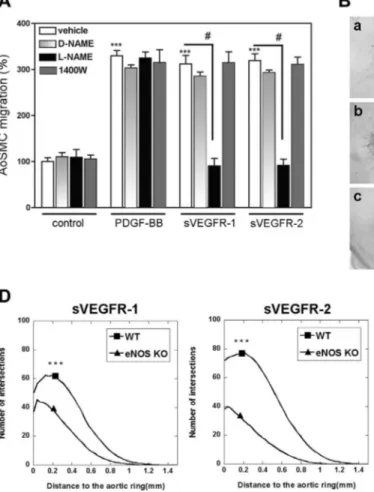
PC recruitment (14, 35, 36). L-NAME, a pan-inhibitor
of NOS, did not modulate basal and PDGF-BB-induced
AoSMC migration, but completely inhibited the
promi-gratory effect induced by sVEGFR-1 and -2 in the
modified Boyden chamber assay (Fig. 7A). L-NAME
also decreased the cellular network outgrowth induced
by sVEGFR-2 (Fig. 7B) in the aortic ring assay. By
immunostaining, the cellular network outgrowth was
identified as being composed mainly of PC/SMCs (data
not shown). Computer-assisted image analysis
quantifi-cation confirmed that L-NAME reduced the mural cell
outgrowth induced by sVEGFR-2 treatment (Fig. 7C).
Similar results were obtained with sVEGFR-1 (data not
shown). On the other hand, D-NAME, the inactive
isomer of L-NAME, and 1400W, a selective inhibitor of
inducible NOS (iNOS), did not inhibit the
promigra-tory effect by sVEGFR-1 and -2. Since neuronal NOS
(nNOS) is not expressed by HUVECs, these data
indicate that eNOS activity is involved in this
promi-gratory process. In addition, the outgrowth of the
mural cell network promoted by sVEGFR-1 and -2 in
wild-type (WT) mouse aorta rings was completely
inhibited with eNOS
⫺/⫺mouse aorta (Fig. 7D).
Collectively, these results demonstrate that eNOS
activity is involved in the recruitment of mural cells
induced by sVEGFR-1 and -2.
EC S1P1 is involved in the process by which
sVEGFR-1 and -2 modulate PC/SMC function
On the basis of its ability to trigger eNOS activation and
NO synthesis by ECs and regarding its key role in
endo-thelium maturation (15, 37–39), we hypothesized that
S1P1 could be involved in sVEGFRs-promoted PC/SMC
migration. Treatment of HUVECs with sVEGFR-1 and -2
but not with PDGF-BB led to an accumulation of S1P1, as
documented by quantified Western blot (Fig. 8A–B). This
overproduction occurred from 60 to 90 min of
incubation in the presence of the sVEGFRs and was
maintained up to 10 h. In addition, 2 different
S1P1-specific antibodies and an S1P1 antagonist
(VPC 23019) that does not modulate
PDGF-BB-induced AoSMC migration completely inhibited the
mural cell promigratory effect observed in the
pres-ence of sVEGFR-1 and -2 (Fig. 8C, D). These data
strongly support that S1P1 is involved in the
para-Figure 6. HUVEC-conditioned media do not modulate mural cell function.
A) Migration assay where AoSMCs were cultured under treatment with or without
PDGF-BB or HUVEC-CM. Media supplemented or not with sVEGFR-1 or sVEGFR-2 (250 ng/ml) were conditioned by culture with HUVECs for 4, 24 or 48 h. Results (means⫾sd) show the percentage of AoSMC migration, n ⫽ 5. ***P ⬍ 0.001 vs. vehicle. Representative data of 3 independent experiments are shown. B) Proliferation assay where AoSMCs were cultured under treatment with or without PDGF-BB or HUVEC-CM. Media supplemented or not with sVEGFR-1 or sVEGFR-2 (250 ng/ml) were conditioned by culture with HUVECs for 48 h. Results (means⫾sd) show the percentage of BrdU uptake, n⫽ 8. ***P ⬍ 0.001 vs. vehicle. Representative data of 3 independent experiments are shown. C) ELISA of PDGF-BB, Ang1, TGF-, bFGF, and S1P performed on media supplemented or not with sVEGFR-1 or sVEGFR-2 (250 ng/ml) and conditioned by culture with HUVECs for 4 or 24 h. Results are means⫾ sd, n ⫽ 5.
crine interactions leading to mural cell migration
under sVEGFRs treatment.
sVEGFR-1 and -2 bind to ECs and modulate VEGFR-2
signaling
To better understand how these soluble VEGF receptor
could promote S1P1 up-regulation and eNOS activity,
we first evaluated if they could bind to ECs. As shown in
Fig. 9
by immunocytochemistry and by Western blot
using FITC-labeled sVEGFR-1 and -2, these soluble
VEGF receptors were able to bind HUVECs (Fig. 9A,
B). sVEGFR-1 and -2 did not completely inhibit the
VEGF-induced phosphorylation of VEGFR-2 and of
eNOS (Fig. 9C), although these soluble receptors, in
the same range of concentration, completely trapped
free VEGF (Fig. 3) and inhibited VEGF-mediated
pro-liferation, migration, differentiation, and angiogenesis
(Fig. 2C–E). As for PMA and VEGF, both soluble VEGF
receptors promoted S1P1 up-regulation through a
PKC-dependent pathway (Fig. 9D). In the aortic ring
assay, the use of a VEGFR-2 tyrosine kinase inhibitor
(ZM323881) did not promote the outgrowth of a mural
cell network (Fig. 9E).
DISCUSSION
In this study, we identify sVEGFR-1 and sVEGFR-2 as
new regulators that contribute to vessel maturation.
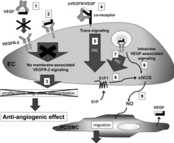
Our results indicate, as summarized in Fig. 10, that
sVEGFR-1 and -2 stabilize the immature endothelium
through VEGF trapping, interaction with ECs, and the
induction of mural cell recruitment. This is mediated
in ECs through a paracrine mechanism where
sVEGFR-1/-2 and VEGF/VEGFR-2 pathway interplay with the
S1P/S1P1 pathway and eNOS activation.
VEGF soluble receptors are 2 physiologically
pro-duced VEGF-sequestering proteins that contribute to
regulate VEGF isoforms bioavailability in physiological
and pathological conditions (24, 40, 41). Even if
natu-ral sVEGFR-2 presents a weaker affinity to VEGF than
Figure 7.sVEGFR-1 and -2 promote PC/SMC migra-tion through eNOS activamigra-tion. A) Migramigra-tion assay where AoSMCs were cocultured with HUVECs, un-der treatment with or without (control) PDGF-BB (20 ng/ml), sVEGFR-1 (250 ng/ml), sVEGFR-2 (250 ng/ml), used independently (vehicle) or in combi-nation with D-NAME (5 mM), L-NAME (5 mM), or 1400W (1 mM). Results (means⫾se) show the per-centage of AoSMC migration of 3 independent ex-periments run in duplicate. ***P⬍ 0.001 vs. vehicle, #P ⬍ 0.001 vs. L-NAME untreated condition. B) Photomicrographs of mouse aortic rings incubated without specific treatment (vehicle) (a), with sVEGFR-2 (250 ng/ml) (b), or with sVEGFR-2 (250 ng/ml) supplemented with L-NAME (5 mM) (c). Representative data of ⱖ3 experiments are shown. Scale bars ⫽ 1 mm. C) Quantification of the cellular network outgrowing from mouse aortic ring. Each curve is a mean of the cellular network distribution obtained by the average ofⱖ5 individual distributions generated for each experimental condition. Mouse aortic rings were incubated without specific treatment (vehicle) or with sVEGFR-2 (250 ng/ml) supplemented or not with L-NAME (5 mM). ***P⬍ 0.05. Representative data ofⱖ3 experiments are shown. D) Quantification of the cellular network outgrowing from WT or eNOS⫺/⫺(eNOS KO) mouse aortic ring. Each curve is a mean of the cellular network distribution obtained by the average ofⱖ5 individual distributions generated for each experimental condition. Mouse aortic rings were incubated with sVEGFR-1 or sVEGFR-2 (250 ng/ml). Representative data ofⱖ3 experiments are shown.
sVEGFR-1 and exhibits an antilymphangiogenic activity
in cornea (42, 43), their antiangiogenic activities are
well described in different tumoral models (21, 25, 44,
45). However, their potential contribution in vessel
stabilization and maturation remained to be
ex-plored. In the aortic ring model of angiogenesis,
these soluble receptors promoted the outgrowth of a
cellular network of mural cells. However, neither
endothelial proliferation, migration, and cord-like
formation nor PC/SMC proliferation and migration
were directly modulated by sVEGFR-1 or -2. Both
sVEGFRs suppressed the bioavailability of VEGF and
completely abrogated VEGF-induced EC
prolifera-tion, migraprolifera-tion, cord-like formaprolifera-tion, and
angiogene-sis in vitro and in vivo. These results indicate that
sVEGFR-1 and -2 can modulate mural cell function in
addition to acting at the endothelial level as VEGF
trappers. Nevertheless, they failed to exert any direct
effect on in vitro ECs or mural cell proliferation,
migration, and differentiation. These observations
led us to speculate that sVEGFR-1 and -2 could
recruit mural cells by modulating interactions
be-tween neighboring ECs and PC/SMCs. This novel
concept is supported by various observations. Indeed,
under sVEGFRs treatment, PC/SMCs were attracted
toward the endothelial cord-like network in a
cocul-tured 3-D model of cord-like formation and in the
aortic ring assay. In addition, we clearly evidenced
that sVEGFR-1 and -2, but not sVEGFR-3, a soluble
receptor binding VEGF isoforms (VEGF-C, VEGF-D)
involved in lymphangiogenesis (33), promoted mural
cell migration only in the presence of neighboring
ECs. This effect reached the same level as that
induced by the PC/SMC promigratory factor
PDGF-BB. Moreover, the mural cell coverage of vessels
developed, in vivo, in a Matrigel plug assay was
increased under sVEGFR-1 and-2 treatment. These
observations correlate with the study of Mazzone et al.
(6), who reported an increase of tumor vascular PC
coverage associated with an increase of sVEGFR-1
transcription in 3 different tumor models applied to
PHD2
⫹/⫺mice. Altogether, our results sustain that
physiological anti-VEGF, sVEGFR-1, is involved in the
interaction between ECs and PC/SMCs that
pro-motes the recruitment of mural cells, similarly to
what was reported for other inhibitors of the VEGF
axis (9, 10, 46). The results obtained with the
recom-binant sVEGFR-2/Fc reach the same conclusions.
However, it must be mentioned that sVEGFR-2/Fc
exhibits a higher affinity to VEGF than the
physio-logically produced sVEGFR-2 (42, 43). Our
observa-tions have therefore to be restricted to a therapeutic
use of sVEGFR-2/Fc and not completely extrapolated
to physiological endogenous interactions.
Recent data suggest that endogenous eNOS-derived
NO is involved in arteriogenesis, angiogenesis, and
mural cell recruitment (35, 43). Accordingly, in the
coculture migration assay and in the aortic ring assay,
the pan-NOS inhibitor L-NAME completely inhibited
the promigratory effect exerted by sVEGFR-1 and -2 on
mural cells. The inability of a selective iNOS inhibitor
(1400W) to inhibit sVEGFR-induced PC/SMC
migra-tion led us to conclude a selective activamigra-tion of EC
eNOS under sVEGFR treatment. This was confirmed by
the loss of effect of these soluble VEGF receptors in an
aortic ring assay performed with eNOS
⫺/⫺mouse
aorta. These results are in agreement with the loss of
vascular PC coverage observed in an ischemic model
performed in eNOS
⫺/⫺mice (35). In addition, they
corroborate the data of Kashiwagi et al. (14), who
Figure 8.sVEGFR-1 and -2 promote mural cell migration by S1P1 overexpression. A) Representative Western blotting of S1P1 (40 kDa) production in HUVECs. Protein loading was controlled by GAPDH (36 kDa) immunolabeling. HUVECs were treated for 60 min; for specific treatments see the figure.
B) Quantification of S1P1 expression obtained by Western
blot. Results (means⫾se) show the intensity (A.U.) of S1P1 bands of 4 independent experiments run in duplicate. *P⬍ 0.05 vs. vehicle. C) Migration assay where AoSMCs were cocultured with HUVECs, under treatment with or without PDGF-BB (20 ng/ml), sVEGFR-1 (250 ng/ml), sVEGFR-2 (250 ng/ml), used independently or in combination with an S1P1-specific antibody 1 (1 g/ml), another S1P1-specific antibody 2 (1g/ml). Results (means⫾se) show the percent-age of AoSMC migration of 3 independent experiments run in duplicate. ***P⬍ 0.001 vs. vehicle,#P⬍ 0.01 vs. anti-S1P1 untreated condition. D) Migration assay where AoSMCs were cocultured with HUVECs, under treatment with or without PDGF-BB (20 ng/ml), sVEGFR-1 (250 ng/ml), sVEGFR-2 (250 ng/ml), used independently or in combination with
VPC 23019 S1P1 antagonist (VPC, 1 M). Results
(means⫾se) show the percentage of AoSMC migration of 2 independent experiments run in duplicate. ***P⬍ 0.001 vs. vehicle,#P⬍ 0.01 vs. VPC untreated condition.
demonstrated that NO derived from eNOS, but not
from iNOS, contributes to increase the mural cell
coverage of B16 melanoma vessels.
S1P through the activation of S1P1, a
G-protein-cou-pled receptor, induces NO synthesis through eNOS
acti-vation in ECs (37). In our experimental conditions, we
showed that 2 different S1P1-blocking antibodies and an
S1P1 antagonist inhibited the promigratory effect exerted
by sVEGFR-1 and -2 on mural cells. In addition, through
a PKC-dependent pathway, sVEGFR-1 and -2 promoted
the overexpression of S1P1 in ECs, albeit in a lesser extent
than PMA or VEGF. All these data evidence that the
S1P/S1P1 axis is implicated in the promigratory effect
induced by sVEGFR-1 and -2. At the present time, the
promoter region of S1P1 gene has not been extensively
characterized in ECs, and mechanistic insights how
ex-pression of S1P1 gene is determined are to be elucidated
more in detail (47). Nevertheless, endothelial-specific
S1P1-knockout mice exhibit mural vessel coverage defect,
demonstrating that S1P/S1P1 signaling contributes to
vessel stabilization (38).
We demonstrated that sVEGFR-1 and -2 were not
only circulating VEGF sequestering proteins, but that
they could bind to ECs. As illustrated in Fig. 9, these
data support that, in addition to trapping VEGF at a
circulating level, sVEGFRs could form a
signaling-inactive membrane-associated complex consisting of
sVEGFR/VEGF/VEGFR-2 that prevents VEGF-driven
angiogenesis. They could also bind ECs through
inter-action with a coreceptor that could subsequently
gen-erate a trans-signaling similar to what is observed for
soluble IL-6 receptor (48). In addition, if they
com-pletely trapped free VEGF and abrogated VEGF-driven
proliferation, migration, differentiation, and
angiogen-esis, they could not completely inhibit VEGFR-2 and
eNOS phosphorylation in ECs. This paradox could be
explained by the presence of an intracrine VEGF
pathway. Indeed, using mice where VEGF was
specifi-cally deleted in ECs, Lee et al. (49) demonstrated that
VEGF acts as an intracrine factor that is crucial for
vascular homeostasis. Their findings uncovered an
im-portant role for intracrine VEGF signaling in survival
mechanisms following hypoxia-mediated stress for
which paracrine sources of VEGF cannot compensate.
This intracrine VEGF/VEGFR-2 pathway, which
re-mains and signals inside ECs, could be recruited when
ECs are deprived of exogenous VEGF by sVEGFR-1 or
-2. Moreover, we did not observed any mural cell
outgrowth in the aortic ring assay when using an
inhibitor that blocks both cytoplasmic
membrane-asso-ciated VEGFR-2 and intracrine VEGFR-2 signaling.
These results support that a residual kinase activity of
endothelial VEGFR-2 is necessary to observe the mural
cell recruitment induced by sVEGFR-1 and -2. They
reconcile our data with the study of Igarashi (47),
which described that S1P1 could be up-regulated by
VEGF. Finally, they underline how the function of a cell
is finely driven by the accurate regulation of its
signal-ing level.
Collectively, our data indicate that when immature
endo-Figure 9.Signaling impact of sVEGFR-1 and -2 binding to ECs. A) Photomicrographs of HUVECs incubated with vehicle or with sVEGFR-1/FITC or sVEGFR-2/ FITC (250 ng/ml). Bound FITC-conjugated proteins were visualized with Alexa488-conjugated FITC specific Ab. Scale bars ⫽ 10 m. B) Representative Western blotting of HUVECs incubated with vehicle or with sVEGFR-1/FITC or sVEGFR-2/FITC (250 ng/ml). Bound FITC-conjugated proteins were visualized with HRP-conjugated FITC-specific Ab (70 kDa). Protein loading was controlled by GAPDH (36 kDa) immuno-labeling. C) Phosphorylation of VEGFR-2 (230 kDa) and of eNOS (140 kDa) was evaluated by Western blot. Protein loading was controlled by GAPDH (36 kDa) or VEGFR-2 (230 kDa) immunolabeling; for specific treatments see the figure. Representative data ofⱖ3 independent experiments are shown. D) S1P1 mRNA expression in HUVECs treated with or without PMA (200 ng/ml), VEGF (50 ng/ml), sVEGFR-1 (250 ng/ml), or sVEGFR-2 (250 ng/ml)⫹ VEGF (0.5 ng/ml). Cells were preincubated with vehicle (basal) or with GF109203X. Results (means⫾se) show the percentage of S1P1 mRNA expression of 2 independent experiments run in duplicate. *P ⬍ 0.05, ***P⬍ 0.001 vs. vehicle;#P⬍ 0.05,##P⬍ 0.001 vs. GF109203X untreated condition (basal). E) Quantification of the cellular network outgrowing from mouse aortic ring. Each curve is a mean of the cellular network distribution obtained by the average ofⱖ5 individual distributions generated for each experimental condition. Mouse aortic rings were incubated without specific treatment (vehicle) or with ZM323881 (ZM); NS, not significant. Representative data ofⱖ3 experiments are shown.
thelium is bathed with sVEGFR-1 or -2 and thus deprived of
circulating VEGF, the proliferative phase of angiogenesis is
stopped, and the mural cell recruitment is engaged to end to
vessel maturation. In these conditions, the recruitment of
mural cells results from interplay between VEGF/VEGFR-2
and S1P/S1P1 pathways that lead to eNOS activation.
In-deed, our data support the following mechanisms of
molec-ular interactions summarized in Fig. 10: 1) in addition to
trapping VEGF at a circulating level, sVEGFR-1 and -2 could
form a signaling-inactive membrane-associated complex
consisting of sVEGFR/VEGF/VEGFR-2 both ending to
pre-vent VEGF-driven angiogenesis, 2) a PKC-dependent
trans-signaling resulting from the binding of sVEGFR/VEGF to a
membrane coreceptor could up-regulate S1P1 expression
and thus favoring eNOS activation through the S1P/S1P1
pathway, 3) intracrine VEGF-associated signaling could
up-regulate S1P1 expression through a PKC-dependent
path-way, and 4) intracrine VEGF-associated signaling could also
directly promote the phosphorylation of eNOS. Our data
complete the study of Greenberg et al. (50), who described
that VEGF was a direct inhibitor of PC function and vessel
maturation. Indeed, they demonstrated that, in mural
cells, the VEGF-mediated activation of VEGFR-2 inhibited
PDGF-R
 signaling through the formation of a receptor
complex consisting of VEGFR-2 and PDGF-R
. In turn
this reduced the mural cell migration. In line with our
data, overexpression of sVEGFR-1 and -2 would reduce
the formation of such complex and sustain
PDGF-BB-dependent mural cell migration. Our results are also in
line with the observations of Mazzone et al. (6), who have
shown that an increase of vessel stabilization is associated
with the enhancement of sVEGFR-1 expression by
quies-cent ECs of newly formed capillaries. In this location the
sVEGFR-1 would, as demonstrated here, inhibit the EC
proliferation and migration and enhance mural cell
cov-erage and vessel maturation.
In summary, we demonstrated here that sVEGFR-1 and
-2 take part in vessel maturation via the induction of
mural cell recruitment through a paracrine effect
involv-ing ECs. Our findinvolv-ings support the idea that endogenously
produced sVEGFR-1 and exogenously administrated
sVEGFR-1/Fc or sVEGFR-2/Fc regulate the balance
be-tween pro- and antiangiogenic factors and that they are
involved in the formation of a stable vasculature by
inducing mural cell migration. In the context of the
currently used anti-VEGF therapies, our results contribute
to clarify clinical observations where agents targeting the
VEGF pathway induce a transient “normalization” of
tumor vessels by increasing their mural cell coverage.
Normalization of tumor vasculature is an emerging
strat-egy to improve radiation and cytotoxic therapies (36).
Contribution of soluble VEGFRs may ultimately prove
beneficial for normalizing tumoral vasculature and
im-proving the response to radiation and chemotherapy.
The authors thank B. Brouwers, I. Dasoul, D. Gabriel-Delapierre, N. Lefin, M.-R. Pignon, E. Konradowski, F. Olivier, P. Gavitelli, and G. Rolland for their excellent technical assistance. Confocal microscopy was performed at CIL- and GIGA-Imaging and Flow Cytometry Facility. This work was supported by grants from the European 7th Research Framework Programme: HEALTH-2007-2.4.1-6 “MICROENVIMET,” Framework Programme 6-NOE LSHM-CT-2004-512040, EMBIC, the Fonds de la Recher-che Scientifique Me´dicale, the Fonds National de la Re-cherche Scientifique (FNRS; Belgium), the Fondation con-tre le Cancer, the Fonds spe´ciaux Recherche (University of Lie`ge), the Centre Anticance´reux pre`s l’Universite´ de Lie`ge, the Fonds Le´on Fredericq (University of Lie`ge), the DGTRE from the Re´gion Wallonne (NEOANGIO), the Fonds Social Europe´en, and the Interuniversity Attraction Poles Programme–Belgian Science Policy (Brussels, Bel-gium). S.L. and C.P. performed all cellular experiments; S. Berndt performed the aortic ring assays; S. Blacher did all quantification based on image analysis; E.G. contributed to the mouse Matrigel plug assay; O.P., E.M., and A.N. contributed to the work with scientific advice; J.-M.F. supervised the study, provided scientific suggestions, and contributed to manuscript preparation and review; C.M. participated to data analysis; and C.P. designed the study, analyzed the data, and wrote the manuscript.
REFERENCES
1. Carmeliet, P. (2005) Angiogenesis in life, disease and medicine.
Nature 438, 932–936
2. Jain, R. K. (2005) Normalization of tumor vasculature: an emerging concept in antiangiogenic therapy. Science 307, 58 – 62 3. Sullivan, R., and Graham, C. H. (2007) Hypoxia-driven selection of the metastatic phenotype. Cancer Metastasis Rev. 26, 319 –331 4. Fukumura, D., and Jain, R. K. (2007) Tumor microvasculature and microenvironment: targets for anti-angiogenesis and nor-malization. Microvasc. Res. 74, 72– 84
Figure 10.Model of VEGF family contribution to EC-PC/SMC dialogue. In addition to trapping VEGF at a circulating level (1), sVEGFR-1 and -2 could form a signaling-inactive mem-brane-associated complex consisting of sVEGFR/VEGF/ VEGFR-2 (2), both ending to prevent VEGF-driven angiogen-esis (3). They could also bind ECs through interaction with a coreceptor (4) that engaged a PKC-dependent trans-signaling (5) up-regulating S1P1 expression and thus favoring eNOS activation through the S1P/S1P1 pathway (6). Intracrine VEGF-associated signaling could up-regulate S1P1 expression through a PKC-dependent pathway (7), thus favoring eNOS activation through the S1P/S1P1 pathway (6) and/or could directly promote eNOS phosphorylation (8). The subsequent NO release induced the migration of neighbor PC/SMCs (9), thus promoting mural cells recruitment.
5. Gerhardt, H., and Semb, H. (2008) Pericytes: gatekeepers in tumour cell metastasis? J. Mol. Med. 86, 135–144
6. Mazzone, M., Dettori, D., Leite de Oliveira, R., Loges, S., Schmidt, T., Jonckx, B., Tian, Y. M., Lanahan, A. A., Pollard, P., Ruiz de Almodovar, C., De Smet, F., Vinckier, S., Ara-gones, J., Debackere, K., Luttun, A., Wyns, S., Jordan, B., Pisacane, A., Gallez, B., Lampugnani, M. G., Dejana, E., Simons, M., Ratcliffe, P., Maxwell, P., and Carmeliet, P. (2009) Heterozygous deficiency of PHD2 restores tumor oxygenation and inhibits metastasis via endothelial normal-ization. Cell 136, 839 – 851
7. Chabottaux, V., Ricaud, S., Host, L., Blacher, S., Paye, A., Thiry, M., Garofalakis, A., Pestourie, C., Gombert, K., Bruye`re, F., Lewandowsky, D., Tavitian, B., Foidart, J.-M., Duconge, F., and Noe¨l, A. (2009) A membrane-type 4 matrix metalloproteinase (MT4-MMP) induces lung metastasis by alteration of primary breast tumor vascular architecture. J. Cell. Mol. Med. 13, 4002– 4013
8. Tong, R. T., Boucher, Y., Kozin, S. V., Winkler, F., Hicklin, D. J., and Jain, R. K. (2004) Vascular normalization by vascular endothelial growth factor receptor 2 blockade induces a pres-sure gradient across the vasculature and improves drug pene-tration in tumors. Cancer Res. 64, 3731–3736
9. Winkler, F., Kozin, S. V., Tong, R. T., Chae, S. S., Booth, M. F., Garkavtsev, I., Xu, L., Hicklin, D. J., Fukumura, D., di Tomaso, E., Munn, L. L., and Jain, R. K. (2004) Kinetics of vascular normalization by VEGFR2 blockade governs brain tumor re-sponse to radiation: role of oxygenation, angiopoietin-1, and matrix metalloproteinases. Cancer Cell 6, 553–563
10. Bergers, G., and Hanahan, D. (2008) Modes of resistance to anti-angiogenic therapy. Nat. Rev. Cancer 8, 592– 603
11. Jain, R. K. (2003) Molecular regulation of vessel maturation.
Nat. Med. 9, 685– 693
12. Armulik, A., Abramsson, A., and Betsholtz, C. (2005) Endothe-lial/pericyte interactions. Circ. Res. 97, 512–523
13. Andrae, J., Gallini, R., and Betsholtz, C. (2008) Role of platelet-derived growth factors in physiology and medicine. Genes Dev.
22,1276 –1312
14. Kashiwagi, S., Izumi, Y., Gohongi, T., Demou, Z. N., Xu, L., Huang, P. L., Buerk, D. G., Munn, L. L., Jain, R. K., and Fukumura, D. (2005) NO mediates mural cell recruitment and vessel morphogenesis in murine melanomas and tissue-engi-neered blood vessels. J. Clin. Invest. 115, 1816 –1827
15. Peters, S. L. M., and Alewijnse, A. E. (2007) Sphingosine-1-phosphate signaling in the cardiovascular system. Curr. Opin.
Pharmacol. 7, 186 –192
16. Allende, M. L., and Proia, R. L. (2002) Sphingosine–phosphate receptors and the development of the vascular system. Biochim.
Biophys. Acta 1582, 222–227
17. Kono, M., Mi, Y., Liu, Y., Sasaki, T., Allende, M. L., Wu, Y. P., Yamashita, T., and Proia, R. L. (2004) The sphingosine-1-phosphate receptors S1P1, S1P2, and S1P3 function coordi-nately during embryonic angiogenesis. J. Biol. Chem. 279, 29367– 29373
18. Shibuya, M. (2006) Differential roles of vascular endothelial growth factor receptor-1 and receptor-2 in angiogenesis. J.
Bio-chem. Mol. Biol. 39, 469 – 478
19. Swendeman, S., Mendelson, K., Weskamp, G., Horiuchi, K., Deutsch, U., Scherle, P., Hooper, A., Rafii, S., and Blobel, C. P. (2008) VEGF-A stimulates ADAM17-dependent shedding of VEGFR2 and crosstalk between VEGFR2 and ERK signaling.
Circ. Res. 103, 916 –918
20. Ebos, J. M., Bocci, G., Man, S., Thorpe, P. E., Hicklin, D. J., Zhou, D., Jia, X., and Kerbel, R. S. (2004) A naturally occurring soluble form of vascular endothelial growth factor receptor 2 detected in mouse and human plasma. Mol. Cancer Res. 2, 315–326
21. Kou, B., Li, Y., Zhang, L., Zhu, G., Wang, X., Li, Y., Xia, J., and Shi, Y. (2004) In vivo inhibition of tumor angiogenesis by a soluble VEGFR-2 fragment. Exp. Mol. Pathol. 76, 129 –137 22. Jacobi, J., Tam, B. Y., Wu, G., Hoffman, J., Cooke, J. P., and Kuo,
C. J. (2004) Adenoviral gene transfer with soluble vascular endothelial growth factor receptors impairs angiogenesis and perfusion in a murine model of hindlimb ischemia. Circulation
110,2424 –2429
23. Szentirmai, O., Baker, C. H., Bullain, S. S., Lin, N., Takahashi, M., Folkman, J., Mulligan, R. C., and Carter, B. S. (2008)
Successful inhibition of intracranial human glioblastoma multi-forme xenograft growth via systemic adenoviral delivery of soluble endostatin and soluble vascular endothelial growth factor receptor-2: laboratory investigation. J. Neurosurg. 108, 979 –988
24. Tsatsaris, V., Goffin, F., and Foidart, J. M. (2004) Circulating angiogenic factors and preeclampsia. N. Engl. J. Med. 350, 2003–2004; author reply 2003–2004
25. Munaut, C., Lorquet, S., Pequeux, C., Blacher, S., Berndt, S., Frankenne, F., and Foidart, J. M. (2008) Hypoxia is responsible for soluble vascular endothelial growth factor receptor-1 (VEGFR-1) but not for soluble endoglin induction in villous trophoblast. Hum. Reprod. 23, 1407–1415
26. Robak, E., Sysa-Jedrzejewska, A., and Robak, T. (2003) Vascular endothelial growth factor and its soluble receptors VEGFR-1 and VEGFR-2 in the serum of patients with systemic lupus erythematosus. Mediators Inflamm. 12, 293–298
27. Wierzbowska, A., Robak, T., Wrzesien-Kus, A., Krawczynska, A., Lech-Maranda, E., and Urbanska-Rys, H. (2003) Circulat-ing VEGF and its soluble receptors sVEGFR-1 and sVEGFR-2 in patients with acute leukemia. Eur. Cytokine Netw. 14, 149 –153
28. Kearney, J. B., Kappas, N. C., Ellerstrom, C., DiPaola, F. W., and Bautch, V. L. (2004) The VEGF receptor flt-1 (VEGFR-1) is a positive modulator of vascular sprout formation and branching morphogenesis. Blood 103, 4527– 4535
29. Kappas, N. C., Zeng, G., Chappell, J. C., Kearney, J. B., Hazarika, S., Kallianos, K. G., Patterson, C., Annex, B. H., and Bautch, V. L. (2008) The VEGF receptor Flt-1 spatially modulates Flk-1 signaling and blood vessel branching. J. Cell Biol. 181, 847– 858 30. Chappell, J. C., Taylor, S. M., Ferrara, N., and Bautch, V. L. (2009) Local guidance of emerging vessel sprouts requires soluble Flt-1. Dev. Cell. 17, 377–386
31. Berndt, S., Perrier d’Hauterive , S., Blacher, S., Pequeux, C., Lorquet, S., Munaut, C., Applanat, M., Herve, M. A., Lamande, N., Corvol, P., van den Brule, F., Frankenne, F., Poutanen, M., Huhtaniemi, I., Geenen, V., Noel, A., and Foidart, J. M. (2006) Angiogenic activity of human chorionic gonadotropin through LH receptor activation on endothelial and epithelial cells of the endometrium. FASEB J. 20, 2630 –2632
32. Masson, V. V., Devy, L., Grignet-Debrus, C., Bernt, S., Bajou, K., Blacher, S., Roland, G., Chang, Y., Fong, T., Carmeliet, P., Foidart, J. M., and Noel, A. (2002) Mouse aortic ring assay: a new approach of the molecular genetics of angiogenesis. Biol.
Proceed. Online 4, 24 –31
33. Bruyere, F., Melen-Lamalle, L., Blacher, S., Roland, G., Thiry, M., Moons, L., Frankenne, F., Carmeliet, P., Alitalo, K., Libert, C., Sleeman, J. P., Foidart, J. M., and Noel, A. (2008) Modeling lymphangiogenesis in a three-dimensional culture system. Nat.
Methods 5, 431– 437
34. Passaniti, A., Taylor, R. M., Pili, R., Guo, Y., Long, P. V., Haney, J. A., Pauly, R. R., Grant, D. S., and Martin, G. R. (1992) A simple, quantitative method for assessing angiogen-esis and antiangiogenic agents using reconstituted basement membrane, heparin, and fibroblast growth factor. Lab. Invest.
67,519 –528
35. Yu, J., deMuinck, E. D., Zhuang, Z., Drinane, M., Kauser, K., Rubanyi, G. M., Qian, H. S., Murata, T., Escalante, B., and Sessa, W. C. (2005) Endothelial nitric oxide synthase is critical for ischemic remodeling, mural cell recruitment, and blood flow reserve. Proc. Natl. Acad. Sci. U. S. A. 102, 10999 –11004 36. Kashiwagi, S., Tsukada, K., Xu, L., Miyazaki, J., Kozin, S. V.,
Tyrrell, J. A., Sessa, W. C., Gerweck, L. E., Jain, R. K., and Fukumura, D. (2008) Perivascular nitric oxide gradients nor-malize tumor vasculature. Nat. Med. 14, 255–257
37. Igarashi, J., Bernier, S. G., and Michel, T. (2001) Sphingosine 1-phosphate and activation of endothelial nitric-oxide synthase: differential regulation of Akt and MAP kinase pathways by EDG and bradykinin receptors in vascular endothelial cells. J. Biol.
Chem. 276, 12420 –12426
38. Allende, M. L., Yamashita, T., and Proia, R. L. (2003) G-protein-coupled receptor S1P1 acts within endothelial cells to regulate vascular maturation. Blood 102, 3665–3667
39. Igarashi, J., and Michel, T. (2008) S1P and eNOS regulation.
Biochim. Biophys. Acta 1781, 489 – 495
40. Kendall, R. L., and Thomas, K. A. (1993) Inhibition of vascular endothelial cell growth factor activity by an endogenously
encoded soluble receptor. Proc. Natl. Acad. Sci. U. S. A. 90, 10705–10709
41. Kamba, T., Tam, B. Y., Hashizume, H., Haskell, A., Sennino, B., Mancuso, M. R., Norberg, S. M., O’Brien, S. M., Davis, R. B., Gowen, L. C., Anderson, K. D., Thurston, G., Joho, S., Springer, M. L., Kuo, C. J., and McDonald, D. M. (2006) VEGF-dependent plasticity of fenestrated capillaries in the normal adult microvasculature. Am. J. Physiol. Heart Circ.
Physiol. 290, H560 –H576
42. Roeckl, W., Hecht, D., Sztajer, H., Waltenberger, J., Yayon, A., and Weich, H. A. (1998) Differential binding characteristics and cellular inhibition by soluble VEGF receptors 1 and 2. Exp. Cell.
Res. 241, 161–170
43. Albuquerque, R. J., Hayashi, T., Cho, W. G., Kleinman, M. E., Dridi, S., Takeda, A., Baffi, J. Z., Yamada, K., Kaneko, H., Green, M. G., Chappell, J., Wilting, J., Weich, H. A., Yamagami, S., Amano, S., Mizuki, N., Alexander, J. S., Peter-son, M. L., Brekken, R. A., Hirashima, M., Capoor, S., Usui, T., Ambati, B. K., and Ambati, J. (2009) Alternatively spliced vascular endothelial growth factor receptor-2 is an essential endogenous inhibitor of lymphatic vessel growth. Nat. Med.
15,1023–1030
44. Ferrara, N., and Kerbel, R. S. (2005) Angiogenesis as a thera-peutic target. Nature 438, 967–974
45. Goldman, C. K., Kendall, R. L., Cabrera, G., Soroceanu, L., Heike, Y., Gillespie, G. Y., Siegal, G. P., Mao, X., Bett, A. J., Huckle, W. R., Thomas, K. A., and Curiel, D. T. (1998)
Paracrine expression of a native soluble vascular endothelial growth factor receptor inhibits tumor growth, metastasis, and mortality rate. Proc. Natl. Acad. Sci. U. S. A. 95, 8795– 8800 46. Dings, R. P., Loren, M., Heun, H., McNiel, E., Griffioen, A. W.,
Mayo, K. H., and Griffin, R. J. (2007) Scheduling of radiation with angiogenesis inhibitors anginex and Avastin improves therapeutic outcome via vessel normalization. Clin. Cancer Res. 13, 3395–3402 47. Igarashi, J., Erwin, P. A., Dantas, A. P., Chen, H., and Michel, T. (2003) VEGF induces S1P1 receptors in endothelial cells: im-plications for cross-talk between sphingolipid and growth factor receptors. Proc. Natl. Acad. Sci. U. S. A. 100, 10664 –10669 48. Rose-John, S., Scheller, J., Elson, G., and Jones, S. A. (2006)
Interleukin-6 biology is coordinated by membrane-bound and soluble receptors: role in inflammation and cancer. J. Leukoc.
Biol. 80, 227–236
49. Lee, S., Chen, T. T., Barber, C. L., Jordan, M. C., Murdock, J., Desai, S., Ferrara, N., Nagy, A., Roos, K. P., and Iruela-Arispe, M. L. (2007) Autocrine VEGF signaling is required for vascular homeostasis. Cell 130, 691–703
50. Greenberg, J. I., Shields, D. J., Barillas, S. G., Acevedo, L. M., Murphy, E., Huang, J., Scheppke, L., Stockmann, C., Johnson, R. S., Angle, N., and Cheresh, D. A. (2008) A role for VEGF as a negative regulator of pericyte function and vessel maturation.
Nature 456, 809 – 813
Received for publication October 30, 2009. Accepted for publication May 6, 2010.

![[PDF] Ebook : support de cours de langage C Christian Bac | Cours langage c](data:image/gif;base64,R0lGODlhAQABAIAAAP///wAAACH5BAEAAAAALAAAAAABAAEAAAICRAEAOw==)