Cdc42 downregulates MMP-1 Expression by inhibiting the
ERK1,2 pathway.
Christophe F. Deroanne
§, Delphine Hamelryckx, T.T.Giang Ho, Charles A.
Lambert, Philippe Catroux°, Charles M. Lapière, and Betty V. Nusgens
Laboratory of Connective Tissues Biology, CBIG/GIGA Research Center, University of Liège, Tour de Pathologie, B23/3,
B-4000 Sart Tilman, Belgium and
°L’Oréal Research and Development, 188 rue Paul Hochart, F-94152, Chevilly-Larue, France
SHORT TITLE: MMP-1 regulation by Cdc42
KEY WORDS: RhoA; Rac1; Cdc42; MMP-1; siRNA; ERK1,2
ABBREVIATIONS: ERK, extracellular signal-regulated kinase; HSF, Human Skin
Fibroblast; MMP-1, Matrix Metalloproteinase-1; MMP-2, Matrix Metalloproteinase-2; MT-MMP, membrane type MMP; PI3K, phosphatidylinositol 3’-OH kinase; PMSF, phenyl methyl sulfonyl fluoride; ROCK, Rho kinase.
§
To whom correspondence should be addressed at: Laboratory of Connective Tissues Biology, University of Liège, Tour de Pathologie, B23/3, B-4000 Sart Tilman, Belgium. Tel. : +32 (0)4 366 24 59; Fax : +32 (0)4 366 24 57; Email : C.Deroanne@ulg.ac.be
SUMMARY
The small GTPases of the Rho family are key intermediates in cellular signalling triggered by activated cell adhesion receptors. In this study, we took advantage of the siRNA technology to define the role of the best characterised members of the RhoGTPase family, RhoA, Rac1 and Cdc42 in the control of MMP-1, MMP-2 and type I collagen expression in normal human skin fibroblasts (HSF). A specific and long lasting repression, up to 7 day post-transfection, of the three GTPases was achieved by transient transfection of specific siRNA. The silencing of Cdc42, but not that of RhoA or Rac1, induced an increased MMP-1 secretion by 15-fold. This up-regulation was confirmed at the mRNA level and observed with 2 different siRNA targeting Cdc42. Such a regulation was also observed in various human cell lines and was rescued by reexpressing wild type Cdc42 encoded by a construct bearing silent mutations impeding its recognition by the siRNA. At the opposite, MMP-2 and type I collagen expression was not affected by the individual silencing of each RhoGTPase. Cytokine proteoarray, ELISA and RT-PCR measurements further revealed that ablation of Cdc42 induced an overexpression of IL-8 and MCP-1. Although these cytokines are known to induce the expression of MMP-1, we proved that they were not involved in the Cdc42-mediated up-regulation of MMP-1. Silencing of Cdc42 also induced an increased phosphorylation of ERK1,2 and p38 MAP kinase. The use of chemical inhibitors on Cdc42-ablated cells revealed that the up-regulation of MMP-1 is dependent on the ERK1,2 pathways while the p38 MAP kinase pathway displayed an inhibitory role. Using simultaneous knock-down of two or three RhoGTPases allowed to demonstrate that the RhoA-ROCK pathway was not involved in this regulation but the silencing of Rac1 reduced the effect of Cdc42 suppression. These data suggest that, in vivo, when the cell-ECM interactions via integrins induce cytoskeleton organisation, the MMP-1 expression is maintained at a low level by Cdc42 via a repression of
the Rac1 and ERK1,2 pathways. Therefore, Cdc42 contributes to ECM homeostasis and connective tissues integrity.
INTRODUCTION
Interactions between cells and the ECM components are primarily mediated through receptors of the integrins family (Hynes, 1992). Engagement of integrin triggers the assembly of focal adhesions, induces the reorganisation of the cytoskeleton and the activation of various signalling cascades that culminate in the control of cell proliferation, survival, differentiation and gene expression (Danen et al., 1998). This includes the expression of genes coding for ECM components and for matrix metalloproteinases (MMPs), a family of zinc-dependent proteases that collectively degrade most components of the ECM and play a major role in physiological and pathological processes involving ECM remodelling. MMP-1, the founding member of this family of enzymes, is barely expressed in normal fibroblasts spread on a rigid support and is dramatically up-regulated by inhibition of the integrins functionality (Kheradmand et al., 1998) or by interfering with integrin-mediated cytoskeleton reorganisation either with cytochalasin D (Lambert et al., 1998; Unemori and Werb, 1986) or by culturing fibroblasts in freely retracting collagen gels (Mauch et al., 1989; Lambert et al., 1992). Other MMPs like MMP-3, MMP-13 and the membrane bound MMP-14 (MT1-MMP), a MMP-2 activator, are similarly up-regulated, while type I and type III collagen expression is inversely controlled (Haas et al., 1998; Lambert et al., 2001a; Lambert et al., 2001b; Mauch et al., 1988).
The small GTPases of the Rho family are at the cross-road of signalling pathways initiated by receptors to diffusible biological mediators as well as by clustered integrins. These are key signaling molecules regulating the architecture of the cytoskeleton (Hall, 1998) and the assembly of proteins into focal adhesions (Hotchin and Hall, 1995). Some of them have been implicated in the control of MMPs expression and activation. It was reported that Rac1 activity was required for the induction of MMP-1 mediated by inactivation of the α5β1
activation (Zhuge and Xu, 2001). It was also observed that constitutive activation of RhoA was sufficient to trigger MMP-1 expression in fibroblasts (Werner et al., 2001; Werner and Werb, 2002).
The function of the RhoGTPases is difficult to assess in cells that are refractory to transfection. Moreover, bacterial toxins but also mutated Rho-proteins are not fully specific for one RhoGTPase (Lerm et al., 2000; Ridley, 2001). In this report, we used the siRNA technology (Elbashir et al., 2001) to analyze the role of the best characterised members of the RhoGTPase family, RhoA, Rac1 and Cdc42 in the control of MMP-1, MMP-2 and type I collagen expression in normal human skin fibroblasts (HSF) cultured on a rigid substrate. The silencing of Cdc42, but not RhoA nor Rac1, in HSF induces a significant overexpression of MMP-1 at both the mRNA and the protein level while the expression of MMP-2 and type I collagen is not affected. Multiple knockdown experiments and the use of chemical inhibitors demonstrate that the up-regulation of MMP-1 is dependent on the Rac1 and ERK1,2 pathways, while the RhoA-ROCK pathway was not involved and the p38 MAP kinase pathway displays an inhibitory role.
MATERIALS AND METHODS Reagents and cells
Y-27632 was kindly supplied by A. Yoshimura (Welfide Co, Japan). PP2 and U0126 were from Calbiochem, Herbimycin A and LY294 from Sigma and SB203580 from Alexis corporation. Antibodies were purchased from the following manufacturers: mouse anti-RhoA (#sc-418), mouse anti-Stat1α (#sc-417), rabbit anti-myosin regulatory light chain (#sc-15370) and goat anti-phospho-myosin regulatory light chain (#sc-12896) from Santa Cruz Biotechnology; mouse anti-Rac1 (23A8) from Upstate Biotechnology; mouse anti-Cdc42 from BD Biosciences; rabbit anti-ERK-1,2 (M-5670) and mouse monoclonal
anti-phospho-ERK1,2 (M-8159) from Sigma, mouse monoclonal anti-human p38 (AHO0782) and rabbit polyclonal phospho-p38 (44-684Z) were from BioSource International, mouse anti-human MMP-1 (Mab3307) from Chemicon, mouse blocking anti-anti-human MCP-1 (Mab279) and anti-human IL-8 (Mab208) from R&D Systems. The blocking antibodies concentration used were 10 times higher than the one required to inhibit either the migration of monocytes induced by 20ng/ml rhMCP-1 or the migration of neutrophiles induced by 20ng/ml rhIL-8. Human primary skin fibroblasts (HSF) were isolated by the explant procedure from normal human dermis and amplified in Dulbecco’s modified Eagle’s medium (Invitrogen) supplemented with 10% FCS (Cambrex). Cells were trypsinized 1:3 every week and used between passages 8 and 13. Human breast adenocarcinoma cell line HS578T, human fibrosarcoma cell line HT1080 and human melanoma cell line A2058 were amplified in Dulbecco’s modified Eagle’s medium supplemented with 8% FCS and trypsinized 1:10 every week. When indicated, cells were starved by serum deprivation for 16h.
siRNA transfection
21-nucleotides long siRNAs were chemically synthesized, desalted, deprotected and PAGE purified (Eurogentec). The GAAGUCAAGCAUUUCUGU-CTT-3’ and 5’-GACAGAAAUGCUUGACUUCTT-3’ oligoribonucleotides were used to inhibit RhoA synthesis; the CACCACUGUCCCAACACUCTT-3’ and 5’-GAGUGUUGGGACAGUGGUGTT-3’ oligoribonucleotides were used to inhibit Rac1 and the 5’-GAUAACUCACCACUGUCCATT-3’ and 5’-UGGACAGUGGUGAGUUAUCTT-3’ oligoribonucleotides (1st siCdc42) or the GACUCCUUUCUUGCUUGUUTT-3’ and 5’-AACAAGCAAGAAAGGAGUCTT-3’ oligoribonucleotides (2nd siCdc42) were used to inhibit Cdc42. As a control we used a randomly mixed sequences of the 1st siCdc42 5’-AUACUUACGCACGCUCCAATT-3’ and 5’-UUGGAGCGUGCGUAAGUAUTT-3’
oligoribonucleotides. Each pair of oligoribonucleotides was annealed at a concentration of 20 µM in 50 mM NaCl, 1mM EDTA, 10 mM Tris-HCl pH 7.5. In some experiments, the two siRNA targeting Cdc42 and the control siRNA were transcribed in vitro with the SilencerTM siRNA Construction Kit from Ambion (#1620). Calcium phosphate-mediated transfection was performed overnight (14-16 hours) on subconfluent cells at a final concentration of siRNA ranging from 0.6 nM to 60 nM. Cells were washed twice with PBS and once with complete medium, this last step was defined as time 0 transfection. At 24h post-transfection, each pool of transfected cells was trypsinized and seeded at sub-confluence in 6 well-plates. In each experiment, a series of wells was dedicated to the evaluation of the silencing of the RhoGTPases by Western blotting analysis.
Immunoblotting and Zymography
Sub-confluent HSF were rinsed twice with and scraped in ice-cold PBS. One third of the cell suspension was used to measure the DNA content by a fluorimetric technique and the remaining was lysed in SDS-PAGE lysis buffer. Samples equivalent to 0.5µg DNA were separated on a 15% gel under reducing conditions. To analyse MMP-1 expression, serum free medium was conditioned by sub-confluent transfected fibroblasts at the indicated post-transfection time. Media conditioned by a number of fibroblasts equivalent to 0.5µg DNA were separated on a 10% gel under reducing conditions. Proteins were transferred to Immobilon-P PVDF membranes and immunodetected with the corresponding antibodies. The bands were visualised with the ECL system (Amrsham). To determine MMP-2 secretion, aliquots of conditioned media corresponding to 0.05 µg DNA were subjected to zymography analysis as previously described (Lambert et al., 2001b).
GTPase assays
The assay was carried out as previously described (Deroanne et al., 2003). Briefly, cells were chilled on ice and lysed in ice-cold buffer containing 1% Triton X-100, 25 mM HEPES pH 7.3, 150 mMNaCl, 4% glycerol, 0.1 mM PMSF, 4 µg/ml aprotinin, 1 µg/ml leupeptin and 1µg/ml pepstatin. Lysates were centrifuged for 8 min. at 13000g. Supernatants were immediately frozen in liquid nitrogen and stored at –80° until used. An aliquot of each supernatant was denatured in SDS-PAGE lysis buffer before freezing to measure the total RhoGTPase content by Western blotting. For pull-down assays, supernatants were incubated for 30 min with 30 µg of GST-PBD protein containing the Cdc42 and Rac binding region of PAK-1B, or GST-RBD protein containing the Rho binding region of rhotekin affinity linked to glutathione-Sepharose beads (Ren et al., 1999; Sander et al., 1999). The beads were washed 4 times in lysis buffer and boiled in 60µl SDS-PAGE lysis buffer.
RT-PCR analysis
The RT-PCR amplifications were performed in an automated thermocycler (GeneAmp PCR system 9600) using a GeneAmp Thermostable rTth Reverse Transcriptase RNA PCR kit, Perkin Elmer) with pairs of primers amplifying mRNA coding for human MMP-1 (GAGCAAACACATCTGAGGTACAGGA-3’ and 5’-TTGTCCCGATGATCTCCCCTGACA-3’), human MMP-2 (5’-AGATCTTCTTCTTCAAGGACCGGTT-3’ and 5’-GGCTGGTCAGTGGCTTGGGGTA-3’), human α1I collagen (CCCACCAATCACCTGCGTACAGA-3’ and
5’-TTCTTGGTCGGTGGGTGACTCTGA-3’), human Stat-1 (CGCACACAAAAGTGATGAACATGGA-3’ and 5’-GGCTGACGTTGGAGATCACCACA-3’), human MCP-1 TAGCAGCCACCTTCATT-CCCCAAG-3’ and 5’-AATGGTCTTGAAGATCACAGCTTC-3’), human IL-8
(5’-GCCAAGGAGTGCTAAAGAACTTAG-3’ and GAATTCTCAGCCCTCTTCAAAAAC-3’), human COX-2 (AGAACTTGCATTGATGGTGACTGTTT-3’ and 5’-TTCTCCTTGAAAGGACTTATGGGTAA-3’), human Cdc42 (5’-GCCCGTGACCTGAAGGCTGTCA-3’ and TGCTTTTAGTATGATGCCGACACCA-3’), mCdc42 (GCCCGTGACCTGAAGGCTGTCA-3’ and 5’-CAGTCGAGGCTGATCAGCGGTTTA-3’) and 28S rRNA (GTTCACCCACTAATAGGGAACGTGA-3’ and 5’-GGATTCTGACTTAGAGGCGTTCAGT-3’). For MMP-1, MMP-2, α1I and the 28S rRNA,
the efficiency of the RT-PCR was controlled by a synthetic RNA transcribed and co-amplified with the same primers as the endogenous RNA to yield an amplification product of larger size (Lambert et al., 2001a; Lambert et al., 2001b). For human Stat-1, MCP-1, IL-8, COX-2, Cdc42 and mCdc42, the length of the RT-PCR product was of 234, 171, 222, 282, 195 and 205-bp, respectively. For quantitative RT-PCR measurements, 10 ng of total RNA and a known copy number of the standard synthetic RNA for MMP-1, MMP-2, α1I or 28S
rRNA were used per 25 µl reaction mixture (final volume). The RT step (70°C for 15 min) was followed by a 2-min incubation at 94°C, for denaturation of RNA-DNA heteroduplexes, and then by amplification for 24 cycles (MMP-1), 25 cycles (MMP-2), 23 cycles (α1I ), 18
cycles (28S rRNA), 30 cycles (Stat1, MCP-1 and IL-8) or 23 cycles (Cdc42 and mCdc42). The PCR conditions were 94°C for 15 s, 66°C for 20 s and 72°C for 10 s. The RT-PCR products were quantified after electrophoresis on a 10% polyacrylamide gel and staining (Gelstar, FMC BioProducts) using a Fluor-STM MultiImager (Bio-Rad Laboratories, Life Science).
ELISA and cytokine proteoarray
The expression of cytokines was measured in serum-free medium conditioned between day 2 and 3 post-transfection. A first screening of 79 cytokines was achieved with RayBioTM Human Cytokine Array Map V (RayBiotech, Inc.; #H0108005) following manufacturer’s instructions. The expression levels of IL-8, MCP-1 and interferon-β were also analysed with ELISA kits from R&D Systems (#D8000C, #DCP00 and #41400-1) following manufacturer’s instructions.
Rescue of Cdc42 knock-down
The entire coding sequence of Cdc42 was amplified by RT-PCR. The amplification product was mutated by mean of a PCR-based approach with mutated primers. Six silent mutations were introduced in the sequence targeted by the 1st siCdc42 in order to make it resistant to this siRNA. The mutated Cdc42 cDNA (mCdc42) was cloned into pShuttle (CLONTECH Laboratories, Inc.). Sequencing confirmed that the 6 expected mutations were introduced into the cDNA. Rescue experiments were carried out with HS578T cells cultured in 6 well plates. The cells were seeded at a density of 1.5x10⁵ cells per well. Eight hours after seeding, they were transfected with 20nM of siScr or 20nM of siCdc42 for 14-16h following the protocol described above. Immediately after the washing step, 1µg of empty pShuttle or 1µg of pShuttle-mCdc42 was transfected into cells for 20-24h with 3µl of Genejuicetm (Novagen) following the manufacturer protocol. Each pool of transfected cells was trypsinized and seeded at sub-confluence in 6 well-plates. In each experiment, a series of wells was dedicated to the evaluation of the expression level of Cdc42 and MMP-1 by Western blotting analysis and another series to evaluate the expression level of both endogenous and mutated Cdc42 mRNA by RT-PCR.
RESULTS
Specific inhibition of RhoA, Rac1 and Cdc42 expression by siRNA.
The RhoA-targeting siRNA designed in a previous work (Deroanne et al., 2003) served as a guideline to define the siRNA target sequences for Rac1 and Cdc42 which were chosen in the same region of the mRNA sequence. As a control, an irrelevant siRNA (siScr), i.e. a randomly mixed sequence of the 1st siRNA targeting Cdc42 was used. Human skin fibroblasts (HSF) were transiently transfected with either 20nM of the respective specific or control siRNA or treated with calcium phosphate alone. As shown in Figure 1A, Western blot analysis of whole cell lysates three days post-transfection revealed a more than 90% reduction in the RhoA and Cdc42 protein level and an 80% reduction in the Rac1 protein level without any significant modulation of the amount of ERK1,2 used as control. The irrelevant siRNA had no effect on any of these proteins. The specific inactivation of the RhoA, Rac1 and Cdc42 pathways was confirmed by pull-down assays which revealed a similar repression of their GTP-bound forms (not shown). Interestingly, the silencing of one RhoGTPase did not alter neither the expression level nor the activation level of the others. Repression of Cdc42 by more than 80% of the control was maintained for up to 7 days (Fig.1B), while Rac1 and RhoA were inhibited respectively for 5 and 9 days (not shown). As observed by phase-contrast microscopy, ablation of RhoA did not induce significant morphological alterations while that of Rac1 decreased lamellipodia formation and ablation of Cdc42 induced a more “dendritic” morphology (Fig.2).
The silencing of Cdc42 up-regulates the expression of MMP-1.
In HSF spread on a rigid support, the expression of MMP-1 is low as compared to experimental conditions where the actin cytoskeleton is disrupted by cytochalasin D (Lambert
et al., 1998, Unemori and Werb, 1986) or by culturing cells in a freely retracting collagen gel (Lambert et al., 1992; Langholz et al., 1995; Mauch et al., 1989). In HSF cultured in monolayer the silencing of Cdc42 induced a 15 fold increased MMP-1 secretion, as measured by Western blot analysis of conditioned media. By contrast, the expression of MMP-1 was not affected by RhoA or Rac1 silencing (Fig.3A and 3B). The induction of MMP-1 was already detectable at a concentration of 2nM of siRNA and reached a maximum at 6nM and then leveled off up to 60nM of siRNA (Fig.3C). The induction of MMP-1 expression by silencing Cdc42 was confirmed at the mRNA level by RT-PCR analysis (Fig.4A-B). By contrast, the expression of α1I collagen and MMP-2 was not affected by the silencing of the RhoGTPases
(Fig. 4A-B). Analysis of serum-free mediums conditioned by the transfected HSF for gelatinase activity by zymography revealed that an equivalent level of the latent form of MMP-2 was present in all tested conditions while the active forms were not detected (not shown). None of the siRNA induced a significant overexpression of the signal transducer and activator of transcription 1, Stat1 (Fig. 4C). On the contrary, the Rac1 siRNA significantly repressed the Stat1 steady-state level. Moreover, interferon-β expression was below the detection limit of our ELISA kit (not shown). This suggests that the phenotypic modulations induced by the siRNA are not related to the activation of the interferon system as recently reported in other models (Sledz et al., 2003). A second siRNA targeting another sequence of Cdc42 repressed Cdc42 as efficiently as the previous one and induced a similar overexpression of MMP-1 at both the mRNA and protein levels (Fig. 5).
The MMP-1 overexpression induced by silencing Cdc42 requires Rac1
It has been reported in various models that a transient activation of either RhoA or Rac1 is a necessary step in the signal transduction cascade leading to MMP-1 production (Kheradmand et al., 1998; Werner et al., 2001). To address their role in the induction of MMP-1 following
Cdc42 silencing, we performed a simultaneous repression of two or the three RhoGTPases by cotransfection of two or three siRNA. In these experiments, each RhoGTPase-targeting siRNA was used at a final concentration of 10 nM. The siScr was added to the transfection mix to reach a final concentration of 30 nM when only one or two specific siRNA were used. Western blot analysis of whole cell lysates collected three days post-transfection confirmed the striking specificity of the repression of each RhoGTPase and revealed a lack of significant interference between the individual silencing after cotransfection with the siRNA targeting the other RhoGTPases (Fig.6). Western blot analysis of media conditioned by the transfected fibroblasts confirmed that only the silencing of Cdc42 induces MMP-1 expression. The co-silencing of Rac1 and Cdc42, but not that of RhoA and Cdc42, significantly reduced the MMP-1 overexpression suggesting that Rac1 participate in the up-regulation of MMP-1 following Cdc42 silencing. The ROCK inhibitor Y27632 used at 10 µM, a concentration inhibiting more than 80% of Myosin regulatory Light Chain phosphorylation, did not antagonise the MMP-1 overexpression induced by silencing Cdc42 (not shown) further suggesting that the RhoA-ROCK pathway is not involved in the process analysed here. It should be noted that Y27632 did not affect Cdc42 silencing (not shown).
The silencing of Cdc42 up-regulates IL-8 and MCP-1 expression.
To analyse the expression profile of cytokines by siCdc42 transfected HSF, proteoarrays were used to evaluate the concentration of 79 different cytokines in the conditioned media. They revealed an overexpression of IL-8, IL-6 and MCP-1 by HSF transfected with the siRNA Cdc42 (Fig. 7A). The up-regulation of IL-8 and MCP-1, two cytokines known to induce MMP-1, was confirmed by RT-PCR and ELISA (Fig. 7B and 7C). It should be noted that IL-1α and IL-1β mRNA were barely detectable in transfected HSF and neither IL-IL-1α nor IL-1β was detected by proteoarray analysis or by ELISA (not shown). The involvement of IL-8 and
MCP-1 in the control of MMP-1 expression by Cdc42 was assessed by using blocking antibodies. Treatment of transfected HSF with up to 10µg/ml of blocking IL-8 or anti-MCP-1 antibodies did not alter the induction of MMP-1 following silencing of Cdc42 (not shown). This suggests that MMP-1 overexpression occurs independently of that of IL-8 or MCP-1.
The induction of MMP-1 following Cdc42 silencing is mediated through the ERK1,2 pathway.
We noted that in starved HSF the electrophoretic pattern of ERK1,2 was modified following Cdc42 silencing, suggesting a shift as observed when these proteins are phosphorylated. The analysis of the phosphorylation status of ERK1,2 and p38 with phospho-specific antibodies revealed that ablation of Cdc42 induced an activation of both ERK1,2 and p38 as compared to HSF transfected with other siRNAs (Fig. 8A). The role of the ERK1,2, the p38 MAP kinase and PI3Kinase pathways in the intracellular signaling mediating the MMP-1 overexpression in Cdc42-ablated cells was evaluated by pharmacological inhibition. Transfected HSF were treated with U0126, a specific MEK1,2 inhibitor, and SB203580, a specific p38 MAP kinase inhibitor. The overexpression of MMP-1 following Cdc42 silencing was significantly decreased with a concentration of U0126 as low as 1µM. Similar results were observed in HSF transfected with the second siRNA targeting Cdc42 (Fig. 9). On the contrary, inhibition of the p38 MAP kinase with SB203580 concentrations ranging from 2 to 6 µM significantly increased the MMP-1 expression (Fig. 8) while barely affecting it at concentrations ranging from 0.01 to 0.3 µM (Fig.9). These observations confirm the involvement of the ERK1,2 pathway in the overexpression of MMP-1 following ablation of Cdc42. The contribution of the PI3K and src signalling pathways was similarly investigated. The PI3K inhibitor LY294 did not affect MMP-1 expression in siCdc42 transfected HSF (Fig.8). The tyrosine kinase inhibitor Herbimycin A, used at 260 nM, completely suppressed the induction of MMP-1,
while a more specific src-kinase inhibitor PP2 (1 µM) was inactive (not shown). It should be noted that these inhibitors did not modify the silencing of Cdc42 (illustrated for U0126, SB203580 and LY294 in fig. 8 and 9).
The induction of MMP-1 following Cdc42 is observed in human cell lines from various origin.
To extend the significance of our results, the control of MMP-1 expression by Cdc42 was also tested in human cell lines from various origin. Human breast adenocarcinoma cell HS578T, human fibrosarcoma cells HT1080 and human melanoma cells A2058 were transfected with either the irrelevant siRNA (siScr) or the first siRNA targeting Cdc42 (siCdc42). Western blot analysis of whole cell lysates 72h post-transfection revealed a significant silencing of Cdc42 by siCdc42. Western blot analysis of the corresponding conditioned media demonstrated a significant overexpression of MMP-1 following Cdc42 in the three cell lines (Fig.10) suggesting that this mechanism of regulation is not limited to HSF.
The rescue of Cdc42 silencing repressed MMP-1 overexpression
To confirm the role of Cdc42 in this control of MMP-1, the silencing of Cdc42 was rescued by expressing a Cdc42 mRNA made resistant to the siRNA by 6 silent mutations and coding for a wild-type Cdc42 protein (mCdc42). While each cell lines was easily transfected with the siRNA, preliminary analysis revealed that HS578T are by far the most efficiently transfected cells with an expression vector for the Enhanced Green Fluorescent Protein (not shown). Rescue experiments were therefore carried out with this cell type. HS578T seeded in 6 well plates were first transfected overnight with 20nM of either siScr or siCdc42. Then, the cells were washed and immediately transfected with 1µg of empty pShuttle or 1µg of pShuttle-mCdc42 for 20-24h. The specific measurement of the pShuttle-mCdc42 mRNA was achieved by RT-PCR using a primer complementary to a nucleotide sequence of pShuttle localised between
the multiple cloning site and the polyadenylation site, i.e. in the 3’UTR specific of mCdc42 mRNA. The lack of amplification product when the RT step was omitted demonstrated that only mCdc42 mRNA was amplified (Fig. 11, top left panel). RT-PCR analysis of total RNA extracted from HS578T 72h post-transfection demonstrated that (i) the silencing of the endogenous Cdc42 mRNA by the siCdc42 was as efficient after transfection of either the empty pShuttle or the pShuttle-mCdc42, (ii) the mutated Cdc42 mRNA was not ablated by the siCdc42 (Fig. 11). Western blot analysis of whole cell lysates and of the conditioned medium of HS578T demonstrated that the reexpression of Cdc42 by transfecting pShuttle-mCdc42 after transfection of siCdc42 significantly decreased the expression of MMP-1 (Fig.11). These results definitively confirm the specificity of the negative regulation operated by Cdc42 on MMP-1 expression.
DISCUSSION
Interactions of cells with their support are primarily mediated by the heterodimeric receptors of the integrin family (Hynes, 1992). These receptors are thought to play a key role in the control of cell behavior by the ECM, in part through their ability to drive cytoskeleton organization which influences the pattern of gene expression (Danen et al., 1998). There is extensive evidence that integrins control actin polymerisation via members of the Rho family of small GTPases (Juliano et al., 2002). Among them, RhoA, Rac1 and Cdc42 are the best characterised and elicit distinct effects on actin structures. RhoA regulates the formation of the actin stress fibres through bundling of pre-existing filaments, while Rac1 and Cdc42 are required for the formation of lamellipodia and filopodia, respectively, via de novo actin polymerisation (Hall, 1998). In fibroblasts, the expression of MMP-1 is tightly controlled by integrin-mediated interactions with ECM components. Fibroblasts spread on a rigid substrate, express low levels of MMP-1. Inhibiting integrin function by mean of blocking antibodies
(Kheradmand et al., 1998) or interfering with integrin-mediated cytoskeleton organisation with cytochalasin D or by culturing cells in floating collagen gels dramatically increases MMPs expression (Mauch et al., 1989; Lambert et al., 1992; Lambert et al., 2001b). We hypothesised that the low expression level of MMP-1 in fibroblasts spread on a rigid substrate involves small GTPases of the Rho family. The conventional methods to target the RhoGTPase pathways include the mutated forms of the Rho proteins (Feig, 1999), the p21-binding domain of their effectors (Nur-E-Kamal et al., 1999) and a class of bacterial toxins that modulate their activity (Lerm et al., 2000). Although widely used, each of these approaches has its own drawbacks. Dominant negative forms could be less specific than expected as some GEFs are shared by several RhoGTPases. Moreover, overexpression of mutated proteins is precluded in cells that are refractory to transfection and may not be adequate to many experimental situations. Similarly, the limited specificity of bacterial toxins did not allow differential analysis of highly homologous Rho proteins. In this report, we took advantage of the siRNA technology to investigate the role of RhoA, Rac1 and Cdc42, in the control of the expression of MMP-1, MMP-2 and type I collagen. This experimental approach proved especially suited for these investigations in the hard-to-transfect primary human skin fibroblasts. It proved very efficient and specific in silencing each GTPase without significant alteration of the overall protein synthesis as suggested by the constant steady-state level of ERK1,2 used as control. It is well-established that activated GTPases translocated to the cell membrane where they bind and activate their downstream effectors (del Pozo et al., 2000). In this cell compartment, the GTPases could have a half-life different from that of the cytoplasmic inactive GDP-bound form. The repression of both the active and inactive forms was confirmed with a pull-down assay and definitively validated the siRNA approach. This targeting was very specific and, further, we did not observe any compensatory modulation of the steady-state level or activation level of one GTPase due to the silencing of another
GTPase was observed. These results are in agreement with recent observations in Rac1-deficient macrophage (Wells et al., 2004).
Recently, Sledz et al. (2003) reported the activation of the interferon system and observed a strong up-regulation of Stat1 upon siRNA transfection in T98G cells. In our system, this side-effect did not occur since the Stat1 protein level was not increased upon transfection with 20nM of siRNAs. Moreover, the expression level of interferon-β was below the detection limit of the ELISA kit used here (not shown). These results suggest that the interferon system is not activated in HSF following siRNA transfection. This discrepancy could be related to the type of cell used and/or to the transfection method, calcium phosphate precipitation in our study versus “OligofectamineTM” in the study of Sledz et al. (2003). The repression of Stat1 by the Rac1 siRNA as we observed here would be worthwhile to further investigate in another context.
Titration of the siRNA targeting Cdc42 revealed that the induction of MMP-1 is observed at concentrations as low as 2 nM, reached a plateau around 6 nM and did not further increase up to 60 nM demonstrating that this effect is saturable. The use of a second siRNA targeting another sequence of Cdc42 confirmed the role of Cdc42 in the control of MMP-1 expression. Silencing of Cdc42 induced a significant increase of MMP-1 expression in human cell lines from various origin suggesting that the negative regulation of MMP-1 by Cdc42 is a widespread mechanism. In HS578T cells, the effect of Cdc42 silencing was rescued by reexpressing wild type Cdc42. The residual expression of MMP-1 upon rescue of Cdc42 silencing is likely due to the 10 to 30% of cells which has not been transfected by pShuttle-mCdc42. These data definitively confirmed the involvement of Cdc42 in the control of MMP-1 expression.
The exquisite flexibility of the siRNA technology allowed to simultaneously knock-down the three RhoGTPases with a specificity that cannot be reached by any other methods currently
used. By mean of multiple knock-down experiments, a role for Rac1 but not RhoA in MMP-1 overexpression following Cdc42 silencing was demonstrated. These results highlight an antagonistic effect of Cdc42 and Rac1 on MMP-1 expression. As Rac1 activity does not seem to be affected by the silencing of Cdc42, we hypothesise that pathway(s) downstream of Rac1 is (are) targeted by Cdc42. WASP and WAVE family proteins are potential candidates as they are common intracellular signalling effectors of the Cdc42 and Rac1 pathways involved in the regulation of the actin dynamics (Takenawa and Miki, 2001). The participation of Rac1 in the overexpression of MMP-1 is in agreement with previous report demonstrating the requirement for Rac1 activity in MMP-1 overexpression mediated by anti-integrin blocking antibodies (Kheradmand et al., 1998). Rac1 is the most “resistant” GTPase to siRNA ablation. It could not be repressed lower than 20-30% of its original steady-state level in our HSF. This might explain why co-silencing of Cdc42 and Rac1 did not result in a complete suppression of MMP-1 overexpression. Finally, the lack of inhibition of MMP-1 expression by the ROCK inhibitor, Y-27362, definitively ruled out the involvement of the RhoA-ROCK pathway in the MMP-1 up-regulation following Cdc42 silencing (not shown).
In fibroblasts, it was reported that IL-1α and IL-1β are “signalling intermediates” acting through autocrine loops to mediate MMP-1 up-regulation triggered either by cytoskeleton disorganisation (West-Mays et al., 1995), phagocytosis (Werner et al., 2001), integrin inactivation (Kheradmand et al., 1998) or interaction with specific ECM components (Utani et al., 2003). However, these cytokines were undetectable at the protein level and barely detectable at the mRNA level in transfected HSF, excluding their involvement in the process studied in this report. The expression profile of cytokines in transfected HSF was further analysed by using proteoarrays. Among the 79 cytokines tested, only MCP-1 and IL-8 were modulated by the silencing of Cdc42. Although these cytokines are potential inducers of MMP-1 expression in HSF (Yamamoto et al., 2000), they were not involved in our
experimental conditions as suggested by the lack of effect of anti-MCP-1 and anti-IL-8 blocking antibodies. As MMPs and prostaglandins levels often parallel each other, the COX-2 mRNA level was also analysed by RT-PCR (not shown). However, the lack of induction of COX-2 mRNA following Cdc42 silencing suggest that the prostaglandins synthesis was not affected.
Our results point to an involvement of ERK1,2 in the overexpression of MMP-1 following Cdc42 silencing while the p38 MAP kinase pathway plays an inhibitory role, as suggested by the increased expression of MMP-1 in fibroblasts treated with the p38 MAP kinase inhibitor (Fig. 8), most likely by inhibiting the activation of ERK1,2 (data not shown). SB203580 has been shown to inhibit MMP-1 expression at low concentrations (Xu et al., 2001). However, in our model, concentrations of SB203580 ranging from 0.01 to 0.3 µM barely affected the MMP-1 expression suggesting that the p38 pathway did not play a key role in the regulation studied here. Several reports point to the divergent regulation of MMPs by the ERK1,2 and the p38 pathways (Ridley et al., 1997; Brauchle et al., 2000; Reunanen et al., 2002). Interestingly, Rac1 and Cdc42 were reported to control p38 MAP kinase activation (Minden et al., 1995) a process responsible for the inhibition of both ERK1,2 activation and MMP-1 expression (Singh et al., 1999; Westermarck et al., 2001), while a specific inhibitor of the p38 MAP Kinase pathway is known to activate the ERK1,2 pathway (Aguirre-ghiso et al., 2003). Moreover, the inhibition of the Rac1 and Cdc42 pathways resulted in ERK1,2 activation (Zugasti et al., 2001). The PI3Kinase and src-kinase pathways are also potentially involved in the regulation of MMPs in fibroblasts (Vincenti et al., 1996; Liao et al., 2003). However, their specific inhibition did not affect MMP-1 expression in our model.
In fibroblasts, an antagonistic regulation of MMP-1 and type I collagen is usually observed by modulating cell-ECM interactions or integrin-mediated cytoskeleton organisation (Lambert et al., 1992; Lambert et al., 1998). However, in three-dimensional collagen gel, the signalling
pathways modulating the expression of these genes are not coupled (Langholz et al., 1995). This is also the case in our model where the type I collagen expression was not regulated following Cdc42 silencing suggesting that Cdc42 do not exert a general control on the integrins-regulated genes.
In summary, our study is an additional evidence of the extreme power of the siRNA technology to analyse RhoGTPases functions. This approach enabled us to underscore the key role played by Cdc42 in the control of the expression of MMP-1, one main enzyme involved in the degradation of the most abundant ECM protein, type I collagen. Our data suggest that, in vivo, when integrins-mediated interactions between cells and ECM induce a cytoskeleton organisation, MMP-1 expression is maintained at a low level by Cdc42 via a repression of the Rac1 and ERK1/2 pathways. The differential regulation of MMP-1 and type I collagen could play a key role in ECM homeostasis and connective tissue remodelling as well as in the control of the mechanochemical information delivered to the cells by the ECM which governs the differentiation state of various cell types (Deroanne et al., 2001; Ingber, 2002). A deregulation of this function of Cdc42 could be at the origin of pathologies associated to excessive matrix degradation such as arthritis, ulcers, atherosclerosis, aneurysms but also tumor progression as it was recently reported that MMP-1 overexpression in invasive melanoma cells is mediated by the ERK1,2 pathway (Huntington et al., 2004).
ACKNOWLEDGEMENTS
The skilful technical assistance of A. Heyeres and M.-J. Nix has been greatly appreciated. This work was partly supported by grants from Belgian FRSM (n° 3.4566.99) and FNRS (n° 1.5.055.01.F) and from the Belspo/Prodex Agency at ESA. Christophe F. Deroanne is a Research Associate of the Belgian FNRS. Y-27632 was a generous gift from Dr Yoshimura (Welfide Co., Osaka, Japan).
REFERENCES
Aguirre-Ghiso, J.A., Estrada, Y., Liu, D. and Ossowski, L. (2003). ERK(MAPK) activity
as a determinant of tumor growth and dormancy; regulation by p38(SAPK). Cancer Res.
63, 1684-1695.
Brauchle, M., Gluck, D., Di Padova, F., Han, J. and Gram, H. (2000). Independent role of
p38 and ERK1/2 mitogen-activated kinases in the upregulation of matrix metalloproteinase-1. Exp Cell Res. 258, 135-144.
Danen, E.H., Lafrenie, R.M., Miyamoto, S. and Yamada, K.M. (1998). Integrin signaling:
cytoskeletal complexes, MAP kinase activation, and regulation of gene expression. Cell
Adhes Commun. 6, 217-224.
del Pozo, M.A.., Price, L.S., Alderson, N.B., Ren, X.D. and Schwartz, M.A. (2000).
Adhesion to the extracellular matrix regulates the coupling of the small GTPase Rac to its effector PAK. EMBO J. 19, 2008-2014.
Deroanne, C.F., Lapiere, C.M. and Nusgens, B.V. (2001) In vitro tubulogenesis of
endothelial cells by relaxation of the coupling extracellular matrix-cytoskeleton.
Cardiovasc Res. 49, 647-58.
Deroanne, C., Vouret-Craviari, V., Wang, B. and Pouyssegur, J. (2003). EphrinA1
inactivates integrin-mediated vascular smooth muscle cell spreading via the Rac/PAK pathway. J. Cell Sci. 116, 1367-1376.
Elbashir, S.M., Harborth, J., Lendeckel, W., Yalcin, A., Weber, K. and Tuschl, T.
(2001). Duplexes of 21-nucleotide RNAs mediate RNA interference in cultured mammalian cells. Nature 411, 494-498.
Feig, L.A. (1999). Tools of the trade: use of dominant-inhibitory mutants of Ras-family
Haas, T.L., Davis, S.J. and Madri, J.A. (1998). Three-dimensional type I collagen lattices
induce coordinate expression of matrix metalloproteinases MT1-MMP and MMP-2 in microvascular endothelial cells. J. Biol. Chem. 273, 3604-3610.
Hall, A. (1998). Rho GTPases and the actin cytoskeleton. Science 279, 509-514.
Hotchin, N.A. and Hall, A. (1995). The assembly of integrin adhesion complexes requires
both extracellular matrix and intracellular rho/rac GTPases. J. Cell Biol. 131,1857-1865.
Huntington, J.T., Shields, J.M., Der, C.J., Wyatt, C.A., Benbow, U., Slingluff, C.L.Jr. and Brinckerhoff, C.E. (2004). Overexpression of collagenase 1 (MMP-1) is mediated by
the ERK pathway in invasive melanoma cells: Role of BRAF mutation and FGF signaling.
J. Biol. Chem. jun 7 [Epub ahead of print]
Hynes, R.O. (1992) Integrins: versatility, modulation, and signaling in cell adhesion. Cell 69, 11-25.
Ingber, D.E. (2002) Mechanical signaling and the cellular response to extracellular matrix in
angiogenesis and cardiovascular physiology. Circ Res. 91, 877-87. Review.
Juliano, R.L. (2002). Signal transduction by cell adhesion receptors and the cytoskeleton:
functions of integrins, cadherins, selectins, and immunoglobulin-superfamily members.
Annu. Rev. Pharmacol. Toxicol. 42, 283-323.
Kheradmand, F., Werner, E., Tremble, P., Symons, M. and Werb, Z. (1998). Role of
Rac1 and oxygen radicals in collagenase-1 expression induced by cell shape change.
Science 280, 898-902.
Lambert, C.A., Soudant, E.P., Nusgens, B.V. and Lapiere, C.M. (1992). Pretranslational
regulation of extracellular matrix macromolecules and collagenase expression in fibroblasts by mechanical forces. Lab. Invest. 66, 444-451.
Lambert, C.A., Lapiere, C.M. and Nusgens, B.V. (1998). An interleukin-1 loop is induced
not involved in the up-regulation of matrix metalloproteinase 1. J. Biol. Chem. 273, 23143-23149.
Lambert, C.A., Colige, A.C., Lapiere, C.M. and Nusgens, B.V. (2001a). Coordinated
regulation of procollagens I and III and their post-translational enzymes by dissipation of mechanical tension in human dermal fibroblasts. Eur. J. Cell Biol. 80, 479-485.
Lambert, C.A., Colige, A.C., Munaut, C., Lapiere, C.M. and Nusgens, B.V. (2001b).
Distinct pathways in the over-expression of matrix metalloproteinases in human fibroblasts by relaxation of mechanical tension. Matrix Biol. 20, 397-408.
Langholz, O., Rockel, D., Mauch, C., Kozlowska, E., Bank, I., Krieg, T. and Eckes, B.
(1995). Collagen and collagenase gene expression in three-dimensional collagen lattices are differentially regulated by alpha 1 beta 1 and alpha 2 beta 1 integrins. J. Cell Biol. 131, 1903-1915.
Lerm, M., Schmidt, G. and Aktories, K. (2000). Bacterial protein toxins targeting rho
GTPases. FEMS Microbiol. Lett. 188, 1-6.
Liao, J., Wolfman, J.C. and Wolfman, A. (2003). K-ras regulates the steady-state
expression of matrix metalloproteinase 2 in fibroblasts. J. Biol. Chem. 278, 31871-31878.
Mauch, C., Hatamochi, A., Scharffetter, K. and Krieg, T. (1988). Regulation of collagen
synthesis in fibroblasts within a three-dimensional collagen gel. Exp. Cell Res. 178, 493-503.
Mauch, C., Adelmann-Grill, B., Hatamochi, A. and Krieg, T. (1989). Collagenase gene
expression in fibroblasts is regulated by a three-dimensional contact with collagen.
FEBS Lett. 250, 301-305.
Minden, A., Lin, A., Claret, F.X., Abo, A. and Karin, M. (1995). Selective activation of the JNK signaling cascade and c-Jun transcriptional activity by the small GTPases Rac and Cdc42Hs. Cell 81, 1147-1157.
Nur-E-Kamal, M.S., Kamal, J.M., Qureshi, M.M. and Maruta, H. (1999). The
CDC42-specific inhibitor derived from ACK-1 blocks v-Ha-Ras-induced transformation. Oncogene
18, 7787-7793.
Ren, X.D., Kiosses, W.B. and Schwartz, M.A. (1999). Regulation of the small GTP-binding
protein Rho by cell adhesion and the cytoskeleton. EMBO J. 18, 578-585.
Reunanen, N., Li, S.P., Ahonen, M., Foschi, M., Han, J. and Kahari, V.M. (2002).
Activation of p38 alpha MAPK enhances collagenase-1 (matrix metalloproteinase (MMP)-1) and stromelysin-1 (MMP-3) expression by mRNA stabilization. J. Biol. Chem. 277, 32360-32368.
Ridley, A.J. (2001). Rho GTPases and cell migration. J. Cell Sci. 114, 2713-2722.
Ridley, S.H, Sarsfield, S.J., Lee, J.C., Bigg, H.F., Cawston, T.E., Taylor, D.J., DeWitt, D.L. and Saklatvala, J. (1997). Actions of IL-1 are selectively controlled by p38
mitogen-activated protein kinase: regulation of prostaglandin H synthase-2, metalloproteinases, and IL-6 at different levels. J. Immunol. 158, 3165-3173.
Sander, E.E., ten Klooster, J.P., van Delft, S., van der Kammen, R.A. and Collard, J.G.
(1999). Rac downregulates Rho activity: reciprocal balance between both GTPases determines cellular morphology and migratory behavior. J. Cell Biol. 147, 1009-1022.
Singh, R.P., Dhawan, P., Golden, C., Kapoor, G.S. and Mehta, K.D. (1999). One-way
cross-talk between p38(MAPK) and p42/44(MAPK). Inhibition of p38(MAPK) induces low density lipoprotein receptor expression through activation of the p42/44(MAPK) cascade. J. Biol. Chem. 274, 19593-19600.
Sledz, C.A., Holko, M., de Veer, M.J., Silverman, R.H. and Williams, B.R. (2003).
Takenawa, T. and Miki, H. (2001). WASP and WAVE family proteins: key molecules for
rapid rearrangement of cortical actin filaments and cell movement. J Cell Sci. 114, 1801-1809.
Unemori, E.N. and Werb, Z. (1986). Reorganization of polymerized actin: a possible trigger
for induction of procollagenase in fibroblasts cultured in and on collagen gels.
J. Cell Biol. 103, 1021-1031.
Utani, A., Momota, Y., Endo, H., Kasuya, Y., Beck, K., Suzuki, N., Nomizu, M. and Shinkai, H. (2003). Laminin alpha 3 LG4 module induces matrix metalloproteinase-1
through mitogen-activated protein kinase signaling. J. Biol. Chem. 278, 34483-34490.
Vincenti, M.P., Coon, C.I., White, L.A., Barchowsky, A. and Brinckerhoff, C.E. (1996).
src-related tyrosine kinases regulate transcriptional activation of the interstitial collagenase gene, MMP-1, in interleukin-1-stimulated synovial fibroblasts. Arthritis Rheum. 39, 574-582.
Wells, C.M., Walmsley, M., Ooi, S., Tybulewicz, V. and Ridley, A.J. (2004).
Rac1-deficient macrophages exhibit defects in cell spreading and membrane ruffling but not migration. J. Cell Sci. 117, 1259-1268.
Werner, E., Kheradmand, F., Isberg, R.R. and Werb, Z. (2001). Phagocytosis mediated
by Yersinia invasin induces collagenase-1 expression in rabbit synovial fibroblasts through a proinflammatory cascade. J. Cell Sci. 114, 3333-3343.
Werner, E. and Werb, Z. (2002). Integrins engage mitochondrial function for signal
transduction by a mechanism dependent on Rho GTPases. J. Cell Biol. 158, 357-368.
West-Mays, J.A., Strissel, K.J., Sadow, P.M. and Fini, M.E. (1995). Competence for
collagenase gene expression by tissue fibroblasts requires activation of an interleukin 1 alpha autocrine loop. Proc. Natl. Acad. Sci. U. S. A. 92, 6768-6772.
Westermarck, J., Li, S.P., Kallunki, T., Han, J. and Kahari, V.M. (2001). p38
mitogen-activated protein kinase-dependent activation of protein phosphatases 1 and 2A inhibits MEK1 and MEK2 activity and collagenase 1 (MMP-1) gene expression. Mol. Cell. Biol.
21, 2373-2383.
Xu, J., Clark, R.A. and Parks, W.C. (2001). p38 mitogen-activated kinase is a bidirectional
regulator of human fibroblast collagenase-1 induction by three-dimensional collagen lattices. Biochem. J. 355, 437-47.
Yamamoto, T., Eckes, B., Mauch, C., Hartmann, K. and Krieg, T. (2000). Monocyte
chemoattractant protein-1 enhances gene expression and synthesis of matrix metalloproteinase-1 in human fibroblasts by an autocrine IL-1 alpha loop. J. Immunol. 164, 6174-6179.
Zugasti, O., Rul, W., Roux, P., Peyssonnaux, C., Eychene, A., Franke, T.F., Fort, P. and Hibner, U. (2001). Raf-MEK-Erk cascade in anoikis is controlled by Rac1 and Cdc42 via
Akt. Mol. Cell. Biol. 21, 6706-6717.
Zhuge, Y. and Xu, J. (2001). Rac1 mediates type I collagen-dependent MMP-2 activation.
FIGURE LEGENDS
Fig. 1. Specific silencing of RhoA, Rac1 and Cdc42 mediated by transient transfection of siRNA. (A) Western blot analysis of whole cell lysates of HSF transfected with calcium
phosphate alone (CaP), with 20 nM of the siRNA targeting RhoA (siRhoA), Rac1 (siRac1) or Cdc42 (siCdc42), or with 20 nM of an irrelevant siRNA (siScr). 72 hours post-transfection, the cells were lysed in SDS-PAGE loading buffer and analyzed by immunoblotting with specific antibodies to RhoA, Rac1, Cdc42 and ERK1,2. (B) Western blot analysis of whole cell lysates of HSF transfected with 20 nM of an irrelevant siRNA (siScr) or with 20 nM of the siRNA targeting Cdc42 (siCdc42). Cells were lysed in SDS-PAGE loading buffer between day 1 and day 9 post-transfection and analysed by immunoblotting with specific antibodies to Cdc42 and ERK1,2.
Fig. 2. Morphological effects of RhoA, Rac1 or Cdc42 silencing in human skin fibroblasts. Representative phase-contrast microscopy of HSF 72h post-transfection with
calcium phosphate alone (CaP), 20 nM of an irrelevant siRNA (siScr) or with 20 nM of the siRNA targeting RhoA (siRhoA), Rac1 (siRac1) or Cdc42 (siCdc42). Bar, 20µM.
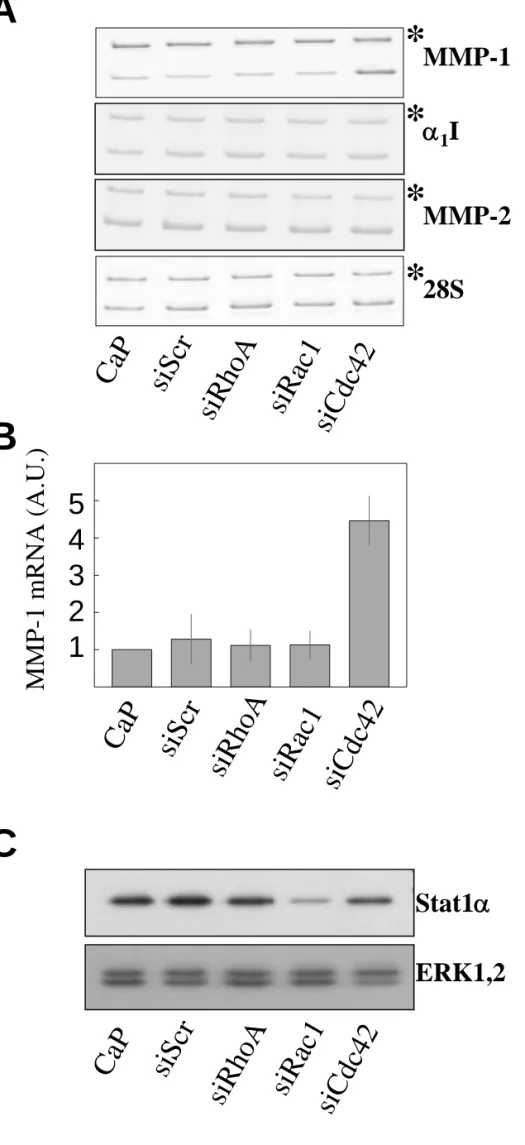
Fig. 3. Cdc42 silencing up-regulates MMP-1 protein level.
(A) Representative Western blot analysis of serum-free media conditioned by HSF for 16h between day 2 and day 3 post-transfection with calcium phosphate alone (CaP), with 20 nM of an siRNA targeting RhoA (siRhoA), Rac1 (siRac1) or Cdc42 (siCdc42), or with 20 nM of an irrelevant siRNA (siScr). (B) Densitometric analysis of (A). Results are the mean+s.d. of three independent experiments. (C) Representative Western blot analysis of cell lysates of
between day 2 and day 3 post-transfection (C.M.). Cells were transfected with the indicated concentrations of either an irrelevant siRNA (siScr) or the siRNA targeting Cdc42 (siCdc42).
Fig. 4. Cdc42 silencing up-regulates MMP-1 but not MMP-2 nor α1I mRNA steady-state level.
(A) Representative quantitative RT-PCR analysis of MMP-1, MMP-2 and α1I mRNA level
and 28S rRNA in total RNA extracted from HSF cultured in DMEM+10%FCS 72h post-transfection with calcium phosphate alone (CaP), an irrelevant siRNA (siScr) or with the siRNA targeting RhoA (siRhoA), Rac1 (siRac1) or Cdc42 (siCdc42). Sample to sample variations in RT-PCR efficiency is controlled by adding a known copy number of synthetic RNA co-transcribed and co-amplified with the same primers which generate a product of larger size (*). (B) Densitometric analysis of (A). Results are the mean+s.d. of three independent experiments. (C) Western blot analysis of whole cell lysates of HSF transfected with calcium phosphate alone (CaP), with 20 nM of the siRNA targeting RhoA (siRhoA), Rac1 (siRac1) or Cdc42 (siCdc42), or with 20 nM of an irrelevant siRNA (siScr). 72 hours post-transfection, the cells were lysed in SDS-PAGE loading buffer and analysed by immunoblotting with specific antibodies to Stat1α and ERK1,2.
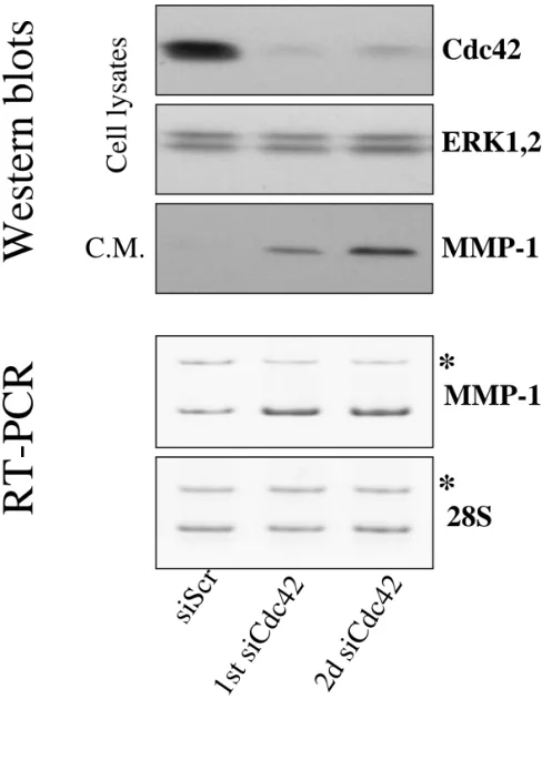
Fig. 5. A second siRNA targeting another region of the Cdc42 mRNA also up-regulates MMP-1. Representative Western blot analysis of whole cell lysates and of serum free media
conditioned by HSF 72h post-transfection with 20 nM of an irrelevant siRNA (siScr), the first siRNA targeting Cdc42 (1st siCdc42) or the second siRNA targeting Cdc42 (2d siCdc42) with specific antibodies to Cdc42, ERK1,2 and MMP-1; RT-PCR analysis of MMP-1 mRNA level and 28S rRNA was perform with total RNA extracted from HSF cultured in DMEM+10%FCS 72h post-transfection with 20 nM of an irrelevant siRNA (siScr), the first
siRNA targeting Cdc42 (1st siCdc42) or the second siRNA targeting Cdc42 (2nd siCdc42). Sample to sample variations in RT-PCR efficiency is controlled by a known copy number of synthetic RNA co-transcribed and co-amplified with the same primers which generate a product of larger size (*).
Fig. 6. Multiple knock-down of RhoGTPases revealed a role for Rac1, but not RhoA, in MMP-1 overexpression following Cdc42 silencing. HSF were transfected with the indicated
siRNA at the concentration of 10 nM. To reach the same final concentration of 30 nM of siRNA in each condition, transfection mix were supplemented with siScr. Representative Western blot analysis of whole cell lysates 72h post-transfection (cell lysates) and of serum free media conditioned for 16h between day 2 and day 3 post-transfection are shown. Cell lysates and conditioned media (C.M.) were analysed by immunoblotting with specific antibodies to RhoA, Rac1, Cdc42, ERK1,2 and MMP-1. The lower panel illustrates the densitometric analysis of MMP-1 measurements by Western blot analysis of C.M.. Results are expressed as the mean+s.d. of three independent experiments. The concentrations of the siRNA in the table below the figures are expressed in nM.
Fig. 7. MCP-1 and IL-8 overexpression following Cdc42 silencing.
(A) Analysis of serum free media conditioned for 16h between day 2 and day 3 post-transfection by Cytokine proteoarray. HSF were transfected with 20 nM of the siRNA targeting RhoA (siRhoA), Rac1 (siRac1) or Cdc42 (siCdc42), or with 20 nM of an irrelevant siRNA (siScr). White arrows indicate the position of the IL-8, IL-6 and MCP-1 signals respectively from the top to the bottom (B) Representative semi-quantitative RT-PCR analysis of MCP-1 and IL-8 mRNA level and 28S rRNA in total RNA extracted from HSF 72h post-transfection with either calcium phosphate alone (CaP), an irrelevant siRNA (siScr) or with
the siRNA targeting RhoA (siRhoA), Rac1 (siRac1) or Cdc42 (siCdc42). The efficiency of the 28S RT-PCR is controlled by a known copy number of synthetic RNA co-transcribed and co-amplified with the same primers which generate a product of larger size (*). (C) ELISA measurements of MCP-1 and IL-8 in serum free media conditioned by HSF for 16h between day 2 and day 3 post-transfection.
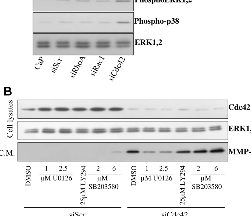
Fig. 8. Overexpression of MMP-1 following Cdc42 silencing is mediated through the ERK1,2 pathway. (A)Western blot analysis of whole cell lysates from starved HSF 72h
post-transfection with either calcium phosphate alone (CaP), 20 nM of an irrelevant siRNA (siScr) or with 20 nM of the siRNA targeting RhoA (siRhoA), Rac1 (siRac1) or Cdc42 (siCdc42). Cell lysates were analyzed by immunoblotting with specific antibodies to phospho-ERK1,2, phospho-p38 and to total ERK1,2 (ERK1,2). (B) Representative Western blot analysis of whole cell lysates of HSF 72h post-transfection (cell lysates) and of serum free media conditioned for 16h between day 2 and day 3 post-transfection (C.M.). HSF were transfected with 20 nM of an irrelevant siRNA (siScr) or with 20nM of the siRNA targeting Cdc42 (siCdc42) and cultured between day 1 and day 3 post-transfection with the indicated concentrations of the MEK kinase inhibitor (U0126), of the PI3Kinase inhibitor (LY294) and of the p38 MAP kinase inhibitor (SB203580). Cell lysates and C.M. were analysed by immunoblotting with specific antibodies to Cdc42, ERK1,2 and MMP-1.
Fig. 9. The ERK1,2 pathway is also required for MMP-1 overexpression following Cdc42 silencing with a second siRNA. (A) Representative Western blot analysis of whole
cell lysates of HSF 72h post-transfection with calcium phosphate alone (CaP) or with the indicated concentrations of an irrelevant siRNA (siScr) or the second siRNA targeting Cdc42 (2nd siCdc42). Cell lysates were analysed by immunoblotting with specific antibodies to
phospho-ERK1,2 and ERK1,2. (B-C) Representative Western blot analysis of serum free media conditioned by HSF for 16h between day 2 and day 3 post-transfection with 20 nM of an irrelevant siRNA (siScr), with 20nM of the siRNA targeting Cdc42 (1st siCdc42) or with 20nM of the second siRNA targeting Cdc42 (2nd siCdc42) and cultured between day 1 and day 3 post-transfection with the indicated concentrations of (B) the MEK kinase inhibitor U0126 or (C) the p38 MAPKinase inhibitor SB203580.
Fig. 10. MMP-1 overexpression following Cdc42 silencing is observed in various human cell lines. Representative Western blot analysis of cell lysates of human breast
adenocarcinoma cell line HS578T, fibrosarcoma cell line HT1080 and melanoma cell line A2058 72h post-transfection and of serum free media conditioned by the same cells for 16h between day 2 and day 3 post-transfection (C.M.). Cells were transfected with 20nM of either an irrelevant siRNA (siScr) or the first siRNA targeting Cdc42 (siCdc42). Cell lysates and C.M. were analysed by immunoblotting with specific antibodies to Cdc42, ERK1,2 and MMP-1.
Fig. 11. The rescue of Cdc42 knock-down represses the MMP-1 overexpression.
RT-PCR analysis of Cdc42 and mutated Cdc42 (mCdc42) mRNA level and 28S rRNA was performed with total RNA extracted from HS578T transfected with 20 nM of either an irrelevant siRNA (siScr) or the first siRNA targeting Cdc42 (siCdc42) and 1µg of either empty pShuttle (vector) or pShuttle-mCdc42 (mCdc42). Total RNA was extracted 72h after transfection with the siRNA. The top left panel illustrates the amplification of mutated Cdc42 mRNA performed with total RNA extracted from HS578T 48h after transfection with pShuttle-mCdc42, with (+RT) or without (-RT) the reverse transcription step. Representative Western blot analysis of whole cell lysates and of serum free media conditioned (C.M.) by
HS578T transfected with 20 nM of either an irrelevant siRNA (siScr) or the first siRNA targeting Cdc42 (siCdc42) and 1µg of either empty pShuttle (vector) or pShuttle-mCdc42 (mCdc42) with specific antibodies to Cdc42, ERK1,2 and MMP-1. The lower panel illustrates the densitometric analysis of MMP-1 measurements by Western blot analysis of C.M.. Results are the mean+s.d. of three independent experiments.