EDITORIAL COMMENT
Chimeric Antigen Receptor T-Cells and
Cardiovascular Toxicity
Cause for Concern?
*
Patrizio Lancellotti, MD, PHD,a,bMarie Moonen, MD, PHD,aMaurizio Galderisi, MDc
I
mmunotherapy is an expanding resource intreating cancer patients, with impressive bene
fi-cial effects on survival in several advanced
ma-lignancies (1). Cancer activates multiple mechanisms
that co-opt host–tumor immune interactions, thus inducing immune evasion. Novel therapeutic ap-proaches have been developed by manipulating and engineering immune cells, which has created great expectations in the oncology community. To date, at least 2 immunotherapeutic strategies, checkpoint inhibition and cellular therapy with autologous chimeric antigen-receptor T cells (CAR-T cells), have clearly demonstrated their efficacy in treating solid tumors and in a few hematologic malignancies. In contrast, checkpoint inhibitors can produce a wide spectrum of immune-related adverse complications, also involving the heart, and episodes of fulminant myocarditis or fatal heart failure have been described
in patients treated with these drugs (2). Adverse
events related to CAR-T cells involve mainly
cytokine-related syndrome (CRS) and neurotoxicity
(3), but little is known about the occurrence of
cardio-vascular (CV) complications.
The study by Alvi et al. (4) in this issue of the
Journal adds valuable information to this issue
through a study of 137 patients treated with CAR-T cells, most of whom had relapsed diffuse large
B-cell lymphoma (5). The definition of cardiotoxicity
was based on a troponin increase or a left ventricular ejection fraction (LVEF) decrease compared to the pre-treatment period. CV events were composites of arrhythmias, decompensated heart failure, and CV
death. The study by Alvie et al. (4) clearly
demon-strated that cardiac damage and CV events represent relatively frequent complications of CAR-T cell ther-apy, as shown by 21% to 59% of the treated patients who experienced cardiac damage, 12% of whom had CV events and 4% of whom had cardiac-related death. The present study pointed out several observations that need commentary. First, CAR-T cell
cardiotox-icity occurred mainly in elderly patients (>60 years
old), often characterized by a high CV risk profile and
prior anticancer treatment including anthracyclines and radiotherapy. Second, there was a strong associ-ation between troponin elevassoci-ation post-CAR-T cell therapy (>0.03 ng/ml or high-sensitivity troponin >14 ng/l) and subsequent CV events. Troponin elevation was observed in 95% (16 of 17) of the pa-tients experiencing CV events and was commonly seen in patients with CRS development (defined as
thefirst appearance of fever after CAR-T cell therapy).
CRS of any grade (using a CRS grading system of 1 to 5) was common, occurring at a median of 5 days after CAR-T cell therapy. Of note, an isolated elevation of troponin without CRS was not associated with the occurrence of CV events. Notably, a clinically signif-icant decrease of LVEF (reduction of at least 10% points to a value below 50%) was detected in 28% of the patients treated with CAR-T cell, but only 29 of the 135 patients had echocardiographic data pre- and during CAR-T cell therapy. For this reason, a parallel between troponin increase and LVEF reduction could
SEE PAGE 3099
ISSN 0735-1097/$36.00 https://doi.org/10.1016/j.jacc.2019.10.028
*Editorials published in the Journal of the American College of Cardiology reflect the views of the authors and do not necessarily represent the views of JACC or the American College of Cardiology.
From theaDepartment of Cardiology, Groupe Interdisciplinaire de
Gen-oproteomique Appliquee Cardiovascular Sciences, University of Liège, Liège, Belgium;bGruppo Villa Maria Care and Research, Anthea Hospital,
Bari, Italy; and thecDepartment of Advanced Biomedical Sciences,
Fed-erico II University, Naples, Italy. Dr. Galderisi has received speaker fees from Bayer, Pfizer, and Sobi. All other authors have reported that they have no relationships relevant to the contents of this paper to disclose.
J O U R N A L O F T H E A M E R I C A N C O L L E G E O F C A R D I O L O G Y V O L . 7 4 , N O . 2 5 , 2 0 1 9 ª 2 0 1 9 B Y T H E A M E R I C A N C O L L E G E O F C A R D I O L O G Y F O U N D A T I O N
not be formally established, although a reduction in LVEF was observed only in patients with increased troponin and higher CRS grade after CAR-T cells were administered. Lack of data for global longitudinal strain and B-type natriuretic peptide release does not support this relationship either. Also, all CV events
occurred in patients with a CRS grade$2, known to be
associated with significant organ toxicity. Finally, a
longer time between recognition of CRS and the administration of tocilizumab was associated with an increase in CV events.
The close relationships among CRS development, troponin release, and the occurrence of CV events
represent a red flag that must be considered with
greater attention in this clinical setting. As observed
by Alvi et al. (4), the increase in troponin occurred in
patients with a CRS grade of at least$2, that is, in
patients with a higher degree of inflammation. Troponin level is a reliable indicator of myocyte injury in oncology patients with cardiotoxic effects of anticancer drugs. In the case of CAR-T cell therapy, the increase in troponin appeared to be primarily related to the hypotension and tachycardia induced by CRS. Treatment with CAR-T cells is associated with a marked increase in interleukin (IL)-6. IL-6 exerts its detrimental effect on several tissues, including
myocardium in CRS (5). This finding is entirely
consistent with the information obtained using
tocilizumab for the treatment of these patients. Tocilizumab is a recombinant humanized anti-IL-6 monoclonal antibody that has been used primarily in the treatment of rheumatoid arthritis. More recently, it has demonstrated great use in success-fully contrasting CRS and blunting its progression
from subclinical to clinical stages (6). It should be
noted that if the main anti-inflammatory properties of
corticosteroids are used to counteract the detrimental
CV effects of immune checkpoint inhibitors (2), then
the cardiotoxicity of CAR-T cells may be specifically counterbalanced by tocilizumab. In the study by Alvi
et al. (4), early administration of tocilizumab was
associated with a significantly lower rate of CV events, without preventing the antitumor effects of CAR-T cells.
Together, these results point to major practical implications that should be considered when using CAR-T cells. First, results confirm the need for close cooperation between oncologists/hematologists and cardiologists in order to maximize the benefits of innovative therapies for malignant tumors while reducing and controlling the burden of CV complica-tions, the characteristics and mechanisms of which
are often not yet well understood (7). Practically,
the combination of old age and evidence of
multiple pre-existing CV risk factors implies the need for a preliminary, comprehensive cardiological
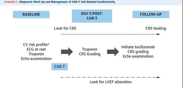
FIGURE 1 Diagnostic Work-Up and Management of CAR–T Cell–Related Cardiotoxicity
BASELINE
FOLLOW-UP
Look for LVEF alteration
DAY 5
POST-CAR-T
CAR-T
Look for CRS
CRS healing
CV risk profile*
ECG at rest
Troponin
Echo examination
Troponin
CRS Grading
Initiate tocilizumab
CRS grading
Echo examination
After a baseline comprehensive cardiovascular assessment, determination of troponin level within 5 days after CAR-T cell therapy should guide patient management. If a significant rise in troponin is detected, echocardiography should be repeated, the CRS grade determined, and tocilizumab initiated promptly. Timing of cardiological follow-up should be established in individual patients according to clinical judgment. *Baseline assessment including a 24-h ECG and an inducible ischemia test, when needed. CAR-T¼ chimeric antigen-receptor T cell; CRS¼ cytokine-related syndrome; CV ¼ cardiovascular; ECG ¼ electrocardiography; Echo ¼ echocardiography; LVEF ¼ left ventricular ejection fraction.
Lancellottiet al. J A C C V O L . 7 4 , N O . 2 5 , 2 0 1 9
CAR-T Cells and Cardiotoxicity in Cancer D E C E M B E R 2 4 , 2 0 1 9 : 3 1 0 9– 1 1
work-up before the beginning of CAR-T cell therapy.
This work-up (Figure 1) should include biomarker
assessment (troponin and B-type natriuretic peptide), a resting 12-lead electrocardiogram (ECG), and a
complete echocardiography-Doppler examination
(including global longitudinal strain) to identify car-diac conditions representing the background of sub-sequent CV complications. Values of LVEF should be determined with good accuracy at baseline in patients who had previously experienced potentially car-diotoxic drugs, such as anthracyclines and radio-therapy, and repeated periodically during CAR-T cell therapy. In this respect, the use of 3D echocardiog-raphy, which allows a more accurate and reproducible assessment of LVEF and, therefore, is more suitable for serial cardiac imaging evaluation, should not be underestimated. In addition, the preliminary assess-ment could include 24-h Holter ECG monitoring and the search for inducible ischemia (stress imaging) in patients with known or suspected coronary artery disease and a high risk of developing episodes of
arrhythmia. During treatment, beyond clinical
assessment (i.e., fever), both troponin and LVEF should be monitored. Any increase in troponin
asso-ciated with a CRS grade of$2 at day 5 after CAR-T cell
administration should prompt the initiation of car-dioprotective therapy with tocilizumab. The same can be done in case of a drop in LVEF. Conversely, an
isolated increase in troponin (a CRS grade of <2)
should not be treated, although caution is warranted in the absence of additional data. Therefore, even if there is a reason for concern, further studies including more patients are needed to better under-stand the relationship between CAR-T cells and car-diovascular toxicity.
ADDRESS FOR CORRESPONDENCE: Dr. Patrizio
Lancellotti, Department of Cardiology, CHU Liège, University of Liège, GIGA-Cardiovascular Sciences, Avenue de l’Hôpital, 1, Bât. B34, B-4000 Liège,
Belgium E-mail: [email protected]. Twitter:
@UniversiteLiege.
R E F E R E N C E S
1.Jain A, Zhang Q, Toh HC. Awakening immunity against cancer: a 2017 primer for clinicians. Chin J Cancer 2017;36:67.
2.Lyon AR, Yousaf N, Battisti NML, Moslehi J, Larkin J. Immune checkpoint inhibitors and car-diovascular toxicity. Lancet Oncol 2018;19: e447–58.
3.Brudno JN, Kovkenderfer JN. Toxicities of chimeric antigen receptor T cells: recognition and management. Blood 2016;127:3321–30.
4.Alvi RM, Frigault MJ, Fradley MG, et al. Cardio-vascular events among adults treated with chimeric antigen receptor T-cells (CAR-T). J Am Coll Cardiol 2019;74:3099–108.
5.Liu D, Zhao J. Cytokine release syndrome: grading, modeling, and new therapy. J Hematol Oncol 2018;11:121.
6.Le RQ, Li L, Yuan W, et al. FDA approval summary: tocilizumab for treatment of chimeric antigen receptor T cell-induced severe or
life-threatening cytokine release syndrome. Oncologist 2018;23:943–7.
7.Lancellotti P, Suter TM, Lopez-Fernandez T, et al. Cardio-oncology service: rationale, organization, and implementation. Eur Heart J 2019;40:1756–63.
KEY WORDS cardiovascular toxicity, CAR-T cell therapy, cell therapy, checkpoint inhibition, immunotherapy
J A C C V O L . 7 4 , N O . 2 5 , 2 0 1 9 Lancellottiet al.
D E C E M B E R 2 4 , 2 0 1 9 : 3 1 0 9– 1 1 CAR-T Cells and Cardiotoxicity in Cancer