Université de Montréal
RNA recurrent motifs: identification and
characterization
par Yury Butorin Département de Biochimie Faculté de Médecine
Thèse présentée à la Faculté des études supérieures en vue de l’obtention du grade de Ph.D.
en biochimie
Avril, 2010
Université de Montréal Faculté des études supérieures
Cette thèse intitulée :
RNA recurrent motifs: identification and characterization
présentée par : Yury Butorin
A été évaluée par un jury composé des personnes suivantes :
Dr Pascal Chartrand, président-rapporteur Dr Serguei Chteinberg, directeur de recherche
Dr Joëlle Pelletier, membre du jury Dr Roscoe Klinck, examinateur externe Dr. François Major, représentant du doyen
Résumé
La détermination de la structure tertiaire du ribosome fut une étape importante dans la compréhension du mécanisme de la synthèse des protéines. Par contre, l’élucidation de la structure du ribosome comme tel ne permet pas une compréhension de sa fonction. Pour mieux comprendre la nature des relations entre la structure et la fonction du ribosome, sa structure doit être étudiée de manière systématique. Au cours des dernières années, nous avons entrepris une démarche systématique afin d’identifier et de caractériser de nouveaux motifs structuraux qui existent dans la structure du ribosome et d’autres molécules contenant de l’ARN.
L’analyse de plusieurs exemples d’empaquetage de deux hélices d’ARN dans la structure du ribosome nous a permis d’identifier un nouveau motif structural, nommé « G-ribo ». Dans ce motif, l’interaction d’une guanosine dans une hélice avec le ribose d’un nucléotide d’une autre hélice donne naissance à un réseau d’interactions complexes entre les nucléotides voisins. Le motif G-ribo est retrouvé à 8 endroits dans la structure du ribosome. La structure du G-ribo possède certaines particularités qui lui permettent de favoriser la formation d’un certain type de pseudo-nœuds dans le ribosome. L’analyse systématique de la structure du ribosome et de la ARNase P a permis d’identifier un autre motif structural, nommé « DTJ » ou « Double-Twist Joint motif ». Ce motif est formé de trois courtes hélices qui s’empilent l’une sur l’autre. Dans la zone de contact entre chaque paire d’hélices, deux paires de bases consécutives sont surenroulées par rapport à deux paires de bases consécutives retrouvées dans l’ARN de forme A. Un nucléotide d’une paire de bases est toujours connecté directement à un nucléotide de la paire de bases surenroulée, tandis que les nucléotides opposés sont connectés par un ou plusieurs nucléotides non appariés. L’introduction d’un surenroulement entre deux paires de bases consécutives brise l’empilement entre les nucléotides et déstabilise l’hélice d’ARN. Dans le motif DTJ, les nucléotides non appariés qui lient les deux paires de bases surenroulées interagissent avec une des trois hélices qui forment le motif, offrant ainsi une stratégie élégante de stabilisation de l’arrangement.
Pour déterminer les contraintes de séquences imposées sur la structure tertiaire d’un motif récurrent dans le ribosome, nous avons développé une nouvelle approche expérimentale. Nous avons introduit des librairies combinatoires de certains nucléotides retrouvés dans des motifs particuliers du ribosome. Suite à l’analyse des séquences alternatives sélectionnées in vivo pour différents représentants d’un motif, nous avons été en mesure d’identifier les contraintes responsables de l’intégrité d’un motif et celles responsables d’interactions avec les éléments qui forment le contexte structural du motif.
Les résultats présentés dans cette thèse élargissent considérablement notre compréhension des principes de formation de la structure d’ARN et apportent une nouvelle façon d’identifier et de caractériser de nouveaux motifs structuraux d’ARN.
Abstract
Although determination of the ribosome tertiary structure has been an outstanding step towards elucidation of the mechanism of protein synthesis, the complexity of this structure does not provide an easy answer of how this large molecular complex works. In order to understand the nature of structure-function relationships in the ribosome, the ribosome structure itself should be subjected to thorough analysis. In the last years, we undertook systematic efforts toward identification and characterization of all recurrent structural motifs existing in the ribosomal RNA and in other RNA-containing molecules.
The analysis of many instances of helix-helix packing in the ribosome structure allowed us to identify a new structural motif which we called “G-ribo”. In this motif, an interaction of the sugar edge of a guanosine in one helix with the ribose of a nucleotide from another helix was found to be at the origin of a complex network of concomitant inter-nucleotide interactions. In total, the G-ribo motif was found at eight locations within the ribosomal RNA. A surprising feature of this motif consists in its ability to favor the formation of pseudoknots of a particular type. In the ribosome structure, there are four pseudoknots whose formation is mediated by the G-ribo motif.
Systematic analysis of the ribosome as well as the RNAseP crystal structures allowed for the identification of a new RNA motif, which we called “DTJ”, or Double-Twist Joint motif. This motif is made of three short RNA double helices, which stack one on top of another. In the contact zone of each pair of helices two consecutive base pairs are over-twisted compared to the regular helical twist of 32° of A-RNA. One nucleotide of the base pair is always directly connected to the one nucleotide of the over-twisted base pair, while the opposite nucleotides of these base pairs are connected with one or several unpaired nucleotides. Introduction of the helical over-twist between two consecutive base pairs breaks the inter-nucleotide stacking and destabilizes the RNA double helix. In the DTJ, the unpaired nucleotides that connect the two over-twisted base pairs interact with one of the three motif-forming helices, providing an elegant strategy for the stabilization of the whole arrangement.
To determine the nucleotide sequence constraints imposed on the structure of recurrent RNA motifs in the functional ribosome we developed a new approach consisting in the selection of functional ribosomes from a combinatorial gene library in which certain nucleotides of the rRNA gene corresponding to a particular motif were randomized. Comparison of the constraints determined for different examples of the same motif allowed us to distinguish between constraints responsible for the integrity of the motif and for its interaction with surrounding elements, including ribosomal proteins.
The work significantly improves our understanding of the principles of RNA structure formation and opens a new way to identify and characterize RNA motifs.
Table of contents
Résumé ... iii
Abstract ...v
Table of contents ... vii
List of Tables ... xiii
List of Figures ...xv
Abbreviations list ...xx
Acknowledgements ... xxiii
1. Introduction ...2
1.1 RNA is a versatile biopolymer ...2
1.1.1 RNA is a carrier of genetic information ...2
1.1.2 RNA can adopt various 3D structures ...3
1.1.3 RNA as an enzyme ...5
1.1.4 RNA in post-transcriptional gene regulation ...7
1.2 The basics of RNA structure ...10
1.2.1 A- and B- helical form of nucleic acids ...11
1.2.2 Non-canonical base pairing ...13
1.3 Primary, secondary and tertiary motifs...16
1.3.1 Primary structure and primary sequence motifs ...16
1.3.2 Secondary structure and secondary structure motifs...16
1.3.3 3D structure of RNA and tertiary motifs ...18
1.3.4 A-minor motif ...20 1.3.5 RNA Bulge ...21 1.3.6 Internal loops ...25 1.3.6.1 Loop-E motif ...25 1.3.6.2 C-loop motif ...27 1.3.6.3 UAA/GAN motif ...28
1.3.6.4 Along Groove Packing motif ...30
1.4 Overview of the basics of protein synthesis ...32
1.4.1 Initiation of protein synthesis ...33
1.4.3 Translocation ...35
1.4.4 Termination ...36
1.5 Ribosomal structures ...38
1.6 An overview of the rRNA structure ...40
1.7 RNA pseudoknots...45
1.7.1 Role of pseudoknots in -1 frameshift ...47
1.8 Principles of molecular dynamics ...50
1.9 Combinatorial approaches in biology ...55
1.10 The main outlines of the project ...57
1.10.1 Identification of new motifs ...57
1.10.2 In vivo studies of motifs...58
1.10.2.1 In vivo systems for studies of rRNA mutagenesis...59
1.10.3 In silico modeling ...60
2. G-ribo: A new structural motif in ribosomal RNA...62
2.1 Abstract ...63
2.2 Introduction ...63
2.3 Definition of the G-ribo motif ...63
2.4 Identification of the G-ribo motif in the ribosome structure ...64
2.5 Tetranucleotide arrangement at the +1 layer ...66
2.6 A-minor interaction at the -1 layer of Helix 1 ...68
2.7 Participation of riboses in the stabilization of the core of the G-ribo motif ... ...68
2.8 The consensus pattern of the G-ribo motif ...69
2.9 Discussion...70 2.10 Acknowledgements ...72 2.11 References ...73 2.12 Figures ...75 2.13 Supplemental Table ...80 2.14 Supplemental Figures ...81
3. G-ribo motif favors the formation of pseudoknots in ribosomal RNA ...87
3.2 Introduction ...88
3.3 Background: the G-ribo motif ...89
3.4 Chain break in strand P ...90
3.5 Wrench pseudoknot in L1024 ...91
3.6 Wrench pseudoknot in S861 ...92
3.7 Wrench pseudoknot in S521 ...93
3.8 Ring pseudoknot...94
3.9 Evolutionary conservation of the G-ribo-based pseudoknots ...96
3.10 Discussion ...98 3.11 Acknowledgements ...99 3.12 References ...100 3.13 Figures ...102 3.14 Supplemental Table ...106 3.15 Supplemental figures ...107
4. Double twist-joints – new recurrent RNA motifs...115
4.1 Abstract ...116
4.2 Introduction ...117
4.2.1 The twist-joint structures ...117
4.2.2 Direct stabilization of TJ ...119
4.2.3 Indirect stabilization of TJ ...120
4.2.4 Double twist-joints ...120
4.3 A-DTJs ...121
4.3.1 A quasi-A-DTJ arrangement ...122
4.3.2 The conglomerate of 2AVY-68 and 2AVY-97...122
4.3.3 Evolutionary conservation of A-DTJs ...123
4.4 B-DTJs ...125
4.4.1. Evolutionary conservation of B-DTJs ...127
4.5 C-DTJs ...127
4.5.1. Evolutionary conservation of C-DTJs ...129
4.6 Discussion...130
4.6.2 Role of DTJs in the long-range interactions ...131 4.7 Conclusions ...133 4.8 References ...134 4.9 Tables ...136 4.10 Figures ...139 4.11 Supplemental figures ...151
5. Recurrent RNA motifs as probes for studying RNA-protein interactions in the ribosome ...163
5.1 Abstract ...164
5.2 Introduction ...164
5.3 Materials and methods ...165
5.3.1 Bacterial strains and media ...165
5.3.2 Plasmids ...166
5.3.3 Design of the combinatorial gene libraries ...166
5.3.4 Plasmid replacement and selection of functional clones ...166
5.3.5 Measurement of the ribosome efficiency and of the growth rates ...167
5.3.6 Sequencing ...167
5.3.7 Molecular dynamics simulations ...167
5.4 Results ...169
5.4.1 Background: general description of AGPM ...169
5.4.2 The motifs studied ...170
5.4.3 Cloning and selection of functional clones ...171
5.4.4 Analysis of the selected clones: the minimal requirement for the integrity of AGPM ...172
5.4.5 Alternative dinucleotide combinations ...174
5.4.6 Molecular dynamics simulations ...175
5.4.7 The symmetry of the central base pairs in motif S296 ...178
5.4.8 Interaction of motif L639 with ribosomal protein L35 ...178
5.4.9 Interaction of motif L657 with ribosomal protein L4 ...179
5.4.10 Exceptional clones A11, B16 and C84 ...182
5.5.1 The power of the approach ...183
5.5.2 New findings about AGPM: principles of RNA structure formation ...184
5.5.3 New findings about AGPM: principles of RNA-protein interaction ...186
5.5.4 The sensitivity of the approach ...187
5.6. Funding ...187 5.7 Acknowledgments ...188 5.8 References ...189 5.9 Table ...193 5.10 Figures ...194 5.11 Supplementary data ...201
5.11.1 Instant evolution versus natural evolution ...201
5.11.2 Potential limitations on the usage of GA as a central base pair in AGPM .... ...203
5.12 Supplementary Methods ...203
5.12.1 Combinatorial gene libraries: primers and cloning ...203
5.13 Supplementary Tables ...205
5.14 Supplementary Figures ...208
5.15. Supplementary References ...211
6. In vivo analysis of the general constraints imposed on the structure of G-ribo motifs in the functional ribosome ...213
6.1 Abstract ...214
6.2 Introduction ...215
6.3 Results ...218
6.3.1 Analysis of the variant sequences of motif S1047 ...218
6.3.2 Analysis of the variant sequences of motif S521 ...219
6.4 Discussion...219
6.4.1 G-ribo motif S1047 can exist without the G-ribo interaction ...220
6.4.2 G-ribo motif S1047 can exist without a stable base pair at -1 layer of H1 221 6.4.3 The role of identity of nucleotide -1T in G-ribo motifs S1047 and S521 ....222
6.5 Conclusions ...223
6.6.1 Bacterial Strains and Media ...224
6.6.2 Plasmids ...224
6.6.3 Combinatorial gene libraries: primers and cloning ...224
6.6.4 Measurement of the ribosome efficiency ...225
6.6.5 Sequencing ...225
6.7 References ...226
6.8 Tables ...228
6.9 Figures ...231
7. Discussion...235
7.1 Identification and characterization of new motifs ...237
7.1.1 G-ribo motif ...237
7.1.2 DTJ-motif ...242
7.2 In vivo studies of the motifs ...244
7.2.1 Functional characterization of the motifs ...245
7.2.2 AGPM ...246
7.2.3 In vivo study of G-ribo motif ...249
8. Conclusions ...257
List of Tables
Chapter 1: Introduction
Table 1. The key aspects that distinguish siRNA and miRNA ...10 Table 2. The 12 geometric families of nucleic acid base pairs with symbols for annotating secondary structure diagrams ...15
Chapter 2: G-ribo: A new structural motif in ribosomal RNA
Supplemental Table 1. The frequency of the occurrence of particular identities for base pairs and individual nucleotides in different G-ribo
motifs ...80
Chapter 3: G-ribo motif favors the formation of pseudoknots in
ribosomal RNA
Supplemental Table 1. The frequency of the occurrence of particular identities for base pairs and individual nucleotides in different G-ribo
motifs ...106
Chapter 4: Double twist-joint – a new recurrent RNA motif
Table 1. The frequency of the occurrence of particular identities for base
pairs and individual nucleotides in different A-DTJ motifs ...136 Table 2. The frequency of the occurrence of particular identities for base
pairs and individual nucleotides in different B-DTJ motifs ...137 Table 3. The frequency of the occurrence of particular identities for base
pairs and individual nucleotides in different C-DTJ motifs ...138
Chapter 5: Recurrent RNA motifs as probes for studying RNA-protein
interactions in the ribosome
Supplementary Table S1. Presence of different tetra-nucleotide
combinations ...205 Supplementary Table S2. Presence of different tetra-nucleotide
combinations as the central base pairs of all AGPMs in the prokaryotic 16S and 23S rRNAs ...206 Supplementary Table S3. Sequences of the oligonucleotides used in this
study ...207
Chapter 6 : In vivo study of the G-ribo motif
Table 1. Nucleotide sequences and activities of the selected variants of
motif S1047 ...228 Table 2. Selection of sequences of clones with GC base pair in position [0P; 0Q] and GU base pair [-1P; -1Q] and either uridine or cytosine -1T ...229 Table 3. Selection of sequences of clones with GC base pair in position
[-1P; -1Q] and GU base pair [0P; 0Q] and either uridine or cytosine -1T ...229 Table 4. Selection of sequences of clones with GC base pair in position [0P; 0Q] and GU base pair [-1P; -1Q] and either pyrimidine or purine in position -1T ...229 Table 5. Selection of sequences of clones with GC base pair in position [0P; 0Q] and AU base pair [1P; 1Q] and either uridine or guanine in position -1T ...229 Table 6. Selection of sequences of clones with either GC or GU base pair in positions [0P; 0Q] and [-1P; -1Q] and pyrimidine in position -1T ...229 Table 7. Selection of sequences of clones that do not have guanosine 0P,
possess a WC base pair [-1P; -1Q] and pyrimidine in position -1T ...230 Table 8. Selection of sequences of clones that have a WC base pair
[0P; 0Q], a noncanonical base pair [1P; 1Q] and pyrimidine in position -1T ...230 Table 9. Selection of sequences of clones that have a non-WC combination in either [0P; 0Q] or [-1P; -1Q] positions and purine in position -1T ...230 Table 10. Sequences of the oligonucleotides that were used in this study ...230
List of Figures
Chapter 1: Introduction
Figure 1. Secondary and tertiary structures of tRNAPhe and bacterial
RNAseP...4
Figure 2. Reactions catalyzed by the known natural ribozymes ... 6
Figure 3. The five bases of DNA and RNA ...11
Figure 4. A-RNA and B-DNA structures ...12
Figure 5. Potential collision of 2’OH of RNA in the B-form ...13
Figure 6. Definition of nucleotide edges ...14
Figure 7. Glycosidic bond orientations ...15
Figure 8. RNA secondary structure example ...17
Figure 9. Four sub-types of A-minor interaction ...21
Figure 10. 3D structures of the RNA bulge ...24
Figure 11. A simplified scheme of an internal loop ...25
Figure 12. E-loop motif ...26
Figure 13. C-loop motif ...28
Figure 14. UAA/GAN motif ...29
Figure 15. Along-groove packing motif ...31
Figure 16. tRNA decoding by the ribosome ...35
Figure 17. Model for the ribosomal hybrid states ...37
Figure 18. Secondary and tertiary structures of 16S, 23S, and 5S rRNAs ...44
Figure 19. A simplified scheme representing a hairpin type (H-type) pseudoknot ...45
Figure 20. Secondary and tertiary structures of various pseudoknots ...46
Figure 21. Secondary structure scheme of the hepatitis C virus internal ribosome entry site ...49
Figure 22. Principle steps of the aptamer selection ...56
Chapter 2: G-ribo: a new structural motif in ribosomal RNA
Figure 1. The definition of different elements of the G-ribo motif ...75Figure 2. The tertiary structure of the G-ribo motifs in the E.coli
ribosome ...76 Figure 3. Secondary structures of the G-ribo motifs identified in the
E.coli ribosome ...78
Figure 4. The consensus structure of the G-ribo motif ...79 Supplemental Figure 1. The special position of nucleotide +1R ...81 Supplemental Figure 2. The tetranucleotide arrangements at the +1 layer in different cases of the G-ribo motif from the E.coli ribosome ...83 Supplemental Figure 3. Interaction of nucleotide -1T with base pair
[-1P;-1Q] in different cases of the G-ribo motif from the E.coli ribosome ...84 Supplemental Figure 4. The structure of the region of motif L1642
encompassing nucleotides -1Q, 0Q, +1Q, +1R and -1T ...85
Chapter 3: G-ribo motif favors the formation of pseudoknots in
ribosomal RNA
Figure. 1. The general description of the G-ribo motif ...102 Figure. 2. The secondary structures of the four G-ribo-based pseudoknots in the E.coli rRNA ...103 Figure. 3. The displacement of base pair [-2P;-2Q] with respect to
[-1P;-1Q] in the G-ribo-based pseudoknots ...105
Supplemental Figure 1. The pseudoknot definition ...107 Supplemental Figure 2.The secondary structures of the G-ribo-wrenches and of the G-ribo ring found in the E.coli ribosome ...109 Supplemental Figure 3. The tertiary structure of the G-ribo wrench
L1024, the superposition of the structures of the G-ribo wrenches L1024 and S861 and the tertiary structure of the G-ribo ring ...110 Supplemental Figure 4. Possible structural variations of the G-ribo
Supplemental Figure 5. The arrangements resembling that of base pairs [-1P;-1Q] and [-2P;-2Q] and nucleotide -1T found in different
pseudoknots not related to the G-ribo motif ...113
Chapter 4: Double twist-joint – new recurrent RNA motifs
Figure 1. Example of the helical over-twist between two base pairs ...139Figure 2. Schematic representation of a TJ arrangement ...140
Figure 3. The C-loop motif as an example of the direct stabilization of TJ 141 Figure 4. An example of the indirect stabilization of TJ ...142
Figure 5. The general scheme of the DTJ arrangement ...143
Figure 6. The secondary structure of A-DTJ motifs ...144
Figure 7. The structure of nucleotide triple [0R;0P;0Q] in A-DTJs ...145
Figure 8. Quasi-A-DTJ motif 2AVY-1142 ...146
Figure 9. The common arrangement of A-DTJs 2AVY-68 and 2AVY-97 .147 Figure 10. The reconstruction of the complete A-DTJ motif 2AVY-1142 in an archaeal nucleotide sequence of the H. marismortui 16S rRNA. ...148
Figure 11. The secondary structures of B-DTJ motifs ...149
Figure 12. The secondary structures of C-DTJ motifs ...150
Supplemental Figure 1. The interaction of nucleotides +1Q and +1P in A-DTJs ...151
Supplemental Figure 2. Deformed base pair [-2P; -2Q] in motif 2AVY-1057...152
Supplemental Figure 3. Secondary structure of homologous arrangement 2AVY-68 - 2AVY-97 from the T. thermophilus 30S subunit ...153
Supplemental Figure 4. The interaction of nucleotide 0R with the major groove of base pair [0P; 0Q] in B-DTJs ...154
Supplemental Figure 5. The interaction of nucleotide -1R with -1P in B-DTJs ...155
Supplemental Figure 6. Additional base-backbone hydrogen bonds found in the dinucleotide stack [0R; -1R] of the B-DTJ motifs ...156
Supplemental figure 7. The interaction of nucleotides +1Q and +1P in B-DTJs ...157 Supplemental Figure 8. The interaction of nucleotide 0R with the major groove of base pair [0P; 0Q] in C-DTJs ...158 Supplemental figure 9. The interaction of nucleotide -1R with the major groove of base pair [-1P; -1Q] in C-DTJs ...159 Supplemental Figure 10. Additional base-backbone hydrogen bonds
found in the dinucleotide stack [0R;-1R] of the C-DTJ motifs ...160 Supplemental Figure 11. Bending of the RNA double helix due to the presence of a DTJ motif ...156
Chapter 5: Recurrent RNA motifs as probes for studying RNA-protein
interactions in the ribosome
Figure 1. Schematic representation of AGPM ...194 Figure 2. Different arrangements of the central base pairs in AGPM ...195 Figure 3. Nucleotide sequences of the three cases of AGPM considered in this study ...196 Figure 4. The structural contexts of motifs L657 and L639 taken from the
E. coli ribosome ...197
Figure 5. Molecular dynamics simulations of the AGPM structure
containing different nucleotide triples ...198 Figure 6. Juxtapositions of the bases in the GA and AC base pairs ...199 Figure 7. Conformational rearrangements associated with the GUÙWC exchange of the central base pairs in AGPM ...200
Supplementary Figure S1. Modeled complex of AGPM used in
molecular dynamics simulations ...208 Supplementary Figure S2. The second molecular dynamics simulation of the AGPM construct containing the central GU-UG dinucleotide
Supplementary Figure S3. The third molecular dynamics simulation of the AGPM construct containing the central GU-UG dinucleotide
juxtaposition ...210
Chapter 6: In vivo study of the G-ribo motif
Figure 1. Definition of the G-ribo motif ...231Figure 2. Secondary structures of G-ribo motifs S521 and S1047 ...232
Figure 3. The over-twist between base pairs [-1P;-1Q] and [-2P;-2Q] in motifs S521 and S1047 ...233
Chapter 7: Discussion
Figure 1. Secondary structures of the alternative G-ribo motifs identified in the E. coli ribosome ...241Figure 2. A typical example of the helical over-twist ...242
Figure 3. Ribose-ribose contacts in AGPM ...248
Abbreviations list
3D Three dimensional
A Adenosine
Å Angstrom (10−10 meters)
AGPM Along-Groove packing motif
Amp Ampicillin
ASD Anti-Sinde-Dalgarno sequence
C Cytidine
CAT Chloramphenicol acetyl-transferase
Cryo-EM Cryo-electron microscopy
DNA Deoxyribonucleic acid
E.coli Escherichia coli
EF-G Elongation Factor G
G Guanosine
GFP Green Fluorescent Protein
H-bond Hydrogen bond
HCV Hepatitis C virus
HDV Hepatitis delta virus
HG Hoogsteen
Hi Helix number i
IPTG Isopropyl-1-thio-β-D-galactopyranoside
IRES Internal ribosome entry site
Kan Kanamycin
LB Luria-Bertani
MD Molecular Dynamics
miRNA Micro RNA
MMTV Mouse mammary tumor virus
PABP poly(A) tail-binding protein
PCR Polymerase Chain Reaction
PDB Protein Data Bank
ps Picosecond (10−12 of a second)
PTC Peptidyl Transferase Centre
Pu Purine Py Pyrimidine
RNA Ribonucleic acid
RNAse P Ribonuclease P
RRF Ribosomal release factor
rRNA Ribosomal RNA
S Svedberg, sedimentation coefficient
SD Shine-Dalgarno sequence
SE Sugar Edge
siRNA Small interfering RNA
Spc Spectinomycin T Thymidine
tRNA Transfer RNA
U Uridine
UTR Untranslated Region
WC Watson Crick
To my wife, my mother and my deceased grand mother for their constant encouragements
Acknowledgements
First, I would like to thank my scientific adviser, Prof. Sergey Steinberg, who guided me through the complex process of the scientific education. His unusual approach to tackling scientific problems allowed me to develop a unique insight, which will be a valuable source of inspiration for the rest of my life. He taught me to ask unusual questions and to find even more unusual answers. With his help I learned what the structure of RNA is. Due to the constant discussions with Sergey about the structure I developed a special skill that allows me to load PDB file directly to my mind and the structural analysis does not require me to have constant access to the computer.
I would like also to thank Matthieu Gagnon, with whom I spent long hours in the wet-lab, while trying to find ways to solve numerous experimental problems. His extremely high level of responsibility made him an invaluable member of our lab for everyone who was dealing with him. Special thanks for Matthieu for teaching me French and correcting my French texts.
I would like to thank Tetsu Ishii, who is able to find a positive aspect of absolutely any event, even totally negative. Special thanks for Tetsu for being my English language teacher. His willingness to help and extreme level of respect he showed to each and every person saved me in the most difficult times of my PhD course.
I would like to thank all members of our lab: François Boulay, Konstantine Bokov and Natalia Kotlova for their help and support.
I am also very grateful to the members of my jury, who took their precious time for reading my Thesis.
I would like also to thank my family: my mother, Marina, who brought me up and who made me who I am; my wife, Liudmila, who crossed the ocean and came to Canada to always stay by me and who helped me to survive immigration; my deceased grand-mother, Zoya, who was my second mother and my best friend; my uncle, Alexander, who was always the model person for me.
Chapter 1
Introduction
1. Introduction
1.1 RNA is a versatile biopolymer
RNA is a high molecular-weight bio-polymer, which consists of a long chain of nucleotide units. Each nucleotide consists of a five-carbon sugar, a phosphate group and one of the four bases: adenine, cytosine, guanine and uracil. RNA is an extremely versatile molecule, which plays diverse functional roles in the cells of all living organisms on Earth. For example, mRNA, tRNA and rRNA are involved in the process of translation (Berg et al., 2002). mRNA participates in the transfer of genetic information from DNA to the ribosome, where this information is converted into protein sequence. tRNA delivers amino acids to the ribosome, while rRNA arranges the sequence-specific catalysis of the reaction of trans-peptidation, resulting in the synthesis of polypeptide chains. miRNA and siRNA participate in the post-transcriptional gene regulation in eukaryotes through the complex mechanism of RNA interference (Carthew & Sontheimer, 2009). Splicing is the process of removal of introns from pre-mRNA. It can proceed either with help of spliceosomes, which contain several small nuclear RNA (snRNAs), or by introns themselves, which can work as ribozymes and catalyze their own excision (Steitz & Steitz, 1993).
I will briefly discuss three major abilities of RNA that explain its functional importance: the ability to transfer genetic information, to form complex 3D structures and to catalyze chemical reactions.
1.1.1 RNA is a carrier of genetic information
As long as both DNA and RNA use the same four-letter genetic code, the primary sequence of RNA can be used for storage and transmission of genetic information. Compared to RNA, DNA is more stable; hence it is used for long-term storage. RNA, on the contrary, is extremely vulnerable and as a result, can be used only for short-time storage of genetic information and for its transmission. The fact that RNA can easily and rapidly degrade is used by cells for post-transcriptional gene regulation (Pillai et al., 2007). Representatives of the three major kingdoms of life – eubacteria, archaea and eukarya, use RNA as an intermediate carrier of genetic information in the form of
mRNA. In non-eukaryotic cells, the newly transcribed mRNA is essentially ready for translation. In contrast, in eukaryotes, a DNA encoded sequence is first transcribed into pre-mRNA, which later undergoes three steps of chemical modifications. These steps are referred to as 5’ capping, 3’ polyadenylation and RNA splicing. The processed strand of mRNA is then translated by ribosomes into the protein sequence (Wilson & Hunt, 2002). In certain viruses, RNA serves as a principal genetic information carrier. For example, the genome of the influenza virus is kept in the RNA form during the whole life-cycle of the virus (Bouvier & Palese, 2008). Thus, in spite of the fact that RNA is much less chemically stable compared to DNA, it is actively used as an intermediate carrier of genetic information or as the principal medium for some viruses.
1.1.2 RNA can adopt various 3D structures
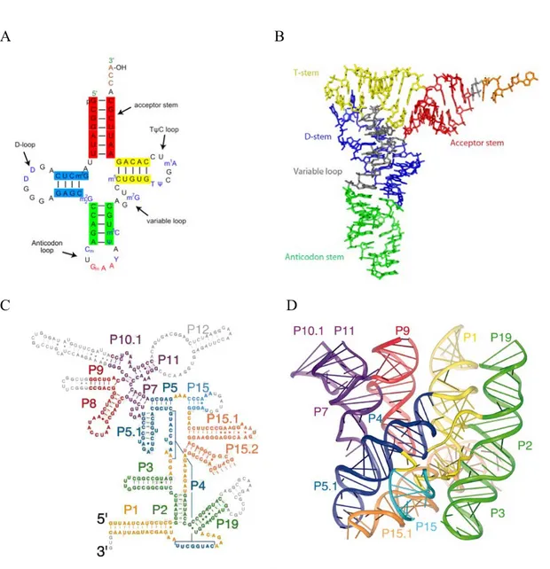
Analysis of structures of various non-coding RNAs showed that in addition to double helices, the RNA chain is capable of forming diverse three dimensional forms and arrangements. The ability to fully exploit the hydrogen-bonding and base-stacking potentials of bases and riboses allows RNA to form a vast variety of structures. Various RNA molecules achieve their active states by adopting specific functional three-dimensional structures. One of the best examples of RNA molecule with a distinct 3D structure is transfer RNA (tRNA) (Figure 1A, B). tRNA is a small RNA molecule of 74-95 nucleotides that delivers a specific amino acid to the growing polypeptide chain on the ribosome. All tRNAs have a similar L-shaped 3D structure that allows them to fit into the P and A sites on the ribosome (Kim et al., 1974; Robertus et al., 1974). Non-canonical base pairs play a critical role in the formation of the specific L-shape of tRNA. For example, the reverse-Hoogsteen base pair A54-U58 was shown to be an indispensable element for the correct interaction between the D- and T- loops of the tRNA, which, in turn, is critical for the formation of the tRNA L-shape (Zagryadskaya et al., 2003).
Analysis of the principles of RNA structure formation is extremely important for better understanding of RNA function. Various aspects of RNA 3D structure will be explored in detail in the text of this manuscript. The ribosome structure will be presented later in the following sections of the Introduction, while a schematic representation of the secondary and 3D structures of the bacterial RNase P is given in the Figure 1 C,D.
A B
C D
Figure 1. Secondary and tertiary structures of tRNAPhe and bacterial RNAseP.
(A) The canonical cloverleaf structure of the tRNAPhe. Modified nucleotides are shown as follows: m2G =>
2-metyhl-guanosine; D => 5,6-Dihydrouridine; m2
2G =>: N2-dimethylguanosine; Cm => O2'-methyl-cytdine; Gm
=> O2'-methyl-guanosine; T => 5-Methyluridine (Ribothymidine); Y => wybutosine (Y-base); Ψ => pseudouridine; m5C => 5-methyl-cytidine; m7G => 5-methyl-guanosine; m1A => 1-methyl-adenosine.
(B) 3D structure of the tRNAPhe forming the canonical L-shape. PDB access code of X-ray structure – 1ehz (Shi
& Moore, 2000).
(C,D) Schematic representation of the secondary and 3D structures of the RNA component of RNase P from Bacillus stearothermophilus (Kazantsev et al., 2005). PDB access code of X-ray structure –2A64.
The elements of phylogenetically refined secondary structure are colored according to the coaxially stacked helical domains in the ribbon representations of the structure. The nucleotides colored in gray in the secondary structure (parts of P9, P10.1, P12, L15, and P19) could not be modeled because of disorder in the crystal.
1.1.3 RNA as an enzyme
There are two major factors that allow RNA to catalyze chemical reactions. As in the case of proteins, RNA possesses a well-defined tertiary structure. On top of that, in an RNA chain, each ribose has a 2' hydroxyl group, which can act as a nucleophilic center. This group makes RNA less stable compared to DNA because it can stimulate self-hydrolysis of the RNA phosphodiester bond. RNA molecules, which are capable of chemical catalysis, are called ribozymes (from ribonucleic acid enzyme) (Kruger et al., 1982). All known natural ribozymes catalyze one of three types of reactions: transesterification, hydrolysis and peptidyl transfer (Figure 2A). The biggest number of ribozymes performs transesterification reaction and is divided in two classes: nucleolytic ribozymes and the splicing introns. Both, nucleotilytic ribozymes as well as self-splicing introns perform different kinds of phosphoryltransfer reactions, which result in breakage of the RNA backbone. In nucleolytic ribozymes the phosphodiester bond is attacked by an adjacent 2’-oxygen atom, whereas in group I (Cech, 1990) and II (Lehmann & Schmidt, 2003) intron ribozymes it is attacked by a remote 3’- and 2’- oxygen atom respectively (Figure 2B, reviewed in (Lilley, 2003)). The class of nucleolytic ribozymes includes several ribozymes with unrelated structure, yet following the same hydrolytic mechanism, such as seen in the following examples: hammerhead and hairpin ribozymes, mostly found in plant viruses, the Varkud satellite (VS) ribozyme in fungal mitochondria and the hepatitis delta virus (HDV) ribozyme, which is present in a human pathogen.
A
Transesterification Hydrolysis Peptidyl transfer
B
Figure 2. Reactions catalyzed by the known natural ribozymes
A. Three types of chemical reactions, catalyzed by the natural ribozymes.
B. Three classess of rybozymes that caralyse reaction of transesterification: nucleolytic, group I and group II introns.
In the nucleolytic ribozymes the phosphorus is attacked by the adjacent 2′-hydroxyl with departure of the 5′-oxygen, generating a cyclic 2′,3′ phosphate.
In the group I intron, the attack comes from the 3′-hydroxyl of an exogenous guanosine that is bound non-covalently by the ribozyme.
In the first step of the group II intron reaction, the scissile phosphorus is attacked by the 2′-hydroxyl of an adenosine that is located in the middle of the intron to be excised.
Introns are shown in red, exons in blue and exogenous guanosine in green.
The figure is taken from (Lilley, 2003) and reproduced with permission from Elsevier Limited, license number 2504960060655.
23S rRNA is a ribozyme as well because it catalyzes the peptidyl-transferase reaction, in which the α-amino group of the aminoacyl-tRNA nucleophillically attacks the ester carbon of the peptidyl-tRNA to form a new peptide bond. An initial proposal for a general acid/base catalytic mechanism involving N3 of A2451—a nucleotide of 23S rRNA in very close proximity to substrate analogues (Muth et al., 2000; Nissen et al.,
2000) - was disproved by the dispensability of A2451 for the peptidyl-transferase reaction (Polacek et al., 2001; Thompson et al., 2001; Katunin et al., 2002; Beringer et al., 2003; Youngman et al., 2004). It had been proposed that binding and orienting of substrates accounts for most of the ribosomal rate enhancement (Nierhaus, 1980). A comparison of the rate of peptide-bond formation by the ribosome and by a ribosome free model system suggested that the ribosome accelerated the reaction (almost 105 fold) solely by entropic effects, which may include substrate positioning, shielding the reaction from bulk solvent, or organization of the active site (Sievers et al., 2004; Trobro & Aqvist, 2005).
The first ribozymes to be discovered were the Tetrahymena thermophila self-splicing intron (Kruger et al., 1982) and the RNA component of RNase P (Guerrier-Takada & Altman, 1984). The discovery of RNA catalytic properties led to the proposition of the hypothesis of the RNA world by Walter Gilbert in 1986 (Gilbert, 1986). The RNA world hypothesis is based on the assumption that in the biotic pre-DNA world, RNA could function as both the storage of genetic information and as the enzyme which catalyzed various chemical reactions required for pre-cellular life.
1.1.4 RNA in post-transcriptional gene regulation
The recent discovery of RNA interference (RNAi) resulted in a significant increase of attention to messenger RNA as a therapeutic target (Bumcrot et al., 2006; de Fougerolles et al., 2007; Pecot et al., 2011). RNAi is a system within living cells, which allows post-transcriptional regulation of the synthesis of protein (Fire et al., 1998). The RNAi pathway is found in many eukaryotes including humans. Some eukaryotic protozoa such as Leishmania major and Trypanosoma cruzi completely lack the RNAi pathway (Robinson & Beverley, 2003; DaRocha et al., 2004). Most or all of the components are also missing in some fungi, most notably the model organism Saccharomyces cerevisiae (Aravind et al., 2000). A recent study however reveals the presence of RNAi in other budding yeast species such as Saccharomyces castellii and Candida albicans (Drinnenberg et al., 2009). There are two types of molecules that are central for RNAi – microRNA (miRNA) and small interfering RNA (siRNA).
Initially, miRNAs and siRNAs appeared to be distinguished in two primary ways. First, miRNAs were viewed as endogenous and purposefully expressed products of an organism’s own genome, whereas siRNAs were thought to be primarily exogenous in origin, derived directly from the virus, transposon, or transgene trigger. Second, miRNAs appeared to be processed from stem-loop precursors with incomplete double-stranded character, whereas siRNAs were found to be excised from long, fully complementary double-stranded RNAs (dsRNAs) (Tomari & Zamore, 2005). Despite these differences, the size similarities and sequence-specific inhibitory functions of miRNAs and siRNAs immediately suggested relatedness in biogenesis and mechanism. Both classes of small RNAs were quickly revealed to depend upon the same two families of proteins: Dicer enzymes to excise them from their precursors, and Ago proteins to support their silencing effector functions (Meister & Tuschl, 2004; Tomari & Zamore, 2005). Thus, these three sets of macromolecules—Dicers, Agos, and 21–23 nt duplex-derived RNAs—became recognized as the signature components of RNA silencing (reviewed in (Carthew & Sontheimer, 2009)).
siRNA are 20-25 nts long, linear, perfectly basepaired dsRNA, introduced directly into the cytoplasm or taken up from the environment (Mello & Conte, 2004). These dsRNAs are processed by Dicer into the siRNAs that direct silencing (Meister & Tuschl, 2004; Tomari & Zamore, 2005). siRNAs were originally observed during transgene- and virus-induced silencing in plants (Mello & Conte, 2004). In 2002 and 2003, centromeres, transposons, and other repetitive sequences were uncovered as another wellspring of siRNAs (Lippman & Martienssen, 2004). Shortly thereafter, functional studies in plants led to the discovery of trans-acting siRNAs (ta-siRNAs) that are diced from specific genomic transcripts and regulate discrete sets of target genes (Vazquez et al., 2004; Allen et al., 2005). More recently, other sources of endogenous siRNAs (endo-siRNAs) have been identified (Golden et al., 2008). These include convergent mRNA transcripts and other natural sense-antisense pairs, duplexes involving pseudogene-derived antisense transcripts and the sense mRNAs from their cognate genes, and hairpin RNAs (hpRNAs). Thus, it has become clear that siRNAs are not solely the products of foreign nucleic acid but arise from endogenous genomic loci as well. During the canonical RNAi pathway, the siRNA guide strand directs RISC to perfectly complementary RNA targets, which are
then degraded via cleaving activity of the Argonaute protein (Carthew & Sontheimer, 2009). One of the two RNA strands of the siRNA molecule must be perfectly complementary to the RNA target in order to promote the cleavage (Carthew & Sontheimer, 2009).
miRNAs are 20-25 nucleotides long single-stranded endogenously expressed RNA molecules that bind to the 3’UTR of target mRNAs, usually resulting in gene silencing (Bartel, 2004). The human genome may encode over 1000 miRNAs (Bentwich et al., 2005), which may target up to 60% of mammalian genes (Friedman et al., 2009), because each miRNA may repress hundreds of different mRNAs (Brennecke et al., 2005; Lim et al., 2005). Most animal miRNAs bind mRNA with mismatches, although the core region of binding (seed region) must include 2-8 Watson Crick (WC) base pairs. In contrast, most plant miRNAs bind to their target sites with near-perfect complementarity. The extent of miRNA-mRNA complementarity has been considered a key factor of the regulatory mechanism. Perfect complementarity leads to cleavage of the mRNA strand, whereas central mismatches inhibit mRNA cleavage and promote repression of mRNA translation (Matranga et al., 2005).
One of the key differences between miRNAs and most siRNAs is in the precision of their ends. Most species of a miRNA have highly exact ends, although there is a little variation. In contrast, siRNAs tend to be much more heterogeneous in end composition. It is this feature of miRNAs that has probably allowed them to interact with greater specificity on substrate mRNAs without a need for stringent complementarity or large overlap. Consequently, the processing machinery is constructed to produce miRNA duplexes with highly exact ends (reviewed in (Carthew & Sontheimer, 2009). The principal differences between miRNAs and siRNAs are listed in the Table 1.
miRNA siRNA (Short interfering RNA) Occurrence Occur naturally in plants and animals Occur naturally in plants and lower
animals. Whether or not they occur naturally in mammals is an unsettled question
Configuration Single stranded Double stranded
Length 19–25 nt 21–22 nt
Complementarity to target mRNA
Not exact, and therefore a single miRNA may target up to hundreds of mRNAs
100% perfect match, and therefore siRNAs knock down specific genes, with minor off-target exceptions
Action Inhibit translation of mRNA Cleave mRNA
Function Regulators (inhibitors) of genes (mRNAs)
Act as gene-silencing guardians in plants and animals that do not have antibody-or cell-mediated immunity Clinical uses Possible therapeutic uses either as
drug targets or as drug agents themselves. Expression levels of miRNAs can be used as potential diagnostic and biomarker tools
siRNAs are valuable laboratory tools used in nearly every molecular biology laboratory to knock down genes. Several siRNAs are in clinical trials as possible therapeutic agents
Table 1. The key aspects that distinguish siRNA and miRNA.
1.2 The basics of RNA structure
In this section I will give a brief overview of the essentials of RNA structure, which will be important for the better understanding of the following text.
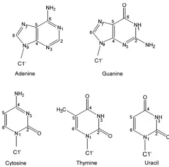
Though RNA and DNA molecules are structurally very similar, there are two structural aspects that make them different. First, while RNA is made of ribonucleotides, DNA is made of deoxyribonucleotides (as a result there is no hydroxyl group attached to the C2’ atom of the deoxyribose in DNA). Second, the complementary base to adenine in RNA is uracyl, while in DNA it is replaced by thymine, which differs by the presence of a methyl group in position 5 of the pyrimidine ring (Figure 3).
Figure 3. The five bases of DNA and RNA.
1.2.1 A- and B- helical form of nucleic acids
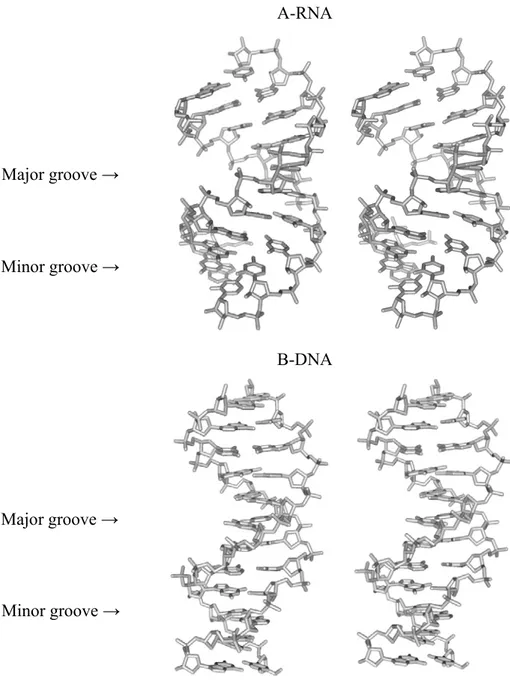
The RNA may form double helices through the formation of the canonical Watson-Crick (WC) base pairing between the complementary bases of the polynucleotide chain. While DNA double helices can adopt both, A- and B- helical forms, the only possible conformation of the RNA double helix is A-form (Figure 4). In DNA, both the major and the minor grooves are well accessible. The wide major groove of DNA is often used for protein binding. A-RNA double helix has a narrow and deep major groove while the minor groove is exposed for the inter-molecular interactions with other substrates and is often called the shallow groove (Figure 4). Due to the fact that the hydroxyl group is attached to the C2’ atom, the ribose is locked into a 3’-endo chair conformation, thus eliminating the possibility of forming the B-helical form. If dsRNA were to form B-form duplexes, the 2’-OH group would inevitably clash with C8 (for a purine) or C6 (for a pyrimidine) as well as O5’ and OP2 of the attached base (Figure 5).
A-RNA
B-DNA
Figure 4. A-RNA and B-DNA structures.
Stereoview of 3D structure of A-RNA and B-DNA. A-RNA has a deep and narrow major groove and the minor groove is significantly exposed. The B-DNA wide major groove is almost identical in depth to the much narrower minor groove
Minor groove → Major groove → Major groove →
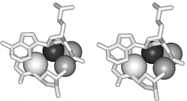
Figure 5. Potential collision of 2’OH of RNA in the B-form (stereo view)
An imaginary situation is presented, where an RNA residue is in the B-form. 2’O (black atom) collides with C6 of pyrimidine, as well as with O5’ and OP2 of sugar-phosphate backbone of the neighboring nucleotide (atoms shown in gray).
1.2.2 Non-canonical base pairing
Although stacking is the most important driving force for the folding of nucleic acids, the edge-to-edge interactions between polarized atoms provide directionality and specificity. In contrast to helical B-DNA structure, RNA exploits the purine and pyrimidine base pairing not only through the Watson-Crick (WC) edge but also through Hoogsteen (HG) and Sugar edge (SE) of the nucleotide (Figure 6), which results in non-canonical base pair formation (Leontis & Westhof, 2001; Leontis et al., 2002; Lee & Gutell, 2004). Analysis of the available RNA structures shows that 60% of bases in structured RNAs participate in WC base pairing while the rest are involved in some other kind of edge-to-edge interaction (Leontis & Westhof, 2001) or even edge-to-sugar-phosphate backbone interaction.
Several attempts have been undertaken to classify the non-canonical base pairs identified in RNA structure (Jeffrey & Saenger, 1991; Tinoco, 1993; G. Dirheimer, 1995). More recent classification by Leontis and Westhof (Leontis et al., 2002) provides more comprehensive information on the non-canonical base pairing in RNA. Moreover, Leontis and Westhof proposed a convenient method of base pair annotation, which will be used throughout the text (Table 2).
Figure 6. Definition of nucleotide edges
(A) Chemical structure of a purine nucleotide illustrating the three edges available for base-to-base interaction. (B) Representation of an RNA base as a triangle, with Hoogsteen (HG), Watson-Crick (WC) and Sugar (SE) edges labeled.
The two-dimensional diagram annotation, proposed by Leontis and Westhof (Leontis & Westhof, 2001), significantly facilitates the secondary structure representation of complex three-dimensional arrangements. As a result, this annotation is widely used in the RNA field for depicting the complex network of inter-nucleotide base pairing.
Given that either the WC, HG or Sugar edge of a nucleotide may interact with one of three available edges of another nucleotide, 6 combinations of pair-wise edge-to-edge interactions are possible. Two nucleotides can interact in cis or trans, depending on the mutual orientation of their glycosidic bonds in respect to an imaginary line drawn along the established hydrogen bonds (Figure 7). As a result all base pairs involving two or more edge-to-edge hydrogen bonds belong to one of 12 geometric families (Table 2).
Figure 7. Glycosidic bond orientations.
(A) An example of cis (left panel) WC base pair versus trans (right panel) WC base pair. The cis and trans orientations are defined relative to a line drawn parallel to the base-to-base hydrogen bonds.
(B) Examples of anti (left panel) and syn (right panel) conformations of a ribonucleotide. The anti conformer has the smaller H-6 (pyrimidine) or H-8 (purine) atom above the sugar ring, while the syn conformer has the larger O-2 (pyrimidine) or N-3 (purine) in that position.
Table 2. The 12 geometric families of nucleic acid base pairs with symbols for annotating secondary structure diagrams (Leontis & Westhof, 2001).
The local strand orientation is given in the last column, assuming that all bases are in the default anti conformation; a syn orientation (Figure 7B) would imply a reversal of orientation.
A
B
1.3 Primary, secondary and tertiary motifs
The title of my thesis “Recurrent RNA motifs: identification and characterization” requires clear understanding of the term “motif”. When speaking about “motifs” one can mean absolutely different subjects. According to the Collins English dictionary, a motif is
“a recurring form or shape in a design or pattern” or “a single added piece of decoration” (Crozier, 2006). In molecular biology we can find several types of motifs.
They can be either primary sequence motifs, secondary or tertiary structure motifs. We will discuss each type of these motifs in the following paragraphs.
1.3.1 Primary structure and primary sequence motifs
The primary structure of RNA is in fact the sequence of nucleotides of the RNA strand. The RNA sequence is the code which determines the secondary and tertiary structure and subsequently the function of any RNA molecule. The primary sequence motif represents a sequence of nucleotides which has a particular biological meaning. Primary sequence motifs are recognized by other nucleic acid sequences or proteins. For example, a G-N combination of a single-stranded RNA is recognized by RNAse T1, while a Py-A combination is recognized by RNAse A, which results in the strand cleavage in the middle of the dinucleotide sequence motif (D'Alessio & Riordan, 1997). The Shine-Dalgarno (SD) sequence is a conserved mRNA sequence motif, which in non-eukaryotic cells allows the correct positioning of the small ribosomal subunit on mRNA via formation of the duplex with the anti-SD sequence of 16S rRNA (Shine & Dalgarno, 1975).
1.3.2 Secondary structure and secondary structure motifs
It is generally known that the primary sequence of homologous RNA molecules may significantly vary. Yet, the three-dimensional structure varies much less. It has been demonstrated in tRNA and rRNA that conservation of the sequences involved in the formation of the double helices is less than for the single-stranded regions (Pace, 1999). The secondary structure can be determined by the alignment of multiple homologous sequences from a large number of organisms (Woese et al., 1980; Noller et al., 1981;
Gutell et al., 1992). Single nucleotide mutations in an RNA strand can occur spontaneously. If this occurs in one strand of RNA double helix, the overall stability of the helix is reduced. The destabilizing effect of a single mutation can be neutralized by a compensatory mutation of the opposite strand of the RNA double helix. As a result, the canonical base pairing is restored and the double helix is re-formed. Therefore, by looking for compensatory base changes in the alignment, it is generally possible to deduce areas forming double helices (Gutell et al., 2002).
Figure 8. RNA secondary structure example.
The secondary structure of the RNase P RNA molecule of Methanococcus marapaludis from the RNase P Database is shown (Brown, 1999). Solid grey lines represent the ribose-phosphate backbone. Dotted grey lines represent missing nucleotides. Solid circles mark canonical WC base pairs. A number of secondary structure motifs can be identified here: A stem is composed of one or more consecutive base pairs; a hairpin loop contains one closing base pair, and all the bases between the paired bases are unpaired; an internal loop is a loop with two closing base pairs, and all bases between them are unpaired; a bulge loop can be seen as a variant of an internal loop in which there are no unpaired bases on one side; a loop is a loop that has at least three closing base pairs; stems originating from these base pairs form a multi-way junction; a pseudoknot contains at least two stem-loop structures in which half of one stem is intercalated between the two halves of another stem.
The secondary structure provides information on the double helices and unpaired regions. The RNA double helix is the most common secondary structure element. The unpaired regions give rise to many other secondary structure elements: pseudoknots, bulges, internal loops, bulge loops, hairpin loops, multi-loops etc (Figure 8). Conserved secondary structures, which are identified by co-variation are often called “motifs” (Gast, 2003). A number of computational methods to predict the secondary structure have been proposed (Zuker, 1989; Gutell, 1995; Mathews, 1998; Rivas & Eddy, 1999; Hofacker, 2003) although information on the tertiary interactions or non-canonical base pairing cannot be obtained by these methods.
RNA secondary structures have been listed in various databases, such as comprehensive database of RNA families, Rfam (Griffiths-Jones et al., 2003), RNA specific databases for ribosomal RNAs (Wuyts et al., 2004), RNAseP (Brown, 1999), tmRNA (Zwieb et al., 2003), SRP (Rosenblad et al., 2003) and others. Various biochemical and biophysical experimental methods can also be used to infer secondary structure (Ehresmann et al., 1987) or in the case of NMR (Furtig et al., 2003) and X-ray crystallographic methods (Holbrook & Kim, 1997) describe secondary and tertiary structure in detail.
Once 3D structure of an RNA molecule is obtained, the secondary structure can be easily incurred by visual analysis. In this case the secondary structure can be useful for 2D representation of a complex 3D shape.
1.3.3 3D structure of RNA and tertiary motifs
The RNA tertiary structure describes the overall three dimensional conformation of a single molecule. The exact structure of RNA is usually determined by crystallography or NMR, although some biochemical methods such as probing and hydroxyl radical cleavage can be used for these studies too (Fox, 1997; Wilson, 2002; Bockelmann, 2004; Tullius & Greenbaum, 2005). The information on the crystal structures of different RNAs can be found in both the Nucleic Acid Database (Berman et al., 1992) and the RCSB Protein Data Bank (Berman et al., 2000).
RNA crystallization has always been difficult due to the problems of preparation of the adequate samples and inherent RNA flexibility. Over the past few years the RNA
crystallography and NMR techniques advanced significantly, which allowed collection of large amounts of information on 3D RNA structure.
Folding of RNA significantly differs from that of proteins. First, there are only four types of monomers used for building of an RNA molecule. The RNA backbone has six degrees of freedom for each residue while the polypeptide chain has only two. RNA structure is not nucleated by a hydrophobic core as in the case of most proteins. In contrast to this, RNA folding is driven by two major forces: hydrogen bonding and base stacking. The extreme flexibility allows single-stranded RNA regions to adopt a wide range of conformations. In spite of the fact that the 3D RNA structures are diverse and sometimes extremely complex, they can be decomposed into smaller size building blocks.
Analysis of the RNA tertiary structure has suggested that RNA molecules are made of conserved structural building blocks or motifs (Leontis & Westhof, 2003). Ability of the ribonucleotides to form a huge variety of non-canonical base pairs allows for the formation of a vast range of different structural arrangements. Generally speaking, RNA structure can be roughly split into the set of repetitive elements and those elements which occur only once and have no analogs (or these analogs have not yet been identified). Our approach to the studies of the 3D structure of RNA consists in identification of the recurrent arrangements and their detailed analysis. There are at least two reasons, which explain our interest in the RNA repetitive elements. The fact that a particular recurrent motif has been selected as a building block for different domains of RNA molecules means that such an arrangement possesses certain important properties which are beneficial for the correct folding of the whole molecule. Thus, by studying repetitive arrangements, we can concentrate on the most important elements of a given RNA structure. Another reason why the systematic study of recurrent RNA motifs is so important is the fact that they are able to fold into similar shapes in spite of different primary sequences. Identification of the rules that allow for the formation of identical 3D arrangements from unrelated sequences represents an important step for better understanding of the principles of RNA structure folding.
In the following sections a few examples of known recurrent RNA motifs will be presented. Introduction of these motifs will help in the better understanding of the matter which is presented in Chapters 2-6 of the thesis.
1.3.4 A-minor motif
The A-minor motif is one of the most simple and at the same time the most abundant tertiary structural motif in large RNA molecules. An A-minor interaction consists in the hydrogen bonding between an unpaired adenosine with the minor groove of the RNA double helix (Doherty et al., 2001; Nissen et al., 2001). Four sub-types of the A-minor interaction are known, depending on the mutual orientation of the 2’ OH group of adenosine and two 2’OH groups of the receptor base pair (Figure 9). The A-minor interactions are present in practically all known structures of large RNA molecules (Cate et al., 1996; Golden et al., 1998; Ban et al., 2000; Wimberly et al., 2000; Adams et al., 2004; Kazantsev et al., 2005; Torres-Larios et al., 2005). A single A-minor interaction is relatively weak while formation of two or three consecutive A-minor interactions significantly increases the stability of the complex. Indeed, in the RNA structure we can often observe formation of consecutive stacks of two or three unpaired adenosines making A-minor interactions with closely or distantly located double helices. Within the stack, adenosines can come from the same or different RNA strands. A-minor motif mediates formation of many other tertiary structural motifs. For example, interaction of the hairpin loops with their receptor helices is mediated by the A-minor interactions of the unpaired adenosines of the loop with the minor groove of the double helix (Nagaswamy & Fox, 2002; Lee et al., 2003). Some other tertiary motifs, such as particularly folded internal loops, also rely on the A-minor interactions for the correct folding (Battle & Doudna, 2002).
A-minor motif forms many important structural contacts and on top of this it plays several important functional roles in the ribosome. For instance, the 3’-terminal adenosines of both the A- and P-site tRNA are positioned in the peptidyl transferase site via formation of A-minor interactions with 23S rRNA (Nissen et al., 2000). Another important functional aspect – monitoring of the correct codon-anticodon base pairing – is
mediated by the adenosines A1492 and A1493 of 16S rRNA with help of the A-minor motif formation (Ogle et al., 2001).
In Chapter 2 a new recurrent motif will be discussed which we identified in the ribosome and named “ribo”. An A-minor interaction is found in the centre of all G-ribo motifs, where it helps to support a particular shape of this complex arrangement.
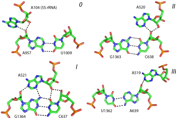
Figure 9. Four sub-types of A-minor interaction.
Four sub-types of A-minor interactions are identified in the crystal structure of 50S ribosomal subunit of H. marismortui (PDB: 1S72): 0, I, II and III. Each type is defined by the position of 2’-OH group of the adenosine in respect to the position of the two 2’-OH groups of the receptor base pair.
1.3.5 RNA Bulge
Incorporation of a single or several nucleotides in the otherwise regular (helical) region results in the formation of the bulge motif (Figure 10A). Although in the secondary structure the bulge motif is represented as an unpaired nucleotide which is flanked with two WC base pairs, on the level of the 3D structure the presence of the bulged nucleotide may result in different structural consequences for the RNA double
helix. (1) The bulged nucleotide may introduce a bend in an RNA double helix, if it intercalates between the 5’ and 3’ flanking nucleotides of the polynucleotide chain (Figure 10B). (2) Being stacked on the 3’ flanking nucleotide, the bulged nucleotide may interact with the minor groove of 5’ flanking base pair, thus stimulating formation of the over-twist between two WC base pairs (Figure 10C). (3) Being stacked on the 5’ flanking nucleotide, the bulging nucleotide may interact with the major groove of the 3’ flanking base pair, thus stimulating the formation of the over-twist between the two WC base pairs (Figure 10D). (4) The bulged nucleotide can be involved in the long-range interactions with other parts of the RNA molecule, stimulating formation of the helical over-twist between the two flanking base pairs (Figure 10E). (5) The presence of the bulging nucleotide may have no effect on the over-all shape of the RNA double helix (Figure 10F). Consequently, the bulge motif is an important structural element that can modulate the shape and direction of the RNA double helix. Different types of the bulge motif are discussed here in order to facilitate understanding of the more complex arrangements that will be presented in Сhapter 4 and are in fact relevant to the structures presented here.
Figure 10B Figure 10C Figure 10D ← 5’ ← 3’ ← 5’ ← 3’ ← 5’ ← 3’
Figure 10E
Figure 10 F
Figure 10. 3D structures of the RNA bulge.
(A) A simplified representation of the bulge. The thick lines represent base pairs in duplex
regions and unpaired bases in the loops. (B-F) Stereo view of the 3D structure of different bulges. Bulged nucleotide is colored black. Two flanking base pairs are colored white. Other nucleotides of the double helix and nucleotides, which do not belong to the helix, are colored gray. “ 5’ “and “ 3’ “ indicate the 5’ and 3’ flanking base pairs of the bulge respectively. (B) Bulged nucleotide intercalates between two flanking nucleotides in the double helix and results in the helix bend. In C, D and E introduction of the bulge initiates formation of the helical over-twist between two flanking base pairs. (C) The bulged nucleotide interacts with minor groove of one of the 5’ flanking base pair. (D) The bulged nucleotide interacts with major groove of 3’ flanking base pair. (E) The bulged nucleotide does not interact with the flanking base pairs and participates in the long-range interactions with nucleotides, which do not belong to the helix. (F) The bulged nucleotide does not interact with the flanking base pairs. No helical over-twist occurs between two flanking base pairs of the bulge.
← 5’
← 3’ ← 3’ ← 5’
1.3.6 Internal loops
An internal loop represents a stretch of unpaired nucleotides in both strands of the RNA double helix which are flanked with regular WC base pairs on each side (Figure 11). These unpaired nucleotides may participate in non-canonical base pairing or can be bulged out of the double helix. Depending on the presence of equal or unequal number of nucleotides in each of the two composing strands, internal loops are referred to as symmetric or asymmetric (Figure 11). Loop-E motif pairing (Wimberly et al., 1993; Correll et al., 1997), C-loop (Lescoute et al., 2005) and UAA/GAN (Lee et al., 2006) are the most common examples of the internal loop-based three dimensional motifs.
Figure 11. A simplified scheme of an internal loop.
Long horizontal rectangles represent WC base pairs of the RNA double helix. The unpaired nucleotides of the internal loops belonging to the strand 1 and 2 are shown in gray and black respectively. If the number of nucleotides in both strands, 1 and 2, is equal, the internal loop is called symmetric. Otherwise it is called asymmetric.
1.3.6.1 Loop-E motif
Loop-E motif is an asymmetric internal loop that includes seven highly conserved nucleotides forming two non-WC base pairs and one base triple (Wimberly et al., 1993; Correll et al., 1997). The first base pair is trans-HG-SE (sheared) AG base pair (Figure 12, shown in red), followed by a trans-WC-HG (reverse Hoogsteen) UA base pair (shown in green), a bulged G, and finally a trans-HG AA base pair (shown in blue on the Figure 12). The three-adenosine stack represents a sticky surface that can easily bind to the minor groove of an RNA helix via formation of the A-minor interaction. In fact, most of
the Loop-E motifs located in the ribosome, indeed participate in long-range interactions with different RNA double helices. Thus, the loop-E motif is an important structural element, which participates in the organization of the three dimensional shape of RNA molecules. The loop-E motif is found in different RNA-containing molecules and has important functional implications. In particular, this motif was found in the sarcin-ricin loop of the 23S rRNA, which is known to mediate the binding of the elongation factors EF-Tu and EF-G to the ribosome. In the E. coli 5S rRNA, loop E represents a binding site for the ribosomal protein L23 (Leontis & Westhof, 1998). Therefore, the loop-E motif seems to be important for formation of the ribosome structure as well as for its function. A
B
Figure 12. E-loop motif.
(A) Secondary structure of the E-loop motif. The E-loop motif represents an asymmetric internal loop. The non-canonical base pairing between pairs of nucleotides are demonstrated using symbols, suggested by Leontis and Westhof (Leontis & Westhof, 2002). (B) Stereo-view of the 3D structure of the E-loop motif. The same color code is used as in (A). The two flanking WC base pairs are not shown for clarity.
1.3.6.2 C-loop motif
The C-loop motif is another example of an asymmetric internal loop (Lescoute et al., 2005). It consists of two helices (H1 and H2) connected by an internal loop and arranged in the way that the first base pair of the second helix is partially stacked to the last base pair of the first helix. Within the C-loop motif, these two base pairs are over-twisted one in respect to the other compared to the juxtaposition of the two neighboring base pairs in the standard A-RNA conformation. The name “C-loop” is given to the whole arrangement because of the first nucleotide of the longer strand (С865 on the Figure 13A), which interacts with the major groove of the last base pair of the H1 and is always cytosine. The other nucleotides of the longer strand make hydrogen bonds with the minor groove of the first and the second base pair of the H2 (Figure 13). Nucleotides of the shorter strand bulge out and participate in long-range interactions with distant segments of the RNA molecule. The C-loop motif significantly increases the helical twist of the stem between the two WC base pairs flanking the internal loop. Together with other internal-loop based motifs, the C-loop motif represents a tool for modulation of the structure of the RNA double helix.