Université de Montréal
The Function oftheSaccharomyces Cerevisiae Ribonucleotide Reductase Second
f3
Subunit in DNA RepairPar Chunyu ZHAO
M.Sc. en sciences biomédicales faculté de Médecine
Mémoire présenté à la Faculté des études supérieures En vue de l’obtention du grade deMaîtrise en Sciences (M.Sc.)
Programme de sciences biomédicales Réparation d’ADN
Août 2006
‘u
LI
r>
u52
3oÔ
L13S
Inde Montréal
Direction des bibliothèques
AVIS
L’auteur a autorisé l’Université de Montréal à reproduire et diffuser, en totalité ou en partie, par quelque moyen que ce soit et sur quelque support que ce soit, et exclusivement à des fins non lucratives d’enseignement et de recherche, des copies de ce mémoire ou de celle thèse.
L’auteur et les coauteurs le cas échéant conservent la propriété du droit d’auteur et des droits moraux qui protègent ce document. Ni la thèse ou le mémoire, ni des extraits substantiels de ce document, ne doivent être imprimés ou autrement reproduits sans l’autorisation de l’auteur.
Afin de se conformer à la Loi canadienne sur la protection des renseignements personnels, quelques formulaires secondaires, coordonnées ou signatures intégrées au texte ont pu être enlevés de ce document. Bien que cela ait pu affecter la pagination, il n’y a aucun contenu manquant. NOTICE
The author of this thesis or dissertation has granted a nonexclusive license allowing Université de Montréal to reproduce and publish the document, in part or in whole, and in any format, solely for noncommercial educational and research purposes.
The author and co-authors if applicable retain copyright ownership and moral rights in this document. Neither the whole thesis or dissertation, nor substantial extracts from it, may be printed or otherwise reproduced without the author’s permission.
In compliance with the Canadian Privacy Act some supporting forms, contact information or signatures may have been removed from the document. While this may affect the document page count, it does flot represent any loss of content from the document.
I
Université de Montréal Faculté des études supérieures
The function of Ribonucleotide Reductase Second
f3
Gene in Yeast DNA Repairprésenté par: Chun-yu Zhao Dr. Euridice Carmona président-rapporteur Dindial Ramotar directeur de recherche
Dong Hao Wang membre dujuiy
RÉSUMÉ
Les réductases de ribonucléotides (RNRs) catalysent la réduction des quatre ribonuclésides diphosphates en deoxyribonucleosides diphosphates et jouent également un rôle central dans le contrôle de la concentration intracellulaire des deoxynucleosides triphosphates (dNTPs). Ces deux étapes sont exigées pour le processus de réplication d’ADN à haute fidélité, ainsi que pour la réparation d’ADN. Dans les saccharomyces cerevisiae, les RNRs constituent des tétramères a2f32 représentés sous forme de deux grandes et de deux petites sous-unités. Parmi ces réductases, Rnr2 codent l’un des 2 petites sous-unités
F3.
Un deuxième gène essentiel codant une petite sous-unité homologue a été identifié en 1997, il s’agit du RNR4. Dans la présente étude, nous sommes les premiers à analyser le rôle de Rnr4 dans la réparation d’ADN, en utilisant les contraintes du mutant rnr4A ainsi que les contraintes parentales (S. cerevisiac BY4741). Ceci était réalisé après un traitement avec différents types d’agents endommageant l’ADN. Voici quelques exemples d’agents de dommage d’ADN Bleomycine (BLM, agent de stress oxydatif), 4-nitroquinoline-1-oxide (4-NQO, inducteur d’effets de dommage identiques à ceux des UV, UV-mimetic DNA damage agent) et Methanesulphonate méthylique (MMS). La contrainte dumutant rnr4z était sensible au BLM et au MMS en comparaison avec la contrainte parentale. L’analyse de la survie a révélé que la contrainte du mutant mr4A avait un taux de survie significativement plus élevé que son parent après une heure de traitement avec le 4-NQO mais moins élevé après seulement 4 minutes de traitement avec les UV. La forte résistance à 4-NQO chez le mutant rnr4A après une heure vs son111
parent est de 50% ± 1.9 vs. 6% ± 1.1 A vs. wt, 3jig/rnl 4-NQO, p<O.O1. L’analyse de la
mutagenèse a démontré que le taux de mutation de rnr4A était significativement plus bas que celui du parent lors de l’administration de 4-NQO. Les résultats suggèrent alors que 4-NQO induit des dommages d’ADN identiques à ceux des UV en plus d’autres dommages. Nous avons démontré que les dommages d’ADN induits par 4-NQO ne soient ni “des dommages UV-MIMETIC d’ADN” ayant lieu principalement dans la voie de la réparation d’excision de nucléotide (REN), ni du stress oxydatif induit par BLM. Nous avons donc suggéré qu’un nouveau mécanisme de lésion d’ADN causée par 4-NQO existe. Cette lésion pourrait être réparée par la méthode error free DNA repair’. Ceci a lieu probablement par une voie de translésion (TLS) peu importe le pooi de dNTP. Un
bas pool de dNTP chez le mutantrnr4A mène à unplus haut taux de survie et un taux bas de mutation en prévenant la voie ILS mais sans aucun effet sur ‘error free DNA repair’. De plus, une grande concentration de dNTP permet aux cellules parentales de réparer la lésion via la voie TLS causant ainsi beaucoup d’erreurs et conséquemment un bas taux de survie en augmentant le taux de mutation.
Nous avons finalement conclu que la faible concentration de dNTPs chez la contrainte mutant RNR4 ne joue pas le rôle dans la réparation de la lésion d’ADN provoquée par ce mécanisme.
Les mots clés
Deuxième sous-unité
f3
de la réductases de ribonucléotide (RNR4) 4-nitroquinoline- 1 -oxide(4-NQO)V
ABSTRACT
Ribonucleotide reductases (RERs) catalyze the reduction of ail four ribonucleoside diphosphates into deoxyribonucieoside diphosphates and piay a centrai roie in controliing the pooi size of ceiiuiar deoxynucleoside triphosphates (dNTPs), required for high fidelity DNA replication and DNA repair processes. In Saccharornyces cerevisiae. ribonucieotide reductase is an a2f32 tetrarner consisting of two large and two smaii subunits. RNR2 encodes one of the 2 smaii f3 subunits. A second essentiai gene RNR4 encoding a hornoiogous small
f3
subunit was found in 1997. In the present study, we investigated for the first time the roie of Rnr4 in DNA repair, using rnr4A mutant strains and parent strains (S. cerevisiae BY4741) afier treatment with different type of DNA damage agents such as bleomycin (BLM, an oxidative stress agent), 4-nitroquinoline- 1-oxide (4-NQO “UV-mimetic” DNA damage agent), methyl methanesuiphonate (MMS). rnr4z\ mutant strain was sensitive to BLM and MMS as compared with parent strain. More interestingly, survivai assay demonstrated that as cornpared with his parent, rnr4A mutant strains have significantiy higher survivai rate afier 1 hour of treatment with 4-NQO (50% ± 1.9 vs. 6% ± 1.1 A vs. parent, 3jig/rni 4-NQO,p<O.Ol),
but has significantiy lower rate afier 4 minutes of treatment with UVC. Mutagenesis assay showed the mutation rate of rnr4A mutant is significantiy iowered as cornpared with the parent when 4-NQO is administrated. Taken together, our resuits suggested that besides “UV-mimetic” DNA damage, 4-NQO could iead to other DNA damage mechanism. This DNA damage is neither a “UV-mirnetic DNA damage, which is processed iargely by the nucieotide excision repair (NER) pathway nor oxidative stress iike BLM induced, but adifferent DNA lesion mechanism causing by 4-NQO. This DNA lesion could be repaired in both an enor free DNA repair maimer (no matter in lower dNTPs pool or in higher dNTPs pool) perhaps using a transiesion (TLS) pathway. The lower dNTPs pool in rnr1z\ mutant strains prevent TLS pathway but doesn’t affect an enor free DNA repair mairner and lead to higher survival rate but lower mutation rate. On the other hand, the higher level of dNTPs pool allowed parent cells to repair this lesion using the TLS pathway, which cause lots of enors and consequently lower survival rate by the higher mutation rate.
Key words:
Ribonucleotide reductase second
f3
subunit (Rnr4) 4-nitroquinoline- 1 -oxide(4-NQO)VII
TABLE 0F CONTENTS
RESUME .11
ABSTRACT. V
TABLE 0f CONTNETS VII
LIST 0f TABLES X
LI$T 0F FIGURES XI
LIST 0F ARBREVIATION$ XII
ACKNOWLEDGEMENT XVII
CHAPTER I: INTRODUCTION 1
SECTION 1.1: General Review 2
SECTION 1.2: Three Different Classes ofRNR 5
PARTi: ClassI 7
PART.2: Classil 9
PART 3: Class III 10
SECTION 1.3: Structure of RNR 11
PART 1: Ri Protein 12
PART 2 R2 Protein 14
PART 3 Holoenzyme 15
SECTION 1.4: Gene Organizations and Regulation of Enzyme Synthesis 18 SECTION 1.5: Anti-tumor Activity of Ribonucleotide Reductase 19
SECTION 1.6: YeastRNR (1-4) 21
SECTION 1.7: Research Objective 24
CHAPTER II: MATERIALS AND METHODS 25
SECTION 2.1: Strains and Media 26
SECTION 2.2: Chemicals and Equipments 27
SECTION 2.3: Survival Curves Assay 29
SECTION 2.4: Mutagenesis Assays 30
SECTION 2.5: Western Blot 31
SECTION 2.6 : Primer Extension Assay 32
SECTION 2.7: Preparation ofYeast Genomic DNA 34
SECTION 2.8: PCR Program 35
SECTION 2.9: Construction ofpRnr4 36
CHAPTERIII: RESULTS 38
SECTION 3.1: Strains RNR4 Mutant Display Extreme Resistance to 4-NQO as
Compared with Parent Strains 39
SECTION 3.2: The rnr4A Strain Is Sensitive to UVC 43 SECTION 3.3: The Frequency of4-NQO-induced Can1 Mutations Risc
SharplyinThe Wildtype Strain 45
Western Blot 47
Ix
SECTION 3.5: Primer Extension Assay .50
SECTION 3.6: Survival Curve Assay for Gst-RNR4 Containing Strain 53
CHAPTER TV: DISCUSSION 55
CHAPTER V: CONCLUSION 62
CHAPTER VI: PROSPECT 64
LIST 0F TABLES
Table 1. Different Classes ofRNR 6
Table 2. PCR Primer for RNR4 Gene Amplification 28 Table 3. Spontaneous Rates and Spectrum ofCantmutation in rnr4
xl
LIST 0F FIGURES
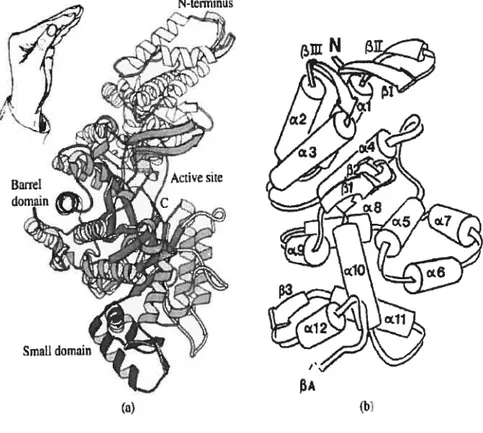
Figure 1. Structure of E. cou Ri protein 13
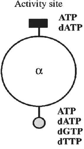
Figure 2. Models for the allosteric regulation of the ribonucleotide reductase
classes 17
Figure 3. Clone ofplasrnid gst-rnr4 37
Figure 4. Survival curve assay afier treatment of 4-NQO 40 Figure 5.. Survival curve assay afier treatment of BLM 41 Figure 6.. Survival curve assay afier treatrnent ofMMS 42 Figure 7. Survival curve assay afier treatment UVC 44 Figure 8. Western blot detection of Rpa32 phosphalation 49 Figure 9. In vitro incorporation of [methyl-3H]dTTP 52 Figure 10. Survival curve assay for gst-rnr4 containing strain 54
LIST 0F ABBREVIATIONS
4-NQO 4-nitroquinoline- 1 -oxide
A adenosine
À
angstrornATP adenosine triphosphate
BLM bleomycin
bp base pair
B$A bovine serum albumin
C cytosine
cDNA cornplementary DNA
degree Celsius
dATP 2-deoxyadenosine 5 ‘-triphosphate dNTP deoxynucleotide triphosphate
DNA deoxyribonucleic acid
DNAse deoxyribonuclease
EDTA ethylenediarninetetra acetic acid
XIII
Fig. figure
G guanosine
g gram
GDP guanosine diphosphate
HE buffer 10 mM Hepes-KOH, pH 7.0, 1 mM EDTA [3H]TTP [3H]thymidine 5’-triphosphate kb kilobase (1000 bp) L litre mM millimolar (1OE3 M) M molar (moles/litre) ml milliliter (1 0 L) MMS methyl methanesulfonate
mRNA messenger RNA
NER nucleotide excision repair
ng nano gram (1 0 gram)
nt nucleotide
0/11 over night (roughtly 12 hours)
radioactive isotope ofphosphorus PAGE polyacrylamide gel electrophoresis
PBS phosphate buffered saline
PCA phenol :choroforrn:isoamyl alcohol (25:24:1)
PCR polymerase chain reaction
pmol picomole
RNA ribonucleic acid
RNase A ribonuclease A
RNR Ribonucleotide reductase
Rnr 1 /R 1 /Y 1 Ribonucleotide reductase a-subunit Rnr3/R3/Y3 Ribonucleotide reductase a-subunit Rnr2/R21Y2 Ribonucleotide reductase 3-subunit Rnr4/R4/Y4 Ribonucleotide reductase E3-subunit RNR Ribonucleotide reductase gene
RNR] Ri gene
RNR2 R2 gene
RNR3 R3 gene
RNR4 R4 gene
RPA Replication protein A
RR inhibitor Ribonucleotide reductase inhibitor
xv
T thymidine
TE 10 mM tris, 1 mM EDTA pH 8.0
Tris tris(hydroxy methyl)aminoethane
TLS Transiesion repair
UVC ultraviolet C (wavelength range 220-280 nrn) microgram (10.6 gram)
microlitre (1OE6 L)
tM micromolar (106 M)
YPD 1% yeast extract, 2% peptone, 2% dextrose
This Thesis Is Dedïcated to My Family for
Their Love, Support and Encouragement
XVII
ACKNOWLEDGEMENT
first, I would like to thank my supervisor Dr. Dindial Rarnotar, for bis excellent guidance and professional attitude in rny master project, which is crucial to my success.
Thanks also go to everybody in the lab 1$, the Guy-Bernier Research Centre,
Maisonneuve-Rosemont Hospital, where I have worked for two years, for their helpful academic discussion and friendship during my graduate study.
$pecial thanks go to jury members Dr. Euridice Carmona and Dr. Dong Hao Wang to review my thesis and their corrections.
This piece of rny academic work is dedicated to my wife $hao-Hong Xu for her contribution to my life and work, her patience to tolerate rny long-hour work and absence of housework. This dedication also goes to my family, far away in China, for their endless understanding, supporting and loving.
2
SECTION Ï.
Ï: General Review
Ribonucleotide reductase (RNR) is a well known and probably the most investigated free radical enzyme (Thelander and Reichard 1979; Jordan and Reichard
199$; EkÏund et al., 2001). Ribnucleotide reductases have been classified into three main
classes according to the free radical generator (sec Section 1.2). A wealth of study on structure and function of the cÏass I and class III enzymes has been donc, that provided detailed views on how these enzymes perform their tasks.
The RNR catalyses the reduction of ribonucleotides to deoxyribonucleotides, which is the rate-limiting step of DNA synthesis, and control of the optimal levels of dNTPs, which are required for DNA replication and DNA repair processes; a failure to control the size of dNTP pools and/or their relative amounts leads to ccli death or genetic abnormalities. An enzyme ofthis type is also beiieved to be needed for the production of the DNA precursors at the time of the transition from the RNA to the DNA (Thelander and Reichard 1979). The common feature of the different classes of RNR is the initiation of the reaction by removai of the 3’-hydrogen of the ribose by a transient cysteinyl radical. The class I enzymes demonstrate a sophisticated pattem as to how the free radical
is used in the reaction, in that it is only deiivered to the active site at exactiy the right
moment (Ekhmd et al., 2001).
The RNR activity has been known to be transcriptionally regulated and is ccii cycle dependent in higher organisms (Bjorklund et ai, 1993). Normally, the overail enzymatic
activity is regulated by ATP (activation) and dATP (feedback inhibition at high concentrations). A unique additional allosteric regulation controls the substrate specfficity such that a balanced supply of ffie different deoxyribonucleotides is present during DNA synthesis (Ekiund et al., 2001).
The role of RNR in DNA synthesis is so important that celi proliferation makes it highly interesting in the context of anti-bacterial, anti-viral and anti-cancer drug development. Just because of this, a great increase in interest on RNR as a target for cancer therapy has been observed recently, since a human ribonucleotide reductase regulated by p53 was identified (Lozano and Elledge 2000; Nakano and Vousden 2000; Tanaka and Nakamura 2000). The p53 protein actively suppresses tumor formation and when it is mutated, several kinds of cancer may develop. Moreover, more than 80% of human tumors have been found to contain mutations in p53 or the pathway that directly regulates it.
Mammals, E. cdli and DNA viruses such as herpes viruses employ class I reductases, in which an iron-containing protein for the generation of the catalytically essential free radical is used. In these reductases, the quatemary organization of the holoenzyme is made up of the a dimer, called protein Rnrl, contains the active sites and binding sites for allosteric effectors, and the 13 dimer, called protein Rnr2, contains one dinuclear iron center and one stable tyrosyl radical per monomer, which are both essential for enzymatic activity. During the passed ten years, most of the structure function studies on class I RNRs have been performed on the E. cou enzyme (Fontecave and Eklund
4
1993) and mouse RNR. Except a normal gene for Rnr2, yeast also contains a homologous gene that codes for a Rnr4 protein lacking important iron ligands, which cannot form an active Rnr2. Although it is known that Rnr2 is unable to fold correctly by itself and is thus unable to form an iron-radical center, instead, Rnr4 has the crucial role of conectly folding and stabilizing an active Rnr2-Rnr4 complex (Chabes and Thelander 2000), but the real role of Rnr4 in DNA synthesis is flot very clear yet.
SECTION 1.2: Three Different Classes of RNR
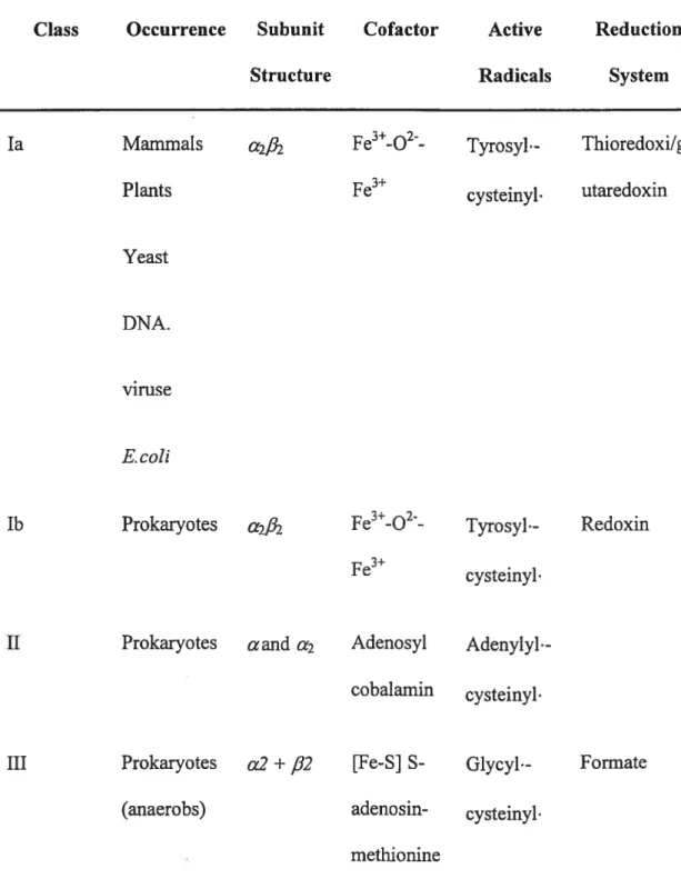
Ail of different RNR have radical generator; this has been the common feature of different RNR. The radical generator produces and stores a radical, which, as a first step of the reaction, is used to oxidize the substrate to a radical form. Surprisingly the radical generator is different for ail RNRs, whereas the reductase component is more similar. Three main classes of ribonucleotide reductases have been described (Table 1), classified according to the radical generator (Reichard 1993). Class I enzymes produce a stable tyrosyl radical on one type ofprotein subunit tbrough the action of a dinuclear iron center. Class II enzymes use a radical on the cofactor cobalamin. Class III enzymes form a stable glycyl radical with the help of an iron—sulfur protein and $-adenosyl methionine.
6
Table 1. Different Classes ofRNR
Class Occurrence Subunit Cofactor Active Reduction
Structure Radicals System
la Mammals fe3-O2- Tyrosyl.- Thioredoxi/gl
Plants fe3 cysteinyl. utaredoxin
Yeast
DNA.
viruse
E.coÏi
lb Prokaryotes a2/32 fe3-O2- Tyrosyl- Redoxin
3+
fe cysteinyl•
II Prokaryotes aand a2 Adenosyl Adenylyl. cobalamin cysteinyl
III Prokaryotes a2 + fi2 [fe-S] S- Glycyl•- Formate
(anaerobs) adenosin- cysteinyl. methionine
PART 1: Class I
Class I enzymes are found practicaiÏy in ail eukaryotic organisms, from yeast and algae to plants and mammals, some prokaryotes and some viruses. They are ail oxygen dependent. Class I is further divided into two subclasses, Ta and lb, according to their polypeptide sequence homologies and their overali allosteric regulation behaviour (Jordan and Reichard 1994). Class la exists in eukaryotes, prokaryotes, viruses, and bacteriophages, whereas lb has only been found in prokaryotes. Class I RNRs are tetrameric enzymes and show ctf3u3 structure (Reichard 2002). The substrate binding active site is located in the large Œ- homodimer, which is called Ri in class Ta. The smali f3-homodimer contains a binding site for two ions in each poiypeptide chain and is calied R2 in class la. Because of the unusual 3-subunit composition in the class la RNR from Saccharomyces cerevisiae, cLcf3f3’ (Huang and Elledge 1997), where only one ofthe R2 subunits (3) can harbour a diiron centre, the class la couid aiso be divided into two subclasses, lai and 1a2. However, the situation in S. cerevisiae seems to be quite exceptional, since even closely related yeast strains, such as Schizosaccharomyces pombe (Wood et al., 2002), won’t be able to express this type ofheterodimers.
For mammalian RNR, ffie mouse enzyme might be the best-studied model. There are a number of significant differences between the E. cou RNR and RNR in higher organisms. The sequence identity between the mouse R2 and the E. cou R2 is only 19%, whereas there is 50%, or higher, identity among the eukaryotic R2 proteins (Kauppi et ai.,
$
1996; Demple et al., 1986) . For class lb, the enzyme from Saïrnonella typhimurium is
PART 2: Class II
The classes II RNRs contain only one type of subunit, which are (L or f3 proteins. The necessary radical is produced by adenosylcobalamin (Booker and $tubbe 1994). The radical is formed by cleaving the adenosyl-Co bond. This type of RNR is found in some microorganisms, while the best-studied example of this type RNR is from Lactobacillus leichmanni. Recently, several class II enzymes have been characterized, the sequences of these enzymes in catalytic domains are distantly related to the class I and III ribonucleotide reductases tRiera and Fontecave 1997).
10
PART 3: Class III
Class III anaerobic reductases use a glycine radical (Sun, and Sjoberg 1996), which is generated with the help of S-adenosyl methionine and an iron—sulfur cluster (Eliasson and Reichard 1990). The active form is a Œ2 dirner (Padovani et al., 2001); it contains the active site glycyl radical and binding sites for the allosteric effectors. It thus corresponds in most respects to the substrate binding Ri subunit of the aerobic reductases. The f32 subunits, a small iron—sulfur-containing protein, is essential for production of the glycyl radical. ($un, and Reichard 1995) The overali reductant is formate (Mulliez, and Reichard 1995) rather than the enzymatic systems employed by class I.
SECTION 1.3: Structure of RNR
E. cou employ class I reductases; it use an iron-containing protein for the generation of the catalytically essential free radical. In this type of reductases, the quaternary organization ofthe holoenzyme is made up ofthe ci dimer, called protein Ri, contains the active sites and binding sites for allosteric effectors, and the
13
dimer, called protein R2, contains one dinuc!ear iron center and one stable tyrosyl radical per monomer; they are both essential for enzymatic activity.High-resolution structures of several forms of E. cou Ri (Uhuin and Eklund i994;Uhlin and Eklund 1996), R2 (Logan, et al., 1996; Aberg and Eklund 1993) and a plausible mode! for the Ri:R2 hoÏoenzyme (Padovani and Fontecave 2001) are now available. Unless stated otherwise the fo!!owing discussion concerns the E. cou enzyme.
12
PARTi: Ri Protein
The Ri protein is the large homodirner (2x85.5kDa) mediates both catalysis and allosteric interactions. Figure 1 shows the high-resolution structure of the Ri protein. Each monomer consists of three domains: one rnainly helical domain comprising the 220 N-terminal residues; a barre! domain (480 residues), novel ten-stranded 13/u barrel; and a small f3/Πdomain comprising 70 residues. The barrel is cornposed of two halves connected in an antiparallel way, each containing five parallel strands and four connecting helices (Uhiin and Ekiund 1 994;Uhlin and Ekiund 1996)
H. Ekhmd et cil, Progrc’ssin Biuphrsi & Molecular Bkilogi 7 (200]) 17?268
N-tcrniinus
t1)
J3A
tb}
PART 2: R2 Frotein
Several excellent reviews (Fontecave and Reichard 1992; Graslund and Sahiin 1996) have discussed the R2 structure. Briefly, four carboxylates and two histidines organized the two t-oxo linked irons of the oxidized center. On reduction the iron center is stiil coordinated by the same carboxylate-dominated ligand sphere, but several of the carboxylate-dominated ligands have been changed in conformation, with loss of the oxpbridge and of two bound water molecules, and a decrease of the coordination number from six to four. The iron center has opened up and become accessible to dioxygen, a prerequisite for radical generation on the neighboring Y 122.
PART 3: The Holoenzyme
Crystals of the Ri :R2 complex suitable for structure determination has flot been obtained yet. However, a plausible model for the holoenzyme was created, mainly from considerations of syrnrnetry, complementarity, and conserved amino acid residues. (Uhlin and Eklund 1994) The C terminus of R2 interacts with a region close to the C terminus of the Ri protein with some species-specificity. Just base on this model form, people designed peptidomimetic drugs that specifically inhibit the activity of the herpes reductase but not the mammalian enzyme. (Liuzzi et al., 1994)
16
PART 4: Allosteric Regulation
In general, allosteric regulation rapidly adapts an enzyme to changing requirernents for its product by binding of effectors, which may efficiently increase or decrease its activity. Most class la reductases are regulated by ATP and dATP when binding either ATP (activating) or dATP (inhibitory) to the activity site of protein RI (Brown and Reichard 1969). However, an additional and unique allosteric mechanism that regulates their substrate specificity and ensures that the enzyme produces equal amounts of each dNTP for DNA synthesis functions in the RNR regulation system. Disturbances in pool sizes may lead ta genetic damage and in severe cases ta ceil death (Brown and Reichard 1969; Kunz et al., 1994). By binding of end products (dATP, dGTP, and dTTP) to the specificity site ofthe reductase this result may be prevented (Brown and Reichard 1969). The allosteric sites communicate with the substrate-binding site and they affect each other at the same time. A detailed model for class la enzymes involving various effectors and substrates was developed in 1979 (Thelander and Reichard 1979). It lias been surviving well over the years. f igure 2 illustrates effector binding ta the allasteric sites from class Ta reducates.
Allosteric regulation ofthe enzymes fram different classes may shows similarities or differences. Here we talk about Class la only.
Figure 2. Models for Hie allosteric regulation of the
ribonucleotide reductase classes
Class la
Spe ci fïci ty
Activity site
ATP
dAT?
AT?
dATP
dGTP
dTTP
site
18
SECTION 1.4: Gene Organizations and Regulation
of Enzyme Synthesis
Ribonucleotide reduction plays a central role in the regulation of the pool sizes of the four dNTPs required for DNA synthesis, even though deoxynucleoside kinases and nucleotidases are also important (Reichard 1988). The small dNTP pools suffice in some ceils for only a few minutes of DNA replication and must therefore be renewed continuously during S-phase. The dNTPs are also required for DNA repair and mitochondrial DNA replication by cells that are not in S-phase. Celis also synthesize dNTPs de novo, albeit at a much slower rate (Bianchi and Reichard 1997). To satisfy the changing demands for dNTPs, the synthesis of ribonucleotide reductases is tightly adapted to the ceil cycle throughout gene organization or regulation.
In S. cerevisiae, four genes involved in ribonucleotide reduction: RNRJ and RNR3 encode Ri proteins, RNR2 encodes an R2 protein (Elledge et al., 1992; Elledge et al.,1993), and RNR4 encodes an R2-like protein (Wang et al., 1997). Rnrl and Rnr2 are essential for normal growth. Rnr3 is not, but when present on a high-copy number plasmid, complements mutations in RNRJ. RNR4 is also required for viability but the coded protein by itself has no R2 activity. It may be required for the formation of a functional holoenzyme complex (Wang et al., 1997).
SECTION 1.5: Anti—tufflor ACtivity of
Ribonucleotide Reductase
As theoretically expected, some RNR inhibitors have been demonstrated to possess antiturnor properties. They have been proved to be able to kiil tumor ceils preferentially with respect to normal celis (i.e., they have a sufficiently high therapeutic index). The high therapeutic index of antimetabolites is generally explained by the commitment of the neoplastic cells to replication and by their decreased adaptability and low responsiveness to regulatory signals making them more vulnerable than normal celis to drug-induced perturbations (Weber 1983). $ome investigators recently used this reasoning specifically to the antitumoral activity of RR inhibitors.
It is known that before the progression of the celi to the next cycle stage, the control of factors, which acts as checkpoints, must ensure that the previous stage has been completed. Just because of this, one hypothesis was developed, that the effect of RR inhibitors (low concentration) on normal mammalian cells is just cytostatic because they are able to control cell cycle progression, while tumor cells have lower or no ability to control ccli cycle progression and are more easily killed by RR inhibitors.
However, it is flot known whether the drug-induced imbalanced growth is the main mechanism responsible for in vivo antitumoral activity of RR inhibitors or it is somehow related to the apoptotic death induced by these agents. Indeed, it is sufficient to note that, despite extensive research and the finding of a great number of powerful
20 cornpounds, hydroxyurea, a relatively weak RR inhibitor, is stili the most used RR i;ihibitor in clinical settings.
SECTION 1.6: Yeast RNR (1—4)
The RNR gene in common bakers yeast S. cerevisiae is cornprised of four genes: RNR], RNR2, RNR3 and RNR4. RNRÏ and RNR3 encode polypeptides for the large subunit, caiied Rnrl and Rnr3 (Elledge and Davis 1990), and RNR2 and RNR1 encode polypeptides for the small subunit, called Rnr2 and Rnr4 (Wang et al., 1997).
The different roles of Rnrl and Rnr3 are flot very clear now. RnrÏ and Rnr3 share 80% amino acid sequence identity, ail the essentiai amino acid residues invoived in catalysis and aliosteric regulation are included in both Rnrl and Rnr3.
It bas been demonstrated that there are three dirneric combinations of these two polypeptides (i.e. R1R1, R1R3, and R3R3). Ail are catalytically active, although the specific activity of R3 dimer is significantly lower than that of the RI dirner (Dornkin et al., 2002). Expression ofRnrl is essential for the ceils to enter mitosis, and the celi cycle regulated the transcription of Rnrl, however, it seems that no expression of RNR3 is expiicitly demanded. The Ïevel ofRNR3 expression is very low during normal ceil living; however, it is strongly inducibie by DNA damage when the expression level can increase by a factor ofup to 100 (Elledge and Davis 1990).
The differences between the two genes encoding small subunit, Rnr2 and Rnr4, are more striking in terms of the presence of functionaily important amino acid residues. While R2 contains ail the 16 critical residues conserved in aÏrnost ail R2 proteins, six of
22 these residues are missing in R4, including three that would be expected to be involved in iron binding.
Moreover, alignments ofthe amino acid sequences ofR2 and R4 reveal only 47% identity, and R4 is about 50 amino acid residues shorter at the N-terminus than normal R2 (Wang et al., 1997).
Deletion of R4 is lethal in some yeast strains (for example, S. cerevisiae) and impairs celi growth in others, indicating an important role of R4 in RNR function.
R2 and R4 can form both homodimeric and heterodimeric complexes, but only complexes involving R2 are catalytically active.
It has been proposed the important role of R4 in RNR is to deliver ions to R2 or/and to stabilize R2 in the proper conformation for ion cluster assembly and radical formation (Ge et al., 2001).
The crystal structure of the yeast R2R4 heterodimer has been solved (Voegtli et al., 2001) and the overail a-helical fold is very similar to other homodirneric class I enzymes, such as E. cou and mouse.
During the normal ceil cycle, RNR] and RNR3 are known to be predominantly localized to the cytoplasm and RNR2 and RNR4 are known to be predominantly present
redistributed to the cytoplasm in a checkpoint-dependent maimer. SubceÏlular redistribution of RNR2 and RNR4 can occur in the absence of the transcriptional induction of the RNR genes but could be happened afier DNA damage and likely represents a post transiational event. These resuits strongly suggest that DNA damage checkpoint modulates RNR activity through the temporal and spetial regulation of its subunits. (Yao et al., 2003)
24
SECTION 1.7: Research Objective
rnr4A strain was initially isolated in genorne wide screen for mutants that are sensitive to Bleomycine. Following cross examination of the mutant against a variety of the DNA damaging agents (MMS, 4-NQO, HU and F1202). We observed that rnr4A strain was extremely resistant to 4-NQO.We therefore decide to characterize this observation. Herein we examined:
1. Whether there is any difference in sensitivity between the wild type ceils and rnr4A mutant ceils, while exposed to different kind DNA damage reagents, for example, 4-NQQ, BLM, MMS.
26
SECTION 2.1: Strains and media
The wild-type strains used were S. cereviskte BY4741. The collection of nonessential haploid rnr1A strains, derived from the S. cerevisiae BY474 1, was obtained from EUROSCARF (frankftirt, Gerrnany).
Standard YPD (1% yeast extract, 2% peptone, 2% dextrose) growth media was used as patch colonies and cultured liquid.
SECTION 2.2: ChemicaÏs and Equipment
Growth culture reagents, yeast extract, peptone and agar, were from Difco (Detroit, MI 4-NQO, MMS, and hydrogen peroxide were from Sigma (Saint Louis, MO).
Bleomycin A5 trihydrochloride was from Calbiochem (La Joua, CA). Pfu DNA polymerase was from Stratagene (La Joua, CA).
Primers (Table.2) were from Invitrogen (Carlsbad, CA)
Running buffer condition: 2.5 mM Tris pH7.O, 19.2 mM Glycine and 0.1% SDS. Nitrocellulose membrane (Hybonc-C+, Amersham)
Transfer buffer (25 mM Tris pH7.O, 192 mM Glycine and 20% methanol)
Blocking buffer (5% non-fat dry milk in 10 mM Tris, 150 mM NaCl, 1 mM EDTA, 0.1% Teween)
Primary antibody (Sigma)
Washing buffer (TBS containing 0.1% Tween 20) Anti-rabbit IgG-HRP conjugate (Sigma)
ECL solution (Amersham) Film (Kodak)
UVC lamp was from Fisher.
Shaker was from New Brunswick Scientific Co., Inc., Edison, N.J.
28
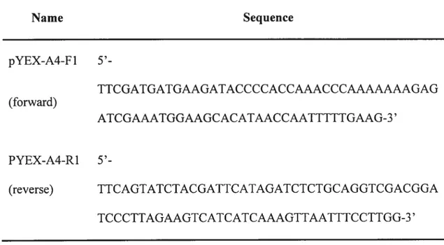
Table 2. PCR primers for RNR4 gene amplification
Name Sequence pYEX-A4-F1 5’-TTCGATGATGAAGATACCCCACCAAACCCAAAAAAAGAG (forward) ATCGAAATGGAAGCACATAACCAATTTTTGAAG-3’ PYEX-A4-R1 5’-(reverse) TTCAGTATCTACGATTCATAGATCTCTGCAGGTCGACGGA TCCCTTAGAAGTCATCATCAAAGTTAATTTCCTTGG-3’
SECTION 2.3: Survival Curves
Ovemight cultures grown to saturation at 30°C in YPD medium were diluted into ftesh YPD medium to an optical density at 600 nm of 0.4 (2 x10⁶ celis per ml) and incubated to an optical density at 600 nrn of 0.8 to 1.0. Aliquots of the cultures were then treated with BLM (with different time course 0, 10, 20, 30, 40 minutes) at 20 tg/ml or MMS (ranging from O to 0.2%) for 1 hour, or 4-NQO (ranging from O to 3 ig/rnl) for I hour, at 30 oc with shaking (250 rpm) in an incubator shaker. Diluted the samples in sterilized water to iO and plated lOOpi on YPD agar plate. Colonies were counted afier 2 to 3 days of growth at 30 °C.
Overnight cultures grown to saturation at 30°C in YPD medium were diluted into ftesh YPD medium to an optical density at 600 nm of 0.4 (2 x 10⁶ cells per ml) and incubated to an optical density at 600 mn of 0.8 to 1.0. Diluted the samples in steriÏized water to iO and plated lOOpi on YPD agar plates, then treated with UVC (0.4 J per second and per square meters with different time courses in 1, 3, 5, 8, and 10 minutes) at
30
SECTION 2.4: Mutagenesis Assays
Yeast cultures were started from single colonies and grown to stationary phase in liquid YPD at 30 °C, then diluted into fresh YPD medium to an optical density at 600 nrn of 0.4 (2x10⁶ cells per ml) and incubated to an optical density at 600 nm of 0.8 to 1.0. freshly dissolved 4-NQO was then added to a final concentration of 1.0 jig/ml. Cells were treated with aeration for 1 h at 30°c, washed, and resuspended in water at a density of lO7cells per ml. Both treated and untreated cells were plated aller appropriate dilutions onto complete medium containing L-canavanine (40 mg/ml) but lacking arginine for canr mutant count and complete medium but lacking arginine for viable cell count. Plates were incubated for 3 to 5 days at 30°c before counting. The frequencies Cant mutants in each culture were calculated by dividing the Cant mutant count by the viable cell count.
SECTION 2.5: Western Blot
Yeast ccli were harvested from liquid culture, washed twice with sterilized water, and centrifttged at 4000 rpm with ss-24 rotor in 4 °C. The pellet was resuspended in yeast extract buffer (50 mM Tris pH 7.0, 30 mM KC1, 10% glycerol, 1 tg/m1 leupeptin, 1 jig /ml aprotinin, I mM PMSf, 1 mM DIT). The proteins were homogenized by using a MINI beadbeater (BioSpec Products, Barttesville, 0K) at 5000 rpm for 20 seconds to six times. cellular debris was partially removed by centrifugation at 9000 g for 10 minutes in SS-24 rotor for 10 minutes. Proteins were quantified according to the method of Bradford.
50 microgram of whole ccli lysate per lane was loaded in an SDS-PAGE mini gel. Run gel at 120V/20 mA per gel untili the dye front vas close to the bottom. Running buffer condition: 2.5 mM Tris pH7.0, 19.2 mM Glycine and 0.1% SDS.
The proteins was transferred to a nitrocellulose membrane (Flybonc-c+, Amersham) at 100V/250 mA in transfer buffer (25 mM Tris pH7.O, 192 mM Glycine and 20% methanol) for 1.5 h. the blot was incubated with blocking buffer (5% non-fat dry milk in 10 mM Tris, 150 mM Naci, 1 mM EDTA, 0.1% Teween) for 2h at room temperature.
The blot was incubated with primary antibody (rabbit anti-Rpa32, diluted to 1:500 in blocking buffer) for 1h in blocking buffer at RT. The blot was washed 3 x 10 min in washing buffer (TBS containing 0.1% Tween 20) with shaking.
32
SECTION 2.6: Primer Extension Assay
Exponentially growing ceils in 5 ml of YPD were either treated or untreated with 4ig/m1 BLM for 1 h at 30°C. CelIs were harvested, washed twice with M9 buffer, and the ceil pellet was stored at -80°C for 1 h. Extraction of the chromosomal DNA was perforrned as usual. b measure the incorporation of [rnethyl-3H] dTMP, 150 iM of chromosomal DNA in 25 tl of HE buffer (10 mM Hepes-KOH, pH 7.0; 1 mM EDTA) was added to 225 pi of an ice-cold reaction mixture. This mixture was consist of 25 mM Hepes-KOH pH 7.6; 25 mM KC1; 10 mM MgC12; 50 ig/m1 bovine serum albumin; 100 iM dATP; 100 tM dCTP; 100 iM dGTP; 30 iM UTTP, and 3 units ofEscherichia cou DNA polymerase per ml. Labeled [methyl-3H] dTTP (NET221X from PerkinElmer Life Sciences; 37.0 MBq) was next added to a specific activity of 1260 cpm/pmol. The reaction was started when the samples were immersed into a 30 °C water bath. At the indicated turnes, 40 jil samples were withdrawn and added to tubes containing 200 pi of 0.1 M sodium pyrophosphate and 1 mg/ml of bovine serum albumin, followed by the addition of 200 pi of 0.8 M trichioroacetic acid. Samples were vortexed and placed on ice for 10 min. The samples were processed on a 1 2-hole filtration apparatus (Millipore, Bedford, MA) using GF/C circle filters (Whatman). The trapped DNA was washed three times with 3 ml of 0.1 M sodium pyrophosphate, briefly rinsed with ethanol, air-dried, and counted with 5 ml of scintillation fluid (BCS, Amersharn Biosciences).
Incubated blot with anti-rabbit IgG-HRP conjugate (Sigma) (diluted I : 10,000 in blocking buffer) for 1 h in blocking buffer at RI.
Washed 3 x 10 min in washing buffer with shaking. Drained washing buffer, added ECL solution (Amersharn) and developed for I min. Exposed to X-ray Kodak film for 1 to 30 min.
34
SECTION 2.7: Preparation ofYeast Genomic DNA
Each single colony of S. cerevisiae BY 4741 strains was cultured into 5 ml YPD liquid at 30°C. In second day, centrifuged at 3600 rpm for 5 minutes, resuspended the celi into 0.5 ml TE in eppendorf tubes. Centrifuged again, resuspended the pellet into 0.5 ml ofspheroplast buffer (1 M sorbital, 10 mM NaHPO4 pH7.0, 10 mM EDTA), and added 5 d 20 ig/m1 of lyticase (Sigma) and 3 t1 beta-mercapthoethonol (Fisher), incubated 37 °C for 20 minutes. Centriftiged 15 second in 14000 rpm. Resuspended into 0.5 ml buffer (50 mM EDTA, 0.3% SDS 5 t1 protease K 20 mg/ml) incubated 60°C for 30 minutes. Added 0.2 ml 5 M Potassium acetic acid on ice for 20 minutes, and centrifuged 10 minutes in 12,000-rpm 4°C. $upernatant were extracted with twice phenol, twice chloroform, precipitated with 2.5 fold of volume 95% ethanol. Centrifuged 10 minutes 12000 rprn 4°C, added 20 pi TE into pellet.
SECTION 2.8: PCR Program
PCR machine: PTC100TM Programmable Thermal Controller Mi research TNC
Master Mixture: DNA or genomic DNA 10-50 ng, lOx taq buffer Siil, oligo upstream 0.5pi (1jig/il), oligo downstream 0.5tl (1ig/pi), 10 mM dNTP 3 pi, 25 mM MgCÏ2 3 pi, Tap DNA polymerase 1 i1, add H20 up to 50 pi.
PCR designed program: 1. 95 °C 5 minutes
2. $0
°c
10 seconds, during this period, add pfu DNA polymerase 1 unit. 3. 94°c
50 seconds4. 50 °C
so
seconds 5. 72 °C 3 minutes6. 10 times repeat go to step 3. Each cycle decrease by i °Cin step 4. 7. 94
°c
50 secondss. so °c
50 seconds 9. 72 °C 3 minutes10. Repeat 20 cycles go to step 7. 11. 72 °C 10 minutes
36
SECTION 2.9: Clone ofplasmid Gst-rnr4
The entire RNR4 coding sequence was amplified by PCR with two primers which contains upstream pYEX-A4-f 1 and downstream pYEX-A4-R1 (Table 2) in vector pTW34O. Co-transformation with fragment and pTW34O vector into yeast strain YW607 and selected with URA mark. The positives clones were confirrned by Western blot and enzyme digestions (Fig. 3).
Figure. 3. Clone of plasmid Gst-Rnr4. The entire RNR4 coding sequence is arnplied by PCR with two primers which contains upstream and downstream in vector pTW34O. Co transformation with fragment and pTW34O vector into yeast strain YW607 and selected with URA mark. The clones are positives confirmed by western blot and enzymes digestion.
38
SECTION 3.1:
$trains rnr4A Mutant Display Extrerne
Resistance to 4-NQO as Compared
With the Parent
In order to determine the contribution of the Rnr4 to the repair of damage sites, we first chose 3 different type of DNA damage agent, 4-NQO, BLM and MMS, and examining whether rnr4A strains conferred the same level of sensitivity to these agents as the parent strains.
In this experiment, exponentially growing cultures were treated with 4-NQO, MMS and BLM then scored for fractional survivors as described in the material and rnethods. Interestingly, strains rnr4A mutant displayed extreme resistance to 4-NQO as compared with the parent strains (50% ± 1.9 vs. 6% + 1.1= A vs. wt, 3tg/m1 4-NQO, 1 hour,
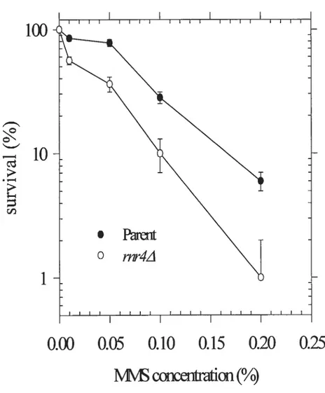
p<O.Ol). (figure .4) On the contrary, rnr4A mutant strains were sensitive to BLM and MMS as compared with the parent strain. (figure .5.6)
40
100
f-’
r:i
10
Figure 4. Survival curve
assay
after treatment of 4-NQO
The Log phase strains were treated with different concentration of4-NQO 1 hour at 30°C.
rnr1/ strains (o) were resistant to 4-NQO as compared with parent strains
(.).
Resuitswere expressed as the mean ± SD from three separate experirnents
O
1
2
3
4
100
I
‘o
FigureS. Survival curve assay after trealment ofBLM
The Log phase ceils were treated with 20 tg/m1 BLM for different tirnes at 3 0°C.
rnr4Astrains (o) were sensitive to BLM as cornpared with parent strains
(.).
Resuits were expressed as the mean ± SD from three separate experiments.0
10
20
30
40
50
42
100
[o
1
ccen&n
(%)
Figure 6.
Survival curve assay after treatment of MMS
The Log phase strains were treated with different concentration of MMS for 1 hour at 30°Crnr4z\mutant strains (o) were sensitive to MMS as compared with parent strains
(.).
Resuits were expressed as the mean ± SD from three separate experirnents.SECTION 3.2: The rnr4z\ Strain Is Sensitive to
UVC
Since the apparent strong similarity in modes of cellular processing (nucleotide excision repair) for UVC light-induced and 4-NQO-induced DNA damage in both prokaryotic and eukaryotic systems, The 4-NQO has been catalogued in “UV-mirnetic” (Felkner and Kadiubar 1968). However, this classification is not accurate enough, because several recent investigations have clearly demonstrated that 4-NQO, unlike 254-nm UV light, can generate a substantial degree of intracellular oxidative stress (Rarnotar et al., 1998). Strains rnr4A mutant display extreme resistance to 4-NQO as compared with the parent, in order to know further if strains rnr4A mutant is resistant to “UV mimetic” DNA damage or the other DNA damage mechanism, the UVC ray was applied for exposure on petri dishes containing 100 t1 of 1 0 diluted wild type or rnr4A strains as described in the methods. The rnr4A mutant showed sensitive to UVC as compared with parent ceils. This resuit suggests that rnr4A strains are specifically resistant to the ceil killing effect, which is not “UV-mimetic”. (Figure.7)
44
100
c .10
>z
cri 1Exposition time (mm)
Figure
7.Survival curve assay Anatysis after Treatment
UVC
Exposure
The parent (wild type BY4741) and strains rnr4A mutant grow in YPD liquid 30°C over night to be saturated. 100 ml of each saturated ceils are made Ï û dilutions and plated on the YPD agar plate. Triple plates of each strain are exposed on UVC on 0.4 J per second and per square meters with different time courses in 0, 1, 3, 5, 8, and 10 minutes. Counting the ail colonies numbers in each plate after 4$ hours incubation in 3 0°C. rnr1A mutant strains (o) were sensitive to BLM compare with parent strains
(.).
Results are expressed as the mean ± SD from three separate experimentsSECTION 3.3: The Frequency of4-NQO-induced can1’
Mutations Rise $harply in the WiYd Type
Strain
Our data showed that strains rnr4A mutant displayed extreme resistance to 4-NQO
as compared with the parent strains. If Rnr4 plays a role in the repair of 4-NQO-induced
DNA lesions, then parent ceils might be expected to show hypermutable phenotype in
4-NQO induced DNA lesions. We therefore measured the reversion mutation rate on -arg plate containing 25 tg /ml canavanine in -arg plate for a wild type and rnr1z\ mutant strains as described in material and methods.
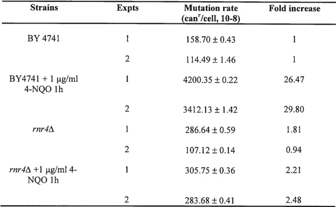
Under normal growth conditions, parent ceils showed no significant difference mutation rate as compared with rnr4A mutant strains (Table.3). The mutation rate was
increased as much as 27-fold when the wild type stains was treated with 4-NQO compare with no treatment. In contrast, the mutation rate increase only 2-fold when the rnr1A
strain were treated with 4-NQO. This resuit suggested that Rnr4 played an important role in allowing 4-NQO-induced mutagenic effects.
46
Table 3. Spontaneous rates and spectrum of can’ mutation in
rnr4A
deficient mutant
Strains Expts Mutation rate Fold increase
(ca&Icell, 10-8) BY4741 1 152.70±0.43 1 2 114.49±1.46 1 BY4741 + 1 jig/ml 1 4200.35 ± 0.22 26.47 4-NQO 1h 2 3412.13±1.42 29.80 rnr4A Ï 286.64±0.59 1.81 2 107.12±0.14 0.94 rnr4zX+1 jig/m14- Ï 305.75±0.36 2.21 NQO 1h 2 283.68 ± 0.41 2.48
SECTION 3.4: Western Blot
Replication protein A (RPA) is essential for multiple processes in DNA metabolism including DNA replication; recombination and DNA repair pathways (including nucleotide excision, base excision and double-strand break repair). It is a heterotrimeric single-stranded DNA-binding protein composed of subunits of 70-, 32-and 14-kDa, which is highly conserved in eukaryotes. RPA binds ssDNA with high affinity and interacts specifically with multiple proteins. Cellular DNA damage causes the N-terminus of the 32-kDa subunit of human RPA (RPA32) to become hyper phosphorylated. Current data indicates that hyper-phosphorylation of human RPA32 causes a change in RPA conformation that down-regulates activity in DNA replication but does flot affect DNA repair processes. This suggests that the role of RPA32 phosphorylation in the cellular response to DNA damage is to help regulate DNA metabolisrn and promote DNA repair. RPA32 is phosphorylated during the normal celi cycle and afier cellular DNA damage in a number of different organisms including humans, Drosophila melanogaster and $accharomyces cerevisiae. By now it has been accepted that changes happen in RPA32 in response to DNA damage induced by various agents and RPA modification may be associated with a loss of replication competence. for review, see ref.(Iftode et al., 1999)
In order to see changes in RPA32 in response to DNA damage induced by 4-NQO, parent 3Y474 1 and rnr4z\ mutant strains were treated with 0, 1, 2 jig/ml of 4-NQO for one hour at 30 degree. The total protein extract was loaded onto 10% PAGE gel, and
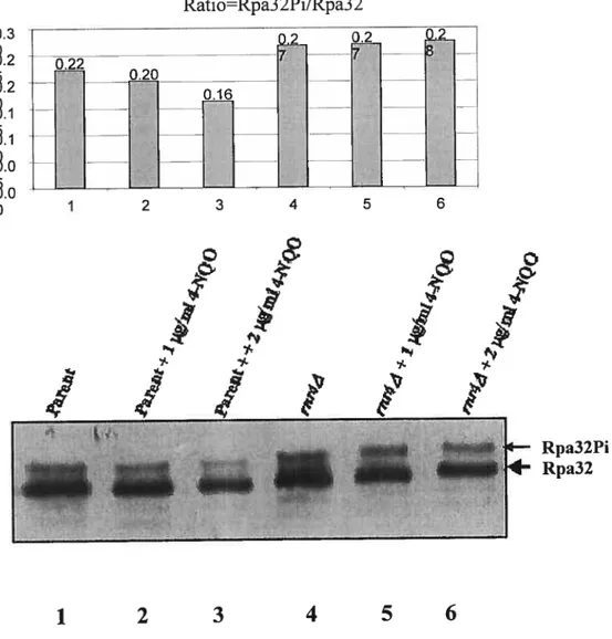
48 probed by western blot using L 5000 dilution of antibody Rpa 32 from rabbit was used. We did not observe any difference between treated and untreated ceils in both strains (Figure 8)
Since 4-NQO has been proved to be a DNA damage agent which easily enters the ceils and that the phosphorylation of Rpa32 was the sarne in both the parent and mutant, we excluded the possibility that resistance of the mutant to 4-NQO is a the level of drug entry.
Ratio=Rpa3 2Pi/Rpa3 2
0.2
Figure 8. Western blot detection of Rpa32 phosphalation
The parent BY 4741 and rnr4A strains were treated with zero concentration, 1
jig/mÏ, and 2 g/mÏ of 4-NQO for one hour in 30°C. 11e 50 jig of total proteins
extract were loaded on the 10% PAGE gel. RPA32 phosphorylated was not significantly increased in rnr4A mutant cells (5.6) as compared with the untreated cells (4); the percentage of RPA phosphorylated was flot significantly increased in parent cells (2,3) compared with the untreated ceils (1).
0.22 0.20 0.3 8.2 8.1 1
8.
o 0.16 2Ï
t .—. 4 5 6F
I
/
I
f
Rpa32Pi Rpa321
2
3
4
5
6
50
SECTION 3.5: Primer Extension Assay
By now we had already known that compared with the parent strain, rnr4A strain was more sensitive to BLM, the inability of the rnr4A deletion mutant to repair BLM damaged DNA could be probably due to a defect in processing DNA strand breaks containing blocked 3’ termini. To test this possibility, we examined whether chromosomal DNA isolated from celis treated with BLM at 40 ig/m1 for 1 h could allow in vitro DNA repair synthesis by E. cou DNA polymerase I (Demple et al., 19$6;Henner et al., 19$3;Levin and Demple 19$8;Masson and Ramotar 1996;Ramotar and Demple
1991;Seki et al., 1991).
In this experiment, chromosomal DNA was isolated from the both strains with or without 40 j.tg/ml BLM concentration in 1-hour 30°C incubation. The 200 ng BLM treated or without treated genomic DNA and 0.05 U of DNA polymerase I (New English BioBar) were added into each reaction. 200 d 0.5M TCA stopped each reaction in 0, 3, 6, and 9 minutes. As shown on the graph: first DNA isolated from untreated ceils (both parent and mutant trains) showed lower [rnethyl-3Hj dTMP incorporation compared with the treated ceils in both parent strain and rnr4z\ mutant strains; secondly, DNA isolated from treated parent celis showed great higher [rnethyl-3H] dTMP incorporation compared with the untreated parent ceil; DNA isolated from treated rnr1A celis showed only slight higher [rnethyl-3H] dTMP incorporation compared with the untreatedrnr4A ceils (Figure. 9). These resuits suggested:
1. BLM-rnediated celÏ killing was a direct resuit of damage to DNA.
2. The defect of the rnr1A deletion mutantto repair BLM-damaged DNA was due to a defect in processing DNA strand breaks containing blocked 3’ termini.
52 12000 10000 8000 C 6000 C Q 4000 H 2000 O
figure
9. In vitro
incorporation of jmethyl-3H] dTTP
To detect accumulation of bleomycin-induced DNA lesions in parent (BY474 1) and
rnr4A strains. The substrat of genornic DNA were prepared from the both strains with or without 40 jig/ml BLM concentration in 1 hour 30°C incubation. The 200 ng BLM treated or without treated genomic DNA and 0.05 U of DNA polymerase I (New English BioBar) were added into each reaction. 200 pi 0.5M TCA stops each reaction in 0, 3, 6, and 9 minutes. Resuits were expressed as the mean ± SD from three separate experiments.
0 2 4 6 8 10
SECTION 3.6: The Survival Curve Assay for Gst-Rnr4
Containing Strain
rnr4Astrain displayed extrerne resistance to 4-NQO as cornpared with the parent strain was the most important resuit in our study. In order to verify this resuit, Gst-Rnr4 inrnr4A strain was created in our lab, as described in material and methods.
Over night grown celis ofrnr4A/ BY 4741 /Gst-Rnr4 inrnr4z\ strain were used for survivalcurve assay. In rnr4A strain, the resistance to 4-NQO aiways showed. Thernr4z\ mutant containing plasmid Gst-Rnr4 lost the resistance to 4-NQO (f ig. 10)
54
100,0
10,0
1,0
0,1
Figure. 10 The survial curve assay for Gst-Rnr4 containing strain.
Inrnr4z\ strain,the resistance of 4-NQO aiways showed. While the rnr4A strain
Containing plasrnid Gst-Rnr4 brought its phenotype with more sensitive with drug 4-NQO. Resuits were expressed as the mean ± SD from three separate experirnents.
0 1 2 3 4
56
RNR catalyze the reduction of ail four ribonucieoside diphosphates into deoxyribonucleoside diphosphates and piay a centrai role in controiiing the leveis of celiuiar deoxynucieoside triphosphates (dNTPs), which are essentiai for high-fidelity DNA replication and DNA repair processes. In S. cerevisiae, RNR is a a2f32 tetramer consisting of two large (Rnrl/Rnr3) and two smail subunits (Rnr2/Rnr4). Rnr4 and Rnr2 appear to have nonoveriapping functions and cannot substitute for each other even when overproduced. To investigate the roie of Rnr4 in DNA repair processes, except the survivai curve assay which show that rnr4z\ mutant strain is resistant to 4-NQO, different kind of techniques (western blot/primer extension assay/rnutation assay) were appiied step by step. As a resuit a new DNA damage mechanism induced by 4-NQO was disclosed and an error free DNA repair mariner may piay an important roie when rnr4A mutant strain is treated with 4-NQO.
First survival curve assay for strains rnr4z\ mutant and parent (S. cerevisiae BY 4741) afier treatment with different type of DNA damage agents (BLM, MM$, and 4-NQO) was appiied in our study.
Bleomycin is used extensiveiy to treat a variety of cancers, inciuding those of the lungs, testicies, head, and neck. The antitumor effect ofBLM is exerted through oxidative iesions in chromosomai DNA, formed via a free radicai-reactive compiex that is produced when BLM binds to iron and oxygen in vivo. The activated Fe-BLM compiex takes a hydrogen atom from the 4’-carbon of deoxyribose, resuiting in two types of
lesions: (i) oxidized apurinic/apyrimidinic (AP) sites and (ii) DNA single-strand breaks that terminate with 3 ‘-phosphoglycolate. Noncoding AP sites lead to the incorporation of incorrect nucleotides by DNA polymerase. While 3’-phosphoglycolate is known to block DNA synthesis, the mutagenic effect of this adduct is unknown. Activated Fe-BLM
complex can also produce double-strand breaks at certain DNA sequences, such as CGCC. In any case, the lesions produced by BLM are toxic and are considered to be mutagenic.
People now use the agent 4-nitroquinoline 1 -oxide (4-NQO) in mammalian systems as a paradigrn for DNA damage-induced carcinogenesis. To damage DNA. 4-NQO must first undergo metabolic activation to the proximate carcinogen 4-hydroxyaminoquinoline 1-oxide, which, following acylation, reacts with DNA to forrn stable quinoline-purine monoadducts, i.e. at the exocyclic N-2 and N-6 positions of guanine and adenine, respectively. In bacteria, yeast, and mammalian celis, these genotoxic “bulky” DNA lesions are repaired largely by the nucleotide excision repair (NER) pathway in a manner analogous to classical dipyrimidine photoproducts. Since the apparent strong similarity in modes of cellular processing (nucleotide excision repair) for UVC light- and 4-NQO-induced DNA damage in both prokaryotic and eukaryotic systems. The 4-NQO is catalogued as “UV-mirnetic”.
In our present study, rnr4A mutant strain was sensitive to BLM and MMS as cornpared with parent. This is flot strange. because even without DNA damage agent,
58
slow growth phenotype as cornpared with parent strains in normal condition (Huang M and Elledge SJ 1997). Moreover, like the other RNR genes, RNR4 is inducible by DNA damaging agents through MEC1, RAD53 and DUN1 transduction pathway. $o the possible mechanisrn for the lethality of deletion of RNR4 is that the mutant ccli bas a lower level of functional RNR complex, resulting in dNTP levels below the thresbold critical for mitoic viability. In fact, the level of dNTPs in yeast increase dramaticaily afier DNA damage and the survival of DNA damage in yeast directly depends on increased dNTP levels (Chabes A et al., 2003).
Interestingly, strains rnr4A mutant display extreme resistance to 4-NQO as compared with the parent, tbis suggests us that RNR4 deletion mutations show strong ability in repair for DNA damage induced by 4-NQO.
$ince 4-NQO is a “UV-rnimetic” DNA damage agent, whose genotoxic “btilky” DNA lesions are processed largely by the nucleotide excision repair (NER) pathway. Does it mean that an RNR4 deletion mutant has a strong ability in NER pathway? We next applied survival curve assay for strains rnr4A mutant and parent (BY 4741) afler treatment with UVC. Ivlore interestingly, tbe strain rnr1A mutant sbowed sensitivity to UVC as compared with parent.
This suggests that the resistant to 4-NQO of rnr4i mutant is not result from a strong ability in NER pathway as compared with parent.
During S phase. when DNA damage is encountered. TLS (transiesion DNA repair) plays a major role in bypassing the lesions. In spite of increased mutagenicity, such a process may be inherently more advantageous than the other repair rnechanisrns where interruptions in the newly synthesized strands persist for long periods. An increase in dNTP concentration above normal S phase levels are essential for transiesion DNA repair (Prakash S and Prakash L, 2002). If rnr4 mutant enhance transiesion DNA repair, then the mutation rate must be raised. We therefore measured the reversion mutation rate on
-arg plate for a wild type andrnr1A mutant strains.
Under normal growth conditions, parent ceils showed no significant difference mutation rate as compared with rnr4A mutant strains (Table.3). The mutation rate was increased by as much as 27-fold when the wild type stains vas treated with 4-NQO as compared with no treatment and 13-fold as compared with rnr1A mutant strains 4-NQO treated. We believe that the lower level of dNTP may prevent the translesion bvpass pathway from ftinction by flot allowing the bypass polymerase from being fully active.
We do not think that the 4-NQO ceil killing effect is the resuit of defect in drug entry or DNA damage, which rnr1A pÏays an important role. In order to answer this, Western-Blot assay was applied. Although, we couldn’t see any difference in Rpa32
between treated and untreated celis in both strains (Figure 8), suggesting that 4-NQO must enter the celi at the sarne rate in both strains to cause induce phosphoryfation of Rpa32.
60 mutant display only resistance to 4-NQO as compared with the parent. Does the celi killing effect of BLM is a direct resuit of DNA damage induced by BLM? Our primer extension assay verifies this resuit and shows that ceil-killing effect of BLM is a direct resuit of DNA damage. The defect of the RNR4 deletion mutant to repair BLM-damaged DNA could be due to a defect in processing DNA strand breaks containing blocked 3’ termini, which is likely regulated by the availability of dNTPs. The ce!! might sense that the level of dNTP is !ow and therefore prevent the action of DNA repair proteins and the subsequent action of DNA po!yrnerase.
Taken together, rnr4A mutant strains disp!ay extreme resistance to 4-NQO induced DNA damage as compared with the parent. In addition, the mutant showed very litt!e increase in 4-NQO-induced mutants when cornpared to the parent. We propose that this DNA damage is neither a “UV-mirnetic” DNA damage. which is processed large!y by the nucleotide excision repair (NER) pathway nor oxidative stress like BLM induced, but a different DNA !esion caused by 4-NQO. This DNA lesion could be repaired in both an error free DNA repair marmer (Prakash S, et al., 2005.) (Huang M and E1!edge SJ 1997), even when the dNTPs !eve! are low. The lower dNTPs pool in rnr4A mutant strains prevent may prevent ILS pathway but doesn’t affect the eior free DNA repair manner and lead to higher survival rate but lower mutation rate; on the other hand, the higher level of dNTPs pool allowed WT cells to repair this lesion in ILS pathway (Chabes and Thelander 2000), but since a lot of error rernained this lead to lower survival rate and higher mutation rate.
At the end the rnost important resuit that rnr1A mutant strains display extrerne resistance to 4-NQO induced ceil killing as compared with the parent was verifled by survival curve assay (Figure 10).
CHAPTER V: CONCLUSION
In conclusion, strains rnr4A mutant display extreme resistance to 4-NQO induced DNA damage cornpared with the parent; this DNA damage is neither a “UV mimetic DNA damage which is processed largely by the nucleotide excision repair NER) pathway nor oxidative stress like BLM induced, but a different DNA lesion mechanism causing by 4-NQO. The lower dNTP pool in rnr1A mutant doesn’t affect the repair of DNA lesion causing by this DNA lesion rnechanism, which could be an error free DNA repair marmer and lead to higher survival rate; on the other hand, the higher level of dNTP pooi in WT allow cells to repair this lesion in TLS, which has a lot of errors and lead to lower survival rate. Base on this result we believe that RNR4 gene play an important role not only in controlling the levels of cellular dNTPs, but also in DNA repair including TL$ and an eior free DNA repair manner. We propose that like some RNR inhibitors, RNR4 gene may play a role in cell antitumor properties.
CHAPTER VI: PROSPECT
In rny project, the strongly 4-NQO resistance phenomenon ofrnr4z\ knockout may corne frorn a third DNA damage role of this drug which is neither a “UV-mimetic DNA darnage which is processed largely by the nucleotide excision repair (NER) pathway nor oxidative stress like BLM induced. For identifying the 4-NQO involved DNA repair pathway in mutant rnr4A strain and fuiiher characterization. The following studies are suggested as future work:
I .Create different gene knockout from parent strain BY4741 and 3Y474l RNR4A.
For example, knockout NER pathway gene radi, rnismatch repair gene msh6, recombination rad5 1.
2. Microarray assay for find related genes. Immunoprecipitation assay to detect
CHAPTER VII: REFERENCE
Aberg,A., S.Hahne, M.Karlsson, A.Larsson. M.Ormo, A.Ahgren, and B .M.Sjoberg. 1989. Evidence for two different classes of redox-active cysteines in ribonucleotide reductase of Escherichia cou. J.Biol.Chern. 264: 12249-12252.
Aberg,A., P.Nordlund, and H.Eklund. 1993. Unusual clustering ofcarboxyl side chains in the core of iron-free ribonucleotide reductase. Nature. 361: 276-27$.
al Khodairy,f. and A.M.Carr. 1992. DNA repair mutants defining G2 checkpoint pathways in Schizosaccharomyces pombe. EMBO J. 11: 1343-1350.
Andreassen,P.R. and R.L.Margolis. 1992. 2-Aminopurine overrides multiple ce!! cycle checkpoints in BHK ceils. Proc.Natl.Acad.Sci.U.S.A. 89: 2272-2276.
Bianchi,V., S .Borella, C .Rampazzo, P.ferraro, F .Calderazzo, L.C .Bianchi, S. Skog. and P.Reichard. 1997. Celi cyc!e-dependent metabolism of pyrirnidine deoxynuc!eoside triphosphates in CEM celis. J.Biol.Chem. 272: 16118-16124.
Bj orklund, S., K.Hj ortsberg, E.Johansson, and L. Thelander. 1993. Structure and prornoter characterization of the gene encoding the large subunit (RI protein) of mouse ribonucleotide reductase. Proc.Natl.Acad.Sci.U.S.A. 90: 11322-11326.
68 ribonucleotide reductase: evidence for the participation of five cysteine residues in ribonucleotide reduction. Biochemistry. 33: 12676-12685.
Brown,N.C. and P.Reichard. 1969. Role of effector binding in allosteric control of ribonucleoside diphosphate reductase. J.Mol.Biol. 46: 39-55.
Chabes A, Georgieva B, Domkin V, Zhao X, Rothstein R, Thelander L. 2003 Survival of DNA damage in yeast directly depends on increased dNTP levels allowed by relaxed feedback inhibition ofribonucleotide reductase Cell.112: 39 Ï-401.
Chabes,A. and L.Thelander. 2000. Controlled protein degradation regulates ribonucleotide reductase activity in proliferating mammalian celis during the normal ce!! cycle and in response to DNA damage and rep!ication b!ocks. J.Bio!.Chem. 275: 17747-17753.
Crissman,H.A., D.M.Gadbois, R.A.Tobey, and E.M.Bradbury. 1991. Transforrned mamma!ian ce!!s are deficient in kinase-mediated contro! of progression through the Gi phase of the cell cycle. Proc.Natl.Acad. Sci.U. S .A. $8: 7580-7584.
Demple,B., A.Jolmson, and D.Fung. 1986. Exonuc!ease III and endonuclease IV remove 3’ b!ocks from DNA synthesis primers in H202-darnaged Escherichia co!i. Proc.Natl.Acad.Sci.U.$.A. 83: 7731-7735.
Domkin,V., L.The!ander, and A.Chabes. 2002. Yeast DNA damage-inducible Rnr3 has a very low catalytic activity strongly stimulated afler the formation of a cross-ta!king Rnrl/Rnr3 complex. J.Bio!.Chem. 277: 18574-1 8578.
Eklund,H., U.Uhlin, M.Farnegardh, D.T.Logan, and P.Nordlund. 2001. Structure and function ofthe radical enzyme ribonucleotide reductase. Prog.Biophys.Mol.Biol. 77: 177-268.
Eliasson,R., M.Fontecave, H.Jornvall, M.Krook, E.Pontis, and P.Reichard. 1990. The anaerobic ribonucleoside triphosphate reductase from Escherichia cou requires S adenosylmethionine as a cofactor. Proc.Natl.Acad.Sci.U.S.A. 87: 3314-3318.
Elledge,S.J. and R.W.Davis. 1990. Two genes differentially regulated in the celi cycle and by DNA-damaging agents encode alternative regulatory subunits of ribonucleotide reductase. Genes Dey. 4: 740-751.
Elledge, S .J., Z.Zhou, and J.B .Allen. 1992. Ribonucleotide reductase: regulation, regulation, regulation. Trends BiochentSci. 17: 119-123.
Elledge,S.J., Z.Zhou, J.B.Allen, and T.A.Navas. 1993. DNA damage and ce!! cycle regulation ofribonuc!eotide reductase. Bioessays. 15: 333-339.


![figure 9. In vitro incorporation of jmethyl-3H] dTTP](https://thumb-eu.123doks.com/thumbv2/123doknet/2054460.5627/72.918.188.704.305.676/figure-in-vitro-incorporation-of-jmethyl-h-dttp.webp)
