HAL Id: pastel-00679718
https://pastel.archives-ouvertes.fr/pastel-00679718
Submitted on 16 Mar 2012HAL is a multi-disciplinary open access
archive for the deposit and dissemination of sci-entific research documents, whether they are pub-lished or not. The documents may come from teaching and research institutions in France or
L’archive ouverte pluridisciplinaire HAL, est destinée au dépôt et à la diffusion de documents scientifiques de niveau recherche, publiés ou non, émanant des établissements d’enseignement et de recherche français ou étrangers, des laboratoires
Development of chemogenomic approaches for
prediction of protein - ligand interactions
Brice Hoffmann
To cite this version:
Brice Hoffmann. Development of chemogenomic approaches for prediction of protein - ligand inter-actions. Quantitative Methods [q-bio.QM]. École Nationale Supérieure des Mines de Paris, 2011. English. �NNT : 2011ENMP0074�. �pastel-00679718�
École doctorale n
O432 : Sciences des Métiers de l’ingénieur
Doctorat ParisTech
T H È S E
pour obtenir le grade de docteur délivré par
l’École nationale supérieure des mines de Paris
Spécialité « Bio-informatique »
présentée et soutenue publiquement parBrice HOFFMANN
le 16 decembre 2011Development of chemogenomic approaches for prediction
of protein-ligand interactions
Directeur de thèse : Véronique STOVEN
Jury
M. Daniel ABERGEL,Directeur de Recherche, Département de Chimie, Ecole Normale Supérieure Examinateur
Mme Hélène DEBAT,Maître de Conférences, Institut de Génétique, Université Versailles Saint-Quentin Examinatrice
M. Dragos HORVATH,Directeurs de Recherche, Laboratoire d’Infochimie, Université de Strasbourg Rapporteur
M. Olivier LEQUIN,Professeur, Laboratoire des Biomolécules, Université Pierre et Marie Curie Rapporteur
R´esum´e en franc¸ais
L’enjeu de cette th`ese porte sur le d´eveloppement de m´ethodes de bio-informatique appliqu´ees `a la pr´ediction des interactions prot´eine - ligand. Le chapitre 1 est une courte introduction qui rappelle le rˆole cl´e que jouent ces interactions dans le fonctionnement de la cellule. Il pr´esente ´egalement le plan de ce manuscrit. Dans le chapitre 2, apr`es avoir rappel´e quelles sont les mol´ecules cl´es du monde vivant ainsi que les principes de l’interaction entre les prot´eines et les ligands, est pr´esent´e l’´etat de l’art de l’´etude de ces interactions, `a la fois exp´erimentale et in silico avec les m´ethodes bas´ees sur l’´etude des ligands (ligand-based) et celles bas´ees sur l’´etude des prot´eines (protein-based). Enfin la m´ethode d’apprentissage statistique des machines `a vecteurs de support (SVM) appliqu´ee au criblage virtuel est pr´esent´ee, car cet algorithme a ´et´e employ´e au cours de cette th`ese pour d´evelop-per une m´ethode de pr´ediction appartenant aux approches dites de ch´emog´enomique. Dans un premier temps (chapitre 3), la th`ese se concentre sur l’´elaboration de cette m´ethode de ch´emog´enomique appliqu´ee aux prot´eines de la famille des GPCRs. Cette m´ethode est bas´ee sur l’utilisation de machines `a vecteurs de support pour pr´edire l’interaction entre des GPCRs et leurs ligands. Cette approche suppose l’emploi de descripteurs pour encoder les prot´eines et les ligands. Plusieurs types de descripteurs ont ´et´e employ´es, afin de comparer leur pertinence dans le cadre de la ch´emog´enomique. Pour les ligands, des descripteurs correspondant `a un en-codage de la structure 2D ou de la structure 3D ont ´et´e test´es. Dans l’approche 2D, une mol´ecule est d´ecrites par un vecteur binaire dont les ´el´ements sont d´etermin´es en fonction d’un graphe qui d´ecrit sa structure chimique. La similarit´e entre deux mol´ecules est alors ´evalu´ee par un co´efficient de Tanimito. Dans le cas de l’approche 3D, les mol´ecules sont d´ecrites par l’ensemble des triplets d’atomes qui la composent, et des distances s´eparant ces atomes. La similarit´e entre deux mol´ecules est ´evalu´ees en comparant les ensembles de leurs triplets respectifs, en
utilisant un noyau appel´e noyau pharmacophore 3D. Les prot´eines sont encod´ees ´egalement de plusieurs fac¸ons: d’une part par leur position dans la hi´erarchie des GPCR telle que cette hi´erarchie est d´efinie dans la base de donn´ees GLIDA, d’autre part par une courte s´equence d’acides amin´es correspondant aux acide amin´ees composant la poche de fixation pour le ligand. Les similarit´es entre prot´eines sont alors ´evalu´ees par plusieurs m´ethodes `a noyaux. Tout d’abord, deux noy-aux relativement basiques, n’employant pas ces encodages pour les prot´eines, sont employ´ees: le noyau Dirac, dans lequel la similarit´e entre deux prot´eines diff´erentes est ´egale `a z´ero, et le noyau Multitask, dans lequel toutes les prot´eines sont ”´egalement diff´erentes”. Le premier correspond en r´ealit´e `a une approche classique par prot´eine: aucune information de ligand n’est partag´ee entre les prot´eines. Le second noyau correspond `a une sorte de base-line pour les m´ethodes de ch´emo-g´enomique, dans laquelle l’information concernant les ligands est partag´ee de mani`ere uniforme entre toutes les prot´eines. Deux noyaux s’appuyant sur l’encodage des prot´eines sont ensuite d´efinis et employ´es: le noyau hi´erarchique qui ´evalue la similarit´e entre prot´eines en fonction de leur distance dans la hi´erarchie des GPCR, et le noyau ”binding pocket” qui ´evalue la similarit´e des acides amin´es formant les sites de fixation des ligands. Ce dernier consiste `a aligner structurale-ment les prot´eines de la famille des GPCRs dont la structure a ´et´e d´etermin´ee exp´erimentalement (deux au moment de la publication) afin de r´epertorier les acides amin´es impliqu´es dans la fixation du ligand. Les s´equences des autres GPCRs ont ensuite ´et´e align´es `a ces deux prot´eines et les acides amin´es corre-spondant au site de fixation ont ´et´e concat´en´es dans un vecteur qui a permis de les comparer. L’espace ch´emog´enomique est encod´e par le produit tensoriel des espaces des prot´eines et des ligands, et les distances entre les paires (prot´eine, lig-and) dans cet espace est ´evalu´e par le produit des noyaux calcul´es sur les prot´eines et sur les ligands. Toutes les combinaisons entre les noyaux pour prot´eines et pour ligands ont ´et´e test´ees. La base de donn´ees d’interactions entre les GPCRs et leurs ligands utilis´ee est la GLIDA. Dans cette base, 4051 interactions ont ´et´e retenues pour ´evaluer la m´ethode. Deux types d’exp´eriences ont ´et´e effectu´ees. La premi`ere consiste, pour chaque GPCR, `a diviser les interactions connues en cinq parties. Le mod`ele est entraˆın´e `a l’aide de quatre des parties ainsi que des
donn´ees de l’ensemble des autres GPCRs puis il est test´e sur la cinqui`eme par-tie. Dans la deuxi`eme exp´erience, pour chaque GPCR l’ensemble de ses lig-ands connus ont ´et´e ignor´es et le mod`ele a ´et´e entraˆın´e en utilisant seulement les donn´ees d’interactions des autres GPCRs. Cette exp´erience revient `a ´evaluer les performances de la m´ethode dans le cas important mais difficile de GPCR orphe-lines, pour lesquelles aucun ligand n’est connu. Les r´esultats ont montr´e d’une part que la m´ethode 2D pour les ligands obtient syst´ematiquement de meilleurs r´esultats que la m´ethode 3D. D’autre part, la m´ethode utilisant la hi´erarchie a obtenu de meilleurs r´esultats pour la premi`ere exp´erience alors que la m´ethode utilisant les vecteurs d´ecrivant le site de fixation a obtenu les meilleurs r´esultats dans la deuxi`eme exp´erience. L’ensemble de ces r´esultats montre par ailleurs que toutes les m´ethodes de chemog´enomique, y compris les plus na¨ıves, pr´esentent de meilleurs performances de pr´ediction des interactions que les m´ethodes classiques qui effectuent les pr´edictions par prot´eine, sans prendre en compte l’information concernant les interactions connues pour d’autres prot´eines de la famille.
L’une des limites de la m´ethode de ch´emog´enomique pr´esent´ee au chapitre 3 est qu’elle n’est applicable que pour des prot´eines apparent´ees, comme les prot´eines de la famille des GPCR. Nous avons souhait´e ´etendre l’application des m´ethodes de ch´emog´enomique `a des prot´eines ne pr´esentant aucune similarit´e de sequence ou de structure. L’id´ee sous-jacente est qu’il serait int´eressant de pouvoir partager l’information sur les interactions prot´eine-ligand entre n’importe quelles prot´eines, afin d’accroitre la taille de la base de connaissance utilisable pour pr´edire de nou-velles interactions. Dans le chapitre 4, pour s’affranchir de l’approche par famille, les m´ethodes propos´ees seront applicables pour pr´edire les interactions prot´eine-ligand par une approche de ch´emog´enomique, pour les prot´eines de structure 3D connue. Ici, les prot´eines sont encod´ees par le nuage de points correspondant aux atomes qui constituent sa poche de fixation pour le ligand. La similarit´e entre deux prot´eines est alors ´evalu´ee par la similarit´e entre les nuages des atomes de leurs poches de fixation pour les ligands. Cette m´ethode implique un alignement en 3D des atomes formant les deux poches, par rotation et translation. Le meilleur aligne-ment est obtenu en favorisant le regroupealigne-ment d’atomes des deux poches ayant des propri´et´es similaires dans des r´egions proches de l’espace. Cet alignement permet ensuite de mesurer la similarit´e entre les poches, et d´efinit la similarit´e entre les
prot´eines. Pour une poche donn´ee, la pr´ediction des ligands est effectu´ee en fonc-tion des ligands connus pour les poches les plus similaires, par une m´etode de ”plus proches voisins”. Plusieurs jeux de donn´ees ont ´et´e utilis´es pour l’´evaluation des performances. Un premier jeu, issu de la litt´erature, est constitu´e d’un ensemble de 100 prot´eines de familles diff´erentes et dont les sites de fixation sont associ´es `a un ligand, parmi une liste de 10 ligands de taille diff´erente. Une version ´etendue de ce jeu de donn´ees a ´et´e cr´e´e et comporte 972 poches fixant l’un des 10 ligands. Un troisi`eme jeu comprenant ´egalement 100 sites de fixations et 10 ligands de taille similaire a ´egalement ´et´e constitu´e et utilis´e. La m´ethode d´evelopp´ee ici a ´et´e compar´ee `a plusieurs m´ethodes issues de la litt´erature. Pour cela deux crit`eres ont ´et´e retenus : le score AUC (Air Under the ROC Curve) et l’erreur de classification. Les r´esultats obtenus ont montr´es que la m´ethode pr´esent´ee ici obtient les meilleurs r´esultats sur les deux jeux de donn´ees comprenant 100 poches, `a la fois en terme d’AUC et d’erreur de classification. D’autre part la version ´etendue du premier jeu de donn´ees nous a permis de montrer que la m´ethode am´eliore ses pr´edictions lorsque la quantit´e de donn´ees augmente. Nous avons montr´e que l’erreur de clas-sification est un meilleur crit`ere d’´evaluation des performances de pr´ediction que l’AUC qui est classiquement utilis´e. Cette m´ethode poss`ede l’avantage de pouvoir comparer le site de fixation de n’importe quel couple de prot´eines, quelque soit leurs familles et leurs similarit´es, `a condition de poss´eder leurs structures 3D. Enfin, le chapitre 5 discute les principales difficult´es rencontr´ees dans les m´ethodes de ch´emog´enomique, comme l’encodage des espaces des ligands, des prot´eines, et des paires (prot´eine, ligand), ainsi que la constitution des bases de donn´ees qui est un ´el´ement crucial dans les m´ethodes d’apprentisage. Le chapitre 3 propose une m´ethode de ch´emo´enomique par famille de prot´eines, et le chapitre 4 propose une m´ethode de mesure de similarit´e pour prot´eines de structure connue, permettant la pr´ediction des interactions prot´eine-ligand par une m´ethode de plus proche voisin. Cependant, si cette derni`ere constitue bien une m´ethode de chemog´enomique, elle ne permet pas l’emploi des SVM car la mesure de similarit´e d´efinie sur les poches ne poss`ede pas les propri´et´es d’un noyau. Le chapitre 5 indique donc des pistes d’exploration possible qui permettraient de ”transformer” cette mesure de simi-larit´e en noyau, afin de disposer d’une m´ethode de ch´emog´enomique b´en´eficiant des performances et des caract´eristiques des m´ethodes SVM `a noyaux. Enfin, nous
´evoquons comment les m´ethodes propos´ees dans cette th`ese sont compl´ementaires d’autres approches de biologie structurale comme la mod´elisation par homologie ou le docking.
Contents
1 Introduction 1
2 Background 3
2.1 Molecules of life . . . 3
2.2 Proteins, machinery of life. . . 4
2.2.1 Enzymes . . . 4
2.2.2 Receptors . . . 5
2.3 Small molecules . . . 5
2.4 Interactions between proteins and small molecules . . . 7
2.5 Experimental methods to study protein-ligand interactions . . . 8
2.5.1 Non structural approaches . . . 9
2.5.1.1 Spectroscopic methods (UV, fluorescence) . . . 9
2.5.1.2 Isothermal Titration Calorimetry (ITC) . . . 9
2.5.1.3 Surface Plasmon Resonance (SPR) . . . 10
2.5.2 Structural approaches . . . 10
2.5.2.1 X-ray diffraction . . . 10
2.5.2.2 Nuclear magnetic resonance (NMR) . . . 11
2.6 Experimental high throughput screening (HTS) . . . 12
2.7 Virtual screening . . . 14
2.7.1 The molecule library . . . 14
2.7.2 Structure-based methods . . . 14
2.7.3 Ligand-based approaches . . . 16
2.7.3.1 Descriptors . . . 16
2.7.3.2 Principle of ligand-based approaches . . . 20
CONTENTS
3 Virtual screening of GPCRs: an in silico chemogenomic approach 31
3.1 Introduction to GPCRs . . . 31
3.2 GPCRs and signal transduction . . . 33
3.3 Targeting GPCRs . . . 35
3.4 Introduction to in silico chemogenomic approach . . . 36
3.5 Methods . . . 41
3.5.1 Encoding the chemogenomic space within the SVM framework . . . . 41
3.5.2 Descriptors and similarity measures for small molecules . . . 43
3.5.3 Descriptors and similarity measures for GPCRs . . . 48
3.5.4 Data description . . . 56
3.5.4.1 Filtering the GLIDA database . . . 56
3.5.4.2 Buliding of the learning dataset . . . 57
3.6 Results . . . 58
3.6.1 Performance of protein kernels . . . 59
3.6.2 Performance of ligand kernels . . . 60
3.6.3 Impact of the number of training point on the prediction performance . 60 3.6.4 Prediction performance of the chemogenomic approach on orphan GPCR 61 3.7 Discussion . . . 63
3.8 Conclusion . . . 67
3.9 Additional files . . . 68
4 Protein binding pocket similarity measure based on comparison of clouds of atoms in 3D 75 4.1 Background . . . 75
4.2 Methods . . . 77
4.2.1 Convolution kernel between clouds of atoms . . . 77
4.2.2 Related methods . . . 80 4.2.3 Performance criteria . . . 82 4.2.4 Data . . . 84 4.3 Results . . . 87 4.3.1 Kahraman Dataset . . . 87 4.3.2 Homogeneous dataset (HD) . . . 94 4.4 Discussion . . . 96
CONTENTS
4.5 Conclusion . . . 103
4.6 additional files . . . 104
5 Discussion and Perspectives 113 5.1 Description of the chemogenomic space . . . 113
5.1.1 Description of the chemical space . . . 114
5.1.2 Description of the biological space . . . 114
5.1.2.1 Sequence-based approaches . . . 114
5.1.2.2 Structure-based approaches . . . 115
5.2 Extension of the proposed methods to SVM-based Chemogenomics methods . 117 5.3 Other structure-based kernels for proteins. . . 119
5.4 The learning database . . . 120
5.5 Extension to proteins of unknown structures by homology modeling . . . 121
5.6 Relation with docking . . . 122 5.7 From prediction of protein-ligand interactions to prediction of biological effects 123
1
Introduction
Identification of ligands for proteins is a major field of research, both at the fundamental level and for many industrial applications. For example, it can help to decipher the function of a protein known to be involved in a disease, and therefore lead to a better understanding of the molecular disorder associated to this pathology. It can also help to discover new drugs for diseases with uncovered needs.
Historically, experimental methods have been developed to identify protein-ligands inter-actions, but since the last two decades, they have been supplemented with computational meth-ods. These methods allow very fast and cheap screening of millions of molecules, in order to reduce the number of actual experimental assays to be undertaken.
In chapter 2, we present the state-of-the art in experimental and in silico approaches to study protein-ligand interactions. We will first remind the main molecules found in leaving cells, and briefly review how protein-ligand interactions can be studied experimentally. We will give a short overview of experimental High Throughput Screening (HTS) methods. We will also recall the principles of the two main in silico strategies, namely ”ligand-based” and ”structure-based” methods to predict protein- ligand interactions. Finally, we will shortly and intuitively present statistical learning methods that can, very generally, be used to predict properties of objects. We will show how these methods can be applied to the question of predicting protein-ligand interactions.
The main contributions of this thesis belong to two different but however related fields: encoding of proteins, and chemogenomics approaches for prediction of protein-ligand interac-tions.
1. INTRODUCTION
Indeed, although encoding of small organic molecules has a long history in the field of cheminformatics, in a form that can be used as input in any computational method, encoding of proteins has been less studied. It has often been restricted to describing the protein by its primary amino-acid sequence. This description is not fully suited to the problem of prediction of protein-ligand interactions, because this description does not point at the protein function in a direct manner. The question of protein encoding is closely related to that of predicting protein-ligand interactions, since the encoded protein is used as input of computational method, and sice the more relevant the encoding with respect to the functional properties of the protein, the better the prediction.
In chapter 3, we propose to encode proteins by a short list of aminoacids expected to be involved in the ligand-binding site of the protein. This method can be applied for proteins within a given family, as long as at least one 3D structure is available in this family. As an example of application, we show that this approach can be used to encode GPCR, a large family of protein receptors of great interest for pharmaceutical industry.
Then, we show the recently introduced chemogenomic approaches, using this encoding of proteins, outperform other state-of-the art ligand prediction methods for GPCR.
In chapter 4, for proteins of known 3D structures, we propose a representation based on the cloud of atoms belonging to the ligand-binding pocket. We have developed a method that allows to compare proteins based on the similarity of their binding pockets (as described by clouds of atoms). We show that this similarity measure can in turn be used to predict ligands for a new pocket, based on known ligands for similar pockets. This method allows to ”learn” from any known protein-ligand complex, whatever its protein family, in order to predict new ligands for any pocket or to propose ligands for ”orphan” pockets, as long as the 3D structures are available.
In chapter 5, we will briefly show how the results obtained in chapter 4 could be generalized and included in a chemogenomic framework similar to that presented in chapter 3.
2
Background
2.1 Molecules of life
Understanding life requires knowing the players of the molecular mechanisms that regulate key cellular functions. The main players are the DNA, RNA and proteins. A link exists between these two types of macromolecules: in fact, DNA is the hereditary material of the cell, contain-ing genes. The sequence of a gene encodcontain-ing a particular protein. Each protein is the product of gene expression, i.e. results from transcription of DNA into RNA, and translation into proteins. Like ourselves, the individual cells that form our bodies can grow, reproduce, process in-formation, respond to stimuli, and carry out an amazing array of chemical reactions. These abilities define life. We and other multicellular organisms contain billions or trillions of cells organized into complex structures, but many organisms consist of a single cell. Even simple unicellular organisms exhibit all the properties of life, indicating that the cell is the fundamental unit of life. We face an explosion of new data about the components of cells, what structures they contain, how they touch and influence each other. Still, an immense amount remains to be learned, particularly about how information flows through cells and how they decide on the most appropriate ways to respond.
Molecular cell biologists explore how all the remarkable properties of the cell arise from underlying molecular events: the assembly of large molecules, binding of large molecules to each other, catalytic effects that promote particular chemical reactions, and the deployment of information carried by giant molecules.
2. BACKGROUND
2.2 Proteins, machinery of life.
Proteins are an important class of biological molecules. They provide most of the cellular functions. Proteins consist of 20 different natural amino acids, also known as residues. A protein is generally capable of one or more specific tasks. These functions are possible through the structure of the protein. It is therefore their structure that allows proteins to fulfill their function, key residues occupying relative positions in space that allow molecular recognition of the biological partners. Therefore, we understand the importance of studying the three dimensional structure of proteins.
Indeed, proteins fold to form a stable structure, driven by a number of non-covalent inter-actions such as hydrogen bonding, ionic interinter-actions, ’ Van der Waals’ forces and hydrophobic packing. These forces give to the protein the cohesion that is necessary to maintain its struc-ture. Determining the 3D structure of proteins is the subject of structural biology, which uses techniques such as X-ray crystallography, nuclear magnetic resonance (RMN) spectroscopy, or electron microscopy.
All freely available 3D structures of proteins are deposited in the Protein Data Bank (PDB). This database grows exponentially, because of the improvement of the technology and the number of researchers involved in this field worldwide. Currently, there are more than 70,000 crystallographic or NMR structures of proteins or nuclear acids available in PDB.
Two main classes of proteins are important for the pharmaceutical industry : receptors and enzymes. In the case of receptors, more than 50% of currently marketed drugs have as main target a protein belonging to this family. For enzymes, although the rate is much lower, this family of proteins will be increasingly targeted by new drugs (1).
2.2.1 Enzymes
An enzyme is a protein (if we exclude the special case of RNA ribozymes) which lowers the ac-tivation energy of a reaction and speeds up millions of times chemical reactions of metabolism occurring in the cellular or extracellular environment without changing the balance formed. Enzymes act at low concentrations and they are found intact at the end of reaction: they are biological catalysts (or biocatalysts). For example, glucose oxidase is an enzyme that catalyzes the oxidation of glucose into gluconic acid.
An enzyme, like any protein, is synthesized by living cells from the information encoded in DNA or RNA in the case of some viruses. There are over 3500 different enzymes listed.
2.3 Small molecules
2.2.2 Receptors
A receptor is a protein from the cell, cytoplasm or nucleus membrane that binds to a specific factor (a ligand such as a neurotransmitter, a hormone or other substances), inducing a cellular response to this ligand. The behavioral changes of the receptor protein induced by the ligand leads to physiological changes that constitute the ”biological effects” of the ligand. There are different types of receptors depending on their ligands and their functions:
• Some receptor proteins are proteins of the outer part of the plasma membrane
• Many receptors for hormones and neurotransmitters are transmembrane proteins em-bedded in the lipid bilayer of cell membranes. These receptors are coupled to either G proteins or holders of an enzymatic activity, or ion channel allowing the activation of metabolic pathways of signal transduction in response to ligand binding.
• The other major class of receptors consists of intracellular proteins such as steroid hor-mone receptors. These receptors can sometimes enter the nucleus of the cell to modulate the expression of specific genes in response to activation by the ligand.
2.3 Small molecules
Much of the cell’s content is a watery soup flavored with small molecules (e.g., simple sugars, amino acids, vitamins) and ions (e.g., sodium, chloride, calcium ions). The locations and concentrations of small molecules and ions within the cell are controlled by numerous proteins inserted in cellular membranes. These pumps, transporters, and ion channels move nearly all small molecules and ions into or out of the cell and its organelles.
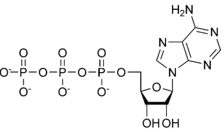
One of the best-known small molecules is adenosine triphosphate (ATP) (Figure 2.1), which stores readily available chemical energy in two of its phosphate chemical bonds. When cells split apart these energy-rich bonds in ATP, the released energy can be harvessed to power an energy-requiring process like muscle contraction or protein biosynthesis. To obtain energy for making ATP, cells break down food molecules. For instance, when sugar is degraded to carbon dioxide and water, the energy stored in the original chemical bonds is released and much of it can be ”captured” in ATP. Bacterial, plant, and animal cells can all make ATP by this process. In addition, plants and a few other organisms can harvest energy from sunlight to form ATP using photosynthesis.
2. BACKGROUND
Figure 2.1: representation of an ATP molecule
Other small molecules act as signals both within and between cells. Such signals direct numerous cellular activities. For example, The powerful effect on our bodies of a frightening event comes from the instantaneous flooding of the body with epinephrine, a small-molecule hormone that mobilizes the ”fight or flight” response. The movements needed to fight or flee are triggered by nerve impulses that flow from the brain to our muscles with the aid of neuro-transmitters.
Certain small molecules (monomers) in the cellular soup can be joined to form polymers through repetition of a single type of chemical-linkage reaction. Cells produce three types of large polymers, commonly called macromolecules: polysaccharides, proteins, and nucleic acids. Sugars, for example, are the monomers used to form polysaccharides. These macro-molecules are critical structural components of plant cell walls and insect skeletons. A typical polysaccharide is a linear or branched chain of repeating identical sugar units. Such a chain carries information: the number of units. However, if the units are not identical, then the or-der and type of units carry additional information. Some polysaccharides exhibit the greater informational complexity associated with a linear code made up of different units assembled in a particular order. This property, however, is most typical of the two other types of biological macromolecules: proteins and nucleic acids.
2.4 Interactions between proteins and small molecules
2.4 Interactions between proteins and small molecules
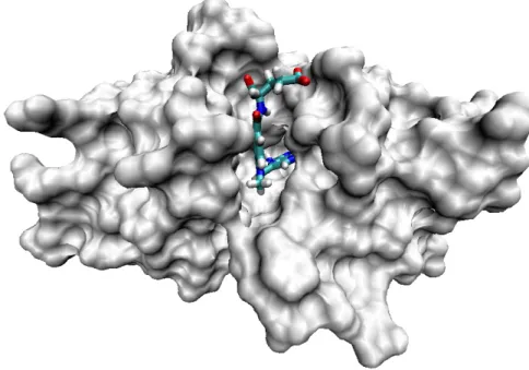
A ligand can be defined as a molecule binding to a biological receptor, which is most often a protein (Figure 2.2). This binding generally triggers an effect: modulation of enzyme activity if the receptor is an enzyme, cellular response in the case of a membrane receptor, a cytoplasmic receptor or a nuclear receptor of a cell. Ligands include all small molecules of low molecular weight, such as a metabolite, peptide, substrate, inhibitor or a small drug molecule and excludes macromolecules such as proteins, lipids or nucleic acids sequences such as DNA and RNA, which we would rather call biological partners.
Figure 2.2: 3D structure of a protein (DHFR) with its ligand (methotrexate)
Ligand binding to the protein occurs by intermolecular forces such as ionic bonds, hydro-gen bonds and Van der Waals forces, and is usually reversible.
The interaction between proteins and small molecules is related to the presence, in the protein structure, of a specific site called the active site. Broadly, it has the shape of a cavity into which the substrates bond. Once bound, small molecules will react and turn into a product in the case of an enzyme, or cause the activation or inhibition of a signaling pathway in the case of a receptor.
2. BACKGROUND
The first model of ligands binding to proteins was the ”lock and key” hypothesis (Figure 2.3). In this model, the affinity of a ligand for a protein is determined by the complementarity between the shapes and the physico-chemical properties of the ligand and of the binding site.
Figure 2.3: The ”lock and key hypothesis”
But this model is not sufficient to describe the interaction. Indeed, proteins and ligands can be very flexible. The model is rather a key capable of deforming the lock when it fits. This phenomenon is called induced fit. The theory suggests that the protein changes its shape to bind the molecule with the proper alignment. The overall effect would be a tighter binding for the molecule and the binding site. Once bound, the molecule intereacts with the protein the same as with the lock and key theory (2).
2.5 Experimental methods to study protein-ligand interactions
The study of protein-ligand interaction is crucial to understand the function of a protein. In fun-damental studies, it allows to describe the biological pathways in which the protein is involved. It is also a key step towards the design of modulator molecules that can interfere (positively or negatively) with the function of a protein target, in the context of drug discovery. Indeed, re-search and discovery of new molecules with activity against proteins has long been the goal of the pharmaceutical industry. There are many experimental techniques to study the interactions between a molecule and a protein. In the following, I will review the most popular methods, distinguishing those that can provide structural information about the complex and the binding mode of the small molecule to the protein, and those that cannot (non structural approaches).
2.5 Experimental methods to study protein-ligand interactions
2.5.1 Non structural approaches
2.5.1.1 Spectroscopic methods (UV, fluorescence)
Spectroscopic methods are often used to highlight molecular interactions. The principle is simple: it is based on the change of the absorbance of a molecule after interaction or rupture of molecular interaction. For example, these methods are often used to monitor the evolution of an enzymatic reaction. The simplicity of implementation, the high sensitivity and the low cost in organic materials are the main advantages of these techniques. However, they can be applied only if one of the molecules involved in a complex have spectral properties and if the specific absorbance of the targeted group is affected by the interaction. In some cases, it is possible to attach fluorescent groups to non fluorescent ligands, although this chemical modification might modify the stability of the complex. Many biological assays involving therapeutic targets have been developed that rely on such spectroscopic methods, as reviewed in (3) for fluorescence and (4) for UV spectroscopy. These methods can be used to measure the complex association constant, i.e. to characterize the strength of the interaction.
2.5.1.2 Isothermal Titration Calorimetry (ITC)
ITC is an analysis technique based on the measurement of heat changes induced during a titra-tion. In practice, a macromolecule located in the measuring cell of a calorimeter is gradually saturated at constant temperature by the injection of a ligand using a syringe. For each addi-tion of ligand, there is a thermal exchange that is characteristic of the macromolecule-ligand interactions. The amount of heat measured during the titration allows to obtain thermody-namic parameters of interaction such as free energy changes (∆G), enthalpy (∆H) and en-tropy (∆S). ITC therefore allows to highlight an interaction, to determine the dissociation constant Kd and the stoichiometry of the system. Furthermore, the thermodynamic data de-rived by microcalorimetry allow to know precisely the enthalpic and entropic contributions in the interaction energy: we can thus specify the nature of the forces contributing to the forma-tion of complexes (hydrophobic or electrostatic) (5). ITC can also be useful to study ionizaforma-tion phenomena, or to highlight and quantify a competition between two ligands. A drawback of this technique is its quite low sensitivity, requiring large amounts of proteins (in the milligram range) to study the complex, which can be a strong limitation for proteins available only in small quantities. In addition, the experiments are relatively lengthy and difficult to perform. Howeveer, the ITC technique has been used in drug discovery applications thanks to a new
2. BACKGROUND
miniaturized, ultrasensitive microcalorimeter. This new microcalorimetry system reduces the quantity of protein (or other macromolecule sample) required to obtain a complete thermody-namic profile by up to 7-fold. The reduction in required sample quantities allows ITC to be effectively utilized at earlier stages of the drug discovery and development process (6). 2.5.1.3 Surface Plasmon Resonance (SPR)
The SPR technology is used to study molecular interactions in real time without labeling one of the two interactants. Without going into many details, the device detects changes in mass at the surface of a sensor chip on which one of the two interactants (for example the ligand) is immobilized, covalently or not. The other interactant (for example the protein) is injected through a microfluidic system in a continuous flow of buffer to the surface of the sensor chip. If the protein and the ligand interact and bind to each other, the device detects an apparent variation in mass for the immobilized molecule. Equilibrium binding constants, kinetic rate constants and thermodynamic parameters are obtained from such study that helps to understand the mechanism of the binding reactions. This information can be directly used to improve binding properties of a drug candidate (see (7) for review).
2.5.2 Structural approaches
Structure based drug design is another method for identifying new drugs and seems to be the most rational way of identifying potential agents. In this approach, the three-dimensional structure of a drug target and its interaction with potential drug molecules is used to guide drug discovery. This structural information can be obtained by various methods, such as X-ray crystallography, NMR, and virtual approaches such as computational chemistry. In the next paragraphs, we will shortly review the two former, which rely or experimental data. Then, virtual approaches will be reviewed in more details, because this techniques are related to the work presented in the following chapters of this manuscript.
2.5.2.1 X-ray diffraction
Crystallography uses X-ray diffraction by the electronic cloud of a molecule to deduce the po-sitions of the atoms constituting the compound to be analyzed. In the case of biomolecules, it is possible to crystallize a protein in complex with its ligand, and to deduce the three dimensional structure of the complex. Such a structure allows to discover the active site of the protein, and
2.5 Experimental methods to study protein-ligand interactions to assess ligand binding modes. This information can be used to conceive optimized molecules that improve their interactions with its target. Crystallography is the technique of choice used in the stages of drug design where an active molecule has been selected and needs to be op-timized to ensure high affinity and selectivity. For instance, the development of successful HIV-1 protease (see (8) for review), reverse transcriptase (see (9) for review), or integrase (see (10) for review) inhibitors was achieved through structure-based drug design using the crystal structures of the corresponding enzymes. In the field of antibiotics, the translational apparatus of the bacterial cell remains one of the principal targets of antibiotics for the clinical treatment of infection worldwide. The high-resolution crystal structures of the bacterial ribosome identi-fying the sites of antibiotic binding are now available, which is central to progress in this area. Experimental assays, coupled with structural studies, have the potential not only to accelerate the discovery of novel and effective antimicrobial agents, but also to refine our understanding of the mechanisms of translation (see (11) for review).
2.5.2.2 Nuclear magnetic resonance (NMR)
Nuclear magnetic resonance is a spectroscopic technique based on the interaction between a magnetic field and the spins of atomic nuclei. The study of protein / ligand interactions at the atomic level by NMR has been made possible through the development of experiments based on observation of the resonance signals of the protein or of the ligand, in presence of each other (12; 13). NMR can provide much information to characterize the interaction between a protein and a ligand. It allows to identify the site of interaction in the protein, i.e. the aminoacids that are in contact with the ligand. It can also provide information about the conformation of the ligand in the active site. NMR can play a critical role in structure determination of many important protein targets such as GPCRs, when they fail to form the single crystals required for X-ray diffraction. NMR can provide valuable dynamic information on proteins and their drug complexes that cannot be easily obtained with X-ray crystallography. These advances suggest that the future discovery and design of drugs might increasingly rely on protocols using NMR approaches (14). However, this technique consumes large amounts of biological material, milligrams per analysis, because of its low sensitivity. In addition, analysis of the spectra is easier in the fast exchange regime, which corresponds to dissociation constant of the protein-ligand complex in the range of millimolar or micromolar, which is not the required range in the context of drug discovery.
2. BACKGROUND
2.6 Experimental high throughput screening (HTS)
In many cases, the search of new drug candidates consists in the identification of molecules that strongly and specifically bind to a therapeutic protein target. Therefore, all methods allowing detection and characterization of protein-ligand interactions are of interest in the context of drug research. Some of the above described experimental methods can be used in screening tests on large scale, using robots, leading to the so called high throughput screening (HTS) approaches (15). In the following, I will briefly review HTS.
The screening of chemical compounds for pharmacological activity has been ongoing in various forms for at least 20 years. The screening paradigm says that when a compound inter-acts with a target in a productive way, that compound then passes the first milestone on the way to becoming a drug. Compounds that fail this initial screen go back into the library, perhaps to be screened later against other targets.
Screening methodologies have improved with time, both in terms of throughput and the amount of information to be derived from the screen. Advances in assay and instrument tech-nologies have provided the means necessary to address these evolving needs.
Using robotics, data processing and control software, liquid handling devices, and sensitive detectors, HTS allows a researcher to conduct biochemical or pharmacological tests. Through this process, one can rapidly identify active compounds which modulate a particular biomolec-ular pathway. The results of these experiments provide starting points for drug design and for understanding the interaction or the role of a particular biochemical process in biology.
Many pharmaceutical companies are screening 100,000 to 300,000 or more compounds per screen to produce approximately 100 to 300 hits. On average, one or two of these become lead compound series. Larger screens of up to 1,000,000 compounds in several months may be required to generate something closer to five leads. Improvements in lead generation can also come from optimizing library diversity. Since its first advent in the early to mid 1990s, the field of HTS has seen not only a continuous change in technology and processes, but also an adapta-tion to various needs in lead discovery. HTS has now evolved into a mature discipline that is a crucial source of chemical starting points for drug discovery. Whereas in previous years much emphasis has been put on a steady increase in screening capacity (’quantitative increase’) via automation and miniaturization, the past years have seen a much greater emphasis on content and quality (’qualitative increase’). Today, many experts in the field see HTS at a crossroad with the need to decide on either higher throughput/more experimentation or a greater focus
2.6 Experimental high throughput screening (HTS) on assays of greater physiological relevance, both of which may lead to higher productivity in pharmaceutical R&D. There will be much more emphasis on rigorous assay and chemical characterization, particularly considering that novel and more difficult target classes will be pursued. In recent years, we have witnessed a clear trend in the drug discovery community toward rigorous hit validation by the use of orthogonal readout technologies, label free and biophysical methodologies. We also see a trend toward the use of focused screening and itera-tive screening approaches. Hit finding strategy also tends to be much more project-related and better integrated into the broader drug discovery efforts. Recently, fragment-based methods have emerged as a new strategy for drug discovery (16). The main advantages are that useful starting points for lead identification for most targets can be identified from a relatively small (typically 1000-member) library of low molecular weight compounds. The main constraints are the need for a method that can reliably detect weak binding and strategies for evolving the fragments into larger lead compounds. The approach has been validated recently, as series of compounds from various programs have entered clinical trials.
However, HTS workflow is hampered by several drawbacks. One can only sample a tiny proportion of the drug-like chemical space. The screening procedure relies on expen-sive robotic equipment, and although many progresses in miniaturization have been made that allow reduction of the experimental volumes, the tests require to consume expensive biological and chemical consumables. The rates of false-positive and false-negative are relatively high owing to the contribution of various factors such as nonspecific hydrophobic binding, poor sol-ubility leading to protein or substrate precipitation, aggregation, presence of reactive functional groups, low purity, incorrect structural assignment or compound concentration, interference of the compounds with the assay or its read-out etc... (17).
In parallel of the HTS approaches, virtual screening strategies have developed rapidly both in academic and industrial research. In particular in silico methods remain an attractive option for prioritizing structures for focussed screening (18). However, it may also be interesting to perform experimental and virtual screens in parallel, on the same chemical databanks, since comparative studies have shown that these two approaches can lead to identification of differ-ent active compounds (19). Today, most pharmaceutical companies have substantial groups devoted to virtual screening approaches (20). In the following, I will briefly review the main topics in the domain of virtual screening.
2. BACKGROUND
2.7 Virtual screening
The main aim of these approaches is to quickly and cheaply select a restricted number of molecules expected to bind to the protein target, from large chemical libraries. This smaller set of molecules (typically 5 percent of the original chemical library) are then experimentally tested. Many methods are available, with different ranges of applications, but they can be classified into two classes: structure-based approaches, and ligand-based approaches. In both cases, they require the choice of a molecule library that will be used in the virtual screen.
2.7.1 The molecule library
A molecule library is a set of molecules, whose sizes may vary from a few hundreds to hun-dreds of thousands of grams per mol, that have been synthesized in large quantities and stored in order to be rapidly available for large scale screening. Chemical libraries can be confi-dential, like those owns by pharmaceutical industries, composed of synthesized or extracted natural molecules. They can also be commercial libraries. Chemdiv, Chembridge or Asinex are examples of some of the most commonly used molecule libraries. The choice of a molecule library is critical in a virtual screen (also in an experimental screen). The content, design and scale of a molecule library needs to be directly related to the purpose of the screen. Because the goal of the screening evolves during the drug discovery process, the design of the libraries to be screened must be related to the advancement of the project. In order to maximize the probability of identifying structurally different hits, early screening assays must involve di-verse libraries giving a broad coverage of the chemical space (21). On the contrary, in the later ”hits to lead” step, targeted libraries made of molecules structurally similar to the identi-fied hits must be designed (22). Because of the development of virtual approaches, molecular libraries are now also provided as virtual libraries, in which molecules are represented in differ-ent formats such as smiles, sdf or mol2, and that encode for their chemical structures. Virtual screening methods will use these molecular descriptions, or other descriptions derived from these standard formats, in order to encode molecules and to manipulate them through various algorithms.
2.7.2 Structure-based methods
Structure-based methods, also called docking, cover a range of approaches that exploit the 3D structure of the protein of interest, to predict its potential ligands. The growing numbers of
2.7 Virtual screening genomic targets of therapeutic interest (23) and macromolecules (proteins, nucleic acids) for which a three-dimensional structure (3D) is available (24) makes docking increasingly attrac-tive for the identification of bioacattrac-tive molecules (25; 26).
The role of molecular docking is to predict the active conformation and relative orientation of each molecule of the chemical library within the binding site of the protein of interest. In other words, docking tries to propose a model for the protein-ligand complex, and to evaluate the stability of this complex. Very generally, the search of possible positions of the ligand in the protein structure focuses on a defined protein pocket that has been experimentally de-termined (for example by directed mutagenesis). These methods use the principle of steric complementarity (Dock, Fred) or of molecular interactions (AutoDock, FlexX, Glide, Gold, ICM, LigandFit, Surflex), to place a ligand in the protein pocket. In most cases, the protein is considered as rigid, although some programs handle flexibility for aminoacid side chains or from small local rearrangements of the backbone. In contrast, ligand flexibility is fully taken into account. Three principles are generally used for the treatment of the flexibility of the ligand:
• a set of conformations of the ligand is calculated beforehand and they are docked to the rigid way the site (eg Fred),
• the ligand is incrementally constructed fragment after fragmentation (eg Dock, FlexX, Glide, Surflex)
• A more or less complete conformational analysis is conducted on the ligand to generate conformations that are most favorable to docking. (eg ICM, Gold, LigandFit).
Typically, several poses for the ligand are generated and ranked by decreasing probability according to a scoring function that tries to estimate the protein-ligand interaction energy. In a docking screen against a protein target, all molecules of a chemical library will be ranked according to their scores, and the 5-10 % molecules of the initial set with the best scores will be viewed as the best molecules for experimental evaluation. In other words, docking can be used as a tool to reduce the size of the molecule library to be experimentally screened, based on the assumption that the best ranked molecules are enriched in true ligands. Docking is an active field of research, because although many programs are available today, there is still a need to improve the methods used to take protein and/or ligand flexibility into account, or to improve the scoring functions. Today, no program has been identified as ”the best” program, and one needs to evaluate the best docking conditions for each project in an ”ad hoc” manner.
2. BACKGROUND
2.7.3 Ligand-based approaches
Ligand-based methods exploit prior knowledge of ligands, and non ligands, for a protein of interest, to predict new or better ligands for this protein. Therefore, they cannot be used in early studies, when no ligands have been identified. However, these methods are quite powerful in later stages of drug development, when optimization of lead compounds is searched. Most ligand-based screening methods rely on the comparison of molecules, with the underlying assumption that similar molecules will have similar behaviors: a molecule that is similar to a known ligand will be predicted to bind to the protein target, which is not expected for random compounds. However, the comparison of molecules is not trivial: it relies on how molecules are encoded, and on the method used to measure the similarity. A large variety of tools have been developed to perform these tasks.
2.7.3.1 Descriptors
Descriptors can be used to encode molecules. They are usually calculated from the molec-ular structure (atoms, bonds, configuration, conformation) or molecmolec-ular properties (physical, chemical, biological) (27; 28). The descriptors may include atoms and bonds, or the presence or absence of fragments, or other 1D or 2D features. Descriptors may also relate to the 3D arrangement of atoms, when 3D conformation information is taken into account. Ideally, the descriptors should be readily calculable and easily interpretable by computers and by users. They should represent the actual chemical system and take the structure of chemical space into account (29).
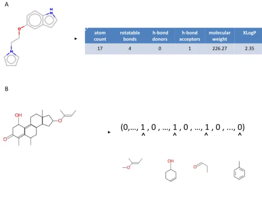
1D descriptors are derived from the empirical formula of the molecule (eg C6H6O for phe-nol). They correspond typically to global molecular properties, such as the molecular weight, hydrophobicity, or general physicochemical properties of the molecule, such as the number of atoms of particular types or hydrogen bond donors and acceptors, solubility (logP). These de-scriptors bear little information about the structure of the molecule and are essentially used to derive filters such as the Lipinski’s rule of five (30), or in combination with other descriptors. It must be noted that these descriptors cannot usually distinguish isomers. The 1D encoding of molecules often take the form of a vector whose elements correspond to these molecular properties used as descriptors (Figure 2.4A). In such vectorial description, similarity between molecules can then be measured according to a Tanimoto coefficient calculated from the ele-ments of these two vectors.
2.7 Virtual screening The Tanimoto coefficient is defined as the ratio :
T (A, B) = �M i=1A(i)B(i) �M i=1A(i) + �M i=1B(i)− �M i=1A(i)B(i)
with A (i) and B (i) are equal to 1 if the i-th descriptor is found in molecule A and B respectively, and 0 if it is absent. M is the total number of descriptors taken into consideration. 2D descriptors are derived from the 2D structure of the molecule, which can also be viewed as a graph. A first class of 2D descriptors consists of general topological indices, related to the notion of graph invariant in graph theory. Seminal examples include the Wiener and Randic connectivity indices, defined respectively from the length of the shortest path between pairs of atoms, and their degrees in the molecular graph (31). In a related approach, topological autocorrelation vectors measure the autocorrelation of atomic physicochemical properties, such as partial charges or polarity, from pairs of atoms separated by a given topological distance, expressed as the length of the shortest path connecting atoms in the molecular graph (32).
A second class of descriptors represents a molecule by a vector indexed by a set of structural features, and relies on the extraction of substructures from the molecular graph. This process defines a molecular fingerprint, and in practice, two different approaches can be adopted.
The first approach considers a limited set of informative predefined substructures to char-acterize the molecule. Each substructure is mapped to a bit of the fingerprint, which either accounts for the presence or absence of the substructure in the molecule. A typical implemen-tation is a bistring indexed by 166 predefined substructures known as the MDL MACCS keys (33). This type of encoding is illustrated in (Figure 2.4B). Among the advantages offered by the structural keys is the expressiveness of the features, and the interpretability retained in the representation, because of the one-to-one correspondence between the bins of the vector and the structural features. However, choosing the features to be included in the substructure rep-resentation may be challenging in practice. While chemical intuition can be helpful for that purpose (34), this task is more generally related to the problem of graph mining that consists in the automatic identification of interesting structural features within a set of graphs. For chem-ical applications, such interesting patterns are typchem-ically defined as non correlated structures frequently appearing in active compounds, and rarely in inactive compounds (35; 36; 37).
In the alternative approach, molecules are represented by simple structural features called linear molecular fragments, defined as successions of covalently bonded atoms. In this case, typical fingerprints, such as the Daylight fingerprints, characterize a molecule by its exhaustive list of fragments made of up to seven or eight atoms.
2. BACKGROUND
In summary, 2D descriptors are calculated from the 2D formula of the molecule, and they provide information about its size, its overall shape and its ramifications. In the case classical of 1D or 2D descriptors encoded in vector representation of molecules, an explicit ”chemical space” is defined in which each molecule is represented by a finite-dimensional vector. These vector representations can be used as such to define similarity measures between molecules such as Tanimoto coefficients.
!"#$#%&%#%"%#%$#%&%#%"%#%$#%&%#%"%#%'''#%"(%%
)%
!"#$%
&#'("% )#"!"!*+,%*#(-.% %/0*#(-%-#(#).% !&&,1"#).%/0*#(-% $#+,&'+!)%2,34/"% 56#47%
&*% +% "% &% ,,-',*% ,'./%
0%
Figure 2.4: Example of descriptors that may be used to encode molecules. (A) Example of 1D descriptors based on physicochemical properties. (B) example of 2D descriptors encoding for presence or absence of predefined chemical fragments.
3D descriptors are derived from the 3D structure of the molecules. A first class of three dimensional descriptors requires a preliminary step of molecular alignment, consisting in plac-ing the molecules in a common orientation in the 3D space through operations of rotations and translations. The quality of the alignment is quantified by a scoring function, and the molecules are said to be aligned when it is maximized. Typical scoring functions consider the number of
2.7 Virtual screening identical atoms superimposed under a skeleton representation (38), or the overlap of the elec-tron clouds surrounding the molecules (39). In order to handle conformational analysis, the alignment can be flexible, in which case additional degrees of freedom are introduced to han-dle rotational bonds, or rigid and based on the optimal alignment of pairs of multi-conformers. Aligning molecules can be a quite complex process, and we refer to Lemmen and Lengauer (40) for a review of the existing techniques. Once the molecules are aligned, 3D descriptors can for instance be defined by sampling molecular surfaces according to rays emanating from the center of mass of the aligned molecules (41; 42), or, in the Comparative Molecular Field Analysis (CoMFA) methodology, by measuring the interaction between the molecules and an atomic probe (e.g., a charged or lipophilic atom) at each point of a discrete box enclosing the molecules (43)
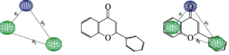
An opposite approach consists in extracting descriptors independent of the molecular orien-tation. Apart from global shape descriptors, such as the Van der Waals volume of the molecule or molecular surfaces areas, most alignment independent descriptors are based on distances between atoms. For example, an early study proposed to characterize a molecule by its matrix of inter-atomic distances (44). While the authors propose several methods to compare such matrices, this approach is not convenient because it does not lead to a fixed size representa-tion of the molecules. Standard vectorial representarepresenta-tions can be derived by considering pairs of atoms of the molecule. Topological autocorrelation vectors can for instance be extended to 3D autocorrelation vectors, computing the autocorrelation of atomic properties from pairs of atoms within a specified Euclidean distance range, instead of a given topological distance on the molecular graph (45). Other representations are based on counting the number of times pairs of atoms of particular types are found within predefined distance ranges in the 3D struc-ture of the molecule (46; 47; 48). Considering molecular feastruc-tures based on triplets or larger sets of atoms leads to the notion of pharmacophore. A pharmacophore is usually defined as a three-dimensional arrangement of atoms - or groups of atoms - responsible for the biological activity of a drug molecule (49). Typical pharmacophoric features of interest are atoms hav-ing particular properties (e.g., positive and negative charges or high hydrophobicity), hydrogen donors and acceptors and aromatic rings centroids (50). In this context, pharmacophore finger-prints were proposed as the three-dimensional counterpart of molecular fragment fingerfinger-prints. Pharmacophore fingerprints represent a molecule by a bitstring encoding its pharmacophoric content, usually defined as the exhaustive list of triplets of pharmacophoric features found
2. BACKGROUND
within a set of predefined distances ranges in its 3D structure (51; 52). Strictly speaking, phar-macophore fingerprints encode putative pharphar-macophores of the molecules, and because the number of potential pharmacophores can be very large, they are usually compressed (53; 54). In chapter 3, we will present and use 2D and 3D pharmacophore representations for molecules that were developed in our laboratory (55) and that we used in this thesis in a chemogenomics framework.
A vast amount of descriptors has therefore been proposed in the literature. The above pre-sentation if far from being exhaustive, and we refer interested readers to the textbooks for a detailed presentation (31; 56). Choosing ”good” descriptors for the task to be performed re-mains nevertheless an open question. For instance, even though the molecular mechanisms responsible for the binding of a ligand to a target are known to strongly depend on their 3D complementarity, different studies account for the superiority of 2D fingerprints over pharma-cophore fingerprints in this context (34; 52; 55). This observation suggests that 2D fingerprints might encode to some extent three-dimensional information (27), and in many cases, they actu-ally constitute the ”gold-standard” representation of chemical structures. Another explanation is that 3D approaches require to know the 3D geometry of the molecule in its ”active” confor-mation, which is not always available. In such cases, the choice of other conformations such as the most free-state conformation might degrade the performance of 3D approaches.
2.7.3.2 Principle of ligand-based approaches
Many ”rational” drug design efforts are based on a principle which states that structurally similar compounds are more likely to exhibit similar properties. Indeed, the observation that common substructural fragments lead to similar biological activities can be quantified from database analysis. A variety of methods, known collectively as Quantitative Structure Activity Relationship (QSAR) have been developed, essentially for the search for similarities between molecules in large databases of existing molecules whose properties are known. The discovery of such a relationship allow to predict the physical and chemical properties of biologically active compounds, and to develop new theories or to understand the phenomena observed. Once a QSAR model has been built to encode this relationship between the chemical space and a given biological activity, this can guide the synthesis of new molecules, limiting the number of compounds to synthesize and test.
The relationship between the structures of molecules and their properties or activities are usually established using methods of statistical learning. The usual techniques are based on
2.7 Virtual screening the characterization of molecules through a set of 1D, 2D or 3D descriptors. A model is estab-lished, that relates the descriptors that encode the molecule, to its biological activity, based on a learning dataset of molecules for which this activity is known. It is then possible to use this model to predict the activity of a new molecule. Numerous studies show that it is impossible to predict accurately the affinity of chemically diverse ligands (57). It is reasonable to hope to discriminate affinity of ligands in the range of nanomolar, micromolar and millimolar.
Decades of research in the fields of statistics and machine learning have provided a pro-fusion of methods for that purpose. Their detailed presentation is far beyond the scope of this section, and we invite interested readers to refer to the classical textbooks (58; 59) for a thorough introduction. In this section we just give general methodological and historical considerations about their application in chemoinformatics.
Models can be grouped into two main categories depending on the nature of the property to be predicted. Models predicting quantitative properties, such as for instance the degree of binding to a target, are known as regression models. On the other hand, classification models predict qualitative properties. In SAR analysis, most of the properties considered are in essence quantitative, but the prediction problem is often cast into the binary classification framework by the introduction of a threshold above which the molecules are said to be globally active, and under which globally inactive. In the following, the term classification implicitly stands for such binary classification.
In order to build the model, the pool of molecules with known activity is usually split into a training set and a test set. The training set is used to learn the model. The learning problem consists in constructing a model that is able to predict the biological property on the molecules of the training set, but without over-learning on it. This overfitting phenomenon can for instance be controlled using cross-validation techniques, that quantify the ability of the model to predict a subset of the training set that was left out during the learning phase. The test set is used to evaluate the generalization properties of the learned model, corresponding to its ability to make correct prediction on a set of unseen molecules. Different criteria can be used for this evaluation. In regression, it is typically quantified by the correlation between the predicted and the true activity values. In the classification framework, a standard criterion is the accuracy of the classifier, expressed as the fraction of correctly classified compounds. However, if one of the two classes is over-represented in the training set, and/or the cost of misclassification are different, it might be safer to consider the true and false positive and negative rates of classification. The true positive (resp. negative) rate account for the fraction of compounds
2. BACKGROUND
of the positive (resp. negative) class that are correctly predicted, and the false positive (resp. negative) rate accounts for the fraction of compounds of the negative (resp. positive) class that are misclassified. In virtual screening applications for instance, where we typically do not want to misclassify a potentially active compound, models with low false negative rates are favored, even it they come at the expense of an increased false positive rate.
Because they usually require a limited set of uncorrelated variables as input, applying these models to chemoinformatics requires to summarize the information about the molecules into a limited set of features, which may not a trivial task due to the vast amount of possible molec-ular descriptors. A popmolec-ular way to address this problem in chemoinformatics is to rely on principal component analysis (PCA), that defines a limited set of uncorrelated variables from linear combinations of the initial pool of features, in a way to account for most of their in-formative content. Alternatively, feature selection methods can be used to identify among an initial pool of features a subset of features relevant with the property to be predicted. Because molecular descriptors are sometimes costly to define, a potential advantage of feature selection methods, over PCA-based approaches, is the fact that they reduce the number of descriptors to be computed for the prediction of new compounds.
Let us now introduce different methods that have been applied to model SAR. The first SAR model was developed in 1964 by Hansch and coworkers who applied a multiple linear regres-sion (MLR) analysis to correlate the biological activity of a molecule with a pair of descriptors related to its electronic structure and hydrophobicity (60). MLR models are still widely ap-plied to model SAR. PCA is commonly used as inputs, in the so-called PC- regression models (61). Moreover, genetic algorithms have been introduced to perform feature selection as an alternative to standard forward selection or backward elimination approaches (62). Related linear approaches can be applied to the classification framework with discriminant analysis al-gorithms (63). However, because this class of models is limited to encode linear relationships, they can be too restrictive to efficiently predict biological properties. While the models can be enriched with the application of nonlinear transformations of the input variables (64), SAR analysis greatly benefited from the development of nonlinear methods, and in particular artifi-cial neural networks (ANN). Early applications of back-propagation ANN accounted for their predictive superiority over standard linear regression techniques. Many studies have demon-strated the strength of ANN to predict biological properties, and they are now a standard tool to model SAR (65; 66).
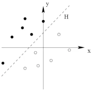
2.7 Virtual screening Despite their predictive efficiency, a major criticism to ANN is their lack of interpretability, which can be of great importance in chemistry in order to understand the biological mecha-nisms responsible for the activity. An alternative class of models builds a classifier expressed as a set of rules relating the molecular structure and the biological activity. Such models have been derived for instance using decision trees algorithms (67). From the practical viewpoint, another criticism that can be made to ANN is the fact that they require some expertise, con-cerning for instance the choice of an architecture, in order to be knowledgeably deployed. Moreover, they are known to be prone to overfitting and are hard to reproduce, because of their random initialization and possible convergence to local minima (68). These theoretical issues are to some extent addressed by the support vector machine (SVM) algorithm, known in particular to avoid the problem of local minima, to prevent overfitting, and to offer a better control of the generalization error (69). Moreover, although its good parametrization remains a crucial point, this algorithm requires less amount of expertise to be deployed. The introduction of SVM in SAR analysis was pioneered by Burbidge and co-workers (68). In this study, the SVM algorithm outperforms several ANN architectures for a particular classification task, re-lated to the ability of molecules to inhibit a biological target. Over the last few years, SVM was shown to be a powerful tool for SAR analysis, often outperforming ANN in classification and regression frameworks. We give in the next section a brief introduction to the SVM algorithm, because we used this algorithm in the chemogenomic approach presented in chapter 3.
2.7.4 Introduction to SVM in virtual screening
One important contribution of this thesis is to explore the use of machine learning algorithms within the newly introduced chemogenomic framework, in order to predict protein-ligand inter-actions. The principle of chemogenomic approaches will be presented in chapter 3. Although this principle is quite simple (i.e. similar proteins are expected to bind similar ligands), to our knowledge, only a very limited number of studies propose computational methods able to han-dle chemogenomic data and to perform predictions. The main reasons are that these data are not trivial to generate, to manipulate, and to be used as input in computational methods in a relevant manner in order to make predictions.
We have proposed to use kernel methods in the the context of Support Vector Machine (SVM) methods, because, as it will be explained in chapter 3, they allow easy manipulation and calculation in the chemogenomic space (i.e. the chemical space of small molecules joined to the biological space of proteins). Presenting the full mathematical framework of SVM and