UNIVERSITÉ DU QUÉBEC À MONTRÉAL
APPLICATION OF GLYCODENDRIMERS AS ANTI-ADHERENCE INHIBITORS AGAINST PSEUDOMONAS AERUGINOSA IN CYSTIC
FIBROSIS
THE SIS PRESENTED
AS PARTIAL REQUIREMENT OF THE MASTER IN CHEMISTRY
BY
SHADY (ASAL) SHOJAEEASL
APPLICATION DE GL YCODENDRIMÈRES COMME INHIBITEURS ANTI -ADHÉRENTS CONTRE LE PSEUDOMONAS AERUGINOSA POUR LA
FIBROSE KYSTIQUE
MÉMOIRE PRÉSENTÉ
COMME EXIGENCE PARTIELLE DE LA MAITRISE EN CHIMIE
PAR
SHADY (ASAL) SHOJAEEASL
UNIVERSITÉ DU QUÉBEC À MONTRÉAL Service des bibliothèques
Avertissement
La diffusion de ce mémoire se fait dans le respect des droits de son auteur, qui a signé le formulaire Autorisation de reproduire et de diffuser un travail de recherche de cycles supérieurs (SDU-522 - Rév.1 0-2015). Cette autorisation stipule que «conformément à . l'article 11 du Règlement no 8 des études de cycles supérieurs, [l'auteur] concède à l'Université du Québec à Montréal une licence non exclusive d'utilisation et de publication de la totalité ou d'une partie importante de [son] travail de recherche pour des fins pédagogiques et non commerciales. Plus précisément, [l'auteur] autorise l'Université du Québec à Montréal à reproduire, diffuser, prêter, distribuer ou vendre des copies de [son] travail de recherche à des fins non commerciales sur quelque support que ce soit, y compris l'Internet. Cette licence et cette autorisation n'entraînent pas une renonciation de [la] part [de l'auteur] à [ses] droits moraux ni à [ses] droits de propriété intellectuelle. Sauf entente contraire, [l'auteur] conserve la liberté de diffuser et de commercialiser ou non ce travail dont [il] possède un exemplaire.»
Je suis reconnaissante à Dieu pour toutes ses bénédictions, le pouvoir qu'il m'a donné afin de surmonter des épreuves et pour toutes les qualifications qu'il m'a contribuées afin de réaliser mes projets. En particulier, dans un pay~ étranger qui est devenu ma deuxiètne patrie.
Maintenant, à ce stade crucial de ma vie, j'ai encore une fois besoin de 1' assistance de Dieu, ainsi que celle de quelques autres personnes remarquables qui n'ontjamais refusé de m'aider tout au long de ma vie. Il est inutile de dire à quel point la vie d'une fille dépendante de la famille peut être difficile à l'étranger. Cependant, j'ai réussi à surmonter et à tolérer les difficultés pour réussir dans mes études. En partie, grâce au Pr René Roy dont les conseils et assistance ont contribué à mon succès dans ce programme de recherche.
Je tiens également à exprimer mes remerciements aux docteurs Rabi Rej et Rahul Bagul pour leurs conseils et leur aide au cours de toutes ces années passées à l'université.
Les mots n'expriment pas mes étnotions et mes sentiments profonds pour ma famille pour leur amour et leur soutien tout au long de ma vie. Je remercie, tout particulièrement mon père qui m'a toujours motivée pour poursuivre mes études; ma sœur aînée, Shilla, dont l'affection a toujours réchauffé mon cœur, ainsi que ma sœur cadette, Dr Pouneh Shojaee Asl, et son mari, Dr Amir Alaie, qui m'ont toujours inspirée. C'est le soutien émotionnel et financier de ma famille qui m'a permis de surmonter les problèmes et d'atteindre mes objectifs.
lV Enfin, je remercie ma mère, la personne la plus chère et la plus belle de ma vie. Diplômée en chimie de l'Université de Téhéran en 1963, elle était sans aucun doute 1na meilleure professeure et conseillère qui m'a aidée à ouvrir la voie à un avenir brillant. Depuis toujours, je rêvais d'aller très loin dans 1nes études comme ma chère sœur. Je dédie ma thèse à ma mère pour exprimer ma sincère gratitude et mon affection pour son amour maternel inestimable.
Je su1s également reconnaissante à mon professeur respectueux, le directeur de Pharmaqam, Dr. Steve Bourgault, et également Dr. Sylvain Canesi, ainsi qu'à tous les membres du personnel de Pharmaqam pour leurs soutiens exceptionnels.
Je souhaite bonne santé, bonheur, paix et prospérité à mes chers professeurs et à ma très chère famille.
Mes reconnaissances les plus sincères,
In the nan1e of God
I am grateful to God for all the blessings and kindness, for all the power given to 1ne to endure hardships in a foreign country which has now become my second homeland, and for all the qualifications which have contributed to my achievements. Now at this crucial stage of life, I once again need special support and concern from God and also sorne other conspicuous people who have never failed to assist me through my life. It is needless to say how difficult life could be for a family-dependent girl abroad; however, I have managed to overcome, endure these difficulties and succeed. I want to thank Pr René Roy for his assistance contributed to my success in this research pro gram.
I shall also express my gratitude to Dr. Rabi Rej and Dr. Rahul Bagul for their kind concern, advice, and assistance through all these years at university.
To appreciate my beloved family, words fail to express my deep emotions and feelings for them and their life-long love and support. I, hereby, thank my father who has al ways motivated n1e to continue my education; my older sister, Shilla, whose sisterly affections has always warmed my heart. My other sister, Dr. Pouneh Shojaee Asl, and her husband, Dr. Amir Alaie, have always inspired me in my life. It was my family's emotional and financial support that enabled me to overcome problems and to reach my goals.
Vl
Finally, 1 thank my mother, the dearest, loveliest person in my life. She was a Chemistry graduate fron1 Tehran University in 1963. She tu1doubtedly was my best teacher and advisor to recognize my talents and paved my way to a bright future. lt has al ways been her life-long dream for me to reach high levels of education like my dear sister. 1 dedicate my thesis to my mother to show my sincere gratitude and affection for her inestimable maternallove.
1 am also grateful to my highly-respected professor and Director of Pharmaqam, Dr. Steve Bourgault and to my other respected professor, Dr. Sylvain Canesi, and to all the staff members in Pharmaqam for their kind concerns and high sense of cooperation. 1 do wish good health, happiness, peace, and prosperity for my dear professors and beloved family.
My sincere regards to all, Shady As al Shoj aee Asl
FIGURE LIST ... x
SCHEME LIST ... xvi
TABLE LIST ... xvii ABBREVIATION AND ACRONYMS LIST ... xviii
RÉSUMÉ ... xxi
ABSTRACT ... 1
CHAPTER I introduction to dendrüners ... 3
1.1 Introduction to dendrimers ... 3
1.1.1 Dendrimers ... 3
1.1.2 Dendrimer anatomy ... 4
1.1.3 Applications ... 5
1.1.4 Dendrimers as n1ultivalent ligands and inhibitors against bacterial biofiln1 formatio11 ... 8
1.2 Strategies for den drim er synthesis ... 9
1.2.1 The divergent method ... 9
1.2.2 The convergent method ... 10
1.2.3 Onion-peel n1ethod ... 10
1.3 "Click chemistry" ... 12
1.4 Glycodendrimers and their role aas inhibitor ... 14
1.5 Aromatic scaffold for glycodendrin1ers ... 15
CHAPTER II Glycobiology and cystic fibrosis ... 20
2.1 Carbohydrate-protein interactions ... 20
Vlll
2.2 Lectin-Carbohydrate bonding ... 21
2.2.1 Hydrogen-bond as noncovalent interactions ... 21
2.2.2 Hydrophobie interaction ... 22
2.2.3 Ionie interactions ... 23
2.2.4 Metal interactio11 ... 24
2.2.5 Role of water ... 25
2.3 Galactose-PA-IL complex interaction ... 26
2.4 Cystic fibrosis and the role of Pseudomonas aeruginosa lectin A ... 27
2.5 Biofilm formation ... 28
2.6 Structures and roles of Pseudomonas aeruginosa lectins ... 29
2. 7 Multivalent interactions via multivalent mechanism~, non-aggregative mechanisms ... · ... 31
2.8 Intra- vs Inter- Molecular cross-linking, aggregative n1echanism ... 33
2.9 Carbohydrate specificity and affinity of the P. aeruginosa lectin A, PA-IL ... 39
2.10 Conclusion ... 41
CHAPTER III Aminooxy role and oxime-ligation ... 43
3.1.1 Aminooxy and Oxime ligation study ... 43
3.1.2 Formation of oxime bonds and their stability ... 44
3.2 Synthetic strate gy ... 46
3 .2.1 Synthesis of three different multivalent glycoclusters in zero generation, Strate gy 1 ... 46
3.2.2 Synthesis of multivalent poly-aldehyde core ... 47
3.2.2.1 Synthesis of the Tri-(4-formacylphenoxy)-1,3,5-triazine (Trif), trivalent core ... 48
3.2.2.2 Synthesis the hexavalent core of hexa-( 4-formylphenoxy)-cyclotriphosphazene ... 51
3.2.2.3 Synthesis the 12-mer aldehyde core using hexachlorocyclotriphosphazene (N3P3Cl6) ... : ... 55
3.2.3 Synthesis of the 2-(aminooxy)ethyl ~-D-galactopyranoside as the monomer ligand 8 ... 59 3.2.3.1 Synthesis of aminooxylated sugar in this study and oxÏlne ligation63
3.3 Synthe sis of higher generation of glycoclusters via "onion peel" strate gy &
oxime-ligation ... 72
3.4 Synthesis of tlnee different multivalent glycoclusters based on the second strategy (Go) ... 82
CHAPTER IV EXPERIMENTAL ... 88
4.1 MATERIALS AND METHODS ... 88
CONCLUSION ... 114
ANNEXE A SPECTRA OF SYNTHESIZED COMPOUNDS ... 116
FIGURE LIST
Figure Page
Figure 1-1. Dendron and dendrimer architecture ... 5
Figure 1-2. Dendrimers applications.1,33 ••••••••.••••••••••••••.•••.••••••.•.••••••••••••••••••••••••••••••••• 7
Figure 1-3. (a) Encapsulation & covalent conjugation for drug delivery on dendrimers (b) Combination drug delivery system on dendrimers: concurrent delivery of water-soluble and -insoluble drugs by adsorption to the surface (ionie interaction), encapsulation within hydrophobie micro cavities inside branching clefts or direct covalent conjugation to the surface functional groups ... 8
Figure 1-4. Dendrimers synthesis strategies: divergent, concergent and onion peel method.41 ...•...••••••.••.•.•.••..•..•...•....•...•.•....••...••.•...•..•.•••..••...•.•.•..•••.•.••••.•....•• 11
Figure 1-5. CuAAC five stages mechanism proposed by Sharpless. 55 ... 14
Figure 1-6. Structure of a glycodendrimer where glycans (mannose, fucose, galactose, etc.) are shown in blue, branching units in black and the core in red (A) Glycan dendron. (B) Glycodendrüner41 .••••••••••.•.••••.•••••••••••••••••..•••••••••••••••••••••••.••••••••..•• 15
Figure 1-8. The n1ultivalent binding of a bacterium or a bacterial toxin to a glycodendri1ner. 87
...•...•...•.•...•..•.•...•... 19
Figure 2-2. (Left) Ionie interactions between sulfated functional group of sulfo-sialic acid with Arg 3 71, Arg 292 and Arg 118 in red dashed lines. (Right) Ionie interactions of sulfo and glycerol moiety in sulfo-sialic acid analogue respectively with Arg 371, Arg 292, Arg 118 and Glu 276 in dashed lines, with stronger electrostatic interaction colored orange. Sulfur atom colored yellow (ref sialic acid).108
••••.••••••••••••••••••••.••••.•••••.•.••••••••..••...•••••••••••••••••••••••••••••.•••••••••••••••••.•••••••••• 24
Figure 2-3. Binding site in the crystal structure of the PA-IL/galactose complex. (Left) Stick model of the amino acids PA-IL coordinated with Ca+2 and in biJ!ding with galactose residue. Ca +2 coordination bond is shown in solid orange line, hydrogen bonds by dashed green line. Col or coding: red for oxygen, blue for nitrogen, black for carbon, pink for calcium. (Right) Electrostatic surface of the protein-binding site related to PA-IL with Ca+2 and galactose. Color coding: from violet for negative to orange for positive of the protein-binding site, large pink sphere for Ca +2 and stick model for galactose residue. lll,ll2 ... 25
Figure 2-4. The initial pro cess of the adhesion schematic description by P. aeruginosa on epithelium cells via galactose/PA-IL complex which leads to tissue damage.44•46 ...•...•...•...•....•... 28
Figure 2-5. Mechanism of bio film formation ... 29
Figure 2-6. (Left) Crystal three-din1ensional view of tetran1eric PA-IL followed by calcium ion on the binding sites of each monomer, Roy. R scheme (Right) The distance measurement between calcium ions in tetrameric PA-IL binding sites
Xll
showing their geon1etrically well- organized structure. (PBD code 1 OK0)42,48,49,lü7 ... 3o Figure 2-7. (left) Chelate aggregative model of tetravalent Calixarene-based in binding
PA-IL from Pseudomonas aeruginosa. (Right) AFM observation of PA-IL filmnents in interaction with the same glycocluster and model of the 1D-network. 73· 122-126 ... 31
Figure 2-8. N onaggregative mechanisms of noncovalent interactions between multivalent ligm1ds and multivalent receptors. 69 Chelate binding model is exceptionally considered as both nonaggregative and aggregative which depends on the binding mode. 115-122 ... 32
Figure 2-9. Multivalency generated cross-linking network of multivalent lectins by multivalent ligands. (a) The long chain polymer due to the interaction of divalent ligand and di valent lectin. (b) The Cross-linked network due to the interaction of tetravalent ligand and di valent lectin. 73 ... 34
Figure 2-10. Glycoclusters are perfectly suited to inhibit bacterial lectins, Roy. R scheme. 122 ... 37
Figure 3-1. Aminooxylated carbohydrates as key building blocks toward therapeutic agents and the various synthetic approach toward sugar-ONH2 derivatives. 171 .. 44
Figure 3-2. The chemistry of C=N double bonds, including aminooxylated spacer. 171 ... 45
Figure 3-3. Relative stabilities of C = N double bonds and the a-effect. 171 ...... 46
Figure 3-5. 1H NMR spectrum (300 MHz, CDC13) of compound 10 and 11. ... 50
Figure 3-6. 13C NMR spectrum (300 MHz, CD Cl) of co1npound 10 and 11 ... 50
Figure 3-7. FT -IR spectrum of compound 11 compared to compound 10 ... 51
Figure 3-8. (a) 31 P NMR spectra (122 MHz) of 6-mer core 14. (b) 31 P-NMR spectra (122 MHz) of the initial N3P3Cl6 13 ... 53
Figure 3-9. 1H NMR spectrum (300 MHz, CDC13) of compound 10 and 14 ... 53
Figure 3-10. 13C NMR spectrum (300 MHz, CDC1 3) of compound 14 and 10 ... 54
Figure 3-11. FT-IR spectrum of compound 10 compare to compound 14 ... 54
Figure 3-12.1 H NMR spectrum (3 00 MHz, CDC13) of compound 19 ... 57
Figure 3-13. FT-IR spectrum of compound 19 ... 57
Figure 3-14. 1H NMR spectrum (300 MHz, DMSO, d6) of compound 20 compare to 1H NMR spectrum (300 MHz, CDC1 3) of compound 19 ... 58
Figure 3-15. (a) 31 P NMR spectra (122 MHz) of 12-mer core 20. (b) 31 P NMR spectra (122 MHz) of the initial N3P3Cl6 13 ... 58
Figure 3-16. Glycosidation mechanism in presence ofneighboring group participation ... 60
XlV
Figure 3-17. Comparison the 1H NMR spectra (CDCb, 300 MHz) of 5 and 7 with appearance of the characteristic signais for aron1atic protons in compound 7 .· .. 62
Figure 3-18. FT-IR spectrum of compound 7 ... 62
Figure 3-19. Comparison of 1H NMR of aminooxylated ligands ... 64
Figure 3-20. 1H NMR (300 MHz, CDCb) between compound 35, 22, and 36 ... 66
Figure 3-21. 13C NMR (300 MHz, CDCb) between compound 35, 22, and 36 ... 67
Figure 3-22. FT-IR spectrum of compound 35 ... 68
Figure 3-23. 1 H NMR (3 00 MHz, CDCb) proton magnetic environment of H y between compound 5 and 22 ... 69
Figure 3-24. FT-IR spectroscopy of compound 22 ... 70
Figure 3-25. FT -IR spectroscopy of compound 36 ... 71
Figure 3-26. 1H NMR (300 MHz, CDCb) of the eliminated product 46 compare to the
linker 44 ... 7 5
Figure 3-27. 1H NMR (300 MHz, CDCb) between compound 24 and 26 ... 77
Figure 3-28.1H NMR (300 MHz, CDCb) of con1pound 27 and 1HNMR (300 MHz,
MeOD) of compound 28 (solubilized better in MeOD than in D20) ... 79
Figure 3-30. HRMS (ESI+) spectru1n of con1pound 30 ... 82
Figure 3-31. Oxime ligation strategy to achieve 3-mer, 6-tner, and 12-mer glycoclusters ... 83
Figure 3-32. 1H NMR (600 MHz, D20) of fraction collected from HPLC for compound 31 ... 85
Figure 3-33. 1H NMR (300 MHz, DMSO) ofremaining residue of compound 33 after dialysis ... ~ ... 86
Figure 3-34. 13P NMR (300 MHz, DMSO) ofremaining residue of compound 33 after dialysis ... 87
SCHEMELIST
Scheme Page
Scheme 3-1. Synthesis of 3-mer aldehyde cluster (Triazine) ustng cyanuric acid
chloride (trichloro triazine) 11 ... 49
Scheme 3-2. Synthesis of 6-mer aldehyde cluster ustng hexachlorocyclotriphosphazene (N3P3Cl6) 14 ... 52
Scheme 3-3. Synthesis of 12-mer aldehyde cluster using (N3P3Cl6) 20 ... 55
Scheme 3-4. Synthesis of an1inooxy building block 8 ... 60
Scheme 3-5. Synthesis of new aminooxylated ligand with longer spacer 37 ... 65
Scheme 3-6. Synthesis of higher generation of glycoclusters via "onion peel" strate gy and oxime-ligation using aminooxylated dendron ... 73
Scheme 3-7. Synthesis of the new monomer of oxime vta oxtme ligation from aminooxy salt ligand ... 76
Scheme 3-8. Synthesis of 3-mer, 6-mer and 12-tner glycoclusters in zero generation ... 84
Table Page
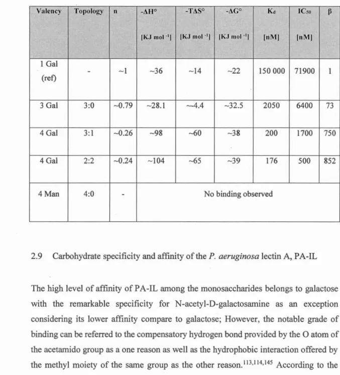
Table 2-1. Microcalorimetry and SPR result related to glycoconjugates of Calixarene fan1ily binding toPA-IL. Monovalent is considered as the reference ... 39
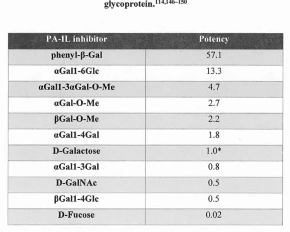
Table 2-2. Carbohydrate specificity of PA-IL. D-Galactose is a reference value of 1.0
for a ICso of 40 nM for inhibition binding of PA-IL to cyst hydatid
glycoprotein.111
Ac Boe DBU DCM DMAP DMF DMSO FCC HPLC NHS NMR Ph th SPE SPR TBDMSCl TFA THF TLC CDCb CM
ABBREVIATION AND ACRONYMS LIST
Acetyl Tert-butoxycarbony 1 1,8-Diazabicyclo[5.4.0]undec-7-ene Dichloromethane 4-(Dimethylamino) pyridine N,N-dimethylformamide Dimethy lsulfoxide
Flash Column Chromathography
High performance liquid chromatography N-Hydroxysuccinimide
Nuclear Magnetic Resonance Phthalimide
Solid Phase Extraction Surface Plasmon Resonace
tert-Butyldimethylsilyl chloride Trifluoroacetic Acid
Tetrahydrofurane
Thin Layer Chromatography Deuterated chloroform Centin1eter
COSY CuAAc Cu Da Equiv.
EhO
EtOAc Et OH FT-IR HSQC Hz ICso L M MALDI-TOF Me OH MeON a Mg MS Nm P-P A-IL PAMAM Rf Correlation SpectroscopyCopper(I)-catalyzed alkyne-azide cycloaddition Copper Dalton Equivalent Diethyl ether Ethyl acetate Ethanol
Fourier-transform Infrared Spectroscopy
Heteronuclear Single Quantum Coherence Spectroscopy Hertz
Isothermal Calorilnetry Litre
Mol ar
Matrix Assistated Laser Desorptionélonisation Time Of Flight Spectroscopy Methanol Sodium methoxide Miligram Mass spectrometry Nanometer Para
Pseudomonas aeruginosa Lectin PolyamidoaJJ?.ine
xx
a Alpha
~ Beta
Ac Acetyl
Ac20 Anhydride acetic
A cO Et Ethly Acetate Asp Ac.Aspartic A sn Asparagine Arg Arginine Phe Phenylalanine Trp Tryptophan Gln Glutamine Thr Threonine Val Valine Tyr Tyrosine His Histidine Pro Praline
oc
Centigrade degree D20 Deuterium oxideDMSO-d6 Deuterated Dimethylsulfoxide MeOD Deuterated 1nethanol
De non1breuses infections débutent par adhésion de 1' agent pathogène sur la paroi cellulaire de l'hôte. Les agents pathogènes, bactériens ou viraux, ciblent les cellules hôtes au moyen d'interactions complén1entaires entre les sucres présents à la surface de la tnembrane cellulaire. Ces sucres liés de manière covalentes sont appelés des glyconjugués. Notamment, le Pseudomonas aeruginosa, le responsable de la fibrose kystique, cible les cellules puhnonaires par 1 'entremise de ses lectines (IL et PA-IlL) qui interagissent avec les galactosides et fucosides présents a la surface de la cellule hôte. À cet effet, il a été démontré que la présence de récepteurs complémentaires aux lectines pathogéniques était capable d'interférer avec le 111écanisn1e de reconnaissance par effet de compétition. Ainsi, les infections pathogéniques peuvent être traitées en inhibant le mécanisme de reconnaissance.
Ma thèse porte sur le développement d'un traitement contre la fibrose kystique en agissant sur le mécanisme de reconnaissance du Pseudomonas aeruginosa. Plus
spécifiquetnent, par le design de dendrünères avec des unités de galactosides capable d'interagir avec la lectine PA-IL du Pseudomonas aeruginosa. Pour ce faire, des dendritnères multivalents avec 3,5 et 12 unités de galactosides furent synthétisés. La synthèse des dendrin1ères furent effectuées par le couplage d'oxime entre des galactopyranoside qui ont été amino oxylé et un squelette de polyaldéhyde. Les galactopyranoside ont été préparés en utilisant la chimie "Click" pour coupler le 2 -azidoéthyle [3-D-galactopyranoside avec des dérivés de 0-propargyle hydroxylamino.
XX Il
Mots-clés: Dendrimère, Glycodendrimère, PA-IL, Chimie "Click", Couplage d'oxime,
Infections by pathogenic agents are frequently triggered by their adhesion on host surfaces. The early step in adhesion strategy involves initial recognition of host cell glycoconjugates by sugar-binding proteins (lectins) on, or released by the viruses or the bacteria that are specifie for the targeted tissues. Pseudomonas aeruginosa is an opportunistic pathogen implicated in the development of lung infections in cystic fibrosis (CF) patients through these sugar-protein complementary interactions. P.
aeruginosa' s mechanism of action is governed by the adhesion of the ir virulence
lectins (PA-IL and PA-IlL) that bind to galactoside and fucoside subunits of glycoconjugates, respectively. Our project is to design potent anti-adhesion inhibitors against the galactoside-dependent PA-IL. To enhance the binding affinities of galactoside residues, nanometer size glycodendrimers have been synthesized.
The synthesis of three multivalent glycodendrimers with 3,. 6, and 12 galactoside moieties were prepared using oxime ligation between aminooxylated galactosides and three different synthesized poly-aldehyde scaffolds. The necessary aminooxylated galactopyranoside has been prepared using efficient and versatile "click chemistry" between the known 2-azidoethyl-P-D-galactopyranoside and a suitably 0-propargylated hydroxylamino derivative (Figure below as model study).
2
Keywords : dendrimers, Glycodendrimers, PA-IL, click chemistry, oxime ligation,
INTRODUCTION TO DENDRIMERS
1.1 Introduction to dendrimers
1.1.1 Dendrimers
Dendrimers are known as a hyper-branched, monodispersed and three-dimensional macromolecules; it is composed of a well-defined core, backbone and multivalent periphery.1'2 As a result of their distinctive architectural morphology and structural
properties, it has been the subj ect of studies for decade in various ~eld of research that spans catalysis, pharmaceutical, biological and many n1ore field of applications. 3-7
The concept of dendrimer as three-dimensional hyper branched architecture was frrst proposed on theoretical evidence by Flory in 1940's.8
-10 A German scientist, Fritz
Vogtle synthesized these molecules successfully at University of Bonn in 1978 based on cascade molecules referring to the polyamines synthesis cascade.11,12 In other words,
the synthetic methodology to generate den drim ers was presented by V 6gtle and his coworkers.3 In 1981, R. G. Denkewalter designed new polylysine dendrimers up to 1 oth generation. 5 The first series of dendrimers were designed and synthesized in 1985 based on the well-established tnethod by pres en ting the pol y( amidoamine) (P AMAM) by Tomalia. 13
4 Significant efforts were made to overcome the synthetic con1plexity of these tnacro-entities by building molecules with diverse and well-designed architectures for different applications. Moreover, the extreme interest of scientists in exploring the dendrimers potential as therapeutic agents has been due to their versatility in architecture and tnultivalency. 14-16
A dendrimer consists of two words "dendron" which means "tree like" and "meras" which means "part of' in Greek language. Dendrimers are designed in a sequential pattern around the central core in layer by layer style where each layer is considered as a new generation. Also, they are star-shaped macromolecules in which each layer could vary in chemical identity.17
The size of the dendrimers is related to the number of their generations; that is, the dendrimers in lower generations are more asymmetric, while higher generations tends to form the spherical style. However, as the size of the dendrimer increases, the steric encombrance between the chemical n1oieties at the periphery becomes more important and restrict the dendrimer growth. This phenomenon is named "starburst limit effect" and vary from dendrimer to dendrimer depending on the chemical entities used to produce the dendrimer.
1.1.2 Dendrimer anatomy
A typical dendrimer anatomy can be divided in four constituents as shown in Figure 1-1: 1) central core, 2) repetitive branching units called generations, 3) peripheral terminal groups, 4) internai cavities.
Thus, all the above-mentioned components have principal roles in the chemical and physical properties of dendrimers which makes them different from linear polymers. The core provides the total shape and direction of the dendrimers; in addition, it determines the number of surface group. In addition, the multiplicity of polymerie
branches is related to the type of central core used to. Dendrimers platform possess multivalent and n1ultifunctional nanoconjugates. The peripheral terminal groups also control the solubility of dendrimers. Moreover, dendritner activity is not restricted to chen1ical interactions of peripheral moieties but they can also trap compounds of interest in their internai cavities. Hence, the interest in dendrin1ers as a drug delivery vesse].
Dendron
Figure 1-1. Dendron and dendrimer architecture.
1.1.3 Applications
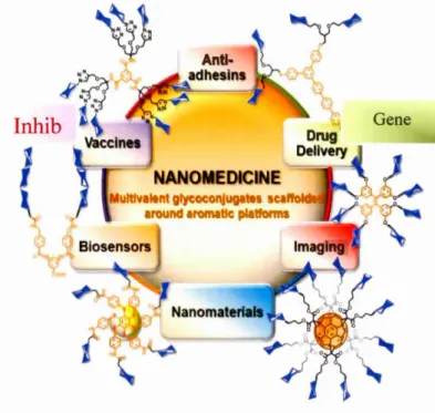
Dendrimers have been explored in a wide range of applications such as drug and gene de li very, 18-21 sensing, 22,23 electronics, 24,25 diagnostics, 26 and nanoengineering2 7 whi ch is shown in Figure 1-2. In addition, dendrimers are majorly used for other principal applications such as bacterial biofilm inhibitors28 which is the main subject of this study 29 as therapeutic agents30 due to their versatility, multivalency and their low polydispersity. Briefly, dendrin1ers are presented as convenient candidates for all different applications mentioned in the schen1e below. Their surfaces and their interior part (as their core) can be utilized to attach different functional moieties.
-
-6 Dendrimers have been largely used in drug delivery due to their monodisperse nature and because they are small enough to travel across biological barriers. Among them,
P AMAM and triazine based dendrimers have been investigated for drug de li very. 31 As shown Figure 1-3 (a) and (b ), drug de li very by dendrimers can occur mainly from three different n1echanisms :
Covalent conjugation: The existence of large functional groups on the surface of dendrimers make them appropriate for covalent conjugation of numerous drugs with relevant functional groups in which the targeting agents and drug molecules are covalently attached to the multivalent surface of the den drim ers.
Electrostatic interaction: The high density of functional groups like amine or carboxyl on the surface of dendrimers have potential applications in enhancing the solubility of hydrophobie drugs by electrostatic attractions.
Encapsulation: Hydrophobie drugs can be solubilized by physical incorporation in their interior part and inserted into internai layers of dendrimers in higher generations. 32
Inhib
Figure 1-2. Dendrimers applications.1 •33
In other words, the internai cavities of the dendrimers can serve as a pocket that can encapsulate a n1olecule of interest through self-assembly. It is kept inside the dendrüner through intern1olecular interaction (hydrophobie or hydrogen bonding) or electrostatic interaction until they reach the ir intended target. In this way, dendrin1er can deliver drugs locally with high efficiency, which allows to reduces drug cytotoxicity and is known as theranostics (c01nbining therapy and diagnostics).34,35
(,enC"r.ll•nn numher
8
Figure 1-3. (a) Encapsulation & covalent conjugation for drug delivery on dendrimers (b) Combination drug delivery system on dendrimers: concurrent delivery of water-soluble and -insoluble drugs by adsorption to the surface (ionie
interaction), encapsulation within hydrophobie micro cavities inside branching clefts or direct covalent conjugation to the surface functional groups.36 Additionaly, dendrimer can serve as a scaffold to increase the drug loading capacity
and improve bio availability due to the n1ultivalent interaction of peripheral moieties
and internai cavities.
For example, dendrimers are highly advantageous for cancer treatment where the tumor can be targeted with high precision. Another application of dendrimers is connected to
gene delivery. DNA and siRNA can be attached to terminal groups of dendriiners through electrostatic interactions to form a c01nplex named dendriplex which leads to a better transfection efficiency.
1.1.4 Dendrimers as multivalent ligands and inhibitors against bacterial biofilm formation
Pathogenic bacteries produces biofiln1 through interaction of surface proteins with oligosaccharides on epithelial of host cells and is known as glycoprotein interaction. Dendrimers with oligosaccharides as peripheral terminal moieties can be used to n1imic
the glycoprotein interaction. In this way, they can as a cotnpetitive pathway inhibit interaction with bacterial protein and inhibit biofilm fonnation.
1.2 Strategies for dendrimer synthesis
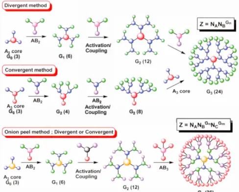
Historically, dendrimers has been synthesized following tinee different strategies: divergent, convergent and "onion peel". Where each method possesses its own advantages and disadvantages as shown in Figure 1.4.
1.2.1 The divergent method
Dendrimers were first synthesized by the divergent method which was developed by Totnalia 37•38 and Newkome. The divergent method involves an inside-outward and
stepwise approach, where a monomer, a molecule which possess one reactive site and multiple dormant groups, is added iteratively to a core molecule.
The dendrimer growth starts with the addition of the monomer reactive site to the core, resulting in the 1 st generation dendrimer where the dom1ant groups of the monomer stands at the periphery and the core at the center of the macromolecule. Then the dormant sites can be activated for subsequent monomer addition, to produce the next generation of dendrimer. The dendrimer growth then proceeds iteratively by addition of mo nom er until it reaches the desired size. At this point, the dendrimer can be capped off with the terminal moieties of interest. 39
The divergent approach offers great control over the size and terminal functionalities of the dendrimers which is in1possible when using the convergent approach. However, at each monomer addition step there is a chance of incomplete reactions, resulting in dendrimers with structural defects. And because it is difficult to purify dendrimers,
10
1nonodispersity is an issue for the divergent approach especially at higher dendrimer generation. Thus, the divergent approach requires high yield monomer reactions, ex cess amount of mono mers and lengthy purification steps to pro duce monodisperse dendrimers.
1.2.2 The convergent method
As oppose to the divergent approach, the convergent n1ethod presented by Hawker and Fréchet is an outside-inward approach.40 In this 1nethod, the branches of the dendrimer called "dendrons" are synthesized separately in earlier steps; where the dendrons can serve as large building blocks that can be attached to a central core to obtain a dendrimer. Using this approach enables the attachment of different dendrons on the same core to design multifunctional dendrimers. Moreover, there is a greater control over the synthesis of dendrons resulting in reduced reagents consumption and simplified purification procedure. Nonetheless, due to steric hindrance, there is a limit to the size of the dendrons that can be attached to a single core. Consequent! y, the convergent method produces smaller dendrimers compared to the divergent method and is not suitable for biological applications.41
1.2.3 Onion-peel method
The onion-peel method is a novel strategy which was developed by R. Roy and co-workers.42-44 This method was mostly used for divergent construction of dendrimers and glycodendrin1ers in which, the various species of building blocks are used at each layer of the dendritic growth. The synthesized dendrimers through this strategy are essentially different from the conventional dendrimers due to their different chemical composition of branches at each generation. Dendrimers provided by this method have heterogeneous layers meanwhile the conventional dendrimers follow the repetitive manner to form the different generations. In addition, they include similar and monotonous chemical units at their scaffold. 42-44 In other words, the novel "onion peel"
approach provides the opportunity to accelerate the coJlection of surface groups
together with different chen1ical entities considering the biophysical versatility
between the dendrimer generations. Recently the construction of dendrimers through
onion peel methodology via convergent and divergent bas been recently reported by R.
Roy and his group.45 According to this prominent strategy, heterogeneous layers were
chemically assembled at each generation by utilization of extremely effective chen1ical
reactions, namely "click chemistry" including the copper-catalyzed azide-alkyne
cycloaddition (CuAAC), thiol-ene (TEC), thiol-yne (TIC).46A7 It also includes coupling
and esterification via diverse orthogonal building blocks. Notably, it enables the use ot
building block to tai lor the dendrimer properties such as the presence of UV -vis
transitions. Divergent method Al core G0 (3) Al core G0 (3) G1 (6) G2 (4) Activation/ Coupling
/
Al core Gl (24) AB2 Activation/ Coupling G2 (8)Figure 1-4. Dendrimers synthesis strategies: divergent, concergent and onion peel method.42
- - - - ---~
12 1.3 "Click chemistry"
"Click chemistry" designate a group of reactions that fulfill a set of criterias, nmnely reactions that are stereospecific, that can be performed under mild conditions, that gives high yield and are easy to purify.48
-50 "Click reactions" are well adapted for the
synthesis of dendrimers and are cri ti cal to the development of new den drim ers. Among the existing click reaction, thiol-ene (TEC), thiol-yne (TYC)47 and copper-catalyzed
azide-alkyne cycloaddition (CuAAC) are the most widely used.46
"CuAAC"is a copper-catalyzed reaction of an azide with an alkyne functional group in order to form stereospecific 1,4 heterocycles of triazole. The first synthesized triazole was reported by Arthur Michael in 1893, but the study of 1 ,3 di polar cycloaddition to form 1,2,3-triazole has been introduced by Huisgen in 1963 based on the reaction kinetics and the conditions. According to Huisgen's studies, the cycloaddition through this process leads to two regioisomers: 1,4 and 1,5-isomers, which basically requires energy such as heat as the essential factor. 51 Later in 2002, the discovery of the copper
(I)-catalysis of 1,3 di polar cycloaddition by K. Barry Sharpless52 and Morten Meldal groups53 solved the problem of Huisgen regiochemistry and kinetic. Significantly, Sharpless and coworkers specified the concept of"click chemistry" as the fast, versatile, regioselective method that thermodynamically is the favored reaction which leads to regiospecific 1 ,2,4-triazole ring. Hawker and coworkers illustrated the synthesis of dendrimers via two types of click methodology:
1. Azide-alkyne reaction followed by halogenation and azido substitution using the convergent method. 54
2. Thiol-ene reaction divergent synthesis of dendrimer up to the 4th generation
Thus, Cu (1) catalyzed alkyne-azide "click reaction" is known as an effective method to synthesiie diverse kind of dendrüners as the fast regioselective reactions in high yield with minimum purification. The major drawback of"click" reaction is due to the copper cytotoxicity in biological, biomedical and pharn1aceutical applications. To address this issue, incubation with EDT A ( ethylenediaminetetraacetic acid) is applied as one of the practical technique, (the lower limit of detection in quantifying methods), to remove the majority of residual copper.
By means of "click chetnistry" reaction via "onion peel" approach, various den drim ers with different building blocks at their periphery can be constructed. Referred to as "multivalent ligands", these synthetic dendrimers offer multiple functionalities on their scaffold with more productivity and efficiency. Thus, they are significantly useful as therapeutic agent in an ti -adhesion therapy.
In 2005, Sharpless and co-worker published the article55 1n which the "click" mechanism via Copper (1)-catalysis has been clarified in 5 stages which is shown in Figure 1-5,51 including stage (A): copper acetylenide formation, stage (B): azide-copper acetylenide coordination, stage (C): cyclization to form an uncom.mon six-member n1etallocycle, stage (D): rearrangement 1,2, stage (E): proteolysis which leads to form the final favored product (1,4-isomer).
N N~ ~N [Cu] [Cu]
~
eD
stage C[cu]a
s
~
[cu]b
[cu]b
@ HFigure 1-5. CuAAC five stages mechanism proposed by Sharpless.56
1.4 Glycodendrimers and their role aas inhibitor
14
Glycodendrimers are dendrimers with sugar moieties as terminal groups and n1ost often
serve as a biomimetic macromolecules, (Figure 1-6). The exposed sugar moieties on
the periphery are responsible for binding-recognition process with their cognate protein
receptors.42,57-62 Typical glycodendrimers are composed of dendrons capped with
glycans e.g. maru1ose, fucose, galactose, etc. that are attached to various multivalent
cores such as cyclotriphosphazene (N3P3) or cyanuric chloride (CJCbNJ). Their
specifie potencies to provide the carbohydrate-protein interaction make them notable
for bion1edical studies as agents in anti-adhesion therapy. Severa} reports including the
Besides, they afford other diverse applications as dendrüners. For instance, they are employed as drug delivery mediators or carrier agents in gene therapy.37
A
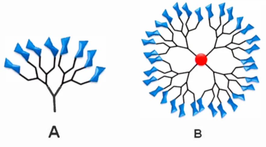
BFigure 1-6. Structure of a glycodendrimer where glycans (mannose, fucose, galactose, etc.) are shown in blue, branching units in black and the core in red
(A) Glycan dendron. (B) Glycodendrimer42
1.5 Aron1atic scaffold for glycodendrüners
The mimetic glycoconjugate provided by glycodendrimer shows superior affinities in
comparison to monovalent glycan-protein interaction. This artificial glycan-protein
interaction is afforded through the "glycoside cluster effect" known as "multivalent
glycoside effect"45·63-66 Accordingly, the synthesis of multivalent glycosylated
structures has been vastly developed.67·60•61·68-74 Various synthetic strategies have been displayed for glycoclusters and glycodendrimers where using multivalent scaffolds
pla ys the principal role th at con tri butes to the formation of the arranged architectures.
Using different multivalent scaffolds in glycoclusters structures allow them to have
better structural diversity with various building blocks which differs in valency and
peripheral functionalities. Their utilization can promote the glycan-protein interaction due to the appropriate spatial orientation of sugar residues as sui table recognition units
16
on their periphery for proper binding. Correspondingly, fitting the topology of this kind of glycoclusters with geometry of proteins of the pathogen such as lectins, enhances the ir role of inhibitors in an ti -adhesion therapy.
The first "aromatic scaffold" has been provided by Yariv and co-workers in the early
1960.61,75 Despite the presence of an hydrophobie aromatic core, the dendrimer is still
soluble in aqueous solution due to the presence of hydrophilic sugar moieties at the periphery. Consequently, dendrimer based on aromatic core can still be characterized in aqueous media by biological surveys and biophysical measurements. The molecules synthesized by using different rigid frameworks such as aromatic rings, heteroaron1atics and complex polycyclic derivatives have various fascinating
biological applications as well as anti-adhesion, vaccines, drug delivery agents, gene
Hex..,h"'ylb T etraphenyl thyl n Cyc:lotripho phaz n
~
'
N-o-Azooonzen Heterocycllc derivatives
pfl~~
!~
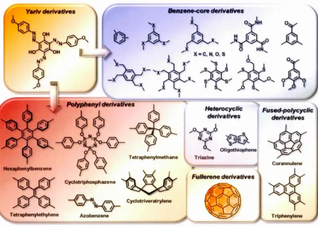
' Oligothioph n Triazl FuJierene derivatives Fused-polycyclic derivativesFigure 1-7. "Aromatic scaffold" used in designed glycodendritic architectures.61
Cyclotriphosphazene and triazine cores as aromatic scaffolds
Cyclotriphosphazene is a weil known building block used in in organic, inorganic and
high-polymer chemistry that can serves as a scaffold for the synthesis of a new class of
glycodendrimers. 76 It has the ad v an tage of being non-toxic and its aromatic n-n*
transitions can be followed by UV -vis spectroscopy.
Allcock and his coworkers 77 synthesized the polyphosphazene compounds as a usage
of carriers in drug delivery systems due to their biodegradability and biocompatibility
which was followed by their thennal and chetnical properties. Also, the star-shaped
r - - -
·--18 prilnary phosphonium cascade molecule was reported by Rengan and Engel80 in the year 1991. Then many dendrimers were made by us~ng the cyclotriphosphazene as the core by Majoral group since 1994. Above all advantages of the cyclotriphosphazene fan1ily, the antünicrobial and biological effects on bacterial cells is considered as an efficient eye-catching characteristic. 81-83 They highly possess potential to
accomtnodate a higher number offunctional units (such as dendrons) on their scaffold which leads to the higher generation in dendrimer synthesis. Triazine and cyclotriphosphazene core possess hydrophobie properties; however, upon functionalization with hydrophilic sugar moieties, they become water soluble which is important factor for glycoconjugate interaction in vivo. Thus, they offer better hydrophilic interaction between sugar residues and protein receptors. Therefore, we have projected our synthetic strategy using cyclotriphosphazene and triazine due to the n1entioned significant properties.
1.6 Multivalency role in anti-adhesion therapy
Biologically, the carbohydrates of cell surface are derived from glycolipids, glycoproteins, and proteoglycans. Mostly, they exist on the cell surface as short units of oligosaccharides generally between one to six carbohydrate residues responsible for carbohydrate-protein interaction process, via sugar recognition site on protein of the pathogens. 84--86 The first carbohydrate-protein interaction between the bacteria and the
host cellleads to the first pathological event. Subsequently the cascade of aggregation of such interaction results in a serious occurrence as infection. Although, the biological carbohydrate-dependent event highly depends on the short oligosaccharide sequences on host cells, it is not dispersed in a wide range. Moreover, according to the reported studies, the binding affinity of individual oligosaccharide groups with pathogen is low, with a dissociation constant (Ko) in the range of ~M to mM.87
,88,89 Therefore, the nature
oflow affinities of carbohydrate-protein interaction, as well as the nature of insufficient.
multivalency of glycodendrimers. In other words, the organized multivalent architectures offers enhanced binding interactions cmnpared to host cells. Consequently, due to mimetic glycoconjugate interactions with their complementary protein receptors of the pathogen with higher affinities, they can be considered as inhibitors to block the biological carbohydrate-protein interactions. Briefly, multivalent glycodendrimers can enhance the glycoconjugate binding force which is the worthful target of this study.87,89 Furthermore, the n1ultivalency concept provided
by glycodendrimers is essential to accomplish the effective recognition and binding, as weil as the improvement oftheir antimicrobial activity90-93 The multivalent binding of
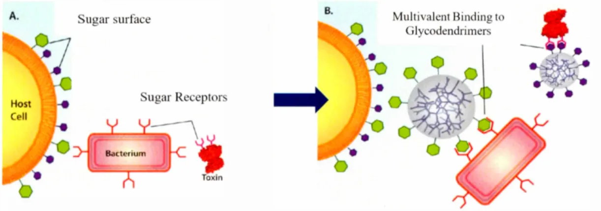
a bacterium or a bacterial toxin to a glycodendrimer is shown in Figure 1-8 where the inhibitory role of glycodendrimer to block the biological carbohydrate-protein interaction is demonstrated.9
°
Figure 1-8A is the depiction of a host cell with two dissimilar carbohydrates interacting with bacterium and bacterial toxin, in the absence of glycodendrimer. Figure 1-8B shows two different glycodendrimers containing the sugar moieties similar to host cells, the binding of the bacterimn and the toxin to glycodendrimers will be achieved through the multivalent interaction to prevent the infection of host cells in a competitive manner.90A. Host Ce li ugar urface ugar Re ept r Toxln B.
Multivalent Binding to Glycodendrimers
Figure 1-8. The multivalent binding of a bacterium or a bacterial toxin to a glycodendrimer.90
CHAPTER II
GL YCOBIOLOGY AND CYS TIC FIBROSIS
2.1 Carbohydrate-protein interactions
2.1.1 Introduction
Both physiologically and pathologically, carbohydrate-protein interactions are considered as the source of several biological processes.94 Additionally, the mentioned
interaction has a principle role in cell adhesion in which, the bacteria, virus, fungi, etc
attach to the host cells resulting in the initial step of infection. That pathological
phenon1enon such as bacterial infection is due to the formation of the glyco-protein
interaction. As explained earlier, the complex carbohydrates which are cmnn1only
situated at the cell surface, can interact \Vith their con1plementary and appropriate
proteinaceous receptors. This formation of carbohydrate-protein interaction is due to the contribution of a specifie amino ac id of a prote in such as Lectin PA-ILwith specifie group of carbohydrate ligands like o-Galactose via two dissin1ilar methods namely
hydrogen bond and hydrophobie interaction. X-ray crystallography study provides us
with the n1ajor data about the co1nbining sites of lectins with their specifie ligands; in
addition, it can offer the detailed information at the atomic lev el for interaction between
the two molecules.94 In this chapter the interaction between Lectin PA-IL as a galactophilic protein and its specifie ligand o-Galactose will be discus ed.
2.2 Lectin-Carbohydrate bonding
Lectins as well as other protein receptors can bind to their specifie ligands through three 1najor hydrogen bonds, hydrophobie interactions and electrostatic through salt bridges. There are other secondary interactions for the formation of such event like ion pairing; correspondingly, the coordination with n1etal ions.94-97 Moreover, water
n1olecules facilitate the bonding which would be described in the following pages. 97
2.2.1 Hydrogen-bond as noncovalent interactions
Hydrogen-bonds are profoundly involved in granting specificity to the protein-carbohydrate interaction and their efficiency in protein-carbohydrate affinity.94•98 The
foundation of hydrogen-bonds is fundrunental for proteins such as lectins to recognize selectively their appropriate carbohydrate ligands shown in Figure 2-1.74•99•100 The
presence of multiple hydroxyls on the carbohydrate architectures as the functional groups allow them to fonn the hydrogen-bonds between the OH groups of sugars and OH, C = 0 and NH functional groups of the amino acid side chains of the protein,
mostly Asp, Asn, Glu, Gln, Arg, Ser, His, Tyr.101 Moreover, the probability of multiple hydrogen-bonds on the same aton1, enhances the interaction with more rigidity and specificity. This interaction is directional and the energy of such an interaction is estin1ated at intermediate lev el between van der Waals interactions and covalent interactions.74•102•103 Hydrogen-bonds are considered as vital phenon1ena for
biochemical events due to their contribution to the dynamic and rapid association/dissociation at room ten1perature in physiological milieu.
22
Figure 2-1. Amino acids involved in mon omer of PA-IL bonding site in
interaction with D-galactose (PDB code lOKO). The interactions include direct
hydrogen bonds, hydrogen bonds with water, interactions with calcium ion and
hydrophobie interactions. Water molecule is shown as a red ba11.38
,44
2.2.2 Hydrophobie interaction
Carbohydrates are highly polar molecules~ however, the st rie disposition of hydroxyl group of the pyranose ring generale additional hydrophobie C-H bond on their surface. This provides them with the ability to interact with their complementary amino acids ofproteins via a and~ sides (30 glycomimic).32·94·104,105·The creation of the apolar face
leads them to form a type of interaction with hydrophobie side chains of the lectin proteins fr quently under ""C-1-1 n stacking'' of the carbohydrate ring with aromatic amino acids of the binding sites of the lectin protein such as Phenylalanine (Phc). Tryptophan (Trp). Tyrosine (Tyr). Hydrophobie stacking is presumed as the foundation of the hydrophobie interaction.74
·94,105 Stacking a carbohydrat ring on a side chain or
an aromatic amino acid is related to the partially positive charges on the aliphatic protons of pyranoside ring and parti ally negative charges from the n electrons of the aromatic ami no ac ids which can be summarized to ··multiple weak C-IL . . n
interactions".103,106,107 In addition, the methyl groups correlated to the 6-deoxy
carbohydrates nonnally provide the hydrophobie interaction with ar01natic amino acid residue as weil as stacking event. Hydrophobie interactions can also occur with the aliphatic amino acids such as valine or leucine. According to the reports and surveys, the hydrophobie interactions play an tmdeniably principal role in stereospecific binding lectin A to the ~-galactoside ligands with various chemical entities which is shown in
Figure 2-1.107-110
2.2.3 Ionie interactions
Carbohydrates are frequently neutral; although, there is vast category of charged sugars which participate in ionie interactions such as sialic, iduronic acids, glucuronic, aminosugars and sulfated sugars. There are also modified-sugars such as sulfated sugars and phosphorylated sugars that are negatively charged or other types of sugars positively charged.32 Basically, there is a strong affinity between the charged sugars and atnino ac ids of lectin pro teins due to the electrostatic forces (Coulomb'
s
law andPauli's repulsion) resulting in ionie interaction as an additional interaction with lectins.
Figure 2-2 presents ionie interactions between the anomeric sulfated functional group
of sulfo-sialic acid with arginine 3 71, arginine 292 and arginine 118 "red dashed lin es" with strong electrostatic forces also ionie interactions between glycerol moiety of
24
v
Figure 2-2. (Left) Ionie interactions between sulfated functional group of sulfo-sialic acid with Arg 371, Arg 292 and Arg 118 in red dashed lines. (Right) Ionie
interactions of sulfo and glycerol moiety in sulfo-sialic acid·analogue respectively
with Arg 371, Arg 292, Arg 118 and Glu 276 in dashed lines, with stronger electrostatic interaction colored orange. Sulfur atom colored yellow (ref sialic
acid).lll
2.2.4 Metal interaction
Metal ions such a Ca2 are situated in the vicinity of the binding sites of bacterial lectins and they are able to be bound indirectly to certain sugars. 74,112 1 he calcium
cations as the complex with lectins, are often coordinated with negatively charged ami no acids oflectins and the oxygen atoms ofhydroxyl groups on carbohydrate . Ca2+
ion has a principal role to enhance the sugar-protein interaction through the relative stereochemistry and to maintain structural integrity ofthe lectin.74,112 Tetrameric Lectin
A and B of the opportunistic Pseudomona..,· uerugino.)·a bacteria are examples of calcium-dependent lectins, in which each monomer binding site is associated respectively with one and two Ca2 atom. The complex of lectin-calcium atom is followed by the network of hydrogen bonds which provide them with the adequate binding site considering the selective specificity that creates the remarkable medium affinity to the 0-galactoside moiety and very higher aftinity in micromolar range to rucose and mannose sugars. 113,114 The galactose -binding site of PA-IL includes one
calcium atom as shown in Figure 2-3.114 According to Figure 2-3, each monomer or
the tetrameric lectin is in direct interaction with one calcium ion in the specifie binding
zone planned for galactoside residue to be locating there. In the PA-IL/galactose complex, th oxygen atoms on the position 3 and 4 of galactose residue are directly involved in the spherical coordination with calcium ion, Figure 2-3.114,115
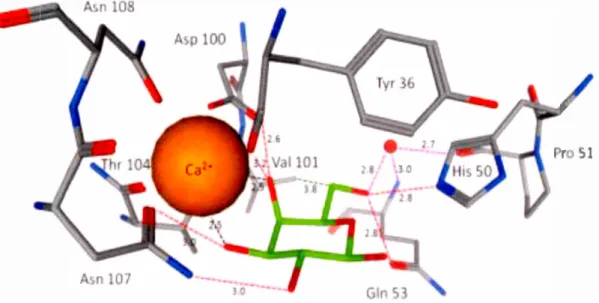
Figure 2-3. Binding site in the crystal structure of the PA-IL/galactose complex (PDB 1 OKO). (Left) Stick model of the amino acids PA-IL coordinated with ca+2 and in binding with galactose residue. ca+2 coordination bond is shown in
solid orange Iine, hydrogen bonds by dashed green Iine. Color coding: red for oxygen, blue for nitrogen, black for carbon, pink for calcium. (Right) Electrostatic surface of the protein-binding site related to PA-IL with ca+2 and
galactose. Color coding: from violet for negative to orange for positive of the protein-binding site, large pink sphere for ca+2 and stick model for galactose
residue. 114•115
2.2.5 Role of water
As shown earlier in Figure 2-1. the extra hydrogen bonds between the ligand and PA-IL are related to the water bridges. The ligand-protein interaction are mostly mediated
by water bridges.116 In aqueous milieu, there are ligand-water and protein-water connections. The overall process of these bindings is replaced by the ligand-protein
26 solvent. The establishment of the hydrogen bond network in carbohydrate-protein complex where water act as hydrogen bond donor or acceptor is essential for ligand recognition. 31
2.3 Galactose-PA-IL complex interaction
The interactions between a galactose ligand and a monomer lectin A of P. aeruginosa,
as an example of the carbohydrate-protein interaction, are shown in Figure 2-1.38 These interactions involve direct and indirect hydrogen bonds, hydrophobie interactions, and other interactions which are bridged to calcium ion and water molecules. Coordination of calcium ion with varions type of lee A amino ac ids include Tyr36, Aspl 00, Thrl 04, Asnl07, Asn108, and with Gal.03 and Gal.04 related to hydroxyl groups. Hydrogen-bonds include Ga1.02 with Asnl07, Gal.03 with Asnl07, Gal.04 with AsplOO, Gal.04 with AsplOO, Gal.06 with His50 and Gal.06 with Gln53. Hydrophobie interactions include Gal. Cl with Tyr36, Gal.C2 with Tyr36, Gal.C4 with Thrl 04 and Gal.C6 with VallOl. Moreover, there are several hydrogen bonds of 4 water tnolecules with Gal.Ol, Gal.02, Gal.03 and Gal.06. Additionally, there are bridging water molecules with Pro51 and Gln53 which help to establish and enhance the hydrogen bond network of the syste1n.44
In conclusion, the lectin-galactoside interactions possess six direct hydrogen bonds between hydroxyl groups of the galactose and lectin A, and one additional water molecule bridge. Moreover, few hydrophobie interactions which can establish the carbohydrate-protein interaction via the contact between the apolar face of galactose residue and specifie hydrophobie an1ino acids which has been explained earlier in Figure 2-1.38,44
2.4 Cystic fibrosis and the role of Pseudomonas aeruginosa lectin A
Cystic fibrosis is the most lethal genetic disease an1ongst the middle-aged Canadian population. The chronic lung infection resulting in the respiratory failure, is considered as the major cause of the mortality in cystic fibrosis and hospitalized patients. The chronic lung infection is due to the colonization and adhesion of the opportLmistic bacteria Pseudomonas aeruginosa on the surface of lung epithelium which leads to the forn1ation of a thick layer ofbacterial aggregation in a tern1 of a bio film. Pseudomonas aeruginosa is a ub:iquitous gram-negative bacteria which is found in various envi.ronment such as soil, water and vegetation. Moreover, it is responsible for numero us nosocmnial infection that can affect people not only with the genetic disorder cystic fibrosis but also with hun1an inlffiunodeficiency such as HIV 1 AIDS. 117,38 The
first adhesion to the host tissues is the initial concerned step toward infection achieved via oligosaccharide-mediated recognition by binding the sites of P. aeruginosa protein as shown in Figure 2-4.28,118 Referred to this event, the initial carbohydrate-protein com1ection i.s fairly sufficient as a key role to pro vide the opening for pathogen for next phase of interactions that ends up with the severe cohesive attaclunents and eventually severe tissue datnage. Correspondingly, galactose plays a fundamental role in carbohydrate recognition by the P. aeruginosa to initiate the adhesion process as shown in Figure 2-4. 114
28
Ctll dO.IOfl
Figure 2-4. The initial process of the adhesion schema tic description by P. aeruginosa on epithelium cells via galactose/PA-IL complex which leads to tissue
damage. 44•46
2.5 Biofilm formation
The colonies of microbial genotypes drive to the respiratory system and after their
aggregation, their growth and division lead th m to form the mature bacterial biofïlm in the airway. Consequently, by di per ing a part of the biofïlm, the fr e-tloating
bacteria would be released for future colonization which enters the cycle and forn1s the biofïlm a a long-lasting infection, resistant to antibiotic-based treatments.2x Figure
2-5 illustrates the mechanism of bio fi lm formation. 119 The uti lization of antibiotics su ch as ciprofloxacin for cystic fïbrosis treatment bas been correspondingly reported as a
temporary treatment due to its short-term effect on the outer layer of microbial
genotypes. However, the depth ofthe layer still includes a vast quantity of al ive bacteria
recolonize and expand the biofïlm layer. Accordingly, seeking for an alternative
strategy is essentially crucial in orcier to avoicl taking the antibiotic as the inefficient
temporary solution. In fact the biofilm formation is followecl by the growth in the
number ofthe galactose 1 PA-IL complexes~ which is clirectly relatee! to the tenclency
of galactophilic LecA to the galactose moi ties exist on the cell surface.
Single
Dispersion to release free bacteria
Mature
Growth & colonization
Aggregation
Figure 2-5. Mechanism of biofilm formation 120
2.6 Structures and roles of Pseudomonas aeruginosa lectins
There are two types of carbohyclrate-bincling proteins of P. oentf{inoso causing cystic
-fïbrosis in which both proteins have been fou nd on the outer membrane of the pathogen,
namecl LecA or PA-IL and LecB or PA-HL with their specifïcity for 0-galactose and
L-fucose, respectively.120-122 Through this stucly we are mainly interestecl in PA-IL, a
calcium dependent lectin that particularly recognizes 0-galactose. PA-IL is consideree!
as the fïrst lectin of P. oeruf{inoso which is isolatecl by affinity chromatography, it is
also the tïrst bacterial prote in which clescribecl galactose specificity.123•124 PA-l L is a
tetrameric protein with total molecular weight of 51 KDa which consists of four
30
PA-IL i furnished with one calcium ion and water molecule which provides them with the adequate binding sites for galactose moicties as displayed in the crystal
structure in Figure 2-6. (left and right).95 Carbohydrate-binding proteins of
Pseudomonas Aeruginosa has a organized geon1etry. The topology of PA-IL based on
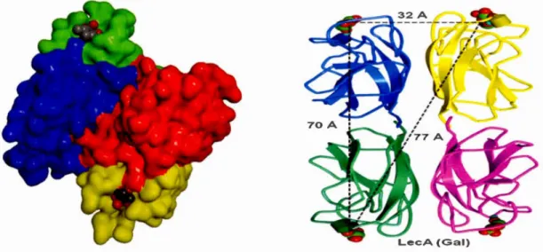
conformational analysis was assessed to be as a tetran1eric protein with rectangular architecture with the distance binding site (Ca+2) ~ 71 A 0 for the long side and ~32 A o for the short side according to their ribbon structure shown in Figure 2-6. (right).125
Figure 2-6. (Left) Crystal three-dimensional view of tetrameric PA-IL followed
by calcium ion on the binding sites of each monomer, Roy. R scheme (Right) The
distance measurement between calcium ions in tetrameric PA-IL binding sites
showing their geometrically weil- organized structure. (PBD code
2. 7 Multivalent interactions via multivalent tnechanisn1s, non-aggregative mechanisms
Mono carbohydrate-protein interaction is a connection between a monovalent ligand
and monovalent receptor considered as a reference for the noncovalent multivalent
glyco-protein interactions through the biological or biomimicry process (glycosylated
cell surface). Such multivalent interactions are followed by the complexity of binding
as an i ue due to the competition between the diffèrent biological mechanisms that
can yield the various biologieal respon~e which some of them are desirable and vice versa. Tetrameric PA-l L proteins possess 4 binding site on a single face. This pote ney
makes them able to interact with multiglycosylated ligands simultaneously uch as
glyco ylated cel! surface. Accordingly, thesc multivalent interactions occur through the
known Chelate As ociation Mechanism (multivalency-driven increase of affinity) as
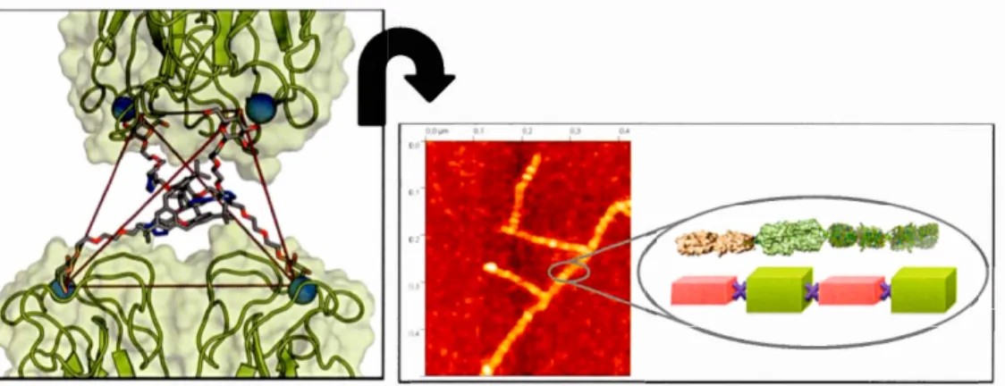
shown Figure 2-8. (a).74Atomic force microscopy (AF M) has confïrmed the chelate aggregative mode! in various cases. particularly for the interaction of tetravalent PA-IL. a galactose specifie lectin. and a tetragalactosylated calixarene-ba ed as its
adequateJy well-matched ligand according to Figure 2-7.12
6-114
Figure 2-7. (left) Chelate aggregative model of tetravalent Calixarene-based in
binding PA-IL from Pseudomonas aeruginosa. (Right) AFM observation of
PA-IL filaments in interaction with the sa me glycocluster and mo del of the lD-network. 74,126-130
32
The other mechanism is named Receptor lustering in which the monomer receptors such as lectins are provoked to interact with multivalent ligands~ therefore. the
monovalent receptors are di ITused through the dy nam ic 1 i pi cl bi layer to pro vide the
maximum interaction with their multivalent partner . Thus, they lorm a clustered figure by the ligands according to Figure 2-8 (b). Receptor clustering i considercd as an intra cellular mechanism.74·131-137 The Sub ite Association as the other mechani m
is known when a heterobivalent ligand is connectee! simultaneou ly with two various binding site correspondingly with two various affinities according to Figure 2-8
(c).74·136,137 Additionally. the interaction between multivalent glycosylatcd ligands and
monovalent receptors such as lcctins can enhance the allinity by the intense density or
ligand adjacent to the binding site region which leads to the higher number of
rea sociation according to Figure 2-8 (d).74·138
(a) Chelate binding mechanism through three possible
(1) Chelate (2) Aggregative chelate
(3) Aggregation
q
(b) Recepto r ( c) Subsite (d) Statistical
Figure 2-8. Nonaggregative mechanisms of noncovalent interactions between
multivalent ligands and multivalent receptors.69 Chelate binding model is
exceptionally considered as both nonaggregative and aggregative which depends