Université de Montréal
Synthesis of Constrained Tricyclic Nucleosides
and the Core of Nagilactone B
par
Robert D. Giacometti
Département de chimie Faculté des arts et des sciences
Thèse présentée à la Faculté des études supérieures et postdoctorales en vue de l’obtention du grade de
Philosophiæ Doctor (Ph. D.) en chimie
Août 2015
Résumé
Cette thèse décrit deux thèmes principaux: 1) la conception, la synthèse, et l'évaluation biophysique des nucléosides tricycliques, et 2) la synthèse de nagilactone B, un produit naturel norditerpenoïde dilactone de la famille de produits naturels “podolactone”.
Le premier chapitre décrit la stratégie de design rationnel des nucléosides nommé “restriction conformationnelle double” basée sur les études de modélisation structurales des duplex ADN–ARN modifiés. Cette stratégie implique un blocage du cycle furanose dans une configuration de type N- ou S, et une restriction de la rotation torsionelle autour de l’angle γ. La première contrainte a été incorporée avec un pont méthylène entre l’oxygène en position 2′ et le carbone 4′ du nucléoside. Cette stratégie a été inspirée par les acides nucléiques bloqués (ou “locked nucleic acid”, LNA). La deuxième contrainte a été réalisée en ajoutant un carbocycle supplémentaire dans l'échafaud de l’acide nucléique bloqué. Les défis synthétiques de la formation des nucléotides modifiés à partir des carbohydrates sont décrits ainsi que les améliorations aux stabilités thermiques qu’ils apportent aux duplex oligonucléïques dont ils font partie.
Chapitres deux et trois décrivent le développement de deux voies synthétiques complémentaires pour la formation du noyau de nagilactone B. Ce produit naturel a des implications pour le syndrome de Hutchinson–Gilford, à cause de son habilité de jouer le rôle de modulateur de l’épissage d’ARN pré-messager de lamine A. Ce produit naturel contient sept stereocentres différents, dont deux quaternaires et deux comprenant un syn-1,2-diol, ainsi que des lactones à cinq ou six membres, où le cycle à six ressemble à un groupement α-pyrone. La synthèse a débuté avec la cétone de Wieland-Miescher qui a permis d’adresser les défis structurels ainsi qu’explorer les fonctionnalisations des cycles A, B et D du noyau de nagilactone B.
Mots-clés: Thérapie antisens, acides nucléiques tricycliques, acides nucléiques bloqués, LNA,
restriction conformationelle, nucléosides, oligonucléotides, acides nucléiques, de la conception basée sur la structure, la stabilité thermique des duplex, nagilactone B, podolactones, cétone de Wieland–Miescher, carbomethoxylation réductrice, oxydation allylique, trioxyde de chrome, l'oxydation Rubottom.
Abstract
The present thesis comprises two major themes: 1) the design, synthesis, and biophysical evaluation of conformationally restricted tricyclic nucleosides for antisense applications, and 2) strategic approaches for synthesizing the core of nagilactone B, a norditerpenoid dilactone from the podolactone family of natural products.
Guided by structural studies of modified DNA–RNA duplexes, Chapter One focuses on a proposed dual-conformational-restriction strategy, in which two modes of conformational restriction are incorporated into a single nucleotide modification: 1) locking the furanose ring in an N- or S-type configuration and 2) restricting rotation around backbone torsion angle γ. The first constraint was incorporated by way of a 2′,4′-anhydro bridge that is found in the scaffold of locked nucleic acid (LNA), while the second was realized by annealing an additional carbocyclic ring to the modified nucleoside. The synthetic challenges associated with preparing these highly constrained molecules from carbohydrate-derived starting materials are described, in addition to the corresponding improvements in duplex thermal stability they provide to oligonucleotide sequences containing them.
Chapters Two and Three describe complementary approaches for the synthesis of the core of nagilactone B, a natural product with implications for Hutchinson–Gilford progeria syndrome, as a consequence of its ability to act as a modulator of splicing events leading to lamin A. This natural product contains seven stereogenic centers overall, including a syn-1,2-diol moiety, a γ-lactone, and a pair of quaternary stereocenters, which are complemented by the presence of an α-pyrone moiety. To address the synthesis of these structural features, the utility of the Wieland–Miescher ketone was explored with an emphasis on synthesizing rings A, B, and D of the core of nagilactone B.
Keywords: Antisense therapy, tricyclic nucleic acids, locked nucleic acids, LNA,
conformational restriction, nucleosides, oligonucleotides, nucleic acids, structure-based design, duplex thermal stability, nagilactone B, podolactones, Wieland–Miescher ketone, reductive carbomethoxylation, allylic oxidation, chromium trioxide, Rubottom oxidation.
Table of Contents
Chapter 1: Synthesis of Highly Constrained Tricyclic Nucleosides ... 1
1.1 Introduction ... 2
1.1.1 Nucleic Acids ... 2
1.1.2 A Brief Overview of Protein Biosynthesis ... 6
1.1.3 Overview of the Antisense Approach ... 11
1.1.4 Modified Oligonucleotides ... 15
1.2 The Design of Tricyclic Nucleic Acid Analogues ... 21
1.3 Synthesis of α-L-TriNA 1 ... 28
1.3.1 Retrosynthetic Analysis of α-L-TriNA 1 ... 28
1.3.2 Synthesis of α-L-TriNA 1 ... 29
1.4 Synthesis of TriNA 1 ... 42
1.4.1 Retrosynthesis of TriNA 1 ... 42
1.4.2 Synthesis of TriNA 1 ... 43
1.5 Duplex Thermal Stability Measurements ... 52
1.5.1 Duplex Thermal Stability Measurements for α-L-TriNA 1 and 2 ... 52
1.5.2 Duplex Thermal Stability Measurements for TriNA 1 and 2 ... 56
1.6 Conclusions and Perspectives ... 60
Chapter 2: First-Generation Strategy for Synthesizing the Core of Nagilactone B ... 62
2.1 Introduction ... 63
2.2 Previous Syntheses of Podolactones ... 66
2.2.1 Adinolfi Group’s Synthesis of LL-Z1271α (1972) ... 67
2.2.2 Welch Group’s Synthesis of LL-Z1271α (1977) ... 68
2.2.3 Hayashi Group’s Synthesis of Nagilactone F (1982) ... 70
2.2.4 Barrero Group’s Synthetic Endeavours (1999–2002) ... 72
2.2.5 Hanessian Group’s Synthetic Approach to Podolactones (2009) ... 76
2.3 A Brief Introduction to Nagilactone B ... 80
2.4 First-Generation Strategy ... 82
2.4.1 Wieland–Miescher Ketone ... 84
2.4.3 Alkylation (Ketal Protective Group) ... 91
2.4.4 Allylic Oxidation (Ketal Protective Group) ... 96
2.4.5 Incorporation of 1,2-syn-Diol (Ketal Protective Group) ... 99
2.4.6 Preparing the 1,2-syn-Diol (tert-Butyldimethylsilyl Protective Group) .... 102
2.4.7 Alkylation and Incorrect Stereochemistry of Quaternary Centre ... 108
2.4.8 Alkylation and Stereochemistry of Quaternary Centre ... 114
2.4.9 Overview of the First-Generation Sequence ... 118
2.5 Future Work, Conclusions, and Perspective ... 121
Chapter 3: Second-Generation Strategy for Synthesizing the Core of Nagilactone B ... 123
3.1 Introduction ... 124
3.2 Synthetic Strategy for Second-Generation Synthesis ... 127
3.3 Pertinent Synthetic Work from the Literature ... 129
3.3.1 Rubottom Oxidation for syn-1,2-Diol: Meiji Seika Kaisha (1999) ... 129
3.3.2 Formal Allylic Oxidation ... 129
3.3.3 Work by Danishefsky’s Group (1996) ... 131
3.4 Second-Generation Approach ... 132
3.4.1 Incorporation of 1,2-syn-Diol ... 132
3.4.2 Functionalizing Ring A Through Dienol Ether (Acetonide) ... 136
3.4.3 Functionalizing Ring A Through Dienol Ether (Silyl) ... 139
3.4.4 Hydroboration–Oxidation Strategy for Oxidizing Ring A ... 141
3.4.5 Alkylation to Establish the Quaternary Stereocenter ... 144
3.4.6 Incorporating the γ-Lactone ... 147
3.4.7 Functionalization of the Enone ... 151
3.4.8 Summary of Current Route ... 155
3.5 Future Work, Conclusions, and Perspective ... 158
Chapter 4: Experimental Section ... 162
4.1 General Experimental Details ... 163
References ... 165
Annex 1: Experimental Data for Chapter 1 ... i
Experimental Procedures for α-L-TriNA 1 ... ii
Experimental Procedures for TriNA 1 ... xxxv Annex 2: Experimental Data for Chapter 2 ... liv Experimental Procedures ... lv Annex 3: Experimental Data for Chapter 3 ... xc Experimental Procedures for Second-Generation Synthesis ... xci Annex 4: Computational Details ... cxx Alkylation of Acetonide-Protected Diol: TS-2.7–2.9 ... cxxi Rubottom Oxidation of Silyl Enol Ether: TS-3.2 & TS-3.3 ... cxxvi Alkylation of Acetonide-Protected Diol: TS-3.5 & TS-3.6 ... cxxx Annex 5: X-ray Crystallographic Data ... cxxxv Benzyl enol ether 1.62 ... cxxxvi α-L-TriNA 1 nucleoside 1.70 ... cxlvi
TriNA 1 nucleoside 1.109 ... clxi α-Hydroxy ketone (±)-2.135 ... clxxiii Enone 2.144 ... clxxxi α-Hydroxy ketone (±)-2.145 ... clxxxix Ketone 2.152 ... cxcviii Diol 2.170 ... ccviii
List of Tables
Table 1.1 – Intramolecular cycloetherification to prepare the 2′,4′-anhydro bridge. ... 34
Table 1.2 – Attempted conditions for allylation reaction. ... 45
Table 1.3 – Ketone reduction with various sources of hydride. ... 46
Table 1.4 – Duplex thermal stability of α-L-TriNA-modified oligonucleotides. ... 53
Table 1.5 – Mismatch discrimination properties of α-L-TriNA-modified oligonucleotides. ... 54
Table 1.6 – Duplex thermal stability of TriNA-modified oligonucleotides. ... 57
Table 2.1 – Alkylation of β-ketoester with methyl iodide. ... 92
Table 2.2 – Alkylative transposition of α,β-unsaturated ester. ... 95
Table 2.3 – Allylic oxidation of α,β-unsaturated ester. ... 98
Table 2.4 – Oxidation of ring B ketone to enone. ... 110
Table 3.1 – Synthesis of methoxy dienol ether. ... 140
Table 3.2 – Attempted isomerization of unsaturated ketal. ... 142
List of Schemes
Scheme 1.1 – Retrosynthetic analysis for α-L-TriNA 1. ... 29
Scheme 1.2 – Synthesis of spirocyclic core of α-L-TriNA 1. ... 30
Scheme 1.3 – Synthesis of cyclization precursor en route to α-L-TriNA 1. ... 31
Scheme 1.4 – Formation of anhydronucleosides from a dimethanesulfonate ester. ... 32
Scheme 1.5 – Synthesis of triflate for the key cycloetherification reaction. ... 33
Scheme 1.6 – Structure elucidation of the constitutional isomer. ... 35
Scheme 1.7 – Proposed mechanism for the cycloetherification reaction. ... 36
Scheme 1.8 – Synthesis of triethylsilyl and acetyl-protected α-L-TriNA 1. ... 37
Scheme 1.9 – Attempted chemoselective deprotection of acetyl-protected C5′-OH. ... 39
Scheme 1.10 – Synthesis of phosporamidite for solid-phase oligonucleotide synthesis. ... 40
Scheme 1.11 – Preparation of the α-L-TriNA 1 phosphoramidite monomer. ... 41
Scheme 1.12 – Retrosynthetic analysis for TriNA 1. ... 42
Scheme 1.13 – Synthesis of precursor for allylation reaction. ... 44
Scheme 1.14 – Oxidation of homoallylic alcohol. ... 45
Scheme 1.15 – Installation of the nucleobase and synthesis of the cyclization precursor. ... 47
Scheme 1.16 – Intramolecular SN2 displacement to form anhydro bridge of TriNA 1. ... 47
Scheme 1.17 – Synthesis of alkynyl alcohol. ... 48
Scheme 1.18 – Completion of the synthesis of a TriNA 1 monomer. ... 50
Scheme 2.1 – Proposed biosynthetic pathway for plant-derived podolactones. ... 64
Scheme 2.2 – Proposed biosynthesis for fungi-derived podolactones. ... 65
Scheme 2.3 – Unexpected reactivity of nagilactone A and nagilactone A diacetate.142 ... 66
Scheme 2.4 – Synthesis of LL-Z1271α via degradation of marrubiin.154 ... 67
Scheme 2.5 –Synthesis of racemic LL-Z1271α from the Wieland–Miescher ketone.155 ... 68
Scheme 2.6 – Bromolactonization of the carboxylic acid enone. ... 69
Scheme 2.7 – Synthesis of nagilactone F by Hayashi’s group.156 ... 71
Scheme 2.8 – Alternative strategy studied by Hayashi’s group. ... 72
Scheme 2.9 - Synthesis of podolactones from trans-communic acid.138,140 ... 73
Scheme 2.10 – Racemic synthesis of the 3β-hydroxy regioisomer of wentilactone B.159 ... 75
Scheme 2.12 – First-generation retrosynthetic analysis for nagilactone B. ... 83
Scheme 2.13 – 2-Methyl-1,3-cyclohexanedione to β-keto ester. ... 89
Scheme 2.14 – Dissolving metal reduction mechanism.250 ... 90
Scheme 2.15 – Carbomethoxylation of lithium enolate with Mander’s reagent. ... 91
Scheme 2.16 – Direct alkylation of β-ketoester. ... 92
Scheme 2.17 – Comparison of transition states for methylation of β-ketoester. ... 93
Scheme 2.18 – Alkylation via exocyclic enolate to set quaternary centre.259,265 ... 94
Scheme 2.19 – Synthesis of α,β-unsaturated ester. ... 95
Scheme 2.20 – Allylic oxidation to functionalize ring A. ... 96
Scheme 2.21 – Allylic oxidation on similar scaffolds. ... 97
Scheme 2.22 – Selenium-dioxide-mediated oxidation to access 1,2-diketone. ... 99
Scheme 2.23 – Preparation of α-hydroxy ketone. ... 100
Scheme 2.24 – Preparation of 1,2-diol. ... 100
Scheme 2.25 – Synthesis of protected 1,2-diol via enol ketone. ... 101
Scheme 2.26 – Improvement of α-hydroxylation reaction with TBS protective group. ... 103
Scheme 2.27 – (+)-Wieland–Miescher ketone to γ-keto-α,β-unsaturated ester. ... 104
Scheme 2.28 – Oxidation of α,β-unsaturated ester with PCC and NHS. ... 105
Scheme 2.29 – Reduction of α,β-unsaturated ester. ... 106
Scheme 2.30 – Oxidation to 1,2-diketone. ... 107
Scheme 2.31 – Synthesis of 1,2-diol. ... 108
Scheme 2.32 – α-Methylation of ester afforded abietic-type stereochemistry. ... 109
Scheme 2.33 – Oxidation to enone and re-protection of 1,2-diol. ... 110
Scheme 2.34 – Attempted alkylation with a more flexible scaffold. ... 114
Scheme 2.35 – Alkylation with benzyl chloromethyl ether to set quaternary stereocenter. ... 115
Scheme 2.36 – Reduction of primary alcohol via mesylate or tosylate. ... 115
Scheme 2.37 - Reduction of neopentylic alcohol to establish quaternary stereocenter. ... 117
Scheme 2.38 – Oxidation to methyl ester and deprotection of silyl ether. ... 117
Scheme 2.39 – Oxidation with Oxone®. ... 118
Scheme 2.40 – First-generation sequence for synthesizing the core of nagilactone B. ... 120
Scheme 2.41 – Conjugate addition to establish quaternary stereocenter. ... 121
Scheme 3.2 – Previous work at Meiji Seika Kaisha.343 ... 129
Scheme 3.3 – Formal allylic oxidation with m-CPBA. ... 130
Scheme 3.4 – The Danishefsky group’s approach for synthesizing a taxol intermediate.362 .. 131
Scheme 3.5 – One-pot procedure for the synthesis of (–)-Wieland–Miescher ketone.245,247 .. 132
Scheme 3.6 – Rubottom oxidation to prepare α-hydroxy ketone. ... 133
Scheme 3.7 – Synthesis of acetonide-protected syn-1,2-diol. ... 136
Scheme 3.8 – Oxidation of methoxy dienol ether and subsequent reactivity of alcohol. ... 137
Scheme 3.9 – Ketal approach to functionalize ring A. ... 138
Scheme 3.10 – Preparing the silyl-protected syn-1,2-diol. ... 139
Scheme 3.11 – Oxidation of methoxy dienol ether with Oxone®/m-CPBA. ... 141
Scheme 3.12 –Protection of secondary hydroxy group with triethylsilyl chloride. ... 141
Scheme 3.13 – Ketal protection with concomitant isomerization of alkene. ... 143
Scheme 3.14 – Hydroboration–reduction sequence to functionalize ring A. ... 144
Scheme 3.15 – Methylation to establish quaternary stereocenter. ... 145
Scheme 3.16 – Oxidation of ring B to prepare γ-lactone precursor. ... 147
Scheme 3.17 – Sulfonylation of secondary hydroxy group. ... 149
Scheme 3.18 – Lactonization via triflate. ... 151
Scheme 3.19 – Options for functionalizing the enone to prepare ring C. ... 152
Scheme 3.20 – Mukaiyama–Michael conjugate addition reaction. ... 153
Scheme 3.21 – Reactivity of tert-butyldimethylsilyl enol ether. ... 154
Scheme 3.22 – Hydrogenation of enone. ... 154
Scheme 3.23 – Second-generation sequence for preparing the core of nagilactone B. ... 156
Scheme 3.24 – Incorporation of isobutyryl group. ... 158
List of Figures
Figure 1.1 – Monomeric subunit of nucleic acids. ... 2
Figure 1.2 – Watson–Crick base pairing of DNA duplex. ... 3
Figure 1.3 – Structures of A-, B- and Z-DNA.7 ... 4
Figure 1.4 – Conformation of sugar moiety in nucleic acids. ... 5
Figure 1.5 – A brief overview of protein biosynthesis.13 ... 7
Figure 1.6 – Alternative splicing.26 ... 11
Figure 1.7 – Small molecule therapeutics versus antisense approach.30 ... 12
Figure 1.8 – Antisense mechanisms.33 ... 14
Figure 1.9 – Representative oligonucleotide modifications. ... 16
Figure 1.10 – Conformational restriction strategies. ... 19
Figure 1.11 – Overlay of LNA and α-L-LNA. ... 20
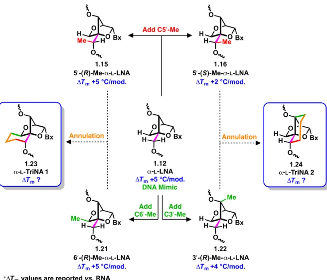
Figure 1.12 – Incorporation of C5′-methyl group on constrained bicyclic scaffolds. ... 21
Figure 1.13 – Design of tricyclic nucleic acids from a LNA scaffold template. ... 24
Figure 1.14 – Design of tricyclic nucleic acids based on α-L-LNA scaffold. ... 25
Figure 1.15 – Overlay of tricyclic nucleosides and NMR structure solutions. ... 26
Figure 1.16 – X-ray crystallographic evidence for the tricyclic nucleic acid core. ... 38
Figure 1.17 – Verification of the tricyclic scaffold of TriNA 1. ... 51
Figure 1.18 – Overlay of α-L-TriNA 1 and 2 on an α-L-LNA-modified DNA–RNA duplex. 55 Figure 1.19 – Overlay of TriNA 1 and 2 on an S-cEt-modified DNA–DNA duplex. ... 58
Figure 1.20 – Summary of the dual-conformational-restriction strategy. ... 60
Figure 2.1 – General scaffold of podolactones. ... 63
Figure 2.2 – Reported biological activity of select podolactones. ... 64
Figure 2.3 – Nagilactone B. ... 80
Figure 2.4 – General analysis of the structure of nagilactone B. ... 82
Figure 2.5 – First enantioselective syntheses of (+)-Wieland–Miescher ketone... 84
Figure 2.6 – Computed transition structures for proline-catalyzed aldol reaction.240,243 ... 86
Figure 2.7 – Catalysts used to synthesize the Wieland–Miescher ketone. ... 87
Figure 2.8 – X-ray crystallographic structure of α-hydroxy ketone. ... 102
Figure 2.10 – X-ray crystallographic structure of enone. ... 106
Figure 2.11 – X-ray crystallographic structure of ketone. ... 109
Figure 2.12 – Transition structures for α-methylation of ester enolate. ... 112
Figure 2.13 – X-ray crystallographic structure of diol from Oxone®-mediated-oxidation. ... 118
Figure 3.1 – Overview of first-generation approach for synthesizing nagilactone B. ... 124
Figure 3.2 – Applying the Wieland–Miescher ketone to the synthesis of nagilactone B. ... 125
Figure 3.3 – Updated analysis of the structure of nagilactone B. ... 127
Figure 3.4 – Transition structures for Rubottom oxidation. ... 135
List of Symbols and Abbreviations
> greater than
>> much greater than α
α stereochemical descriptor
[αα]t
D specific rotation at temperature t and wavelength of sodium D line
Å angstrom
AC50 concentration at which compound exhibits half-maximal efficacy
Ac acetyl AcO acetate aq aqueous β stereochemical descriptor Bn benzyl BOM benzyloxymethyl BSA bis(trimethylsilyl)acetamide Bu butyl
Bx general placeholder for a nucleobase
CDI 1,1′-carbonyldiimidazole
CPTS collidinium p-toluenesulfonate
CSA camphor-10-sulfonic acid
ΔTm difference in melting point between oligonucleotide containing the
modification of interest duplexed with a complementary strand of RNA and the corresponding RNA–RNA duplex
d day (for experimental details)
DBN 1,5-diazabicyclo[4.3.0]-5-nonene
DBU 1,8-diazabicyclo[5.4.0]undec-7-ene
DCE dichloroethane (e.g., 1,2-DCE = 1,2-dichloroethane)
DDQ 2,3-dichloro-5,6-dicyano-1,4-benzoquinone DMAP 4-dimethylaminopyridine DME 1,2-dimethoxyethane DMF N,N-dimethylformamide DMI 1,3-dimethyl-2-imidazolidinone DMP Dess–Martin periodinane
DNA deoxyribonucleic acid
EDC 3-(ethyliminomethyleneamino)-N,N-dimethylpropan-1-amine
ent enantiomer of the given compound that immediately follows the descriptor
ESI electrospray ionization
Et ethyl
HATU 1-[bis(dimethylamino)methylene]-1H-1,2,3-triazolo[4,5-b]pyridinium 3-oxid hexafluorophosphate
HMPA hexamethylphosphoramide
hν indicates light; h is Planck’s constant, and ν is the photon frequency
i iso (as in i-Pr)
IBX 2-iodoxybenzoic acid
IC50 half maximal inhibitory concentration KHMDS potassium bis(trimethylsilyl)amide
LDA lithium diisopropylamide
Lev levulinyl (i.e., 4-oxopentanoyl)
m meta
m-CPBA 3-chloroperoxybenzoic acid
M molar (mol dm-3, mol L-1)
Me methyl
min minutes (for experimental details)
mol mole
mol % mole percent
MOMCl chloromethyl methyl ether (or chloro(methoxy)methane)
MPO 4-methoxypyridine-N-oxide
mRNA messenger RNA
Ms mesyl (methylsulfonyl)
MS molecular sieves
m/z mass-to-charge ratio
n normal (as in n-butyl or n-Bu)
Nap 2-naphthylmethyl
NCS N-chlorosuccinimide
NMR nuclear magnetic resonance
OAc acetate
OTf triflate (as in trifluoromethanesulfonate)
% percent
p para
PCC pyridinium chlorochromate
PDC pyridinium dichromate
PG protective group
pH negative logarithm of hydrogen ion concentration
Piv pivaloyl
Ph phenyl (C6H5)
pNB para-nitrobenzoyl
PPTS pyridinium p-toluenesulfonate
q quartet (spectra)
RNA ribonucleic acid
RNase ribonuclease
r.t. room temperature (refers to ambient temperature of the surroundings)
s singlet (spectra)
s secondary (as in s-Bu)
t triplet (spectra)
t tertiary (as in t-Bu)
TMS trimethylsilyl
TBAF tetrabutylammonium fluoride
TBAI tetrabutylammonium iodide
TBDPS tert-butyldiphenylsilyl
TBS tert-butyldimethylsily
TES triethylsilyl
Tf trifluoromethanesulfonyl
TFAA trifluoroacetic anhydride
THF tetrahydrofuran
Tm melting temperature of oligonucleotide duplex: corresponds to the temperature at which 50% of a duplex is unwound into single strands
TMS trimethylsilyl
tosyl 4-toluenesulfonyl (also Ts)
triflate trifluoromethanesulfonate
Ts tosyl (also 4-toluenesulfonyl)
v/v volume per volume
w/v weight per volume
w/w weight per weight
wt weight
“Your time is limited, so don't waste it living someone else's life. Don't be trapped by dogma – which is living with the results of other people's thinking. Don't let the noise of others' opinions drown out your own inner voice. And most important, have the courage to follow your heart and intuition, they somehow already know what you truly want to become. Everything else is secondary.”
Steve Jobs, 2005 Stanford Commencement Address
Acknowledgements
Thank you Professor Hanessian for accepting me into your research group at the University of Montreal and in particular, for providing me with an opportunity to work in collaboration with Isis Pharmaceuticals on the nucleoside projects. It’s certainly inspiring to see the longevity you’ve had pursuing your passion for chemistry, and in that spirit I would like to thank you for giving me a great deal of perspective on life.
Professor Houk, I am incredibly grateful for the opportunity that you provided me to work with you and your group at UCLA for four months. Your mentorship and collaborative approach to tackling challenging problems were truly inspirational. I consider myself fortunate to have worked with you on so many diverse and interesting projects, which are not described in the present document. Moreover, I am indebted to all of the wonderful people in your group, who helped a synthetic organic chemist transition to the computational chemistry world and explore the American Southwest, particularly: Colin Lam, Stephen Lopez, Ashay Patel, Jessica Grandner, Mareike Holland, Hung Pham, Buck Taylor, Ilhan Yavuz, and Raghu Ramabhadran. The time I spent at UCLA was truly too short in duration.
A big thank you to Professor Collins and Professor Wuest for the guidance you have both provided during my studies, as members of my thesis committee. Your suggestions, critiques, and overall support are very much appreciated. Professor Collins, thank you for accepting me as an honorary member of your group (if only because of Anna) – and for the delicious pulled-pork sandwiches. I appreciate the advice you offered when times were tough and for the frank, but constructive criticisms you provided throughout my time at UdeM.
I am also grateful to all the members of the department who have helped make this research possible: the NMR group (Dr. Minh Tan Phan-Viet, Dr. Cédric Malveau, Silvie Bilodeau and Antoine Hamel), the Centre for Mass Spectral Analysis (Alexandra Furtos, Karine Venne, Marie-Christine Tang, and Christophe Camy), and the machine shop (Louis Beaumont, Yves Teasdale, Jean-François Myre). A big thank you to Michel Simard, Francine Bélanger-Gariépy, and Thierry Maris for taking the time (a lot of time!) to teach me how to acquire and analyze X-ray crystallographic data, as well as allowing me to use the instruments during the weekends – I truly appreciate it.
I was extremely fortunate to receive funding from the National Sciences and
Engineering Research Council of Canada, Université de Montréal, and the Schmeelk Canada Foundation to support my studies, and I am truly grateful to all three organizations for their
I have been fortunate to be surrounded by many wonderful colleagues throughout my studies; it would take at least another volume to describe the positive impact each of you have had on my studies and life – either directly or otherwise – and your insights have hopefully made me a better scientist/colleague/friend/person. The following list is by no means exhaustive, and I sincerely apologize in advance for anyone that I have missed. A big thank you to: Etienne Chenard, Benoît Deschênes Simard, Stéphane Dorich, Eduardo Sánchez-Larios, William Bechara, Benjamin Schroeder, Jernej Wagger, Rebecca Fransson, Bradley Merner, Martin Bueschleb, Michael Perryman, Jean-Philippe Cusson, Benoît Sicard, Oscar M. Saavedra, Juan Carlos Salinas Hernandez, Lorena Rico, Miguel Vilchis-Reyes, Helge Menz, Thomas Jennequin, Eli Stoffman, Francesco Scorzelli, Vu Linh Ly, Bin Chen, Jérémie Tessier, Gabrielle St-Pierre, Phoebe Yap, Gwendal Grelier, Mike Mulholland, Anne-Catherine Bédard, André Bessette, Augusto Hernandez, Marie-Eve Mayer, Sylvain Petit #1, Sylvain Petit #2, Dominique Brossard, Alexandre Giguere, Shashidhar Jakkepalli, Sudip Pal, Amit Kumar Chattopadhyay, Laksiri Weerasinghe, all of the external collaborators at Isis
Pharmaceuticals, particularly Punit Seth and Michael Østergaard, and of course to Michèle U.
Ammouche, for your immeasurable ability to simply all of our lives so we can focus on our studies – and the delicious lunches!
A special thank you to my wonderful family and friends outside of chemistry, who have provided unwavering support throughout these past five years – and frankly, during the preceding undergraduate years as well. I have not been present nearly as frequently as I would have liked during these times, and I cannot thank you enough for the constant encouragement, inspiration, and perspective each of you have unceasingly provided, especially in the face of the rather bizarre hours and schedules that our time together often revolved around. I look forward to thanking each of you in person during the coming months and celebrating the start of a new chapter together.
Аня, большое спасибо за всë. I remember when we embarked on this graduate school adventure together (albeit, unknowingly at the time), and I am so very glad that you are here at the end of it. You really complement me in so many different ways and I cannot thank you enough for the love, support, and optimism you have continually provided. I look forward to many more adventures together.
To my mother and sister, I have not always vocalized my appreciation as much as I could, but your constant love and encouragement has truly sustained me through these years. Without you, none of the work described in this document would have been realized by my hands. This thesis is as much yours as it is mine.
Chapter 1:
1.1 Introduction
The first chapter describes the design, synthesis, and biophysical evaluation of highly constrained tricyclic nucleosides, which have particular relevance to the field of antisense therapeutics, and whose study was performed in collaboration with Isis Pharmaceuticals. Complementary to the traditional small-molecule approach to drug design, antisense therapeutics provide a promising platform for selectively targeting ribonucleic acid (RNA) and have, within the past three decades, emerged as a legitimate approach for selectively modulating gene expression. While traditional small molecule drugs inhibit disease-causing proteins based on the shape of the protein, antisense drugs inhibit the production of proteins based on the protein’s mRNA and gene sequence. A brief description of the role of nucleic acids is given below, for the purpose of providing proper context for the potential application of the tricyclic nucleosides that were studied.
1.1.1 Nucleic Acids
Nucleic acids such as deoxyribonucleic acid (DNA) and ribonucleic acid (RNA) are polymeric macromolecules that are essential for life as we know it. They are comprised of monomeric subunits termed nucleotides (Figure 1.1), which contain a furanose sugar moiety,
Figure 1.1 – Monomeric subunit of nucleic acids. N NH2 O N O OH H H O P O O O Furanose sugar Nucleobase (Cytosine) Phosphate Nucleoside Nucleotide R (R = H, DNA) (R = OH, RNA) 1! 2! 3! 4! 5! Pyrimidine Nucleobases N N NH2 O N N NH O O N N NH NH2 O Cytosine (C) Adenine (A) N N N N H2N Purine Nucleobases N NH O O
Thymine (T) Uracil (U)
Guanine (G) Me
a phosphate group, and a nitrogenous heterocyclic base (nucleobase); devoid of the phosphate group, the subunit is referred to as a nucleoside. In the case of DNA, the pentose-derived sugar is deoxyribose, and the nucleobase is one of adenine, guanine, cytosine, or thymine, while natural RNA is comprised of a ribose-based monosaccharide and the same nucleobases, save for the substitution of thymine with its C5-demethylated analog, uracil.1 The monomeric nucleotides in the nucleic acid scaffold are connected to one another through a phosphodiester linkage between the 3′ and 5′ position (i.e., the phosphorus atom attached to the C5′ oxygen atom is covalently bonded to the C3′ oxygen atom of the adjacent nucleotide); refer to Figure
1.2 for an illustration.
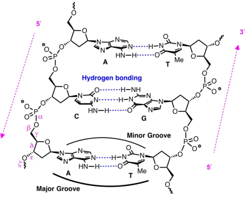
Figure 1.2 – Watson–Crick base pairing of DNA duplex.
As a consequence of the hydrogen-bonding donor and acceptor moieties present within each nucleobase, it is favourable for individual strands of DNA to pair up with one another (Figure 1.2). The strands are complementary and align in an antiparallel orientation, held together by a specific Watson–Crick base-paired hydrogen-bonding network*: adenine pairs with thymine and guanine pairs with cytosine, in agreement with the Chargraff group’s base
* Base pairing that does not follow the Watson–Crick model is also known (i.e., Hoogstein hydrogen bonding),
and the interested reader is directed elsewhere for a more thorough discussion.2 O N O O N N N O NH H H N N O HN H O P O O O O P O O O O PO O O O O PO O O O A C T N N N N HN H H N N O O Me G 3! 3! 5! 5! O O O O O A T N N N N HN H H N N O O Me Major Groove Minor Groove Hydrogen bonding α β γ δ ε ζ
composition data.3-5 Notably, guanine–cytosine pairs have three hydrogen bonds, while adenine–thymine pairs have only two, which results in extended regions of the former being more thermally stable than regions containing the latter. In addition to the antiparallel base-paired structure, the stereochemistry and conformation of the sugar moiety6 (Figure 1.4, p. 5), as well as the torsional degrees of freedom along the backbone (i.e., along angles α through ζ,
Figure 1.2) impart another structural feature – helicity.
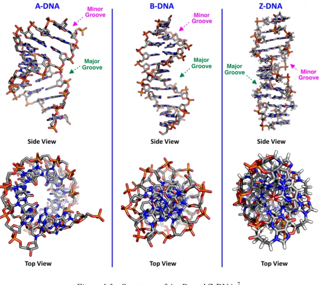
Figure 1.3 – Structures of A-, B- and Z-DNA.7
For DNA, three major helical structures have been observed with implications in biological processes: A-DNA, B-DNA, and Z-DNA (Figure 1.3).2,8 The most commonly observed form of DNA under physiological conditions and in vivo is the right-handed B-DNA duplex, in which the deoxyribose sugar is found in an S-type sugar pucker (Figure 1.4) and the
Minor Groove Major Groove Major Groove Minor Groove Minor Groove Major Groove
base pairs are effectively perpendicular to and centered over the helical axis (top view, Figure
1.3). In contrast, A-type duplexes are characterized by a comparatively thicker right-handed
helix, with a shorter distance between adjacent base pairs and a marked tilt and displacement of the base-pairs away from the helical axis (A-DNA, Figure 1.4). The A-type duplex contains a pentose sugar that is in an N-type sugar pucker (Figure 1.4), and it is commonly observed for dehydrated samples of DNA–DNA duplexes, as well as RNA–RNA and hybrid RNA– DNA duplexes. The remaining Z-DNA motif is a more significant departure from the other two motifs in that it is left-handed and is characterized by a zigzagging backbone as a consequence of the alternating sugar puckers for adjacent nucleosides; the sugar moiety of deoxyguanosine is found in an N-type sugar pucker, while those of thymidine, deoxycytidine, and deoxyadenosine are found in an S-type conformation. Furthermore, the guanine base is in a syn-conformation (i.e., its bulk extends over the pentose moiety rather than away from it), rather than the anti-conformation observed for A- and B-form nucleic acids. Z-DNA is less commonly observed in the cell, although it does occur in regions of alternating purine– pyrimidine sequences and has been observed as part of a junction within a strand of B-DNA, the so-called B-to-Z junction box, that is stabilized by Z-DNA-binding proteins.9
Figure 1.4 – Conformation of sugar moiety in nucleic acids.
Each duplex is also characterized by major and minor grooves that are adjacent to the base pairs and provide binding sites for proteins via hydrogen-bonding donor and acceptor motifs, as well as hydrophobic groups (i.e., methyl groups), where the latter is exclusive to the major groove.10 Owing to the previously described conformational differences, the major groove of A-DNA is effectively deeper and narrower than the corresponding major groove of
N-type (C3!-endo) Favoured for RNA
A-Type Duplexes R Bx O O OP S-type (C2!-endo) Favoured for DNA
B-Type Duplexes Bx O O R O P R = H, DNA R = OH, RNA O O O O O O
B-DNA, while the minor groove is shallower and wider. Overall, the B-form of the duplex is considered to be universal, in the sense that it can accommodate any known sequence of naturally-occurring DNA and is stable under a broad variety of conditions.2 Nevertheless, given that substantial variability in structural parameters (i.e., base pair tilt, rotation of helix per residue, pitch of the helix) has been observed with only mild changes to the environmental conditions, it would appear that the idealized structure of B-DNA does not represent a deep local energetic minimum.2
1.1.2 A Brief Overview of Protein Biosynthesis
The importance of nucleic acids, namely DNA and RNA, stems from their prominent role in encoding, transmitting, and expressing genetic information. This genetic information is used to direct the synthesis of proteins, which are macromolecular structures consisting of one or more chains of amino acid residues, that are ultimately responsible for performing a vast array of functions within living organisms, including transport, providing structural support, allowing movement, facilitating biochemical reactions as enzymes, and defending the body from antigens as antibodies. As such the collection of proteins within a cell will directly determine the function of a cell and is ultimately responsible for the overall health of an organism. Protein biosynthesis (Figure 1.5, p. 7) occurs through a highly-regulated sequence that may be conceptually separated into two major steps: transcription and translation. The former describes the flow of information from DNA to RNA, while the latter defines its propagation from RNA to protein. Interestingly, the movement of genetic material within biological systems follows the fundamental description put forth by Crick who stated that,
“[detailed residue-by-residue transfer of sequential] information cannot be transferred back from protein to either protein or nucleic acid.”11,12 In other words, once the genetic information from DNA has been used to synthesize a protein, the same protein cannot be used to arrive back at DNA or RNA; this does not, however, rule out the reverse flow of information from RNA to DNA.
Transcription occurs in the nucleus of the cell and is mediated by a family of nucleotidyl transferase enzymes referred to as RNA polymerases (Figure 1.5, p. 7).14 To initiate transcription in eukaryotic cells, well over 100 individual protein subunits must assemble in a promoter region along the DNA backbone. After initiation, the RNA polymerase is released from this large complex of proteins and moves stepwise along one strand of the unwound DNA backbone (i.e., the antisense strand) in the 5′ to 3′ direction at a pace of approximately 50 nucleotides per second.1 As it moves along the antisense strand, RNA polymerase catalyzes the formation of phosphodiester bonds between nucleotides on the RNA transcript and incoming ribonucleotide triphosphates (i.e., ATP, CTP, UTP, and GTP).1 Since the polymerase is only active in a segment of the gene in which the nucleobases are exposed and the helix unwound, as it moves along the DNA backbone, the RNA polymerase continues to unwind portions of the DNA double helix ahead of the polymerization active site in order to expose a new region of the template. Furthermore, the polymerase actively reforms the DNA double helix in the region behind the active site, by dynamically displacing the newly-formed RNA chain; in this way, only a small portion of a particular gene is unwound at any given time and the RNA transcript that forms is effectively single-stranded.
The RNA that forms is referred to as pre-messenger-RNA (pre-mRNA) because there are a number of post-transcriptional processing events that must occur in order to produce a mature mRNA molecule that can leave the nucleus and interact with the ribosomal machinery responsible for protein synthesis. Specifically, it is necessary to: 1) modify both ends of the pre-mRNA transcript, and 2) separate the sequence of nucleotides that codes for a protein (exons) from the intervening non-coding regions (introns) that are present. The first step is involves capping the 5′-end of the pre-mRNA transcript with a 7-methylguanosine moiety that is connected to the adjacent nucleoside through a 5′–5′ triphosphate linkage15; this is followed closely by polyadenylation of the 3′ end. Together, the capping and polyadenylation modifications assist the cell in discriminating between mRNA and other types of RNA, while serving as a way to verify that the mRNA produced is complete and the corresponding genetic information intact. The 5′-cap serves the additional role of assisting the cell in leaving the nucleus and plays an important role in the translation of mature mRNA into the corresponding protein.
The remaining post-transcriptional modification involves a series of splicing events, each of which effectively removes a single non-coding sequence (intron) through two sequential phosphoryl-transfer/transesterification reactions. Naturally, the process itself is significantly more intricate as a consequence of the need to effect splicing at specific sites. Accordingly, each splicing event is mediated by a RNA-protein complex (vis., the spliceosome), in which five additional RNA molecules and several hundred proteins are implicated.16 Unlike the previously described transcription sequence, the key steps of the splicing sequence are actually performed by the RNA molecules, rather than proteins; in addition to being responsible for recognizing the sequences that specify the site of splicing, the RNA molecules also participate in the phosphoryl-transfer/transesterification reaction itself.
Following successful splicing events, the mature mRNA transcript is ready to be exported to the cytosol through nuclear pore complexes, where it may be translated into protein.1 To ensure the mRNA has been properly processed, the cell can analyze the proteins that are bound to it, since it is expected that a characteristic presence (and corresponding absence) of certain proteins should be observed as a consequence of the sequence of processing the mRNA has gone through. The mRNA should only be released from the nucleus to the cytosol once the proteins bound to the mRNA collectively signal that transcription and the post-transcriptional modifications were successful.
To this point, the transfer of information is conceptually straightforward: since DNA and RNA are structurally and chemically similar, the former can serve as a template for the latter and direct the copying through complementary base pairing. In the case of protein synthesis, the information contained in RNA must effectively be translated into a different language, comprised of amino acids. Since there are only four unique nucleotides in mRNA and twenty different amino acids in a protein, a direct one-to-one translation of each “letter” is not possible. The rules that govern this translation are referred to as the genetic code and effectively state that the sequence of nucleotides in mRNA are read in consecutive groups of three, referred to as a codon. Each codon is recognized through the action of molecules known as transfer RNA (tRNA), which are precisely-folded single-stranded molecules of RNA, with a unique 3D structure. At one end of their scaffold, tRNA molecules can covalently bond with a single amino acid through an ester bond, while they simultaneously recognize and bond to
the nucleobases in each codon through complementary hydrogen-bonding base-pairing interactions that occur at another site (Figure 1.2, p. 3). Amino acids are covalently coupled to the appropriate tRNA molecule through the action of aminoacyl-tRNA synthetases, and it occurs through a two-step mechanism involving initial attachment of the amino acid and a subsequent discrimination step to ensure that the correct amino acid has been attached.
The mechanism by which amino-acid-carrying tRNA molecules link those amino acids together in a specific order to produce a protein – based on the sequence of codons in mRNA – is summarized in Figure 1.5 (p. 7). The mRNA sequence is decoded within a well-studied structure known as the ribosome, which comprises two major subunits that are together composed of more than 50 different proteins and several strands of ribosomal RNA; there are also three major sites within the ribosome where each tRNA may be bound and specific reactions/events occur.17-19 The small subunit provides a framework for the tRNA molecules to accurately pair with the strand of mRNA, while the large subunit catalyzes the formation of peptide bonds that link together amino acids in the forming polypeptide chain. Protein synthesis is initiated through a start codon, AUG, close to the 5′-end that codes for methionine and continues as the strand of mRNA is read in the 5′ to 3′ direction, with the individual amino acids added to the C-terminus of the growing polypeptide.20 In general this occurs through a multistep process: 1) an incoming tRNA molecule will bind to the mRNA scaffold through hydrogen-bonding interactions with the codon, 2) the growing polypeptide is transferred to the adjacent tRNA molecule as a new peptide bond forms, and 3) the large and small ribosome subunits shift towards the 3′-end of the mRNA strand, creating space in the ribosome for another amino-acid-containing tRNA molecule, while ejecting the amino-acid-depleted tRNA molecule another from the 5′-end. This process continues until a stop codon is encountered, at which point the two ribosome subunits separate and the polypeptide chain is released. Next, the polypeptide chain must be correctly folded into its appropriate 3D conformation, bound to additional cofactors, and assembled with other protein subunits (if required).21-25 It is only at this point that the protein is considered to be mature and functional.
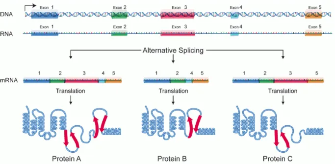
Figure 1.6 – Alternative splicing.26
The complexity of the biosynthetic sequence for protein synthesis is remarkable, yet despite the significant molecular machinery in place for error-checking, sometimes it does not proceed as expected. For example, aberrant splicing events (Figure 1.6) may lead to the production of protein isoforms that have different biological properties, particularly their ability to effect catalysis, their subcellular localization, and the protein–protein interactions they can participate in.27 While the effects of the abnormal splicing events are difficult to predict, they become much more obvious when they lead to diseases; examples of diseases related to aberrant splicing events include spinal muscular dystrophy, Hutchinson–Gilford progeria syndrome, and Prader–Willi syndrome, amongst others.27 Numerous studies have also shown that alternative splicing patterns are quite pervasive in cancerous cells.28,29
The following section describes a therapeutic approach for addressing these challenges as well as those associated with the regulation of gene expression.
1.1.3 Overview of the Antisense Approach
The pursuit of potential cures and treatments for diseases and challenges related to gene expression has frequently centered around developing small molecule therapeutics (molecular weight of less than 800 Daltons) that inhibit or increase the activity of proteins through the interactions of those small molecules with amino acid residues in binding pockets
Figure 1.7 – Small molecule therapeutics versus antisense approach.30
of the protein; consequently, the interaction is based, at least partially, on shape complementarity with the protein (Figure 1.7). However, as additional information has surfaced on the biosynthetic pathways for gene expression (Figure 1.5, p. 7), a number of other targets began to emerge, including nucleic acids themselves. One such approach – referred to as the antisense approach* – is based on the premise that gene expression can be regulated by targeting RNA and gene sequences themselves, rather than proteins. In other words, while traditional small molecule therapeutics inhibit disease-causing proteins based on the shape of the protein, antisense therapeutics can directly inhibit the production of the protein itself by binding directly to the protein’s mRNA and gene sequence through
* The term “antisense” is favoured since the nucleotide sequence of a particular therapeutic oligonucleotide is
complementary to the corresponding target RNA; therefore it also has a sequence that is analogous to the DNA antisense strand that serves as the source code for a given protein (Figure 1.5, p. 7).
established Watson–Crick base-pairing interactions (Figure 1.7). The antisense oligonucleotides are typically 8 to >50 nucleotides in length, with an average length of 20 nucleotides, which corresponds to a molecular weight of roughly 7000 Daltons.
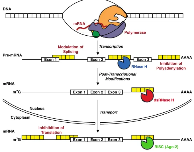
The appeal of the antisense approach is that there is significant potential to create gene-selective therapeutics using well-established concepts: base-pairing provides an opportunity for rational drug design based on hybridization (Figure 1.2, p. 3) and as our knowledge of the molecular biology of the cell increases, so too does the opportunity for rationally designing antisense oligonucleotides based on increasingly accurate models and validated RNA targets. For the purpose of modulating gene expression, RNA transcripts may be targeted by antisense oligonucleotides at many different points during protein biosynthesis; a number of examples are shown in Figure 1.8 (p. 14). Following the binding of an antisense oligonucleotide to an RNA transcript, there are two primary mechanisms by which inhibition can occur: 1) interfere with the function of RNA, without promoting its degradation (i.e., modulation of RNA splicing, inhibition of translation or polyadenylation), or 2) promoting the degradation of RNA through endogenous enzymes (i.e., RNase H or RISC/Argonaute 2) by incorporating cleavage sequences that are directly designed into the antisense oligonucleotide. Notably, the mechanisms that result in degradation of the target RNA have been found to be more robust, particularly that of RNase H, which is a sequence-nonspecific endonuclease that cleaves RNA strands in RNA–DNA hybrids.31
The first explicit disclosure of a therapeutic antisense strategy that selectively targeted RNA was described in 1978 by Zamecnik and Stephenson, who demonstrated the ability of a synthesized 13-nucleotide-long oligodeoxyribonucleotide to inhibit the viral activity of Rous sarcoma virus 35S RNA, by binding to the viral RNA through complementary base-pairing.32 At the time the synthesis itself was no small feat, and Zamecnik and Stephenson had the additional foresight to recognize the potential of this strategy for designing therapeutic agents; they proposed potential binding sites for the oligonucleotide in RNA, other targets (i.e., influenza, measles, and rabies), and even described practical approaches for stabilizing synthetic oligonucleotides through modifications to the 3′- and 5′-termini to protect against exonucleases that degrade nucleic acids.
Figure 1.8 – Antisense mechanisms.33
Their attempts to improve the stability of oligonueotides were particularly insightful, since they further highlight one of the potential challenges associated with using antisense oligonucleotides to target RNA or DNA: unmodified RNA and DNA are inherently unstable molecules in biological systems. Despite the explicitly described potential for oligonucleotides reported by Zamecnik and Stephenson, essentially no medicinal chemistry research was performed on oligonucleotides to improve their therapeutic profile until the late 1980s. While significant strides have been made since that time, to date only two antisense drugs have been approved by the U.S. Food and Drug Administration: 1) formivirsen34-36 for cytomegalovirus retinis (i.e., inflammation of the retina of the eye) and 2) mipomersen37-40 for homozygous familial hypercholesterolemia (i.e., cholesterol). A brief description of modifications that have been made to oligonucleotides to improve their therapeutic potential is provided in Section 1.1.4, but it is apparent that a great deal remains to be discovered.
mRNA Polymerase DNA Pre-mRNA Transcription Post-Transcriptional Modifications Exon 3 Exon 2 Exon 1 RNase H Modulation of Splicing Inhibition of Polyadenylation AAAA mRNA Transport Exon 2 Exon 1 dsRNase H Exon 3 AAAA m7G Nucleus Cytoplasm mRNA Exon 2 Exon 1 RISC (Ago-2) Exon 3 AAAA m7G Inhihibition of Translation
1.1.4 Modified Oligonucleotides
Unmodified oligonucleotides are not ideal therapeutic agents as a consequence of their instability within biological systems. In particular, they are susceptible to cleavage by ubiquitous nucleases and have rather poor pharmacokinetic properties; unmodified oligonucleotides are small enough to be filtered by the glomerulus and are only weakly bound to plasma proteins, which leads them to be rapidly filtered and excreted.41 Furthermore, the ability of a strand of nucleic acid to discriminate between a natively complementary construct and a synthesized oligonucleotide is expected to be rather poor, since the base-pairing interactions in both cases are quite similar. Likewise, the affinity of DNA for RNA is lower than the affinity RNA has for itself,42 which presents another challenge if one is targeting RNA using DNA-like antisense oligonucleotides to activate the robust RNase H pathway. Fortunately, the nucleic acid scaffold has a number of sites amenable to modification that can be used to improve the therapeutic profile of the antisense constructs, including the phosphodiester backbone, the nucleobase, and the sugar moiety (Figure 1.9, p. 16).
Thus far, the most useful modification has proven to be the substitution of a non-bridging oxygen atom in the phosphodiester backbone with a sulfur atom, forming a phosphorothioate backbone (1.7, Figure 1.9).43 The introduction of the phosphorothioate linkage is particularly beneficial because: 1) it greatly increases the stability of the oligonucleotide to nucleolytic degradation, 2) it induces RNase H cleavage of the target RNA, and 3) it increases binding to plasma proteins, which prevents rapid excretion, while further facilitating binding to other acceptor sites.44 Although the inclusion of the phosphorothioate linkage decreases the affinity of an antisense transcript for its intended RNA target (ΔTm ≈ –2 °C/modification),* this drawback can be significantly attenuated by including modified nucleosides that increase the affinity of the antisense construct for its complementary strand. Overall, the benefits gained by including the phosphorothioate linkages greatly outweigh the downside, and for this reason they are generally included alongside other classes of
* T
m values refer to the midpoint on a curve of UV-absorption versus temperature, and are indicative of the point
at which 50% of an oligonucleotide duplex has been unwound into the corresponding single strands.45,46 The values provided in this chapter are given as the difference (ΔTm) between DNA sequences containing the modified nucleotide and an analogous unmodified sequence of deoxyribonucleotides that serves as a control, when each is hybridized to complementary strands of DNA or RNA.
oligonucleotides in order to achieve the improved therapeutic properties required for use as a drug. Other modifications to the backbone have also been explored with varying levels of success and appeal, including boranophosphates,47 phosphorodithioates, methylphosphonates, and phosphoramidates, amongst others.48
Figure 1.9 – Representative oligonucleotide modifications.
Another site that has been extensively modified within the nucleic acid scaffold is the nucleobase itself.49,50 Typically, nucleobase modifications have focused on increasing the binding affinity for complementary nucleic acids, since the preservation of the Watson–Crick base-pairing interaction is crucial for the recognition of complementary nucleic acid targets.
O P R = H, DNA R = OH, RNA O O O Bx O O R O P 1.1 Phosphorothioate ΔTm –2 °C/mod. vs. RNA Backbone modification O S O Bx O O 1.5 2"-O-Methyl ΔTm +1 °C/mod. vs. RNA Sugar modification O Bx O O OMe 1.4 2"-Fluoro ΔTm +2 °C/mod. vs. RNA Sugar modification O Bx O O F 1.6 2"-O-Methoxyethyl (MOE) ΔTm +2 °C/mod. vs. RNA Sugar modification O Bx O O OMOE O P 1.3 Morpholino ΔTm 0 °C/mod. vs. RNA Backbone & sugar modification
O Me2N N O O Bx O 1.7
Locked Nucleic Acid ΔTm +5 °C/mod. vs. RNA Sugar modification O Bx O O 1.2 5-Propynyl ΔTm +2 °C/mod. vs. RNA Nucleobase modification O N O O NH O O Me
Sites Amenable to Modification: 1) Phosphodiester backbone 2) Nucleobase
3) Sugar: •substitution
•conformational restriction
Overall, as a consequence of the need to maintain comparable hydrogen bond donor–acceptor regions and accommodate the nucleobase, there are substantial restrictions on the portions of the nucleobase that may be productively modified. One prototypical example of a nucleobase modification is the inclusion of a propynyl moiety at the C5 position of the uracil (1.2, Figure
1.9), which results in an overall extension of the π-rich surface and an increase in the available
hydrophobic surface.51 This modification effects an overall increase in the stability of duplexes as a consequence of the enhancement of intrastrand stacking interactions between the nucleobases. Unfortunately, 5-propynyl-pyrimidine-containing oligodeoxynucleotides with a phosphorothioate backbone induce severe liver toxicity in vivo, which could not be attenuated through additional modifications.52 Further changes to the nucleobase moiety have also been explored, including the incorporation of 5-thiazoylpyrimidines,51 diaminopurines,53 and phenoxazines,54-57 but despite extensive efforts only modest progress has been made to address the ability of nucleobase modifications to support RNase H activity and to attenuate their often poor in vivo pharmacological profiles.
In contrast, modifications to the pentose sugar moiety of the nucleic acid scaffold have been markedly more successful overall. Interestingly, even complete substitution of the furanose sugar with a morpholine ring was found to be well-tolerated (1.3, Figure 1.9), affording scaffolds that have similar affinity to DNA–DNA duplexes and are also stable to nucleases as a consequence of the phosphoramidite bond.58-61 The morpholino phosphoramidites do not, however, activate RNase H and are primarily used in steric blocking mechanisms (e.g., for alternative splicing or to prevent translation). Replacement of the sugar moiety with hydroxyproline or even peptides has also been explored, although many obstacles remain to be overcome for each.
By comparison, modifications to the C2′-position of the furanose ring have been much more successful than complete replacement of the sugar moiety, owing in part to the ability of substituents at that position to effectively pre-organize the pentose moiety into an N-type sugar pucker (Figure 1.4, p. 5) as a consequence of their electronegativity or steric bulk.62 This results in an increase in binding affinity and also confers the additional benefit of nuclease resistance by virtue of the proximity of the C2′-substituent to the C3′-phosphodiester bond. Incorporating a (R)-configured fluorine atom at the C2′-position (1.4, Figure 1.9) favours the
N-type sugar conformation as a consequence of the electronegativity of the fluorine atom,
while improving stability of the nucleoside relative to RNA. Although this modification does not activate RNase H or improve nuclease resistance beyond that displayed by DNA, the corresponding (S)-configured analogue was shown to activate RNase H.63
The incorporation of C2′-alkyl ethers (e.g., 2′-O-methyl 1.5 and 2′-O-methoxyethyl
1.6, Figure 1.9) represents another group of modifications, which are particularly appealing in
that they improve binding affinity, while also imparting on the corresponding antisense transcripts a substantial increase in resistance to degradation by nucleases. The 2′-O-methoxyethyl substitutent (1.6, Figure 1.9) is one of the most studied and oft-used of this class of modifications, and is often referred to as one of the representative modifications of second-generation antisense drugs since it is present in mipomersen; phosphorothioate linkages (1.1,
Figure 1.9) exemplify the characteristic first-generation modification, and they are present in
fomivirsen as well as mipomersen. Although RNase H activity is significantly attenuated for many nucleotides containing substituents at the C2′-position, a gapmer strategy can be used to address this limitation. The gapmer strategy involves including a sequence of unmodified deoxyribonucleotides (typically with a phosphorothioate backbone) between regions containing the modified nucleotides.41 In this way, the central portion of the antisense oligonucleotide can recruit RNase H, while the flanking regions effectively improve nuclease resistance and increase affinity for complementary strands.
In contrast to incorporating discrete substituents at the C2′ position to confer nuclease resistance and a conformational bias to the sugar pucker, one can also imagine pursuing a complementary strategy for inducing the desired conformational bias: restricting rotation around torsional bonds along the nucleotide scaffold (Figure 1.10). Constraining the phosphodiester backbone (i.e., α,β-constrained nucleic acid 1.8, Figure 1.10)64 or torsion angles γ and δ (i.e., tricyclo-DNA 1.9, Figure 1.10)65,66 conferred a significant increase in duplex thermal stability (ΔTm ≈ +3 °C/mod.). However, the most promising increase was observed when the furanose sugar was locked in an N-type sugar pucker by virtue of including a methylene tether between the C2′-oxygen atom and the C4′ position.67-71 The resultant monomer, which has been dubbed Locked Nucleic Acid (LNA, 1.7, Figure 1.10), has a sugar moiety that is effectively locked into the same conformation found in RNA, and as such,
oligonucleotides that incorporate LNA monomers tend to form A-type duplexes (Figure 1.3, p. 4). As a consequence of the constrained scaffold of LNA,72-74 the oligonucleotides also display a remarkably high increase in affinity and specificity for the complementary strand relative to the corresponding DNA–RNA duplex.75 Furthermore, oligonucleotides that include LNA monomers demonstrate high in vivo stability and a general lack of toxicity. While poly-LNA oligonucleotides do not inherently activate RNase H, implementation of a gapmer strategy has been successfully used to overcome this limitation.
Figure 1.10 – Conformational restriction strategies.
Following their disclosure of the promising hybridization properties of LNA, the group of Wengel evaluated the seven other stereoisomers of this locked scaffold to determine whether the isomeric forms had comparable properties.71 While all but two of the stereoisomers displayed similar binding affinity for complementary RNA relative to a DNA reference, the truly surprising observation came in the form of one of these stereoisomers exhibiting an increase in affinity for RNA that was comparable to the remarkably high value measured for LNA. This nucleoside was revealed to be the C1′ epimer of the enantiomer of
O 1.7
Locked Nucleic Acid ΔTm +5 °C/mod. vs. RNA Sugar modification O Bx O O O Bx O P O O α β γ δ ε ζ b a) Restrict rotation around α and β a O Bx O c) Lock furanose ring in N-type sugar pucker γ δ R c 1.9 tricyclo-DNA ΔTm +2 to 4 °C/mod. vs. RNA R = H, DNA R = OH, RNA O O PO O O O 1.8
α,β-constrained nucleic acid ΔTm +3 °C/mod. vs. RNA O Bx O O P O O O β α O Bx O O O b) Restrict rotation around γ and δ