Université de Montréal
L’infestation palpébrale et faciale au
Demodex folliculorum
par Sarah Aumond
École d’Optométrie Faculté des études supérieures
Mémoire présenté à la Faculté des études supérieures en vue de l’obtention du grade de
Maîtrise en Sciences de la Vision option Sciences fondamentales et appliquées
octobre 2018
Résumé
Le Demodex folliculorum [D. folliculorum] est l’ectoparasite le plus fréquent dans les follicules des cils et de la peau. Son rôle précis dans la microflore n’est pas encore déterminé, mais il semble agir comme un commensal. Par contre, lorsque sa quantité dépasse un certain seuil, il devient pathogénique. Son infestation est associée à une blépharite antérieure et/ou postérieure, une sécheresse oculaire et plusieurs types d’affectation du visage tels que l’acné rosacée. Comme il peut se retrouver autant dans les paupières que la peau du visage, cela suggère qu’une infestation palpébrale pourrait être associée à une plus grande densité faciale dudit parasite. Pour vérifier cette hypothèse, des participants ayant diverses sévérités de blépharite antérieure à D. folliculorum ont été recrutés. Une épilation de cils infestés a été effectuée, permettant un décompte des parasites. D’un autre côté, l’infestation faciale a été évaluée par une biopsie superficielle standardisée de la peau du front, prélevant ainsi quelques couches de l’épiderme superficiel et le contenu des follicules pileux. L’analyse des résultats révèle que la présence d’une blépharite à D. folliculorum est associée à une densité faciale du parasite au-delà du seuil de normalité. De plus, le degré d’érythème facial augmente avec la sévérité de l’infestation des paupières et du visage. Certains signes et symptômes oculaires sont aussi affectés par la présence de la blépharite. Par exemple, on retrouve une réduction de la hauteur du ménisque lacrymal et une augmentation des yeux larmoyants lors d’une infestation légère à modérée, de même qu’une augmentation de la sensation de picotement à la bordure des paupières en présence d’une infestation sévère.
En conclusion, comme les infestations faciale et palpébrale sont concomitantes, autant les professionnels de la vue que les dermatologistes doivent être impliqués dans le diagnostic et la prise en charge de cette condition. Une approche multidisciplinaire serait certainement bénéfique pour les individus atteints.
Mots-clés : Demodex folliculorum, blépharite, pathophysiologie du cil, démodécie, follicule, biopsie superficielle faciale
Abstract
Demodex folliculorum [D. folliculorum] is the most common ectoparasite in the eyelash and skin follicles. Its precise role in the microflora is not yet determined, but it seems to act as a commensal. However, when its quantity exceeds a certain threshold, it becomes pathogenic. Its infestation is associated with anterior and/or posterior blepharitis, dry eye disease and several types of facial conditions such as rosacea. Since D. folliculorum can be found as much in the lids as in the facial skin, this suggests that a palpebral infestation may be associated with a higher facial mite density. To verify this hypothesis, participants with various severities of D. folliculorum blepharitis were recruited. The epilation of infested lashes was performed, to assess mite counts. The facial infestation was quantified by a standardized skin-surface biopsy of the forehead, thus extracting a few layers of the superficial epidermis and the content of the hair follicles. The results revealed that the presence of Demodex blepharitis is associated with a facial mite density beyond the normal threshold. In addition, the degree of facial erythema increases with the severity of palpebral and facial infestations. Some ocular signs and symptoms are also affected by the presence of Demodex blepharitis. For example, a mild to moderate infestation is associated with a tear meniscus height reduction as well as more watery eyes, and a severe infestation with increased itching along the lids.
In conclusion, since facial and palpebral infestations are concomitant, both eye care professionals and dermatologists should be involved in the diagnosis and management of this common parasite. In addition, a multidisciplinary approach would ultimately benefit the affected individuals.
Keywords :
Demodex folliculorum, blepharitis, eyelash pathophysiology, demodicosis, follicle, skin-surface biopsy
Table des matières
Résumé ... i
Abstract ... ii
Table des matières... iii
Liste des tableaux ... iv
Liste des figures ... v
Liste des sigles ... vi
Liste des abréviations ... vii
Remerciements ... viii
1. Introduction ... 1
1.1 L’ectoparasite Demodex folliculorum ... 1
1.2 L’infestation palpébrale au Demodex folliculorum ... 2
1.2.1 Article 1: The eyelash follicle features and anomalies, a review ... 4
1.2.2 Prise en charge de l’infestation palpébrale ... 33
1.3 L’infestation faciale au Demodex folliculorum ... 34
1.3.1 Prise en charge de l’infestation faciale ... 37
1.4 Le lien entre l’infestation palpébrale et faciale ... 38
2. Méthodologie ... 39
2.1 Objectifs ... 39
2.2 Raisonnement ... 39
2.3 Hypothèses ... 40
3. Résultats ... 41
3.1 Article 2: Palpebral and facial skin infestation by Demodex folliculorum ... 42
4. Discussion ... 73
5. Conclusion ... 77
Liste des tableaux
Article 1
Tableau 1. Hair anomalies and their associated pathologies……….14
Tableau 2. Etiologies and management of lash conditions………15
Article 2 Tableau 1. Exclusion criteria………..46
Tableau 2. Clinician Erythema Assessment (CEA)………51
Tableau 3. Facial skin conditions reported with an effect or not on facial mite density………51
Tableau 4. Demographics of each severity group of Demodex blepharitis……….53
Tableau 5. Percentage (%) of each dermatological condition per severity group………55
Tableau 6. Ocular and lid variables……….57
Tableau 7. Mean mite count for each lid……….57
Tableau 8. Ocular Surface Disease Index, Non-Invasive Break Up Time and Tear Meniscus Height for each Demodex blepharitis severity………58
Liste des figures
Article 1Figure 1. The general morphology of the eyelash and its surrounding skin………6
Figure 2. The anatomy of the eyelash……….7
Figure 3. The life cycle of the eyelash………...10
Figure 4. Eyelash debris assessment by eyecare professionals……….22
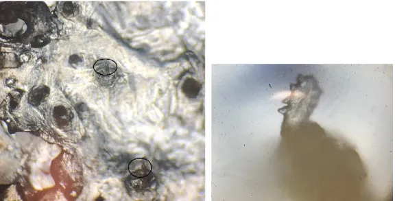
Article 2 Figure 1. Demodex folliculorum (D.f) from the lashes under 40-400x magnification………..50
Figure 2. Demodex folliculorum (D.f) from the forehead skin under 40-400x magnification...50
Figure 3. Forehead Demodex densities (mite count/cm2) expressed as marginal mean ± standard error (SE)……….54
Figure 4. Scatterplot of the degree of facial erythema and the forehead Demodex density [mite count/cm2]………..………55
Figure 5. Lash mite count expressed as mean ± standard error (SE) for each severity group...56
Figure 6. Percentage (%) of participants with different symptoms (itching along the lid margin, ocular itching and watery eyes) for each severity group………..……59
Liste des sigles
ANCOVA: A one-way analysis of covariance ANOVA: A one-way analysis of variance ROSCO: global ROSacea COnsensus
Liste des abréviations
CD: cylindrical dandruffCEA: clinician erythema assessment D. brevis: Demodex brevis
DED: dry eye disease
D. folliculorum: Demodex folliculorum ECP: eye care professional
EGFR: epidermal growth factor receptor
IVCM: in vivo confocal microscopy, microscopie confocale in vivo MG: meibomian gland; glandes de Meibomius
MGD: meibomian gland dysfunction NIBUT: non-invasive break-up time OSDI: ocular surface disease index SSSB: standardized skin-surface biopsy TBUT: tear breakup time
TLR: receptors toll-like TMH: tear meniscus height
Remerciements
Si quelqu’un m’avait dit lors de ma graduation en 2011 que j’allais revenir à l’école d’optométrie pour me lancer dans une maîtrise en Sciences de la vision, je ne l’aurais pas cru. Pourtant, me voilà à discuter avec mon amie Audrey d’un possible retour à l’école, lors d’une randonnée de ski de fond entre Noël et le jour de l’an 2016. Merci Audrey de m’avoir mis cette idée en tête et de m’avoir convaincu de contacter Etty Bitton, afin qu’elle soit ma directrice de maîtrise. Merci Etty pour ta confiance, ton support et toutes les heures que tu as passées à me relire… En plus, tu m’as aidé à obtenir des bourses d’études, ce qui m’a permis de me concentrer à 100% sur mon projet de recherche. Je n’aurais pas pu avoir une meilleure personne pour me guider durant ces deux dernières années. C’est stimulant de côtoyer quelqu’un qui s’investit autant dans tout ce qu’elle fait, et qui réussit à te transmettre sa passion. Grâce à toi Etty, j’aime lire des articles scientifiques et faire de la recherche!
J’aimerais aussi remercier le personnel et les professeurs de l’école d’optométrie, qui m’ont encouragé dans mon parcours académique, et merci tout spécialement à Walter Wittich, qui m’a fait aimer les statistiques, ce qui n’est pas une mince affaire. Merci à Julie, Anton, Vanessa et Diane, aussi étudiants au cycle supérieur, pour votre motivation et surtout, pour m’avoir diverti!
Je dois remercier mon amoureux des 17 dernières années, qui m’a supporté dans cette aventure, autant financièrement qu’émotionnellement. Lorsque j’avais des doutes, il était toujours là pour m’écouter, me rassurer, me pousser à continuer… et m’offrir un soutien informatique, et ce, jour et nuit ! Merci d’avoir été si compréhensif… Et finalement, un merci tout spécial à Mathéo, qui m’a accompagné 24h sur 24 durant les neuf derniers mois de mon projet de recherche, et qui me laisse un peu de temps chaque jour, pouvant varier de quelques minutes à quelques heures, pour finaliser mon mémoire.
1. Introduction
1.1 L’ectoparasite Demodex folliculorum
Les êtres humains ont une flore qui leur est propre et qui leur permet de réguler et maintenir l’homéostasie. La microflore cutanée peut contenir entre autres deux ectoparasites spécifiques aux humains, soient le Demodex folliculorum [D. folliculorum] et le Demodex brevis [D. brevis] (Nicholls et al., 2017). Les deux espèces sont des arthropodes qui ont une portion antérieure composée d’une tête et de quatre paires de pattes, ainsi qu’une région postérieure composée d’un long opisthosoma sans anus. Leur cycle de vie est d’une durée de 15 à 18 jours, de l’éclosion des œufs pondus par les femelles jusqu’à leur pleine maturité (Rufli et al., 1981). Les parasites sembleraient se transférer d’une personne à l’autre par contact direct avec des articles domestiques, tels que la literie et les serviettes, de même que la poussière (Tarkowski et al., 2015). Bien que leur rôle précis dans la microflore n’est pas complètement établi, ils agiraient comme des parasites commensaux (Lacey et al., 2011). Le D. Brevis se loge profondément dans les follicules ciliaires et pileux, ainsi que dans les glandes de Meibomius (Nicholls et al., 2017). L’espèce la plus prévalente est le D. folliculorum, retrouvé surtout dans les couches superficielles de la peau du visage et des paupières, ainsi que dans les follicules qui lui sont rattachés, soient les follicules pileux et ciliaires (Lacey et al., 2011; Nicholls et al., 2017). Il a été établi que sa fréquence augmente avec l’âge et qu’il serait même présent chez 100 % des gens âgés de plus de 70 ans (Post et al., 1963). Une association avec l’âge n’a pas été démontrée quant au D. Brevis. Ce projet de recherche se concentrera sur le D. folliculorum. Une description détaillée des différentes couches de la peau ainsi que des follicules pileux et ciliaires est de mise afin de mieux comprendre l’interaction du D. folliculorum avec son milieu. Une revue de littérature a donc été réalisée et publiée à ce sujet. La base de données Ovid MEDLINE a été utilisée pour rechercher les mots-clés « eyelash physiopathology /abnormalities/pathologies » et quelques articles complémentaires ont été dénichés sur Embase. Cette revue de littérature non-systématique, aussi appelée « scoping review », est donc présentée à la sous-section 1.2.1.
Bien que le D. folliculorum fait partie de la microflore normale, il devient pathogénique lorsqu’il se retrouve en quantité excessive (Lacey et al., 2011). Il y a alors une infestation du tissu sous-jacent, que ce soit les paupières ou la peau du visage. Les infestations palpébrale et faciale à D. folliculorum sont décrites aux sections 1.2 et 1.3, respectivement.
1.2 L’infestation palpébrale au Demodex folliculorum
Une infestation des paupières par le D. folliculorum se traduit entre autres par une blépharite antérieure [cils] et postérieure [glandes de Meibomius] (Bhandari et al., 2014; Chen et al., 2017; Lopez-Ponce et al., 2017). Des analyses avec microscopie confocale in vivo [IVCM] démontrent que le parasite est surtout présent à plusieurs niveaux du follicule ciliaire et parfois, à l’extérieur du follicule entre les cils (Randon et al., 2015). De plus, les cils ayant des follicules ciliaires colonisés par une grande quantité de D. folliculorum sont entourés à la base seulement par des gaines gélatineuses appelées « cylindrical dandruff [CD] » (Gao et al., 2005). Les mêmes chercheurs ont établi que les CD sont un signe pathognomonique d’une quantité excessive de Demodex. Une infestation à D. folliculorum peut donc être l’étiologie d’une blépharite antérieure. Plusieurs autres types de blépharite antérieure existent, dont la séborrhéique (Paulino, 2017; Wolf et al., 2014) ou la bactérienne [surtout par les staphylocoques] (Bernardes et al., 2010). Moins fréquemment, elle peut être causée par un autre parasite, le Phthiriasis palpebrarum (Turgut et al., 2009).
D’un autre côté, il n’a pas été démontré que le D. folliculorum serait l’une des causes de la blépharite postérieure. En effet, le D. folliculorum n’est pas retrouvé à l’intérieur des glandes de Meibomius, contrairement à D. brevis (Randon 2015). La prévalence du D. brevis augmente en présence d’une dystrophie des glandes de Meibomius et/ou de chalazion (Liang et al., 2014; Liang et al., 2018). Il est tout de même possible de diagnostiquer une blépharite antérieure à D. folliculorum concomitante à une blépharite postérieure. De même, une blépharite à Demodex peut déséquilibrer le film lacrymal, causant ainsi une sécheresse oculaire. Des concentrations plus élevées de cytokines dans les larmes, surtout IL-17, sembleraient expliquer l’inconfort oculaire ressenti et les télangiectasies fréquemment retrouvées lors d’une infestation palpébrale (Kim et al., 2011).
La blépharite à Demodex peut être asymptomatique ou associée à des signes et symptômes non spécifiques tels qu’un picotement, une rougeur sur les paupières, des yeux larmoyants ou une sensation de sécheresse oculaire (Sedzikowska et al., 2016). De plus, comme tous les autres types de blépharite antérieure, elle peut avoir à long terme des répercussions négatives sur les cils telles que des changements de pigmentation, de direction, de position ou de croissance. Ces anomalies sont associées à des pathologies des cils telles que la poliose [perte de pigmentation], la trichiase [mauvaise direction] et le milphosis [perte des cils] (Ferreira et al., 2010; Kumar et al., 2012; Vij et al., 2015). Ces anomalies peuvent aussi être associées à l’application de produits cosmétiques sur les cils. Plusieurs recommandations ont donc été émises afin de réduire l’impact des cosmétiques sur les cils et sur la surface oculaire (O'Dell et al., 2017). La description de plusieurs pathologies ciliaires, leurs étiologies, les facteurs iatrogéniques qui affectent les cils tels que les cosmétiques de même que l’implication du D. folliculorum sur l’homéostasie des paupières font partie d’une revue de littérature. Cette dernière met aussi en évidence que les recherches sur le cil et son follicule ont un retard considérable par rapport à celles sur les cheveux. Les cils ont longtemps été considérés comme un simple poil sans rôle particulier. De ce fait, peu d’intérêt leur était porté par la communauté scientifique. De plus, le prélèvement d’un follicule ciliaire humain est très complexe, étant donné qu’une portion de la paupière doit être retirée. Ce sont surtout les modèles animaux qui ont permis de démontrer les caractéristiques spécifiques des follicules ciliaires et ainsi, en faire des entités distinctes. Dans les dernières années, de plus en plus de recherches s’orientent sur le cil et son follicule, ce qui comble graduellement l’écart avec la recherche sur les cheveux (Paus et al., 2016; Thibaut et al., 2010). La revue de littérature se retrouve à la section suivante (1.2.1).
1.2.1 Article 1: The eyelash follicle features and anomalies, a review
[publié dans Journal of Optometry (2018), 11, 211-222]Sarah Aumond1, Etty Bitton1
1École d'optométrie, Université de Montréal, CP 6128 succursale centre-ville, Montréal, Qc, Canada
Abstract
The primary role of eyelashes is to protect and maintain the health of the lid margin. However, the mechanisms to fulfill this role are not fully understood. Unravelling these mechanisms will stand to greatly improve the efficiency of eye care professionals’ interventions in anomalies of the eyelashes. The aim of this article is to provide a review on eyelashes including highlights and new avenues for research; the biology of both the lash and its follicle; the pathophysiology and management of lash anomalies by eye care professionals; and the effect of iatrogenic factors on lashes. Using the database of Ovid MEDLINE, we reviewed studies specifically directed on human/mammalian eyelashes and key articles on current trends in scalp hair methodologies that can be applicable to lash research. The eyelash morphology, pigmentation and growth rate have been documented using techniques ranging from lash imaging to follicle immunohistochemistry. Furthermore, studies have demonstrated that the lash follicle is sensitive to many factors of the external environment, a variety of systemic/topical medications and cosmetics. Recently, aerodynamic studies using a mammalian eye model confirmed that an optimal lash length was needed so that eyelashes serve a protective role in reducing the number of particles that can reach the eye. Despite recent advances in lash research, studies are still scarce, due to the limited availability of the human lid for sampling. This review brings awareness that further research is needed with respect to eyelashes and will hopefully reduce the gap with scalp hair research.
Keywords
Introduction
Little research has been done on the human eyelash on account that most of the attention has been directed to research on hair for people suffering from scalp hair loss. However, recent discoveries on the role of eyelashes and its distinctive characteristics have led to an increased scientific interest. Moreover, eyelashes are now considered an important aspect of the facial aesthetic and are the object of various beauty treatments to enhance them (Draelos, 1991; Jones, 2011). Since eyelashes form a barrier between the external and internal environment of the eye, they are extremely sensitive to a variety of threats and irritants and are highly innervated to perform that function (Montagna et al., 1969). Eyelashes are an integral part of the lid margin anatomy, much like the Meibomian glands, eyelid skin and biofilm, each contributing to the overall homeostasis of the ocular surface. As such, it is important to maintain their integrity. As a whole, the lid margin is responsible for the production of the tear film lipid layer and the protection of the eye from external trauma. Via the blink, it distributes the tears towards the nasolacrimal puncta found in the inner portion of the lid margin (Willcox et al., 2017). If any part of the lid margin is inflamed, it can induce a tear film disturbance or instability which can, in turn, affect the ocular surface (Craig et al., 2017). Left untreated, this inflammatory cascade can develop into dry eye disease (Craig et al., 2017). Therefore, studying the eyelash and its pathophysiology is valuable for researchers and eye care professionals [ECPs] alike, to maintain ocular surface homeostasis.
An Ovid MEDLINE search for eyelash physiopathology/abnormalities/pathologies has led to 419 human and 59 non-human publications. The articles that were selected for this non-systematic review concentrated on the general biology of the human lash, the prevailing methods in lash research, lash anomalies and the resulting pathologies with their associated clinical management by an ECP. Also, we reviewed relevant articles on current trends in scalp hair research that can be applicable to eyelashes, and the iatrogenic factors that can affect the lashes, such as cosmetics.
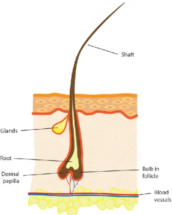
The human lower lid contains 75-80 lashes dispersed in three to four rows, whereas the upper lid has 90-160 lashes scattered on five to six rows (Liotet et al., 1977; Montagna et al., 1969; Na et al., 2006). The anatomy of the lash and hair has some similar characteristics (Marieb, 2005). Both have a hair shaft [the visible part] that extends outside the skin, a root that is under the skin and a bulb, which is the enlarged terminal portion [Figure 1]. The inferior portion of the bulb is in direct contact with the dermal papilla, which brings key mesenchymal-epithelial interactions in follicle cycling.
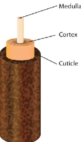
The lash itself is made up of three structures that fit into one another [Figure 2] (Liotet et al., 1977). The innermost structure, the medulla, consists of loose cells. A thicker cortex surrounds the medulla to ensure its strength and stability. The pigmentation of either the lash or hair is the result of the melanin contained in the cortex. Finally, the cuticle, composed of several cell layers, forms the outermost portion, offering protection to the internal structures by its impermeability.
Figure 2. The anatomy of the eyelash
The anatomy and physiology of the lash follicle are distinctive from other hair follicles. Consequently, the lash follicle is worthy of a detailed study and appreciation of its specific characteristics and surrounding skin; its influence on the lash life cycle, curvature, and pigmentation; and the age-related changes of the lashes.
The surrounding skin of the follicle and its features
To appreciate the main characteristics of the lash follicle and its surrounding skin, it is imperative to compare these structures to that of the scalp hair, which have been studied extensively. The scalp skin contains three layers: the epidermis [external], the dermis [middle] and the hypodermis [internal] (Marieb, 2005) whereas the skin of the lids has two layers: a thinner epidermis and a dermis (Thibaut et al., 2010). All follicles on the human body are rooted in their deepest skin layer, notably the hypodermis for the scalp and the dermis for the lid. Consequently, the lash follicle is shorter than the scalp hair follicle (Thibaut et al., 2010). Another major characteristic that distinguishes lash follicles from scalp hair follicles is that they have no arrector pili muscles, which are responsible for straightening the hair in response to cold or intense emotions (Marieb, 2005), producing what is commonly referred to as ‘goose bumps’. Therefore, lashes do not require individual mobility (Montagna et al., 1969).
Lash follicles are connected with two types of secretory glands: Zeiss and Moll. They produce different substances released through channels that flow into the follicle. The Zeiss glands use a holocrine mechanism of action, thus liberating their complete cell content, which is a sebum (Smith et al., 2008). It has antimicrobial and lubrication properties, just as it allows the transport of antioxidants, although the exact function of the sebum is unknown (Smith et al., 2008).The Moll glands, only found in the lids and active from birth, are apocrine glands that produce secretions by fragmentation from one side of their cells (Stoeckelhuber et al., 2003). Their secretions, which contain a variety of sugar components, might play a critical role in the defense against microorganisms (Stoeckelhuber et al., 2003).
The life cycle, curvature and pigmentation
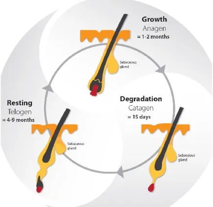
Lashes have a life cycle consisting of three phases : the growth phase [anagen], the degradation phase [catagen] and the resting phase [telogen] (Kloepper et al., 2010). Following the telogen phase, the lash falls out and the life cycle begins again with a new lash in the anagen phase [Figure 3] (Kloepper et al., 2010). The daily growth rate of a lash is 0.12 mm to 0.14 mm (Na et al., 2006; Thibaut et al., 2010). The anagen phase duration varies from four to ten weeks and the complete life cycle is from four to eleven months (Na et al., 2006; Paus et al., 2016; Thibaut et al., 2010). The lash length rarely exceeds 12 mm, as the growth rate and anagen phase duration are shorter than the ones observed in scalp hair research (Thibaut et al., 2010). The lash growth rate is influenced by several factors, including the topical prostaglandin analogs used to reduce the intraocular pressure in glaucoma patients (Paus et al., 2016). Nesher et al. (2015) demonstrated that the prostaglandin analog F2α receptors were expressed in several layers of the lash follicle during its anagen phase. Further studies are needed to confirm the expression of the prostaglandin analog F2α receptors with the use of prostaglandin analogs eyedrops.
The morphology of the eyelashes is such that they are curved in all individuals, regardless of ethnicity. This curvature is initiated at the bulb of the lash and continues until the tip of the shaft (Thibaut et al., 2005). Thibaut et al. (2010) have shown that several cell types of the lash follicle alongside the bulb location are asymmetric [i.e. certain sheaths along the concave side of the
bulb are thicker than the ones on the convex side]. While studying the biology of curly hair in vitro, the same phenomenon of asymmetry has been observed (Thibaut et al., 2005). Curly hair’s markers are also found in the cuticle and cortex of the lash itself (Thibaut et al., 2007; Thibaut et al., 2010). These studies offer possible explanations of the mechanisms involved in lash curvature. Nevertheless, the amplitude of the curvature can vary among individuals. Ethnic studies have identified a difference in the degree of lash curvature [or curl] revealing that the lift-up and curl-up angles are more pronounced among Caucasians as compared to Asians (Na et al., 2006).
The skin and all hair on the human body acquire their pigmentation mostly by melanogenesis, which is the synthesis of different types of melanin by the melanocytes (Commo et al., 2004), these cells being under the influence of several enzymes, including tyrosinase-related protein 2 [TRP-2] (Schallreuter et al., 1998). When it comes to lashes, their degree of pigmentation is defined by the quantity of melanocytes in the lash follicle structure (Thibaut et al., 2010). The lash becomes greyish at a very advanced age and rarely whitens (Liotet et al., 1977). The maintenance of the lash pigmentation may be explained by a sustained expression of TRP-2 (Thibaut et al., 2010). Further studies are needed to confirm if this association is a direct cause-effect relationship.In contrast, many studies have shown that scalp hair will progressively lose its pigmentation due to a gradual depletion of melanocytes (Commo et al., 2004). Several mechanisms have been demonstrated to explain this phenomenon: melanocyte apoptosis, oxidative stress and a reduced expression of certain enzymes, such as TRP-2 (Arck et al., 2006; Commo, Gaillard, Thibaut, et al., 2004; Tobin, 2008).
Figure 3. The life cycle of the eyelash
Age-related changes
There are limited studies about age-related changes on eyelashes. Procianoy et al. (2015) have studied women’s lower lid lashes and found that the curvature of the lashes on the medial and central eyelid portion increased with age, while those of the lateral portion remained in the same direction. Glaser et al. (2014) reported that there was a reduction of length, thickness and pigmentation of the lashes with age.
Research methods
Compared to scalp hair, little research has been dedicated to the study of eyelashes, namely due to the limitation inherent to the in vivo access of the lash follicle. This section explores the techniques employed in lash research and their challenges.
Sampling and organ culture of the lash follicle
There are only a few studies on human lash follicle sampling because it is limited to cadaveric studies and eyelid surgeries, such as ectropion repairs (Montagna et al., 1969; Nesher et al., 2015; Thibaut et al., 2010). It was in 1969 that the first research on the histology and cytochemistry of human lids and lashes was published (Montagna et al., 1969). The function and microscopic anatomy of the follicle cells were studied post mortem following sudden accidental deaths. Other studies that have succeeded in preserving a portion of the human lid have used immunohistochemistry to perform analysis (Nesher et al., 2015; Thibaut et al., 2010). This technique consists of detecting certain antigens [proteins] in the cells of the tissue, using antibodies that bind to them specifically. This technique allowed researchers to describe the morphology, curvature and pigmentation of the lash follicles (Thibaut et al., 2010), and the presence and characteristics of some prostaglandin receptors in the follicles (Nesher et al., 2015).
As sampling on human lash follicles is limited, it is even more complex for scientists to perform lash follicle cultures, hence existing publications on that topic are limited to animal studies. Yet, human scalp hair cultures are common and have been successfully achieved for the past several decades, whereby tissues are obtained during face-lift procedures (Harries et al., 2013; Kloepper et al., 2010; Philpott et al., 1990). A research guide has been proposed to standardize the scalp hair follicle culture, which is of interest to researchers in different fields: experimental dermatology, genetics, developmental biology and endocrinology (Langan et al., 2015). It is even possible to observe human hair follicles in vivo with xenotransplantation [the follicles in their anagen phase or full-thickness skin portions are removed from the human scalp and transplanted on the back of mice] (Gilhar et al., 2013; Hashimoto et al., 2000; Oh et al., 2016). All of these studies highlight how hair research has progressed as compared to eyelash research. To overcome this gap, other mammals had to be considered. In the past, primate tissues have been used due to their resemblance with respect to anatomy and physiology. However, animal protection laws have evolved and primates have a restricted accessibility (Weatherall et al., 2007). Previous hair studies have demonstrated that both human and porcine hair follicles are anatomically close (Debeer et al., 2013; Mangelsdorf et al., 2014). Consequently, preliminary
trials on cultivated porcine lash follicles were performed as recently as 2016, whereby follicles from pigs intended for human consumption were used for organ culture assays (Paus et al., 2016). An elongation of the follicles was obtained within a few days. Hopefully, this study and future ones will enhance our knowledge on the lash and help to reduce the gap between studies on lashes and hair.
Imaging of lashes
Imaging is a minimally invasive technique to quantify various parameters of human lashes. Observational studies with different age categories demonstrated that lashes change with age (Glaser et al., 2014; Procianoy et al., 2015). Characteristics such as the orientation, length, thickness and pigmentation were measured from photographs of the lower and upper lids (Glaser et al., 2014; Procianoy et al., 2015). Human and mouse eyelash life cycle phases have been estimated with photographs in several studies (Na et al., 2006; Paus et al., 2016; Tauchi et al., 2010; Thibaut et al., 2010). However, the measures were variable from one study to another due to non-standardized methodologies. To improve accuracy, Thibault et al. (2010) observed preselected lashes over a nine-month period by measuring their length each week. Once again, while imaging of lashes estimates the parameters of the life cycle, more advanced techniques described above, such as xenotransplantation and follicle cultures, are routinely used to measure the same parameters for hair (Oh et al., 2016).
Imaging techniques have also been utilized for ethnic differences between the lashes of Caucasian and Asian women (Na et al., 2006). Digital photographs were used to calculate the lift-up and curl-up angles of the upper lashes and phototrichogram [allows in vivo study of the growth cycle] (Dhurat, 2006) for the number, length and thickness of lashes (Na et al., 2006). Smaller lash details such as the cuticle textures and layer density were observed ex-vivo with electron microscopy (Na et al., 2006).
Study on aerodynamics
No in vivo studies of human lash aerodynamics exist to date in the literature. Only one in vitro study exists using a model of a mammalian eye to study the aerodynamics of particles around
eyelashes (Amador et al., 2015). The biophysics research team designed a wind tunnel in which air flow, that could affect the ocular surface, was recreated. The model had dimensions of an adult human eye, with a depth of 4 mm and a diameter of 20 mm, and was made with an aluminum dish. Water filled in the dish was used to mimic the tear film and a mesh was chosen to represent the lashes, after obtaining the same results with commercially available lashes made with human hair. Measurements of water evaporation on the ocular surface and calculation of particle deposition on the lashes were obtained using various mesh lengths. Optimal lash lengths that decreased evaporation and particle deposition were determined. Using fluid mechanic principles, the flow on the model eye’s surface, as well as around and through lashes, were calculated. The optimal lash length determined was compared with other mammalian eyelash lengths, obtained by photographs from phylogenetically diverse preserved mammalian heads. They established that the optimal lash length was one third of the width of the eye. Aerodynamic analysis confirmed that this was the optimal length, because it reduced tear evaporation and deposition of particles on lashes by half. Short lashes created a zone of airflow stagnation above the ocular surface, while long lashes pushed the airflow towards the ocular surface. These aerodynamic studies highlighted the important role of lashes in the protection of the eye. In vivo studies should be carried out to validate these results on humans, but the wind tunnel effect would most undoubtedly induce an increased frequency of reflex blinking to protect the ocular surface. Based on this study, any alterations of lash length, be it with certain pharmaceuticals or cosmetic procedures [ie. lash extensions], would have an impact on the protective effect of the ocular surface. Further studies will be needed to validate this impact. Although the study provided an interesting insight into the role that lashes play in the aerodynamics at the front surface of the eye, there remained limitations. Some examples of those include the plane surface used in the model, instead of a convex surface, and water as the chosen composition to represent the tear film, which is more complex in mammals. Studying the protective role of lashes was unexplored before this study, and the authors have paved the way for future investigations in the aerodynamic role of in vivo human lashes.
Pathophysiology of the lashes and their management
Definitions of lash anomalies
During an evaluation of the anterior segment of the eye, ECPs need to assess the normalcy of several structures including eyelashes. There are several terms in the literature that define anomalies of hair anywhere on the body, however some are specific for eyelashes. Table 1 summarizes the different types of hair anomalies and their associated pathologies.
Table 1. Hair anomalies and their associated pathologies
Anomaly Pathological condition Lashes only Description Pigmentation Poliosis
Decreased/absence of pigmentation in hair from any area of the body (Sleiman et al., 2013), by decreased/absence of melanin and/or hair follicle melanocytes (Meyer et al., 2009)
Direction and position Trichiasis Distichiasis X X
Primary trichiasis: misdirected lash, by misdirection of the hair shaft (Dutton et al., 2000)
Secondary trichiasis: secondary to entropion, normal orientation of the hair shaft (Dutton et al., 2000)
Abnormal row of lashes near or in the Meibomian glands; rare condition (Ferreira et al., 2010)
Growth
Hypotrichosis Milphosis Madarosis
X
Reduced hair density in any area of the body (Law, 2010)
Lash loss (Vij et al., 2015)
Lash and/or eyebrow loss (Vij et al., 2015)
Mixed
Hypertrichosis
Trichomegaly
An increased of hair in any area of the body, beyond the normal variation for age, sex or race (Wendelin et al., 2003)
Increased lash [>12 mm] and/or eyebrow length, curl, stiffness, pigmentation and thickness (Almagro et al.,
2003; Paul et al., 2012; Santmyire-Rosenberger et al., 2005)
There is a consensus on most definitions on eyelash anomalies with the exception of hypertrichosis and trichomegaly. The term hypertrichosis is commonly misinterpreted as a synonym for trichomegaly, even though they have distinct definitions. Hypertrichosis is defined as an increase in hair in any part of the body, whereas trichomegaly is specific for eyelashes and eyebrows (Almagro et al., 2003; Paul et al., 2012; Santmyire-Rosenberger et al., 2005). The definitions of hypertrichosis and trichomegaly adopted for this review reflect the ones used in recent publications (Almagro et al., 2003; Paul et al., 2012; Santmyire-Rosenberger et al., 2005). Also, the terms madarosis and milphosis are often confounded. In a clinical setting, the term madarosis is used to describe lash loss, however milphosis is the more appropriate term. Management of lash anomalies
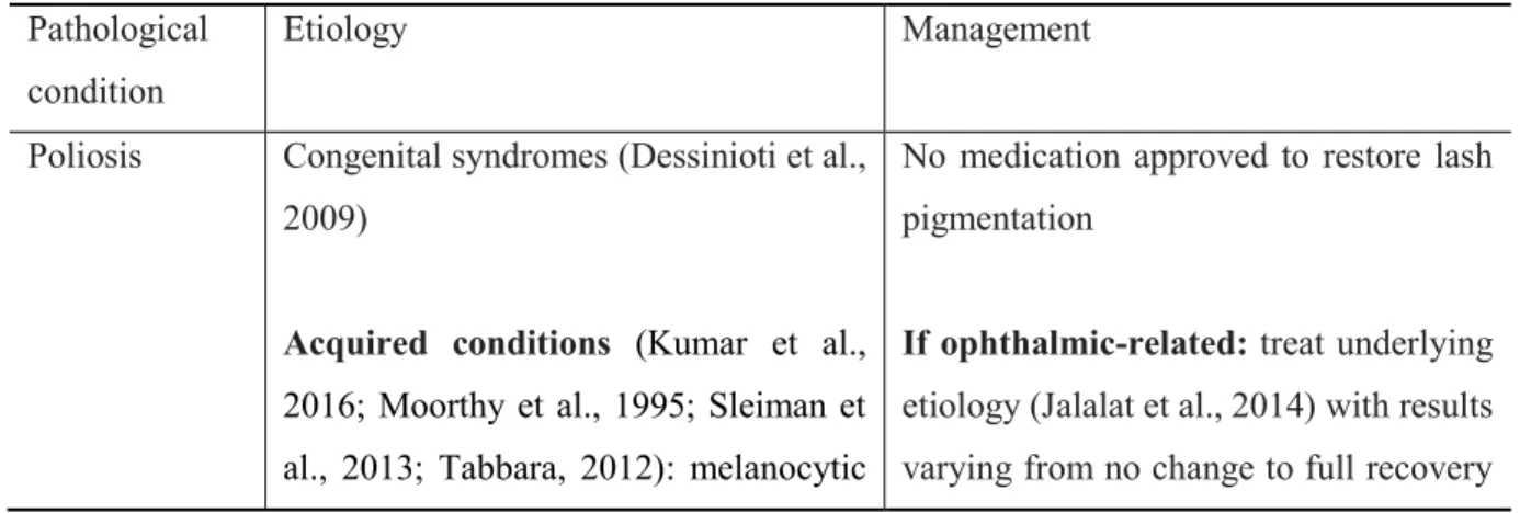
The etiology and management of poliosis, trichiasis, milphosis and trichomegaly have been summarized from the written literature, mainly from case reports [Table 2]. Depending on the etiology, a single pathology or several lash pathologies may be present simultaneously. As a result of the lack of substantive clinical trials on lash pathologies, Table 2 emphasizes the most frequently reported management strategies, and as such, is non-exhaustive.
Table 2. Etiologies and management of lash conditions
Pathological condition
Etiology Management
Poliosis Congenital syndromes (Dessinioti et al., 2009)
Acquired conditions (Kumar et al., 2016; Moorthy et al., 1995; Sleiman et al., 2013; Tabbara, 2012): melanocytic
No medication approved to restore lash pigmentation
If ophthalmic-related: treat underlying
etiology (Jalalat et al., 2014) with results varying from no change to full recovery
lesions, inflammatory systemic disorders, blepharitis and vernal keratoconjunctivitis (VKC)
Less common: systemic/ophthalmic drug-induced (side effect of all topical prostaglandin analogues) (Chen et al., 2004; Nakakura et al., 2015; Ozyurt et al., 2015; Waheed et al., 2001)
of the pigmentation (Chen et al., 2004; Tabbara, 2012; Waheed et al., 2001)
If systemic implication (Jalalat et al.,
2014): referred to the patient’s primary care doctor
Trichiasis
Ophthalmic (Ferreira et al., 2010): lid
margin scars from trauma/surgery; lid margin inflammation; conjunctival diseases; conjunctival burns
Various skin diseases (Ferreira et al., 2010)
Initial episode: epilation of the affected
lash(es) and soft contact lenses for short-term relief of induced conjunctival or corneal irritation (Chiou et al., 1998)
Recurrent episode: refer to an
ophthalmologist for an ablation procedure [depends on the severity]: laser argon ablation (Basar et al., 2000; Yung et al., 1994), electrolysis (Benson, 1882), trephination (McCracken et al., 2006), direct internal lash bulb extirpation (Dutton et al., 2000),
folliculectomy (Tirakunwichcha et al., 2006), cryotherapy (Elder et al., 1994), entropion surgery (Ballen, 1964)
If skin disease suspected: refer to a
dermatologist
Ophthalmic (Vij et al., 2015); lid infestation [staphylococcal spp., Demodex folliculorum, trachoma] and/or lid inflammation [posterior
Reversible follicle damage (Khong et
al., 2006): lash(es) regrowth possible; consider pharmaceutical approach to increase the regrowth rate [bimatoprost
Milphosis (loss of lashes)
blepharitis, ocular rosacea, seborrheic blepharitis]
Associated with eyebrow madarosis
(Kumar et al., 2012): several dermatological diseases and inherited conditions
Associated with eyebrow and hair madarosis (Kumar et al., 2012):
Extensive list of systemic and drug-induced conditions
ophthalmic solution 0.03% is the only approved drug in patients with healthy lashes and hypotrichosis] (Barron-Hernandez et al., 2017; Fagien, 2010; Smith et al., 2012; Zaleski-Larsen et al., 2017)
Irreversible follicle damage: no lash
regrowth
Severe irreversible milphosis: refer to
an oculoplastic surgeon for lash grafting consideration (Kumar et al., 2012)
Trichomegaly
Congenital syndromes (Kaur et al.,
2015): Oliver-McFarlane and Cornelia de Lange
Familial
Acquired (Paul et al., 2012): Allergic
rhinitis, atopic dermatitis, HIV infection, uveitis and VKC
Drug-induced (Paul et al., 2012):
Topical prostaglandin analogues and epidermal growth factor receptor [EGFR] inhibitors used in oncology
Trimming of the affected lashes if visual disturbance or smudging with inner surface of eyeglass lens (Dueland et al., 2003; Lacouture et al., 2006; Robert et al., 2005)
With concomitant trichiasis (Chiou et al., 1998): refer to trichiasis management [above]
It is essential for ECPs to first determine the etiology of the lash pathology to select the appropriate management. In the particular case of milphosis without a clear etiology, ECPs must consider a psychiatric disorder called trichotillomania as a possible causal factor, regardless of
the concomitance of eyebrow and/or hair madarosis. Trichotillomania is characterized by an uncontrollable urge to pull out hair from any part of the body (Christenson et al., 1991) and a referral for a psychological evaluation should be considered. This condition can be managed by investigating the cause of the behaviour and, in some cases, pharmacotherapy is recommended (Johnson et al., 2016).
The etiology of the lash pathology also affects the appearance of the lashes, which may affect the patient’s esthetic appearance. For example, common causes of trichomegaly, such as HIV infection and allergic rhinitis, will induce the regrowth of long and smooth lashes, while epidermal growth factor receptor [EGFR] inhibitors make them rougher and more dispersed (Paul et al., 2012). The appearance of eyelashes also has a major impact on the quality of life, as hair loss can cause various degrees of psychological distress (Phillips et al., 2017). Dunnill et al. (2017) have demonstrated that the most distressing side effect of chemotherapy is the loss of hair. Other systemic diseases that induce hair loss can be stressful for the affected person, such as alopecia areata, which is characterized by a patchy loss of scalp hair. This disease can progress to other parts of the body, including eyelashes (Liu et al., 2016). Although the exact pathophysiological mechanism is not established yet, it is thought to be caused by an autoimmune attack on hair follicles (Dainichi et al., 2017). Patients with alopecia areata have poor health-related quality life scores similar to what is established in other chronic skin diseases, as demonstrated in two systematic studies (Liu et al., 2016; Rencz et al., 2016).
As seen in Table 2, the management of lash pathologies will be influenced by their causal agent. When the lash pathology severity ranges from mild to moderate, this can be managed by an ECP in most cases. To date, there is only one pharmaceutical option approved for eyelash loss [lash hypotrichosis], consisting of a topical application of bimatoprost ophthalmic solution 0.03% [Latisse, Allergan] along the superior lash base. It’s mode of action is to prolong the anagen phase of the lash life cycle (Barron-Hernandez et al., 2017), hence resulting in an increased lash length, thickness and pigmentation (Barron-Hernandez et al., 2017; Fagien, 2010; Smith et al., 2012; Zaleski-Larsen et al., 2017). Before using this product, patients should always seek the advice of an ECP. If adverse events occur, such as discomfort and skin pigmentation, discontinuation of the product will typically reverse the effects. Some cases of poliosis have
been reported following prostaglandin analogues use (Chen et al., 2004). To date, there is no medication approved to restore the pigmentation of lashes.
Severe lash pathologies must be referred to specialists for an ablation procedure (Basar et al., 2000; Benson, 1882; McCracken et al., 2006; Tirakunwichcha et al., 2006), eyelash grafts (Kumar et al., 2012), lid reconstruction (Choo, 2002; Khafagy et al., 2012) or systemic therapies (Dainichi et al., 2017). Promising therapies are emerging for hair loss, which can lead to the discovery of new treatments for eyelashes specifically. Dainichi and Kabashima (2017) have reviewed all current therapies and treatments that have an effect on hair regrowth in alopecia areata and universalis [subtype of alopecia that also affect the lashes]. In alopecia universalis, hair regrowth has been observed with medications such as topical immunotherapy and corticosteroids (Dainichi et al., 2017). However, these studies focused on scalp hair regrowth and did not collect data on the regrowth rate of lashes. Data from scalp hair studies may guide future therapies for eyelashes.
Lid margin microflora
The inherent microflora of the lid margin also plays a vital role in maintaining the lashes free of anomalies. The lid margin typically contains a microflora composed of commensal bacteria and parasites. A disruption of the balance of this microflora can lead to various types of blepharitis (Nicholls et al., 2017). Lee et al. (2012) have demonstrated that the most common bacteria found in lash samples were Propionibacterium, Staphylococcus, Strepophyta, Coryne- bacterium and Enhydrobacter. The variety of the lash microflora is unique to each individual. The study further highlighted that when blepharitis was diagnosed, Staphylococcus, Streptophyta, Corynebacterium, and Enhydrobacter were increased whereas Propionibacterium was
decreased. Due to the constant contact of the lids with the ambient air, the lid microflora is particularly influenced by environmental factors, such as pollen, dust, and soil particles. Furthermore, touching our lids with our fingers can inoculate other varieties of the microflora, contributing to the dynamic nature of the lid margin microflora.
Bacterial overpopulation can be observed clinically with the presence of debris on the lashes.
Acute Staphylococcal blepharitis is identified with collarettes, which are hard crusts on lashes, and with other non-specific signs [squame/scale, telangiectasia vessels, Meibomian gland dysfunction] (Bernardes et al., 2010). Chronic signs are madarosis, trichiasis, poliosis, tylosis or scars on the lid margin (Bernardes et al., 2010; Nicholls et al., 2017). Blepharitis can also be caused by other micro-organisms, such as parasites. Three types of parasites, all arthropods, can inhabit the lid margin: Demodex folliculorum, Demodex brevis and Phthirus pubis (Padhi et al., 2017). Demodex parasites are part of the natural eye microflora (Nicholls et al., 2017) and their number increases with age, so much so that one study has reported 100% prevalence in people over 70 years old (Roth, 1979). D. folliculorum is mostly found in the lash follicle and other parts of the body such as eyebrows, scalp, nose and ears. D. Brevis is found mainly in the Meibomian glands and other sebaceous glands, such as the face (Liu et al., 2010). The anatomy of the Demodex parasite consists of a head with four pairs of legs on either side and an elongated body/tail that contains the digestive system. The parasite has no anus, hence all the ingested material remains in the gut, along with its own microflora of Streptococcus spp., Staphylococcus spp. and Bacillus oleronius (Szkaradkiewicz et al., 2012). At the end of its life cycle, approximately 15-18 days in length, the parasite bursts and releases its content, which can provoke an inflammatory response (Liu et al., 2010; Szkaradkiewicz et al., 2012). An overpopulation of the parasite is called a demodicosis, and on the lids, this would be termed blepharitis secondary to Demodex. Since Demodex has the potential to affect both the anterior [lashes] and posterior [Meibomian glands] portion of the lid margin, some (Duncan et al., 2015) have proposed the term marginal blepharitis to describe a demodicosis of the lid margin. Clinical signs of demodicosis include follicular hypertrophy (Liu et al., 2010), gelatinous debris surrounding the base of the lashes termed cylindrical dandruff (Gao et al., 2005), and non-specific blepharitis signs as stated above. Cylindrical dandruff is a pathognomonic sign for Demodex (Gao et al., 2005). On the other hand, the parasite Phthirus pubis is not found in the normal lid microflora, as it originates in pubic hair. It can be transmitted by sexual contact and less often through linens and bedding (Anane et al., 2013). Transfer of the parasite from the infested region to the hand can reach any hair on the body, including the eyelashes (Turgut et al., 2009). The resulting blepharitis is termed Phthiriasis palpebrarum and its clinical presentation, in one or both eyes, includes the translucent parasite’s body firmly attached to
lashes, brown scales corresponding to the parasite’s feces, and multiple nits (unhatched parasites) fixed on the lashes and the lid margin (Anane et al., 2013; Turgut et al., 2009). Symptoms include itchiness and irritation of the lid margin (Anane et al., 2013).
Finally, blepharitis can be the result of seborrheic dermatitis, a dermatological condition affecting the skin and, in some cases, the lid margin. Many etiologies have been proposed and researchers are still looking for the exact mechanism (Paulino, 2017). An imbalance in the skin micro-organisms might be the cause (Paulino, 2017). Other than the non-specific signs of blepharitis detailed above, ECPs can observe some flaky scales on the lids that can fall on the lashes (Wolf et al., 2014). When the condition affects the anterior portion of the lid, it is called seborrheic blepharitis.
Management of anterior blepharitis
The aim in blepharitis management is to restore the normal microflora (Nicholls et al., 2016). A complete eradication of Phthiriasis palpebrarum is desired and can be achieved by mechanical removal of the parasites and nits (Dagdelen et al., 2013; Padhi et al., 2017). Blepharitis secondary to the imbalance of the microflora are chronic conditions. Hence, they are not expected to be completely eradicated. First line therapies of blepharitis include warm compresses to soften the debris and eyelid hygiene to manually dislodge them (Lindsley et al., 2012). Targeted lid margin therapies should be adopted by ECPs, including antibacterial for Staphylococcal infestation (Liu et al., 2010) and anti-parasitic formulations for Demodex parasites (Gao et al., 2005; Ngo et al., 2017), in order to limit lid margin anomalies that come with chronic blepharitis. To target properly seborrheic blepharitis, a reference to dermatology is required to establish the diagnosis of seborrheic dermatitis and treat the affected skin with the appropriate pharmaceutical (Paulino, 2017).
ECP assessment of lashes
Figure 4 summarizes in a flowchart the management of debris on eyelashes. This tool can be used when teaching students or residents to identify and localize debris on lashes and select the appropriate management.
Visible debris?
Any evidence of trichiasis, milphosis or poliosis?
Any evidence of lash pathologies?
Is it manageable by ECPs?
To answer this, you have to review: - Topical/systemic medications
- Past and present ocular/ systemic history - Other eye structures involved
No treatment Concomitant eye disorders? Refer to the appropriate medical specialist Management of the concomitant eye disorder Management of the lash pathologies (see table 2) Anterior blepharitis identification (see below) and management
(see the corresponding subsection)
- Translucent debris at the base of the lashes:
Demodex (cylindrical dandruff-CD; most common) or Phthiriasis palpebrarum (less frequent; associated with visible nits)
- Hard crusts all along the lashes (collarettes):
Staphylococcal (most common); other types of bacterial infestation
- Flaky scales on the lashes, coming from the lid: Seborrheic blepharitis no yes yes no no no yes yes yes no
Figure 4: Eyelash debris assessment by eyecare professionals
Iatrogenic factors
The appearance of eyelashes is an increasingly growing preoccupation in many people who are seeking an aesthetic enhancement. Cosmetics have been used since biblical times to improve the aesthetic of the eye and the thickness, length and color of eyelashes (Draelos, 2001). Lash tints and extensions are newer and popular, surpassing expectations in the cosmetic marketplace. Over $55 million in 2014 were dedicated to eyelash extension and adhesives alone in the US (Statista). Synthetic lashes are glued individually on the natural lashes with different adhesives, which have been linked to ocular problems such as keratoconjunctivitis (Amano et al., 2012). In most cases, the glue and removing agents have induced inflammation on the eye, by direct contact with the ocular surface. Furthermore, the vapors associated with the application, or the dissolving, of the glue afterwards have been reported as ocular irritants. Lash tinting is done with dyes that contain p-phenylenediamine, a sensitizer that can provoke an allergic reaction and contact dermatitis (Ali et al., 2017; Teixeira et al., 2006). Water-based mascara is made of several waxes, types of pigment and resins dissolved in water, whereas solvent-based mascara, known as waterproof, has its pigments and waxes added in petroleum distillates (Draelos, 2001). Fukami et al. (2014) found a positive correlation between the frequency of mascara use and the degree of cracking in the lash cuticles. Also, long-term use of mascara led to milphosis, possibly due to the rubbing, with fingers and water only, by the users (Kadri et al., 2013). Needless to say, eye cosmetics in general can have an impact on the lids, lashes, tear film and ocular surface (Ng et al., 2016). Consequently, patients need to be educated appropriately about the application, removal, shelf life and associated precautions of cosmetic use.
Conclusion
This review brings a deeper awareness on the eyelashes and their follicles. The current literature has numerous cases of how lashes are affected by systemic/eye diseases, pharmaceuticals and cosmetics, and how they change the lash morphology. In addition, an imbalance of the lash microenvironment can lead to a variety of blepharitis and impact negatively the adjoining lid margin. Many studies have demonstrated that lashes do not serve only a cosmetic function, but
also a protective role on the lid margin and the ocular surface. The lash follicle structures are mostly studied with ex vivo techniques, which are limited by the poor availability of the human lid sampling. This review further highlights the challenges when studying the lash follicle and the need to develop newer techniques. Increasing lash research will certainly improve the efficiency of ECP’s interventions in lash anomalies.
Acknowledgements
A special thanks to Micheline Gloin for her graphic assistance.
Funding
This research did not receive any specific grant from funding agencies in the public, commercial, or not-for-profit sectors.
Conflict of interest
SA has received honorarium for conferences/consulting from Allergan and Shire. EB has received funding from Alcon, Allergan, Canadian Optometric Trust Fund, I-Med Pharma Inc, Shire and honorarium for conferences/consulting from Akorn, Alcon, Allergan, American Academy of Optometry, Canadian Association of Optometry, CooperVision, Labtician, Jobson Publishing, Novartis, Santen, Shire.
References
Ali, L., Foulds, J. S. et Abdul Ghaffar, S. (2017). Severe eyelid allergic contact dermatitis secondary to eyelash tint: two case reports. Contact Dermatitis, 77(1), 59-60.
Almagro, M., del Pozo, J., Garcia-Silva, J., Martinez, W., Castro, A., et al. (2003). Eyelash length in HIV-infected patients. AIDS, 17(11), 1695-1696.
Amador, G. J., Mao, W., DeMercurio, P., Montero, C., Clewis, J., et al. (2015). Eyelashes divert airflow to protect the eye J R Soc Interface (Vol. 12).
Amano, Y., Sugimoto, Y. et Sugita, M. (2012). Ocular disorders due to eyelash extensions. Cornea, 31(2), 121-125.
Anane, S., Malek, I., Kamoun, R. et Chtourou, O. (2013). Phthiriasis palpebrarum: diagnosis and treatment. Journal Français d'Ophtalmologie, 36(10), 815-819.
Arck, P. C., Overall, R., Spatz, K., Liezman, C., Handjiski, B., et al. (2006). Towards a "free radical theory of graying": melanocyte apoptosis in the aging human hair follicle is an indicator of oxidative stress induced tissue damage. FASEB Journal, 20(9), 1567-1569. Ballen, P. H. (1964). A Simple Procedure for the Relief of Trichiasis and Entropion of the Upper
Lid. Archives of Ophthalmology, 72, 239-240.
Barron-Hernandez, Y. L. et Tosti, A. (2017). Bimatoprost for the treatment of eyelash, eyebrow and scalp alopecia. Expert Opin Investig Drugs, 26(4), 515-522.
Basar, E., Ozdemir, H., Ozkan, S., Cicik, E. et Mirzatas, C. (2000). Treatment of trichiasis with argon laser. European Journal of Ophthalmology, 10(4), 273-275.
Benson, A. (1882). On the Treatment of Partial Trichiasis by Electrolysis. British Medical Journal, 2(1146), 1203-1204.
Bernardes, T. F. et Bonfioli, A. A. (2010). Blepharitis. Seminars in Ophthalmology, 25(3), 79-83.
Chen, C. S., Wells, J. et Craig, J. E. (2004). Topical prostaglandin F(2alpha) analog induced poliosis. American Journal of Ophthalmology, 137(5), 965-966.
Chiou, A. G. Y., Florakis, G. J. et Kazim, M. (1998). Management of Conjunctival Cicatrizing Diseases and Severe Ocular Surface Dysfunction. Survey of Ophthalmology, 43(1), 19-46.
Choo, P. H. (2002). Distichiasis, trichiasis, and entropion: advances in management. International Ophthalmology Clinics, 42(2), 75-87.
Christenson, G. A., Popkin, M. K., Mackenzie, T. B. et Realmuto, G. M. (1991). Lithium treatment of chronic hair pulling. Journal of Clinical Psychiatry, 52(3), 116-120. Commo, S., Gaillard, O. et Bernard, B. A. (2004). Human hair greying is linked to a specific
depletion of hair follicle melanocytes affecting both the bulb and the outer root sheath. British Journal of Dermatology, 150(3), 435-443.
Commo, S., Gaillard, O., Thibaut, S. et Bernard, B. A. (2004). Absence of TRP-2 in melanogenic melanocytes of human hair. Pigment Cell Research, 17(5), 488-497. Craig, J. P., Nichols, K. K., Akpek, E. K., Caffery, B., Dua, H. S., et al. (2017). TFOS DEWS
Dagdelen, S., Aykan, U. et Cetinkaya, K. (2013). Phthriasis palpebrarum can resemble tick larva infestation in an eyelid. Journal of AAPOS, 17(4), 440-442.
Dainichi, T. et Kabashima, K. (2017). Alopecia areata: What's new in epidemiology, pathogenesis, diagnosis, and therapeutic options? Journal of Dermatological Science, 86(1), 3-12.
Debeer, S., Le Luduec, J. B., Kaiserlian, D., Laurent, P., Nicolas, J. F., et al. (2013). Comparative histology and immunohistochemistry of porcine versus human skin. European Journal of Dermatology, 23(4), 456-466.
Dessinioti, C., Stratigos, A. J., Rigopoulos, D. et Katsambas, A. D. (2009). A review of genetic disorders of hypopigmentation: lessons learned from the biology of melanocytes. Experimental Dermatology, 18(9), 741-749.
Dhurat, R. (2006). Phototrichogram. Indian Journal of Dermatology, Venereology and Leprology, 72(3), 242-244.
Draelos, Z. D. (2001). Special considerations in eye cosmetics. Clinics in Dermatology, 19(4), 424-430.
Draelos, Z. K. (1991). Eye cosmetics. Dermatologic Clinics, 9(1), 1-7.
Dueland, S., Sauer, T., Lund-Johansen, F., Ostenstad, B. et Tveit, K. M. (2003). Epidermal growth factor receptor inhibition induces trichomegaly. Acta Oncologica, 42(4), 345-346.
Duncan, K. et Jeng, B. H. (2015). Medical management of blepharitis. Current Opinion in Ophthalmology, 26(4), 289-294.
Dunnill, C. J., Al-Tameemi, W., Collett, A., Haslam, I. S. et Georgopoulos, N. T. (2017). A Clinical and Biological Guide for Understanding Chemotherapy-Induced Alopecia and Its Prevention. Oncologist.
Dutton, J. J., Tawfik, H. A., DeBacker, C. M. et Lipham, W. J. (2000). Direct internal eyelash bulb extirpation for trichiasis. Ophthalmic Plastic and Reconstructive Surgery, 16(2), 142-145.
Elder, M. J. et Bernauer, W. (1994). Cryotherapy for trichiasis in ocular cicatricial pemphigoid. British Journal of Ophthalmology, 78(10), 769-771.
Fagien, S. (2010). Management of hypotrichosis of the eyelashes: Focus on bimatoprost. Clinical, Cosmetic and Investigational Dermatology, 3, 39-48.
Ferreira, I. S., Bernardes, T. F. et Bonfioli, A. A. (2010). Trichiasis. Seminars in Ophthalmology, 25(3), 66-71.
Fukami, K., Inoue, T., Kawai, T., Takeuchi, A., Uesugi, K., et al. (2014). Internal structure changes of eyelash induced by eye makeup. J Cosmet Sci, 65(4), 217-224.
Gao, Y. Y., Di Pascuale, M. A., Li, W., Baradaran-Rafii, A., Elizondo, A., et al. (2005). In vitro and in vivo killing of ocular Demodex by tea tree oil. British Journal of Ophthalmology, 89(11), 1468-1473.
Gao, Y. Y., Di Pascuale, M. A., Li, W., Liu, D. T., Baradaran-Rafii, A., et al. (2005). High prevalence of Demodex in eyelashes with cylindrical dandruff. Investigative Ophthalmology and Visual Science, 46(9), 3089-3094.
Gilhar, A., Keren, A. et Paus, R. (2013). A new humanized mouse model for alopecia areata. Journal of Investigative Dermatology. Symposium Proceedings, 16(1), S37-38.
Glaser, D. A., Jones, D., Carruthers, J., Campo, A., Moench, S., et al. (2014). Epidemiologic analysis of change in eyelash characteristics with increasing age in a population of healthy women. Dermatologic Surgery, 40(11), 1208-1213.
Harries, M. J., Meyer, K., Chaudhry, I., J, E. K., Poblet, E., et al. (2013). Lichen planopilaris is characterized by immune privilege collapse of the hair follicle's epithelial stem cell niche. Journal of Pathology, 231(2), 236-247.
Hashimoto, T., Kazama, T., Ito, M., Urano, K., Katakai, Y., et al. (2000). Histologic and cell kinetic studies of hair loss and subsequent recovery process of human scalp hair follicles grafted onto severe combined immunodeficient mice. Journal of Investigative Dermatology, 115(2), 200-206.
Jalalat, S. Z., Kelsoe, J. R. et Cohen, P. R. (2014). Alopecia areata with white hair regrowth: case report and review of poliosis. Dermatology Online Journal, 20(9).
Johnson, J. et El-Alfy, A. T. (2016). Review of available studies of the neurobiology and pharmacotherapeutic management of trichotillomania. J Adv Res, 7(2), 169-184. Jones, D. (2011). Enhanced eyelashes: prescription and over-the-counter options. Aesthetic
Plastic Surgery, 35(1), 116-121.
Kadri, R., Achar, A., Tantry, T. P., Parameshwar, D., Kudva, A., et al. (2013). Mascara induced milphosis, an etiological evaluation. Int J Trichology, 5(3), 144-147.
Kaur, S. et Mahajan, B. B. (2015). Eyelash Trichomegaly. Indian Journal of Dermatology, 60(4), 378-380.
Khafagy, A., Mostafa, M. M. et Fooshan, F. (2012). Management of trichiasis with lid margin split and cryotherapy. Clinical Ophthalmology (Auckland, N.Z.), 6, 1815-1817.
Khong, J. J., Casson, R. J., Huilgol, S. C. et Selva, D. (2006). Madarosis. Survey of Ophthalmology, 51(6), 550-560.
Kloepper, J. E., Sugawara, K., Al-Nuaimi, Y., Gaspar, E., van Beek, N., et al. (2010). Methods in hair research: how to objectively distinguish between anagen and catagen in human hair follicle organ culture. Experimental Dermatology, 19(3), 305-312.
Kumar, A. et Karthikeyan, K. (2012). Madarosis: a marker of many maladies. Int J Trichology, 4(1), 3-18.
Kumar, S. et Al Khars, W. (2016). Vitiligo in association with vernal keratoconjunctivitis. Saudi J Ophthalmol, 30(2), 128-129.
Lacouture, M. E., Boerner, S. A. et Lorusso, P. M. (2006). Non-rash skin toxicities associated with novel targeted therapies. Clinical Lung Cancer, 8 Suppl 1, S36-42.
Langan, E. A., Philpott, M. P., Kloepper, J. E. et Paus, R. (2015). Human hair follicle organ culture: theory, application and perspectives. Experimental Dermatology, 24(12), 903-911.
Law, S. K. (2010). Bimatoprost in the treatment of eyelash hypotrichosis. Clinical Ophthalmology (Auckland, N.Z.), 4, 349-358.
Lee, S. H., Oh, D. H., Jung, J. Y., Kim, J. C. et Jeon, C. O. (2012). Comparative ocular microbial communities in humans with and without blepharitis. Investigative Ophthalmology and Visual Science, 53(9), 5585-5593.
Lindsley, K., Matsumura, S., Hatef, E. et Akpek, E. K. (2012). Interventions for chronic blepharitis. Cochrane Database Syst Rev(5), CD005556.
Liotet, S., Riera, M. et Nguyen, H. (1977). Les cils. Physiologie, structure, pathologie. Archives d'Ophtalmologie, 37(11), 697-708.
Liu, J., Sheha, H. et Tseng, S. C. (2010). Pathogenic role of Demodex mites in blepharitis. Current Opinion in Allergy and Clinical Immunology, 10(5), 505-510.
Liu, L. Y., King, B. A. et Craiglow, B. G. (2016). Health-related quality of life (HRQoL) among patients with alopecia areata (AA): A systematic review. Journal of the American Academy of Dermatology, 75(4), 806-812 e803.
Mangelsdorf, S., Vergou, T., Sterry, W., Lademann, J. et Patzelt, A. (2014). Comparative study of hair follicle morphology in eight mammalian species and humans. Skin Research and Technology, 20(2), 147-154.
Marieb, E. (2005). Le système tégumentaire (Traduit par R. Lachaine). Dans ERPi (dir.), Anatomie et physiologie humaines (3e éd., p. 156-208).
McCracken, M. S., Kikkawa, D. O. et Vasani, S. N. (2006). Treatment of trichiasis and distichiasis by eyelash trephination. Ophthalmic Plastic and Reconstructive Surgery, 22(5), 349-351.
Meyer, K. C., Brzoska, T., Abels, C. et Paus, R. (2009). The alpha-melanocyte stimulating hormone-related tripeptide K(D)PT stimulates human hair follicle pigmentation in situ under proinflammatory conditions. British Journal of Dermatology, 160(2), 433-437. Montagna, W. et Ford, D. M. (1969). Histology and cytochemistry of human skin. 3. The eyelid.
Archives of Dermatology, 100(3), 328-335.
Moorthy, R. S., Inomata, H. et Rao, N. A. (1995). Vogt-Koyanagi-Harada syndrome. Survey of Ophthalmology, 39(4), 265-292.
Na, J. I., Kwon, O. S., Kim, B. J., Park, W. S., Oh, J. K., et al. (2006). Ethnic characteristics of eyelashes: a comparative analysis in Asian and Caucasian females. British Journal of Dermatology, 155(6), 1170-1176.
Nakakura, S., Yamamoto, M., Terao, E., Nagatomi, N., Matsuo, N., et al. (2015). Prostaglandin-associated periorbitopathy in latanoprost users. Clinical Ophthalmology (Auckland, N.Z.), 9, 51-56.
Nesher, R., Mimouni, M., Elnaddaf, H., Nemet, A. et Kidron, D. (2015). Characterization of prostaglandin F2alpha receptors in human eyelids. European Journal of Ophthalmology, 25(2), 81-84.
Ng, A., Evans, K., North, R. V., Jones, L. et Purslow, C. (2016). Impact of Eye Cosmetics on the Eye, Adnexa, and Ocular Surface. Eye Contact Lens, 42(4), 211-220.
Ngo, W., Jones, L. et Bitton, E. (2017). Short-Term Comfort Responses Associated With the Use of Eyelid Cleansing Products to Manage Demodex folliculorum. Eye Contact Lens.

![Table 2. Clinician Erythema Assessment [CEA] (Tan et al., 2014)](https://thumb-eu.123doks.com/thumbv2/123doknet/11317653.282540/60.918.117.605.328.529/table-clinician-erythema-assessment-cea-tan-et-al.webp)
![Figure 3. Forehead Demodex densities [mite count/cm 2 ] expressed as marginal mean ± standard error [SE]](https://thumb-eu.123doks.com/thumbv2/123doknet/11317653.282540/63.918.118.593.294.625/figure-forehead-demodex-densities-count-expressed-marginal-standard.webp)
![Figure 4. Scatterplot of the degree of facial erythema and the forehead Demodex density [mite count/cm 2 ]](https://thumb-eu.123doks.com/thumbv2/123doknet/11317653.282540/64.918.118.644.206.586/figure-scatterplot-degree-facial-erythema-forehead-demodex-density.webp)
