SINZIANA AVRAMESCU
CELLULAR AND HOMEOSTATIC NETWORK
MECHANISMS OF POSTTRAUMATIC EPILEPSY
Thèse présentée
à la Faculté des études supérieures de l’Université Laval dans le cadre du programme de doctorat en neurobiologie pour l’obtention du grade de Philosophiae Doctor (Ph.D.)
DÉPARTEMENT D’ANATOMIE ET PHYSIOLOGIE FACULTÉ DE MÉDECINE
UNIVERSITÉ LAVAL QUÉBEC
2008
Résumé
Suite aux traumatismes crâniens pénétrants, le cerveau devient graduellement hyperexcitable et génère des activités paroxystiques spontanées. Les mécanismes qui sous-tendent l’épileptogénèse demeurent cependant peu connus. La ligne directrice de nos travaux consiste en l'hypothèse que la diminution de l'activité corticale engendrée par la déafférentation déclenche des mécanismes homéostatiques agissant tant au niveau cellulaire qu’au niveau du réseau cortical, et qui mènent à une excitabilité neuronale accrue culminant en crises d’épilepsie.
Nous avons testé cette hypothèse chez des chats adultes, lors de différents états de vigilance ou sous anesthésie, ayant subits une déafférentation partielle du gyrus suprasylvien. Nous avons évalué les effets de la déafférentation corticale aigue et chronique sur la survie des neurones et des cellules gliales et nous avons investigué comment la privation chronique d'afférences neuronales pourrait modifier les propriétés du réseau cortical et déclencher des crises d’épilepsie.
Après la déafférentation du gyrus suprasylvien, les neurones situés dans les couches corticales profondes, en particulier les neurones inhibiteurs GABAérgiques, dégénèrent progressivement et parallèlement à une fréquence croissante des activités paroxystiques, notamment pendant le sommeil à ondes lentes. La privation chronique d'afférences neuronales et la perte de neurones activent les mécanismes homéostatiques de plasticité qui favorisent une plus grande connectivité neuronale, une efficacité plus élevée des connexions synaptiques excitatrices et des changements des propriétés neuronales intrinsèques. Ensemble, ces facteurs favorisent une excitation accrue du réseau cortical. L'activité corticale spontanée, mesurée par les taux moyens de décharge, augmente progressivement, en particulier pendant le sommeil à ondes lentes, caractérisé par des périodes silencieuses alternant avec des périodes actives. Ceci soutient, en outre, notre hypothèse concernant la participation des mécanismes de plasticité homéostatique. La dégénération des neurones des couches corticales profondes produit des changements importants dans la distribution laminaire de l'activité neuronale, qui est déplacée vers les couches plus superficielles, dans la partie déafferenté du gyrus. Ce changement dans la distribution de profils de profondeurs de décharges neuronales modifie également le
déclenchement de l'activité corticale spontanée. Dans le cortex normal et dans la partie relativement intacte du gyrus suprasylvien, l'activité corticale est générée dans les couches corticales profondes. Pourtant, dans le cortex chroniquement déafferenté, l'oscillation lente et les activités ictales sont générées dans les couches superficielles et puis diffusent vers les couches plus profondes. Le traumatisme cortical induit également une importante gliose réactive et une altération de la fonction normale des cellules gliales, ce qui cause l’enlèvement dysfonctionnel du K+ extracellulaire et qui augmente l'excitabilité des neurones favorisant ainsi la génération d’activités paroxystiques.
En conclusion, les mécanismes de plasticité homéostatique déclenchés par le niveau diminué d'activité dans le cortex déafferenté produisent une hyperexcitabilité corticale incontrôlable et génèrent finalement les crises d’épilepsie. Dans ces conditions, l’augmentation de l'activité corticale plutôt que la diminution avec des médicaments antiépileptiques pourrait être salutaire pour empêcher le développement de l'épileptogenèse post-traumatique.
Abstract
After penetrating cortical wounds, the brain becomes gradually hyperexcitable and generates spontaneous paroxysmal activity, but the progressive mechanisms of epileptogenesis remain virtually unknown. The guiding line of our experiments was the hypothesis that the reduced cortical activity following deafferentation triggers homeostatic mechanisms acting at cellular and network levels, leading to an increased neuronal excitability and finally generating paroxysmal activities.
We tested this hypothesis either in anesthetized adult cats, or during natural sleep and wake, using the model of partially deafferented suprasylvian gyrus to induce posttraumatic epileptogenesis. We evaluated the effects of acute and chronic cortical deafferentation on the survival of neurons and glial cells and how long-term input deprivation could shape up the properties of neuronal networks and the initiation of spontaneous cortical activity.
Following cortical deafferentation of the suprasylvian gyrus, the deeply laying neurons, particularly the inhibitory GABAergic ones, degenerate progressively in parallel with an increased propensity to paroxysmal activity, mainly during slow-wave sleep. The chronic input deprivation and the death of neurons activate homeostatic plasticity mechanisms, which promote a gradual increased neuronal connectivity, higher efficacy of excitatory synaptic connections and changes in intrinsic cellular properties favoring increased excitation. The spontaneous cortical activity quantified by means of firing rate augments also progressively, particularly during slow-wave sleep, characterized by periods of silent states alternating with periods of active states, which supports furthermore our hypothesis regarding the involvement of homeostatic plasticity mechanisms. The degeneration of neurons in the deep cortical layers generates important changes in the laminar distribution of neuronal activity, which is shifted from the deeper layers to the more superficial ones, in the partially deafferented part of the gyrus. This change in the depth profile distribution of firing rates modifies also the initiation of spontaneous cortical activity which, in normal cortex, and in the relatively intact part of the deafferented gyrus, is initiated in the deep cortical layers. Conversely, in late stages of the undercut, both the cortical slow oscillation and the ictal activity are initiated in the more superficial layers and
then spread to the deeper ones. Cortical trauma induces also an important reactive gliosis associated with an impaired function of glial cells, responsible for a dysfunctional K+ clearance in the injured cortex, which additionally increases the excitability of neurons, promoting the generation of paroxysmal activity.
We conclude, that the homeostatic plasticity mechanisms triggered by the decreased level of activity in the deafferented cortex, generate an uncontrollable cortical hyperexcitability, finally leading to seizures. If this statement is true, augmenting cortical activity rapidly after cortical trauma rather than decreasing it with antiepileptic medication, could prove beneficial in preventing the development of posttraumatic epileptogenesis.
Foreword
The following thesis is presented in the form of a collection of scientific articles - published or submitted for publication. The general introduction describes the theoretical context and the experimental strategies we used in the presented studies. A review of the results and a general discussion finalizes the thesis. The bibliography used for both introduction and discussion is presented at the very end of the manuscript, while the bibliography for each chapter follows the text of the corresponding paper, with a formatting of the reference list in accordance to the rules of the journal where the paper was published or submitted.
As Carl Sagan liked to say, “Science is a way of thinking much more than it is a body of knowledge”, and I feel honored to be part of a group of privileged people who do science and think science.
I would like to use this opportunity to express my gratitude to my thesis supervisor - Prof. Igor Timofeev, for his help during the complex and laborious experiments, for the fruitful discussions, critics and support throughout the progression of my thesis, without which the present studies would not have been possible. I will always remember him for his excellent electrophysiology skills and knowledge, for his friendly and encouraging attitude.
I thank the directorial board of the Neurobiology Program for giving me the opportunity to write this thesis in English.
Many thanks also to Mr. Pierre Giguère and Mr. Serge Ftomov for their excellent technical support and to all the colleagues from the lab for their friendship and for the productive atmosphere, which made my scientific journey much more agreeable. In addition, I would like to acknowledge the contribution of Dr. Dragos Nita to three of the papers included in the thesis.
Equally, I would like to thank the organisms which provided financial support for the development of these projects: Canadian Institutes of Health Research, Natural Science and Engineering Research Council of Canada and Savoy Foundation.
I am grateful to my fiancé, Dragos, for teaching me to “never stop exploring”, for making everyday look brighter and for just being there every time I needed.
Last but not least, I would like to thank my family for their affection and continuous support - my mother, Emilia, for giving me the courage to dream, but also to fulfill my dreams; my father, Victor, who taught me the meanings of determination and self-esteem; and my brother, Radu, from whom I learned that adults are allowed to be kids and that braking the rules once in a while makes you feel better.
“There is no scientific study more vital to man than the study of his own brain. Our entire view of the Universe depends on it.” Francis Crick (Scientific American, 1979)
Table of contents
Résumé...i
Abstract... iii
Foreword...v
Table of contents... viii
List of tables...xi
List of figures... xii
List of abbreviations ...xiv
Introduction...1
1 Seizures and Epilepsy ...1
1.1 Definitions ...1
1.2 Diagnostic overview ...2
2 Epileptic syndromes characterized by SW/PSW complexes and/or fast runs ...6
2.1 Absence seizures...6
2.1.1 Typical absences ...6
2.1.2 Atypical absences ...7
2.2 Absence status epilepticus ...8
2.3 Electrical status epilepticus during sleep ...8
2.4 The Lennox-Gastaut syndrome...10
2.5 The West syndrome ...12
2.6 Posttraumatic epilepsy ...13
2.6.1 Epidemiology of PTE ...14
2.6.2 Frequency of PTE ...15
2.6.3 Risk factors for PTE ...16
3 Sleep and epilepsy ...18
3.1 Effect of sleep on epilepsy...18
3.2 Effect of epilepsy on sleep...20
4 Cortically generated seizures ...21
4.1 Evidence for cortical origin of SW seizures ...22
4.2 Cellular correlates of SW complexes and fast runs ...23
5 Ions in the brain ...31
5.1 Neuronal activity alters ion distributions...31
5.2 Extracellular potassium in the brain ...31
5.3 Potassium regulation in the cortex...32
5.3.1 Recapture by the neurons...32
5.3.2 Diffusion ...32
5.3.3 Glial regulation ...34
5.4 Potassium and seizures ...35
5.4.1 Potassium can induce seizures...35
5.4.2 Potassium accumulates in the interstitial space during seizures...37
5.4.3 Too much potassium can stop seizures...37
6 Reactive gliosis in epilepsy...38
6.1 K+ Channels ...40
7 Experimental post-traumatic epilepsy ...43
7.1 Acute traumatic injury ...44
7.1.1 Mechanisms of acute trauma ...47
7.2 Chronic traumatic injury...48
7.2.1 Mechanisms of chronic trauma...49
8 Rationale for the thesis ...60
9 Synaptic strength modulation following cortical trauma: a role in epileptogenesis...62
9.1 Résumé...63
9.2 Abstract...64
9.3 Introduction...65
9.4 Materials and Methods...67
9.5 Results...72
9.6 Discussion...79
9.7 Acknowledgements:...83
9.8 References...84
9.9 Figures ...92
10 Neocortical post-traumatic epileptogenesis is associated with loss of GABAergic neurons...102
10.1 Résumé...103
10.2 Abstract...104
10.3 Introduction...105
10.4 Materials and methods ...107
10.5 Results...111
10.6 Discussion...115
10.7 Acknowledgements...117
10.8 References...118
10.9 Figures ...126
11 Laminar distribution of spontaneous cortical activity following cortical deafferentation 137 11.1 Résumé...138
11.2 Abstract...139
11.3 Introduction...140
11.4 Materials and methods ...142
11.5 Results...146
11.6 Discussion...151
11.7 Acknowledgements...155
11.8 References...156
11.9 Figures ...162
12 Posttraumatic reactive gliosis is associated with paroxysmal activity and impaired potassium clearance ...171
12.1 Résumé...172
12.2 Abstract...173
12.3 Introduction...174
12.4 Materials and methods ...176
12.5 Results...182
12.6 Discussion...186
12.8 References...191
12.9 Figures ...199
13 Conclusions...207
13.1 Posttraumatic neuronal loss and cortical disorganization...208
13.2 Homeostatic plasticity following chronic cortical deafferentation...209
13.3 Laminar redistribution of spontaneous cortical activity ...211
13.4 Glial dysfunction and K+ clearance impairment...212
13.5 Final remarks ...213
List of tables
Table 1. Epileptic seizure types and precipitating stimuli for reflex seizures ...3 Table 2. Epilepsy syndromes and related conditions...5
List of figures
Figure 4-1. Field potential and intracellular features of ideopatic electographic seizure
recorded from cortical area 5 of cat anesthetized with ketamine-xylazine. ...24
Figure 4-2. Evolution from slow oscillation to isolated PDSs and SW seizure...25
Figure 4-3. Synaptic excitability to cortical stimuli during the pre-seizure epoch, different components of the seizure consisting of SW complexes over a depolarizing envelope, and post-seizure epoch.. ...26
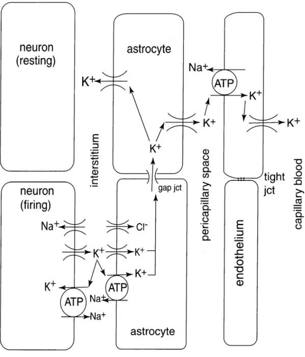
Figure 5-1. Diagram of the fate of K+ released from neurons. ...33
Figure 5-2. Uptake of K+ and Cl- in glial cells. ...35
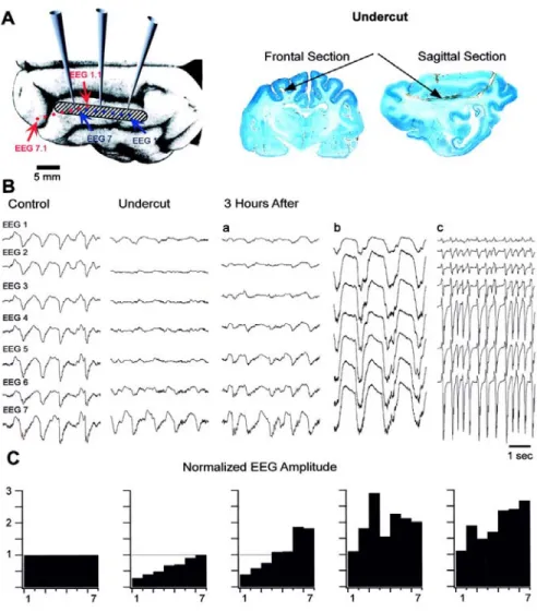
Figure 7-1. Slow oscillation in intact suprasylvian gyrus is modified by cortical undercut.. ...45
Figure 7-2. Spontaneous electrographic seizures in the suprasylvian gyrus were generated at the border between intact and undercut cortex...46
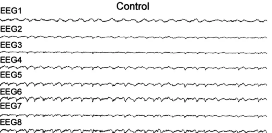
Figure 7-3. Patterns of EEG activity under ketamine–xylazine anesthesia.. ...50
Figure 7-4 Sleep modulation of paroxysmal activities...51
Figure 9-1. Experimental paradigm and methodology of analysis.. ...93
Figure 9-2. Different types of responses and possible designs of synaptic topology...94
Figure 9-3. Examples of responses and failures in direct synaptic connections.. ...95
Figure 9-4. Changes of synaptic interactions following cortical deafferentation.. ...96
Figure 9-5. Modulation of synaptic interactions in acute and chronically deafferented cortex...98
Figure 9-6. Increased neuronal input resistance in chronically undercut cortex.. ...99
Figure 9-7. Progressive increase neuronal excitability following cortical undercut...100
Figure 9-8. Increased duration of hyperpolarizing periods following cortical trauma. ...101
Figure 10-1. Increased propensity to seizures after chronic cortical deafferentation.. ...127
Figure 10-2. Penetrating brain wounds cause a reduction of grey matter's thickness and the disorganization of cortical architecture.. ...128
Figure 10-3. Immunohistochemical labeling of cortical neurons.. ...130
Figure 10-4. Examples of cortical depth profiles showing the distribution of excitatory and inhibitory neurons in control and undercut cortex...132
Figure 10-5. Depth profile distribution of neuronal densities in control and after cortical trauma.. ...134
Figure 10-6. Changes in the balance between excitation and inhibition towards excitation in chronically deafferented cortex. ...136
Figure 11-1. Modulation of ictal events by the state of vigilance in early and chronic undercut.. ...163
Figure 11-2. Laminar profile of multi-unit activities in the anterior part of the undercut in A) early and B) chronic stages.. ...164
Figure 11-3. Laminar profile of multi-unit activities in the posterior part of the undercut in A) early and B) chronic stages.. ...165
Figure 11-4. The dynamics of the total neuronal firing rate following cortical deafferentation during wake and SWS. ...166
Figure 11-5. Higher discharge rate at the beginning of the active phase of the slow oscillation, in chronic undercut. ...167 Figure 11-6. Local field-potentials (LFP) and current-source density (CSD) analysis of
the slow oscillation in the anterior undercut in early and chronic stages.. ...168 Figure 11-7. Local field-potentials (LFP) and current-source density (CSD) analysis of
the slow oscillation in the posterior undercut in early and chronic stages...169 Figure 11-8. Local field-potentials (LFP) and current-source density (CSD) analysis of
the paroxysmal activity in the anterior and posterior undercut.. ...170 Figure 12-1. Immunocytochemical labeling of GFAP+ cells. ...199 Figure 12-2. Relationship of ictal (4 Hz) activities with different states of vigilance..200 Figure 12-3. Incidence of ictal events during different phases of the sleep-wake cycle.
...201
Figure 12-4. Phasic variation of the extracellular potassium concentration during sleep and ictal events...202 Figure 12-5. Steady increase of the extracellular potassium concentration ([K+]o)
following cortical activation...204 Figure 12-6. Average increase of the extracellular potassium concentration ([K+]o)
following cortical activation...205 Figure 12-7. Intracellular recordings of presumed glial cells during cortical activation
from slow-wave sleep (SWS) to REM sleep in intact (A) and chronically
deafferented (B) cortex. ...206 Figure 13-1. Integrated mechanisms of epileptogenesis following chronic cortical
List of abbreviations
AED antiepileptic drugs
AMPA α-amino-3-hydroxy-5-methyl-4-isoxazolepropionic acid
AQP4 aquaporin-4
CNS central nervous system
DSWS deep slow-wave sleep
EC extracellular
ECS extracellular space
EEG electro-encephalogram
EMG electro-myogram
EOG electro-oculogram
EPSP excitatory postsynaptic potential FFT Fast Fourier Transformation
FRB fast-rhythmic-bursting
FS fast-spiking
GABA gamma-amino butyric acid
GAD glutamate decarboxylase
HP homeostatic plasticity
IB intrinsically-bursting
IC intracellular
IEA inter-ictal epileptiform activity
Ih H-potassium current
ILAE International League Against Epilepsy INa(p) persistent Na+ current
IPSP inhibitory postsynaptic potential
IR input resistance
ISF interstitial fluid
IS interictal spikes
ISI inter-spike interval
[K+]o extracellular K+ concentration [K+]i intracellular K+ concentration KCC2 K+/Clcotransporter
Kir inward rectifying K+ channels LSWS light slow-wave sleep
mEPSC mini excitatory postsynaptic current
NE network excitation
NI network inhibition
NKCC1 Na+/K+/Cl- cotransporter NMDA N-methyl-D-aspartate
PDS paroxysmal depolarizing shift
PSW poly spike-waves
PTE posttraumatic epilepsy
PTS posttraumatic seizures
REM rapid-eye movements
SD spreading depression
STA spike-triggered averages
SW spike-waves
SWS slow-wave sleep
TBI traumatic brain injury
TC thalamo-cortical
TLE temporal lobe epilepsy
Introduction
1 Seizures and Epilepsy
1.1 Definitions
Modern epileptology emerged from John Hughlings Jackson's 1870 paper "A study of convulsions" in which he spectacularly pointed out that seizures do not equal epilepsy: “A convulsion is but a symptom, and implies only that there is an occasional, an excessive and a disorderly discharge of nerve tissue on muscles. This discharge occurs in all degrees; it occurs with all sorts of conditions of ill health, at all ages, and under innumerable circumstances” (Jackson, 1870).
According to the latest consensus of the International League Against Epilepsy (ILAE) and of the International Bureau for Epilepsy, an epileptic seizure is a clinical, transient event, with clear start and finish, characterized by an abnormal excessive or synchronous neuronal activity in the brain (Fisher et al., 2005).
Epilepsy is not one condition, but a diverse family of disorders, having in common an abnormally increased predisposition to seizures. The definition of epilepsy requires the occurrence of at least one unprovoked epileptic seizure (Fisher et al., 2005). Therefore, a predisposition, as determined for example by a family history or by the presence of epileptiform activities on the electroencephalogram (EEG), is not sufficient to determine epilepsy. The definition requires in addition to at least one seizure, the presence of an enduring alteration in the brain which increases the likelihood of further seizures (Fisher et al., 2005).
Epileptogenesis refers to the cascade of biological events altering the balance between excitation and inhibition in the brain and leading to the development of an epileptic disorder (Clark and Wilson, 1999). It has been shown both in animal models of epilepsy (Cavalheiro et al., 1991; Mello et al., 1993) and in patients (Annegers et al., 1980; Marks et al., 1992) that there is a latent period between the induction of a localized cerebral insult and the appearance of a chronic epileptic condition. The progressive pathological processes occurring during the latent period, such as neuronal loss and abnormal synaptic
reorganization (Mello et al., 1993; Leite et al., 1996; Mathern et al., 1996), lead to abnormally increased excitability and synchronization and eventually to the generation of spontaneous seizures (Cavalheiro et al., 1991; Isokawa and Mello, 1991). This latent period may offer a therapeutic window for the prevention of epileptogenesis and the development of unprovoked seizures and epilepsy (Herman, 2002).
Even after its development, epilepsy should not be viewed as a random succession of seizures but as a dynamic process which results in both ictal phenomena and interictal functional and structural abnormalities in the brain (Rodin, 1972). Patients who develop chronic intractable epilepsy demonstrate progression in both the number of seizures and in seizure-related neurological symptoms such as cognitive and behavioral disorders (Elwes et al., 1984; French et al., 1993; Williamson et al., 1993; Cole, 2000). In the view of these data, the challenge we are facing is to fill in the gaps of the basic mechanisms that induce epilepsy, so that we will be able to prevent epileptogenesis and the progression of disease.
1.2 Diagnostic overview
The ILAE Commission on Classification and Terminology proposed in 2001 a revised diagnostic scheme intended to provide the basis for a standardized description of individual patients, consisting of five levels, or Axes. The Axes are organized to facilitate a logical clinical approach to the development of hypotheses necessary to determine the diagnostic studies that need to be performed, and the therapeutic strategies to be undertaken.
Axis 1 – Ictal semiology – includes the description of the ictal event, without reference to etiology, anatomy, or mechanisms. It can be very brief or extremely detailed, as required for clinical or research purposes, because the communication among clinicians, and among researchers, would be greatly enhanced by the establishment of standardized terminology for describing ictal semiology.
Axis 2 – Epileptic seizure type – indicates the type of seizure derived from a list of accepted seizure types which represent diagnostic entities with etiologic, therapeutic, and/or prognostic implications (Table 1). Localization within the brain should be specified when this is appropriate, and in the case of reflex seizures, the specific stimulus will also be specified here.
_________________________________________________________________________
Table 1. EPILEPTIC SEIZURE TYPES AND PRECIPITATING STIMULI FOR REFLEX SEIZURES
[Modified from: ILAE (Epilepsy, 1989)] Self-limited seizure types
Generalized seizures
• Tonic-clonic seizures • Clonic seizures
o Without tonic features o With tonic features • Typical absence seizures • Atypical absence seizures • Myoclonic absence seizures • Tonic seizures
• Spasms • Myoclonic seizures
• Massive bilateral myoclonus • Eyelid myoclonia
o Without absences o With absences • Myoclonic atonic seizures • Negative myoclonus • Atonic seizures
• Reflex seizures in generalized epilepsy syndromes • Seizures of the posterior neocortex
• Neocortical temporal lobe seizures
Focal seizures
• Focal sensory seizures
o With elementary sensory symptoms o With experiential sensory symptoms • Focal motor seizures
o With elementary clonic motor signs o With asymmetrical tonic motor seizures o With typical (temporal lobe) automatisms o With hyperkinetic automatisms
o With focal negative myoclonus o With inhibitory motor seizures • Gelastic seizures
• Hemiclonic seizures
• Secondarily generalized seizures
• Reflex seizures in focal epilepsy syndromes Continuous seizure types
Generalized status epilepticus
• Generalized tonic-clonic status epilepticus • Clonic status epilepticus
• Absence status epilepticus • Tonic status epilepticus • Myoclonic status epilepticus
Focal status epilepticus
• Epilepsia partialis continua of Kojevnikov • Aura continua
• Limbic status epilepticus (psychomotor status) • Hemiconvulsive status with hemiparesis Precipitating stimuli for reflex seizures
• Visual stimuli
o Flickering light -color to be specified when possible o Patterns
o Other visual stimuli • Thinking Music • Eating • Praxis • Somatosensory • Proprioceptive • Reading • Hot water • Startle
• Precipitating stimuli for reflex seizures
_________________________________________________________________________
Axis 3 – Epilepsy syndrome – the syndromic diagnosis is also derived from a list of accepted epilepsy syndromes (Table 2), although it is understood that a syndromic diagnosis may not always be possible.
Axis 4 – Etiology – specifies the etiology when this is known. The etiology could consist of a specific disease derived from a classification of diseases frequently associated with epileptic seizures or syndromes, a genetic defect, or a specific pathological substrate.
Axis 5 – Degree of impairment – this axis is optional and designs the degree of impairment caused by the epileptic condition. Classification of impairment will be derived from the World Health Organization’s International Classification of Impairments, Disabilities and Handicaps (ICIDH-2) which is currently in preparation.
Next, I will discuss in detail only those epileptic syndromes and types of seizures characterized by Spike-Waves/Poly-Spike-Waves (SW/PSW) and/or fast runs pertinent to my experimental approach.
_________________________________________________________________________
Table 2. EPILEPTIC SYNDROMES AND RELATED CONDITIONS
[Modified from: ILAE (Epilepsy, 1989)]
EPILEPTIC SYNDROMES
• Benign familial neonatal seizures • Early myoclonic encephalopathy • Ohtahara syndrome
• Migrating partial seizures of infancy*
• West syndrome
• Benign myoclonic epilepsy in infancy • Benign familial infantile seizures • Benign infantile seizures (non-familial) • Dravet's syndrome
• Hemiconvulsions-Hemiplegia syndrome (HH syndrome) • Myoclonic status in nonprogressive encephalopathies • Benign childhood epilepsy with centrotemporal spikes
• Early onset benign childhood occipital epilepsy (Panayiotopoulos type) • Late onset childhood occipital epilepsy (Gastaut type)
• Epilepsy with myoclonic absences • Epilepsy with myoclonic-astatic seizures • Lennox-Gastaut syndrome
• Landau-Kleffner syndrome
• Epilepsy with continuous spike-and-waves during slow-wave sleep • Childhood absence epilepsy
• Progressive myoclonus epilepsies
• Idiopathic generalized epilepsies with variable phenotypes o Juvenile absence epilepsy
o Juvenile myoclonic epilepsy
o Epilepsy with generalized tonic-clonic seizures only • Reflex epilepsies
o Idiopathic photosensitive occipital lobe epilepsy o Other visual sensitive epilepsies
o Primary reading epilepsy o Startle epilepsy
• Autosomal dominant nocturnal frontal lobe epilepsy • Familial temporal lobe epilepsies
• Generalized epilepsies with febrile seizures plus* • Familial focal epilepsy with variable foci*
• Symptomatic (or probably symptomatic) focal epilepsies o Limbic epilepsies
- Mesial temporal lobe epilepsy with hippocampal sclerosis - Mesial temporal lobe epilepsy defined by specific etiologies - Other types defined by location and etiology
o Neocortical epilepsies - Rasmussen syndrome
- Other types defined by location and etiology * Syndromes in development
CONDITIONS WITH EPILEPTIC SEIZURES THAT DO NOT REQUIRE A DIAGNOSIS OF EPILEPSY
• Benign neonatal seizures • Febrile seizures
• Reflex seizures
• Alcohol withdrawal seizures
• Drug or other chemically-induced seizures • Immediate and early post traumatic seizures • Single seizures or isolated clusters of seizures • Rarely repeated seizures (oligo-epilepsy)
_________________________________________________________________________
2 Epileptic syndromes characterized by SW/PSW
complexes and/or fast runs
2.1 Absence seizures
Absence seizures are broadly divided into (a) typical absences of mainly idiopathic generalized epilepsy with generalized, greater than 2.5 Hz spike or polyspike-and-slow waves, and (b) atypical absences of symptomatic or cryptogenic epilepsies with slower, less than 2.5 Hz generalized discharges (International League Against Epilepsy (Commission on Epidemiology and Prognosis, 1981).
2.1.1 Typical absences
Typical absences are brief, generalized epileptic seizures with sudden onset and termination. They have two essential components: clinically the impairment of consciousness (absence) and EEG generalized 3 Hz to 4 Hz spike and slow wave discharges (International League Against Epilepsy (Commission on Epidemiology and Prognosis, 1981). Impairment of consciousness may be severe moderate, mild, or inconspicuous (the detection of which may require special cognitive testing). The discharge
SW frequency varies from onset to termination. It is usually faster and unstable in the opening phase (first second), becomes more regular and stable in the initial phase (first 3 seconds), and slows down towards the terminal phase (last 3 seconds).
2.1.2 Atypical absences
Atypical absences are associated with a high incidence of changes in postural tone. The beginning and end are usually difficult to identify because they are more progressive and because this type of seizure affects children whose mental function is altered. It is, therefore, difficult to determine the duration that ranges from 5 seconds to 20 seconds. The axial tone is affected, and this may cause the patient to fall. Eyelid clonus, mild tonic or autonomic features, or automatisms may also be observed. There is, therefore, a whole spectrum of clinical manifestations varying from typical absence to mild manifestations (Loiseau et al., 1995). Because the clinical features may be mild in an intellectually impaired child, it is often difficult to count such seizures, even with video EEG monitoring. Attention may decrease seizure frequency, whereas drowsiness may increase it. The frequency of atypical absences varies from a few a day to nearly continuous. The frequency of generalized SW discharge on EEG is less than 3Hz (Panayiotopoulos, 1997). Only an EEG can identify this type of seizure. Many patients have, in addition to clinical seizures, subclinical discharges. Counting the seizures is, therefore, an unresolved challenge since isolated clinical observation omits subclinical discharges that can affect cognition. Counting EEG discharges would ignore differences in the impact of clinical versus subclinical discharges, and video EEG is reliable only if permanent clinical observation is also used to determine whether the discharge is clinical or subclinical. Such observation would alter the frequency of seizures, though, since it would raise the vigilance of the child and, therefore, prevent the occurrence of absences and discharges. However, the practical implications of this difficulty are moderate because the aim of treatment, including those used in clinical trials, should be the disappearance of seizures and of spikes on EEG (Dulac, 2003). Atypical absences may be combined with tonic seizures and slow spike waves, and this combination defines the Lennox-Gastaut syndrome, which is usually symptomatic and may follow West syndrome in 40% of the cases. Atypical absences may be atonic or tonic. They may occur as the only type of seizure in a patient who exhibits continuous SW in slow wave sleep (SWS). In such patients, the absences are mainly atonic. Atypical absences
may be combined with generalized tonic-clonic and myoclonic seizures in myoclonic-astatic epilepsy. In this condition, the absences are also mainly atonic (for review see Panayiotopoulos, 2002c).
2.2 Absence status epilepticus
Absence status epilepticus is a prolonged, generalized absence seizure, which is defined as lasting more than half an hour, but usually lasts for hours and even for days. It is associated with typically regular and symmetrical generalized discharges of 1 Hz to 4 Hz spike or multiple spike-and-slow wave complexes. Though the sharing symptom of absence status epilepticus is impairment of cognition, this is often associated with other clinical manifestations that may be syndrome-related. It should be emphasized that absence status epilepticus, like the brief absence seizure, is not one but many types of a prolonged, generalized, absence seizure. The cardinal symptom shared by all cases of absence status is the altered content of consciousness in a patient who is usually fully alert. Memory and higher cognitive intellectual functions such as abstract thinking, computation, and personal awareness are the main areas of disturbance. This varies from extremely mild to extremely severe with intermediate states of severity occurring more often (for review see Andermann and Robb, 1972; Guberman et al., 1986; Agathonikou et al., 1998; Panayiotopoulos, 2002a).
The pathophysiology of absence status (typical or atypical) is unknown. It is likely that the generating mechanisms of absence seizures and absence status are the same because their clinico-electroencephalographic features show marked syndrome-related similarities (Agathonikou et al., 1998). The difference is in the duration of the discharge, which may also perpetuate more severe cognitive impairment. Absence seizures last for only a few seconds, whereas absence status epilepticus is prolonged for hours and days (Panayiotopoulos, 2002b).
2.3 Electrical status epilepticus during sleep
A disorder in which sleep induced an EEG pattern characterized by "subclinical" SW occurring almost continuously during SWS and appearing every night for a variable length of time in children was reported in 1971 by Patry and coworkers under the title of
"subclinical electrical status epilepticus induced by sleep in children" (Patry et al., 1971). The disorder was later termed "electrical status epilepticus during sleep" (Tassinari et al., 1985).
The clinical manifestations of this syndrome include: (1) a heterogeneous epileptic disorder, (2) a deterioration of neuropsychological functions associated with or independent from the epileptic disorder and (3) a deterioration of motor functions. The typical EEG pattern of continuous SW during SWS is also an essential and absolute feature for the recognition of the syndrome (Tassinari et al., 1999).
The seizure types occurring in the disorder can be both partial and generalized. They include unilateral or bilateral clonic seizures, generalized tonic-clonic seizures, absences, partial motor seizures, complex partial seizures or epileptic falls. They may occur during wakefulness or sleep. Tonic seizures, however, never occur. The first seizure is reported to be nocturnal and of unilateral type in almost one half of the cases reported. At onset, the frequency of seizure attacks is low. At the time of discovery of the typical nocturnal EEG pattern, however, the epileptic seizures frequently change in severity and frequency. Absences and epileptic falls herald the appearance of continuous SW during SWS and seizure frequency increases, both during wakefulness and sleep (Tassinari et al., 1985; Tassinari et al., 1999). Most researchers assert that more than 85% of SWS is occupied by SW discharges; however, quantitative studies of different sleep stages and of temporal evolution of this EEG disturbance have not been carried out (Jayakar and Seshia, 1991). The typical EEG changes appear 1 year to 2 years after the first seizure and are associated with behavioral deterioration. Focal and generalized interictal spikes (ISs) occur before this time and persist during wakefulness and REM sleep after the appearance of continuous SW during SWS (Tassinari et al., 1999).
After the seizures appear, but before the continuous SW during SWS develop, the EEG during wakefulness may show both diffuse SW, sometimes in bursts, and focal abnormalities such as spikes and slow spikes, with or without associated slow waves, which usually involve the fronto-temporal or the centro-temporal region. Sleep EEG performed at an early stage shows an increase of the aforementioned abnormalities without the features of the continuous SW during SWS (Tassinari et al., 1985).
2.4 The Lennox-Gastaut syndrome
An EEG with a 2 Hz ("slow") SW pattern was first described by Gibbs in 1939 (Gibbs et al., 1939). It was associated with a special type of absence seizure characterized by incomplete loss of consciousness. By contrast, classic petit mal absence seizures had been known to be associated with rhythmic 3 Hz SWs. The term "petit mal variant" was therefore used to describe the EEG pattern and the clinical seizure complex. Lennox and Davis were the first to correlate the slow SW EEG pattern with a distinctive group of clinical manifestations (e.g., mental retardation and specific seizure types), including myoclonic jerks, atypical absences, and astatic seizures (drop attacks) (Lennox and Davis, 1950). Thereafter, Gastaut and his colleagues described the clinical manifestations and EEG patterns of 100 patients with slow SW (Gastaut et al., 1966). They called this syndrome "Lennox syndrome" or "childhood epileptic encephalopathy with diffuse slow SW." Finally, the term "Lennox-Gastaut syndrome" first appeared in the literature in 1969 (Niedermeyer, 1969).
The Lennox-Gastaut syndrome may result from a variety of diffuse encephalopathies. It is characterized by the clinical triad of diffuse slow spikes-and-waves on EEG, mental retardation, and multiple types of generalized seizures, including especially atypical absences and tonic and atonic seizures (Aicardi and Levy Gomes, 1992; Dulac and N'Guyen, 1993). The Lennox-Gastaut syndrome is, with rare exception, a condition of children. The age of onset is between 2 and 8 years in most cases. Boys are affected more frequently than girls. Symptoms can appear de novo without apparent cause (cryptogenic Gastaut syndrome) or result from obvious brain insult (symptomatic Lennox-Gastaut syndrome). Cryptogenic cases start later than symptomatic cases do (for review see Dulac and Engel, 2003).
In young children, the Lennox-Gastaut syndrome usually begins with episodes of sudden falls. In the school-age group, behavioral disturbances may be the heralding signs, along with drop attacks. This is soon followed by frequent seizures, episodes of status epilepticus, progressively deteriorating intellectual functions, personality disturbances, and chronic psychosis (Roger et al., 1989).
Tonic seizures occur in most affected children. They are usually brief, lasting only seconds. Depending on the extent and groups of muscles involved, they may appear as axial (characterized by flexor movements of the head and trunk), axial rhizomelic (characterized by elevation and adduction of proximal upper limbs, stiffening of posterior neck muscles, elevation of shoulders, opening of the mouth, upward deviation of the eyes and brief apnea), or global, leading to sudden falls if the patient is in an upright position (Gastaut et al., 1966).
Atypical absence seizures occur in approximately two-thirds of patients. Both the onset and the termination are gradual in contrast to the strikingly sudden lapses in typical absence. Whereas typical absences are usually brief (less than 10 seconds) and consciousness returns immediately and completely afterward, atypical absences are usually longer and are often followed by some postictal cognitive impairment. During the atypical absence there is "clouding" rather than loss of consciousness so that patients can continue their activity to some degree (Gastaut et al., 1966). Associated manifestations are more common in atypical than in typical absences and include eyelid or perioral myoclonus, progressive flexion due to loss of postural tone, and localized motor phenomena, such as neck-stiffening or head-nodding. Other kinds of seizures, including atonic, partial and generalized tonic-clonic seizures, are less frequent (Roger et al., 1989).
Most patients with the Lennox-Gastaut syndrome have one or more episodes of status epilepticus (Dulac and N'Guyen, 1993). A variety of forms of status epilepticus occur that represent a continuum from absence status, consisting of an insidious confused state that can last for days or weeks, to pure tonic status epilepticus, which is more often seen in adolescents or adults than in children.
The slow SW consists of a blunt, slow spike (approximately 150 ms), followed by a slow wave (approximately 350 ms). The amplitude ranges from 200 μV to 800 μV. When 2 spikes to 3 spikes precede the slow wave, they constitute a PSW complex. The frequency (1.5-2.5 Hz) is relatively slow and arrhythmic compared to that of classical absence seizures (rhythmic 3 Hz). Occasionally, bursts of rapid spike-and-waves at 3 Hz or even 4 Hz may occur in combination with the slow SW (Gastaut et al., 1966). Generalized PSW discharges or lower voltage fast activity (generalized paroxysmal fast activity), lasting 1
second or more without obvious clinical correlates, are common during SWS. Relaxation, drowsiness, sleep, and hyperventilation facilitate the appearance of slow SW. During sleep, the abnormal patterns become more prominent, symmetrical, and synchronous, with even slower SW or PSW complexes. Photic stimulation, on the other hand, has no effect on these EEG events (Markand, 1977). When evolution in EEG patterns occurs in patients previously affected by infantile spasms, the direction is from hypsarrhythmia to multifocal interictal spikes to generalized spike discharges to slow SW, with the last representing a stable pattern that characterizes children with the Lennox-Gastaut syndrome (Kotagal, 1995).
The EEG correlate of tonic seizures consists of a 10 to 13 Hz recruiting rhythm, usually followed by high amplitude slow activity rather than postictal EEG depression. Although the EEG during absence seizures often shows irregular SW discharges, these patterns are not necessarily different from interictal EEG discharges and do not clearly demarcate the occurrence of an ictal event. Similarly, there is no characteristic pattern of absence status; this prolonged confused state may be associated with an increase in slow SW activity or with irregular slowing resembling hypsarrhythmia (Dulac and N'Guyen, 1993).
2.5 The West syndrome
West syndrome comprises a triad of spasms in clusters, mental retardation, and diffuse and profound paroxysmal EEG abnormalities. The onset is insidious in either an otherwise normal or an already handicapped infant. It is an age-dependent epilepsy syndrome that begins in infancy, mostly between 4 months and 6 months of life, before the age of 12 months in over 90% of cases (Kellaway et al., 1979).
Infantile spasms are characterized by usually symmetrical, bilateral, brief, and sudden contractions of the axial muscle groups. The features of the seizures depend on whether the flexor or extensor muscles are predominantly affected and also on the number and distribution of the muscle groups involved. Thus, spasms may vary from extensive contractions of all flexor or extensor muscles to contractions of only neck muscles or abdominal recti (Hrachovy and Frost, 1989).
In fact, the type of spasms, whether in flexion, extension, or mixed does not seem to be affected by etiology or the prognosis. In contrast, whether the spasms are symmetrical or not is important because asymmetry, consisting of lateral deviation of the head or eyes, contributes to indicate some kind of cortical brain damage (Fusco and Vigevano, 1993). Spasms tend to occur soon after awakening or on falling asleep. Most of the spasms occur in clusters, so the interval between successive spasms is less than 60 seconds. Usually the intensity of spasms in a given cluster will peak gradually and then decline (Hrachovy and Frost, 1989).
The usual EEG abnormalities consist of diffuse, high amplitude, asynchronous paroxysmal and slow wave theta and delta activity with loss of background features that is continuous when awake and fragmented in sleep. This hypsarrhythmic pattern may be symmetrical or asymmetrical because of additional foci, or unilateral. In other conditions, it consists of one or several spike foci when awake with secondary generalization in sleep (Dulac, 2003). The interictal EEG of infantile spasms is usually characterized by hypsarrhythmia with a continuous, irregular, random, ever-changing, and disorganized, high-voltage spike and slow wave activity. This is sufficiently characteristic to be easily identified, and the term “chaos” may be inappropriate from this point of view. It may be present during wakefulness and SWS sleep or may be present only during sleep (Watanabe et al., 1993).
2.6 Posttraumatic epilepsy
The posttraumatic seizures (PTS) are the seizures occurring after head trauma that are thought to be causally related to the trauma itself, while posttraumatic epilepsy (PTE) is defined as one or more unprovoked seizures after head trauma (Frey, 2003).
An unprovoked seizure is a seizure occurring in the absence of one or more precipitating factors. By contrast, a provoked (acute symptomatic) seizure is a seizure occurring in close temporal relation with an acute systemic, toxic, or metabolic insult (including traumatic brain injury - TBI), which is expected to be the underlying cause. Unprovoked seizures include events occurring in patients with antecedent stable (nonprogressing) insults of the central nervous system (CNS), including TBI (remote
symptomatic seizures) (Beghi, 2003). After TBI, the difference between provoked and unprovoked seizures is relevant to the assessment of the indications for and effects of treatment, as suggested by The Brain Injury Special Interest Group of the American Academy of Physical Medicine and Rehabilitation.
PTS are usually divided into three categories: "immediate" seizures, early seizures, and late seizures. Early seizures are those occurring while the patient is "still suffering from the direct effects of the head injury" (Annegers et al., 1980), a period commonly defined as 1 week after head injury. Approximately 90% of seizures occurring within the first 4 weeks after head injury will happen during this first week (Jennett, 1975). In general, seizures that happen at or minutes after impact – "immediate”, "contact," or "concussive" seizures – are not included in studies of early PTS. The exact pathophysiology behind immediate seizures and their exact clinical significance therefore remains unclear. Late PTS are usually defined as seizures occurring >1 week after injury (for review see Frey, 2003).
By definition, seizures occurring within 24 h of TBI (immediate seizures) and seizures occurring between the first 24 h and the first 7 days after injury (early PTS) are provoked seizures, whereas seizures seen after 7 days (late PTS) are unprovoked seizures. Based on these definitions, the effects of antiepileptic drugs (AED) in patients with TBI must be assessed separately in terms of prevention and control of provoked seizures (which include immediate and early PTS) and prevention of subsequent unprovoked seizures (late PTS or PTE).
2.6.1 Epidemiology of PTE
In individuals younger than 45 years, injury is the primary cause of death in the developed nations and the general incidence of TBI in developed countries is frequently stated to be 200 per 100,000 population at risk per year (Bruns and Hauser, 2003).
The overall risk of seizures is as high as 53% after war injuries (Salazar et al., 1985) and ranges from 1.8% to 5% in general population (Jennett, 1975; Annegers et al., 1980), with a significant proportion of cases resulting in lifelong disability (Goldstein, 1990). Of mild head injuries, 10% are thought to result in permanent disability, as well as 66% of moderate head injuries and 100% of severe head injuries (Jallo and Narayan, 2000). The
higher incidence of PTSs following wartime injuries is explained by the correlation between the occurrence of PTSs and the severity of injury. Compared with series done in the civil sector, a consistently higher proportion of severe head injuries was seen in series of wartime injuries (Salazar et al., 1985). This disproportionate severity of wartime injuries is in large part due to a higher proportion of injuries that involve dural penetration and widespread brain damage in military patients, as mentioned earlier. For example, 40.9% of the veterans involved in the Vietnam Head Injury Study (Caveness et al., 1979) had head injuries with damage to multiple lobes of the brain, whereas only 7.1% of the civilian patients studied by Annegers et al. (Annegers et al., 1980) qualified for a diagnosis of severe brain injury. It only makes sense, then, that these series of wartime injuries, with their higher proportion of severe head injuries, would show greater PTS frequencies than would civilian series.
2.6.2 Frequency of PTE
2.6.2.1 Early seizures
The incidence of early seizures (usually within 1 week of injury) ranges from 2.1 to 16.9% and, in general, is correlated with the distribution of head-injury severity within the specific group being studied. Of the patients in one series, 10% were in status epilepticus, a presentation more common in children (Jennett, 1975).
2.6.2.2 Late seizures
Depending on the series, the incidence of late seizures ranges from 1.9% to more than 30%. Like the incidence of early PTSs, the variability in this finding is likely due to variability within the patient populations being studied, especially with respect to injury severity. In general, most late PTSs occur during the first year after injury (Jennett, 1975; Annegers et al., 1998), although they can also occur for many years afterward. Exactly how long the increased risk of seizures persists after head injury remains still unclear. Investigators for the Vietnam Head Injury Study concluded that, in their population, PTSs incidence does not reach that of the general uninjured population until well after 15 years after injury (Weiss et al., 1986). However, with extended follow-up of the same population of head-injured patients, other authors concluded that even unprovoked seizures occurring more
than 10 years after a severe traumatic brain injury can be attributed in large part to the initial injury (Annegers et al., 1998).
2.6.3 Risk factors for PTE
The risk factors that have been found to be significant for the development of both early and late PTSs are described below.
2.6.3.1 Early seizures
The most consistent risk factor is the presence of an intracerebral blood collection, which confers approximately 30% increase in the risk of early PTSs, regardless of the patient's age or other features of their injury (Jennett, 1975; Desai et al., 1983). This increased risk is especially true for subdural hematomas in children (Hahn et al., 1988). In this same age group, intraparenchymal and epidural hematomas do not confer the same risk of early seizure development, presumably because of the lesser degree of direct cortical irritation from blood products (Hahn et al., 1988).
The second most predominant risk factor for the occurrence of early PTSs is a higher injury severity. It has been reported a five-year cumulative probability of developing PTSs of 0.7% in patients with mild injuries, 1.2% in those with moderate injuries, and 10.0 percent in those with severe injuries. Moreover, the 30-year cumulative incidence of seizures was 2.1% for mild injuries, 4.2% for moderate injuries, and 16.7% for severe injuries (Annegers et al., 1998).
In many studies, early seizures occurred 50–100% more frequently in children than they did in adults with comparable injuries (Kollevold, 1979; Annegers et al., 1998). Nevertheless, younger age is not an independent risk factor for the occurrence of early PTSs if the incidence is adjusted for the increased risk of seizures that exists for this population, regardless of antecedent head injury (Annegers et al., 1998).
Other risk factors that likely influence the occurrence of early PTSs include diffuse cerebral edema (in children), intracranial metal-fragment retention, residual focal neurologic deficits, and depressed or linear skull fractures (adults only) (Frey, 2003).
2.6.3.2 Late seizures
Although some studies state that the presence of early PTSs is the most consistently significant risk factor for the development of late PTSs (Hauser et al., 1996; Olafsson et al., 2000; Olsen, 2001), others found that the occurrence of early seizures was not an independent risk factor in multivariate analysis (Annegers et al., 1998), or even that there was no relationship whatsoever between early seizures and the incidence of late seizures (Temkin, 2001; Herman, 2002). Nevertheless, the relation between early PTSs and the development of late PTSs appears to vary based on age. Indeed, Jennett (Jennett, 1975) found that the risk of late PTSs is not significantly increased in children younger than 16 years who only have focal early seizures. In another study, no children with early PTSs were at increased risk of developing late seizures, regardless of their early seizure type (Annegers et al., 1980).
Intracranial hemorrhage is also a risk factor for the occurrence of late PTSs and may confer as much as a 10 fold increase in risk (Annegers et al., 1998). Subdural hematomas are likely responsible for most of this increased risk in both children and adults. The presence of a brain contusion was as strong of a predictor of late seizure occurrence as was the presence of a subdural hematoma (Frey, 2003).
Premorbid chronic alcoholism likely increases also the risk of the development of late PTSs (Kollevold, 1979; Hauser et al., 1984; Ng et al., 1988). In one study, a higher percentage of patients with chronic alcohol use had late PTSs than did comparable patients who did not drink (Kollevold, 1979). In addition, the same study suggested that chronic alcohol use may decrease the chance of seizure remission, although the data were not necessarily tested for significance. Conversely, patients with seizures after head injury have been shown to be at higher risk for both drug and alcohol abuse (Hauser et al., 1984). The relation between alcohol use and seizures (of any type) was studied more specifically by Ng et al. (Ng et al., 1988), who concluded that heavy alcohol use is an independent, dose-related risk factor for seizures.
Other risk factors for the development of late PTSs include metal-fragment retention, skull fracture, residual cortical neurologic deficits, a single CT lesion in the temporal or
frontal regions, and persistent focal abnormalities on EEG for more than one month after injury (Frey, 2003).
3 Sleep and epilepsy
Although it is obvious that clinical absences can only be detected in the waking state, the electrographical correlates of these seizures (i.e. SW complexes at ~3 Hz) preferentially occur during SWS and are absent or dramatically decreased during REM sleep (Sato et al., 1973a; Sato et al., 1973b; Kellaway, 1985; Shouse et al., 2000), suggesting a profound effect of sleep on epilepsy. Nevertheless, we should also acknowledge that epilepsy influences sleep in several ways, such as decreasing sleep efficiency and increasing sleep stage shifts (Malow, 2007).
3.1 Effect of sleep on epilepsy
The intimate relationship between epilepsy and sleep has been recognized since antiquity, when both Hippocrates and Aristotle observed the occurrence of epileptic seizures during sleep. However, the relationship between sleep and epilepsy was not investigated until the end of the 19th century when Gowers studied the effect of the sleep/wake cycle on grand mal epilepsy (Gowers, 1885). An early study looking at the time of occurrence of nocturnal seizures described two peaks, the first peak occurring approximately two hours after bedtime and the second between 4-5 am (Langdon-Down and Brain, 1929). Nevertheless, these reports were based on clinical observations alone since they were published before the discovery of the human electroencephalogram (EEG) (Berger, 1929), when the EEG was not yet incorporated into studies of sleep and epilepsy.
It was in 1947 when Gibbs and Gibbs first reported on the relationship of interictal epileptiform activity (IEA) and sleep using EEG (Gibbs and Gibbs, 1947). They observed an increase in the IEA during sleep compared to the frequency of the IEA recorded during the waking state.
Recognizing the close relationship that exists between sleep and epilepsy, many authors have classified seizures based on the time of occurrence of seizures with regard to
the sleep/wake cycle. Three distinct groups were created (Gowers, 1885; Langdon-Down and Brain, 1929; Janz, 1962):
1. Seizures restricted to sleep (sleep epilepsy)
2. Seizures occurring only when awake (waking epilepsy)
3. Seizures occurring in both the sleep and waking states (diffuse epilepsy)
In the early studies based on clinical observation alone, it was found that 42-45% of the patients experienced seizures that occurred predominately during the daytime, 19-24% experienced seizures that occurred only during nocturnal sleep, and 33-37% experienced seizures that occurred both during the daytime and nighttime hours (Gowers, 1885; Langdon-Down and Brain, 1929; Patry, 1931). It soon became apparent to investigators that the various types of seizure are affected differently by the circadian sleep-wake cycle, but also by the ultradian rapid-eye-movement/slow-wave-sleep (REM/SWS) cycle, and new studies emerged.
It appears that according to the kind of epilepsy, sleep may have a facilitating or precipitating effect on seizures or else a protective effect against epilepsy (Baldy-Moulinier, 1986). The protective effect is suggested by the occurrence of seizures after sleep deprivation. This effect was well established for the grand mal epilepsies of awakening defined by Janz (Janz, 1953).
The facilitating effect of sleep on seizure occurrence is suggested by:
1. The existence of sleep epilepsies, called ‘morpheic epilepsies’ (Passouant et al., 1951). A prospective study restricted to patients with partial seizures showed that overall, 20% of seizures occurred during sleep (Herman et al., 2001). Nevertheless, seizures that occur only during sleep may represent a distinct class with a particularly good prognosis, except in patients with a history of head trauma or central nervous system lesions (Yaqub et al., 1997; Park et al., 1998).
2. The increase of IED during sleep (Gibbs and Gibbs, 1947). More recently, Malow et al. compared interictal discharges in routine awake EEGs with overnight EEG recordings
in 24 patients with refractory temporal lobe epilepsy. All showed IEA in the overnight study, as opposed to only 46% during daytime recordings (Malow et al., 1999).
3. The relationship between the production of sleep spindles and epileptic discharges (Gloor, 1979; Kostopoulos et al., 1981). In these studies, electrophysiological data obtained in generalized penicillin epilepsy of the cat indicate that bilaterally synchronous SW discharge represents an abnormal response pattern of cortical neurons to afferent thalamocortical (TC) volleys normally involved in the elicitation of spindles.
The different stages of sleep may have a different effect on interictal discharge production. In SWS, particularly in stage 2, a synchronizing effect is shown by an increase in frequency and a spreading of seizures, while REM sleep prevented generalized discharges of generalized epilepsies (Bazil and Walczak, 1997; Herman et al., 2001). Furthermore, it has been shown that focal epileptiform spikes that occur during REM sleep are more accurate for seizure localization compared with other sleep states (Malow and Aldrich, 2000).
3.2 Effect of epilepsy on sleep
Reciprocally, epilepsy alters the organization and microarchitecture of sleep: firstly, by the acute effect of a seizure during sleep, disrupting continuity, and secondly, by the chronic effect of epilepsy, impairing the organization and altering the microarchitecture of sleep. These modifications differ according to the kind of epilepsy.
From an organizational point of view, epilepsies generate a decrease in total sleep time, wakefulness after sleep onset and sleep efficiency index, an increase in sleep onset latency, first REM delay and percentage of the stage 2 SWS rate with a decrease in the percentage of REM sleep rate have been reported (Touchon et al., 1991; Crespel et al., 1998; Crespel et al., 2000). Similarly, decreased sleep efficiency and increases in sleep stage shifts, entries to wakefulness, and the number and duration of awakenings were noted in 15 patients with recently diagnosed (less than 3 months) and untreated temporal lobe epilepsy (Touchon et al., 1987). Carbamazepine treatment improved sleep stability in these patients and this improvement could play a role in the therapeutic effect of the drug. Apparently, seizures themselves suppress REM sleep and increase SWS stage 1 sleep, as
reported in a study of 87 recordings in 21 patients that compared nights with seizures and seizure-free nights (Bazil et al., 2000). In this study, patients in an epilepsy monitoring unit were recorded with polysomnography under baseline conditions (seizure free) and compared with the same patients following complex partial or secondarily generalized seizures. With daytime seizures, there was a significant decrease in REM sleep the following night without significant changes in other sleep stages or in sleep efficiency. When seizures occurred at night, this decrease in REM was more pronounced and there were increases in stage 1 and decreases in sleep efficiency, and these effects were even more pronounced when seizures occurred early in the night (Bazil et al., 2000). This reciprocal acute and chronic action of sleep and epilepsy may constitute a vicious circle, enabling the facilitation of seizure and/or diurnal sleepiness by chronic sleep deprivation.
The physiopathological mechanisms that underlie this relationship between sleep and epilepsy remain unclear and hypothetical. An action of the inputs of the ascending neurotransmitter systems and their changes with sleep-waking states and the subcortical synchronizing systems or inhibitory systems may be involved. The TC volleys, the local conditions and the different degrees of responsiveness to sleep-regulating factors may play a role in this interrelationship between sleep and epilepsy, but future studies are needed in order to better understand this complex association.
4 Cortically generated seizures
In the light of the above information, one understands that although seizures with SW/PSW complexes at about 3 Hz are the signature of petit-mal or absence epilepsy, the SW complexes should not be equated with absence epilepsy since they occur also in other types of epilepsy (Niedermeyer, 1999).
Clinical and experimental studies pointed out that SW seizures are not “suddenly generalized and bilateral synchronous” as traditionally regarded according to the initial “centrencephalic” hypothesis (Jasper and Kershman, 1941). Instead it is now proven that they are locally generated and progressively built up within cortical and cortico-thalamic networks and then they propagate from one hemisphere to another with time-lags as short as 15 ms (Lemieux and Blume, 1986), which obviously cannot be estimated by
visual inspection. In another study, the investigation of apparently bilateral synchronous SW complexes in children with sleep-activated seizures similarly showed an inter-hemispheric time difference during SW activity of 12-26 ms (Kobayashi et al., 1994).
Indeed, even early studies in humans show focal and multi-focal clinical and EEG abnormalities in a significant percentage of patients with absence seizures associated with SW complexes at ~3 Hz (Gibbs and Gibbs, 1952). Some SW/PSW seizures display focal paroxysmal activity (O'Brien et al., 1959) confined to one cortical area or to few contiguous cortical fields, and this is in line with the concept that SW/PSW seizures originate in the neocortex and are thereafter disseminated through intra-cortical circuits, before they spread to the thalamus and exhibit generalized features (Neckelmann et al., 1998).
4.1 Evidence for cortical origin of SW seizures
Recent experimental studies strongly suggest that seizures characterized by an EEG aspect of SW complexes at ~3 Hz originate in the neocortex (for review see Steriade, 2003; Timofeev and Steriade, 2004). The findings that support this conclusion are:
1. The presence of such paroxysms in neuronal pools within the cortical depth, even without reflection at the cortical surface (Steriade, 1974), and in isolated cortical slabs in
vivo (Timofeev et al., 1998; Timofeev et al., 2000).
2. Total hemithalamectomy did not prevent the occurrence of SW seizures elicited by local infusion of the gamma-amino butyric acid GABAA-receptor antagonist, bicuculline, in the cortex (Steriade and Contreras, 1998).
3. Absence of SW seizures after intra-thalamic injections of bicuculline, which rather induced a pattern of highly synchronous slowed spindling in cat (Ajmone-Marsan and Ralston, 1956; Steriade and Contreras, 1998) or rat (Castro-Alamancos, 1999) thalamus in vivo, in ferret slices in vitro (Bal et al., 1995), but not seizures.
4. Most of the TC neurons are hyperpolarized and do not fire spikes during paroxysmal discharges recorded in corresponding cortical areas (Steriade and Contreras, 1995; Pinault et al., 1998; Timofeev et al., 1998; Timofeev and Steriade, 2004).
Frequently, the SW/PSW complexes on EEG correspond clinically with the clonic components of seizures, while the runs of fast spikes lasting longer than 5 seconds correspond to the tonic components (Niedermeyer, 1999).
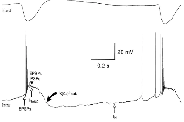
4.2 Cellular correlates of SW complexes and fast runs
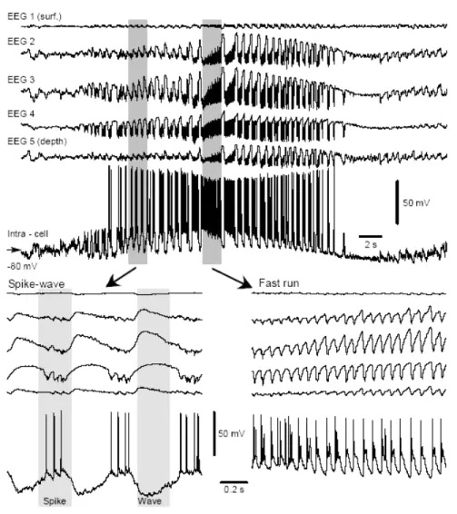
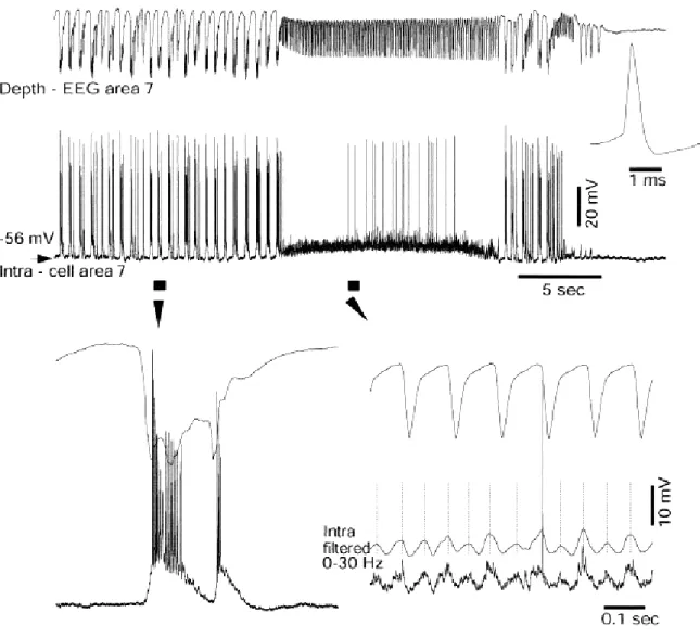
The neuronal mechanisms underlying the generation of electroencephalographic SW complexes are similar in seizures consisting of only SW and in those paroxysms resembling the EEG patterns of the clinical Lennox-Gastaut syndrome, in which SW complexes at slightly lower frequencies are intermingled with fast runs (Halasz, 1991; Yaqub, 1993; Niedermeyer, 1999; Timofeev and Steriade, 2004). During SW discharges the cortical neurons are depolarized and fire spikes during depth-negative (EEG spike) components and hyperpolarized during depth-positive (EEG wave) components. The typical seizure consisting of SW/PSW complexes recurring with frequencies 1-3 Hz and fast runs with frequencies of oscillations at 8-14 Hz is shown in the Figure 4-1. The seizure starts with SW/PSW discharges, which progressively increase in duration and the seizure displays a prolonged period of fast runs, followed again by PSW complexes and the seizure ends with SW discharges (Timofeev and Bazhenov, 2005).
The electrographic “spike”-components of SW complexes correspond intracellularly to paroxysmal depolarizing shifts (PDS) (for review see McNamara, 1994; Traub et al., 1996; Timofeev and Steriade, 2004). Interictal EEG “spikes” (IS) or PDS are similar to those that occur during SW seizures and firstly isolated PDS may progressively evolve into full-blown SW paroxysms (Figure 4-2) (Neckelmann et al., 1998; Timofeev and Steriade, 2004). Initially, PDS have been regarded as giant excitatory postsynaptic potentials (EPSP) (Johnston and Brown, 1981, 1984), enhanced by activation of voltage-gated intrinsic currents (Wong and Prince, 1978; Dichter and Ayala, 1987). Nevertheless, recent in vivo and in vitro data demonstrate the presence of inhibitory processes during different types of seizure activity (Traub et al., 1996; Esclapez et al., 1997; Prince and Jacobs, 1998) and the important inhibitory component of PDS (Cohen et al., 2002; Timofeev et al., 2002a; Fujiwara-Tsukamoto et al., 2003). This inhibitory component of PDS accounts, at least partially, for the diminished excitability of neocortical neurons to antidromic and synaptic volleys during the EEG “spike” component of SW complexes (Steriade and Amzica, 1999).