HAL Id: dumas-01074142
https://dumas.ccsd.cnrs.fr/dumas-01074142 Submitted on 13 Oct 2014
HAL is a multi-disciplinary open access
archive for the deposit and dissemination of
sci-L’archive ouverte pluridisciplinaire HAL, est destinée au dépôt et à la diffusion de documents
Aortic dissection with acute malperfusion syndrome :
endovascular fenestration by the funnel technique
Anne VendrellTo cite this version:
Anne Vendrell. Aortic dissection with acute malperfusion syndrome : endovascular fenestration by the funnel technique. Human health and pathology. 2014. �dumas-01074142�
UNIVERSITE JOSEPH FOURIER FACULTE DE MEDECINE DE GRENOBLE
Année : 2014 N°
THESE
PRESENTEE POUR L’OBTENTION DU DOCTORAT EN MEDECINE DIPLÔME D’ETAT
ANNE VENDRELL
Né(e) le 05/05/1985 à Montpellier
THESE SOUTENUE PUBLIQUEMENT A LA FACULTE DE MEDECINE DE GRENOBLE* Le Vendredi 26 septembre 2014
DEVANT LE JURY COMPOSE DE
Président du jury : M. le Professeur Gilbert FERRETTI Membres
M. le Docteur Frédéric THONY M. le Professeur Olivier CHAVANON M. le Professeur Ivan BRICAULT M. le Professeur Jean-Philippe BAGUET M. le Docteur Mathieu RODIERE
*La Faculté de Médecine de Grenoble n’entend donner aucune approbation ni improbation aux opinions émises dans les thèses ; ces opinions sont considérées comme propres à leurs auteurs.
AORTIC DISSECTION WITH ACUTE MALPERFUSION
SYNDROME: ENDOVASCULAR FENESTRATION BY THE
Remerciements
Au Jury
Monsieur le Docteur Frédéric Thony :
Merci d’avoir accepté d’encadrer ce travail, d’avoir supporté mes
mails incessants et mes irruptions inopinées dans ton bureau. Je sais
que ce sujet te tenait à cœur, et j’ai été honorée que tu me le confies.
Merci également pour les heures d’apprentissage en angiographie, et
ton coté toujours physiopathologique des choses. Tu trouveras dans ce
travail l’expression de mon plus grand respect.
Monsieur le Professeur Gilbert Ferretti :
Merci de me faire l’honneur de présider cette thèse. Merci pour votre
disponibilité, vos conseils, votre soutien dans mes démarches. Je vous
remercie également pour les heures de travail, d’enseignement, parfois
même de mathématiques. Veuillez trouver dans ce travail l’expression
de ma gratitude et de ma considération.
Monsieur le Professeur Olivier Chavanon :
Je vous remercie d’avoir accepté de juger ce travail. Veuillez accepter
l’expression de toute ma gratitude.
Monsieur le Professeur Jean-Philippe Baguet :
Je vous remercie de votre présence au sein de ce jury. Merci
également pour votre gentillesse et votre accessibilité. Je vous
exprime ma reconnaissance et mon plus grand respect.
Monsieur le Professeur Ivan Bricault :
Je te remercie d’avoir accepté de juger ce travail. Merci pour ton
soutien, ton encadrement pédagogique et universitaire : tu es une vraie
référence pour nous. Trouve ici l’expression de ma plus grande
Aux plus proches…
A Julien, mon plus fidèle soutien
Merci de ton aide pour ce travail toute cette année… La patience, les
conseils, les heures de relecture ensemble….Merci d’être là, toujours
présent et fier de moi. Avec toi, j’ai l’impression que rien n’est
impossible, merci de me donner tant de force. Je t’aime.
A mes parents, d’avoir toujours été là pour moi :
Maman, ton éternelle franchise est parfois dure à entendre… mais si
souvent véridique!!! J’espère que je serai une aussi bonne maman que
toi, car tu es la meilleure.
Papa, je te dédie ce travail. Tu m’as élevée à devenir une adulte, mais
aussi une jeune médecin. Nos longues conversations médicales
nocturnes ont profondément marqué ma vision de la médecine. Je
serai heureuse si j’arrive à la cheville du médecin que tu es.
A mes frères,
Jean-François, car tu n’as jamais vraiment grandi, et que tu es
atypique. Merci pour la relecture de ce travail : ta vision synthétique et
la pertinence de tes conseils m’ont impressionnée. Merci à Karine de
te supporter depuis si longtemps.
Vincent, pour les projets de fusée, les petits contrats et l’affaire JFK…
j’espère que tu sauras un jour qui a assassiné le président !
A mes trois soleils : Éloïse, Anna et Alice, je vous aime tellement !
Au petit Nicolas, enfin un garçon dans cette famille de filles.
A Montpellier, qui m’a vue grandir…
Aux copines de toujours :
Ma Boudou… mon alter ego, mon 2eme doigt de la main. Tu n’as pas
directement contribué à ce travail, mais tu as contribué à celle que je
suis devenue. Merci pour cette si grande amitié, elle durera toute la
vie.
Tahon, ton excentricité nous a toujours tellement fait rire…Reste telle
que tu es c’est comme ça qu’on t’aime !!! Merci de garder le contact
avec tes petits skypes ;).
A l’externat :
A mes « grosses » !!!
Aurélouve, pour t’avoir 10 fois par jour au téléphone. Quel recoin de
ma vie ne connais tu pas à force ?? Merci d’être si présente !!!
Potos (hihihi), pour les fous rires et la choré du chien en pédiatrie…
N’oublie jamais que les réunionnais ont un os dans le nez !
A ma Bergogne et ma Delph, pour les voyages et les fous rires. Lise je
te reverrai toute ma vie t’envoler de la portière de ma voiture… Delph,
14 ans que l’on se connaît !
A vous quatre, je suis heureuse que l’internat ne nous aie pas séparées.
On se retrouve dans le Sud dans pas longtemps !!
A Stéfano…on se suit depuis tant d’années, et je crois que tu me
désespères de plus en plus !! Je me demande bien si on a changé
depuis l’ère du « caps derrière les impôts ». A Lidia, car je ne sais pas
comment elle fait !
A Pierrot, le « calbar mocas » est inoubliable. Merci de me faire tant
rire à me « torturer ». Je mettrais un point d’honneur à ne plus pleurer
en voyage !
A Grenoble qui m’a vu m’épanouir…
A mes copppinnnes !!
Ma poule Hitier, pour les rires, les coups de blues, les histoires de
services… et les jacuzzi nocturnes !!
Mathildos, pour ton calme, ta tolérance, tes conseils avisés...tu es
vraiment une maman.
Au Dr Savoye (à prononcer à la halmut) : Merci pour tout mon
pierrot !!!! J’ai tellement rit en vacation avec toi, quand tu shootais
derrière ma chaise… J’ai longtemps cru que ce serait toujours comme
ca, mais non…Je suis contente que l’on soit restés proches et d’avoir
mieux découvert Leila. Vivement les prochains fous rires !!
A la Mama : Je ne te l’ai jamais dit, mais tu es une sorte de modèle
pour moi… Merci pour les apéros, les longues conversations persos,
d’avoir été si présente à une époque difficile, tu me manques ici!!
Malgré ton départ, j’espère que l’on ne se perdra pas de vue et que
l’on restera amies.
A Béatou : Comment te dire merci ??? Tu m’as ouvert ton cœur et ta
maison… On est deux sensibles toutes les deux, mais je crois que tu es
plus tarée que moi!! Casque de moto sur la tête, carte black, et c’est
bon on peut se faire un rencard !! Sinon salade de lentilles, au choix.
A mes cointernes :
Ma Delouche, je t’adore !! Coucou, c’est moi !!
Mon fiston, cette bonne vieille Ghelf, ça te dis un veau aux olives ? A
Soso dudu !
Maillot, le bourgeois détente
Moumoune, mais putain ferme la !
Chapuitos, dit tchap
Ju Cohen pour tes diagnostics quotidiens de maladies rarissimes
Lucky Luke Moncharmus, tu préfères l’escalade à la radio !
A Nico, Lionel, Tristano, Béné, Finassier, Rouchy
A Marie M pour les bons et mauvais moments en angio,
A Elo et Marie Lorette
Aux différents services qui m’ont accueillie…
La radiopédiatrie, stage pas toujours facile… A Cornepoux, Jean Mich
et Annabelle pour les bons moments.
La neuroradio, le stage à deux (ou presque) avec Marco. Merci à
Omer d’avoir toujours été en backup de la salle d’angio… A ma sister
pour les histoires de cœur.
Aux sudistes, pour la bonne humeur et les bons gâteaux !! A Chris ma
secrétaire préférée.
A la Centrale, le vaisseau Mère de la radio… quand on revient en
Centrale, on rentre un peu à la maison ! Merci à tous : manips,
secrétaires, ASH d’avoir été la pendant ces 5 semestres, et d’avoir
supporté mes humeurs et/ou mes rires incessants.
Dédicace à Maryse pour tous tes compliments, Alain Charabie pour
tes « AAAnne », Ju pour les « SOS d’un terrien en détresse » en
pleine garde.
Merci aux Docteurs Seinturier et Francois-Joubert de m’avoir
accueillie dans leurs services respectifs. Merci à leurs équipes pour
tous les bons moments.
A l’ensemble de l’équipe de radiologie vasculaire du CHU de
Montpellier, pour les vacations de TAP de la mort et les IRM
cardiaques bien trop matinales. A mes co-internes Marie-Hélène,
Juliette et Laure, je me suis régalée à vos côtés ! J’espère que l’on
continuera à se croiser dans des congrès à l’autre bout du monde ou
aux soirées blanches… !! A Casalonga, pour les 10 décas par jour et
les blagues pourries…
Au Pr Hélène Vernhet, merci de m’avoir accueillie durant ces six mois
et de m’avoir fait partagé votre expérience, en vasculaire et en
Tables des matières
RESUME
ARTICLE
Abstract
Introduction
Material and methods
Table 1
Figure 1
Figure 2
Results
Table 2
Table 3
Discussion
Conclusion
References
CONCLUSION SIGNEE
SERMENT D’HIPPOCRATE
Résumé
Objectif : Analyser les résultats à court et long terme d’une méthode originale de fenestration
aortique (FA), la technique de l’entonnoir, lors de dissections aortiques (DA) compliquées de syndrome de malperfusion (SMP).
Matériels et méthodes : La technique de l’entonnoir consistait à réaliser une fenestration
aortique par perforation du flap intimal, puis à déployer une endoprothèse non couverte du faux vers le vrai chenal. Vingt-huit patients atteints de DA (types A n=19, types B n=9) ont étés traités par la technique de l’entonnoir en raison d'un SMP par ischémie dynamique (16 rénaux, 20 digestifs, 14 membres inférieurs). Neuf patients étaient en état d’ischémie sévère à l’arrivée (6 digestives, 7 rénales, 3 membres inférieurs).
Résultats : La procédure a été réalisable chez 27/28 patients (96%). La symptomatologie
ischémique a disparu chez 25/28 patients (89%) à J +30, tandis que deux patients (7%) atteints d’ischémie sévère sont décédés de complications ischémiques digestives. Cinq patients ont présenté des complications post-procédure : 1 majeure (thrombose artérielle sur l’introducteur, conduisant à un échec technique) (3,5%) et 4 mineures.
A 4 ans de suivi, aucune récidive ou complication liée à la fenestration n’a été observée, et le diamètre aortique thoracique est resté stable chez 21/22 patients suivis (91%).
Conclusion : Le traitement par la technique de l'entonnoir dans les SMP au cours des DA a
Article
Aortic dissection with acute malperfusion syndrome:
Endovascular fenestration by the funnel technique
A. Vendrell, J. Frandon, M. Rodière, O.Chavanon, JP. Baguet, V. Bach, G. Ferretti, F.thony
Abstract:
Purpose: To analyze the short- and long-term results of an original aortic
fenestration (AF) method using the funnel technique (FT) during aortic dissection (AD) complicated by malperfusion syndrome (MPS).
Materials: The FT consists in an intimal aortic flap fenestration completed by
the deployment of an uncovered aortic stentgraft placed from the false to the right lumen through the AF. 28 patients presenting with an AD (type A n=19, type B n=9) were treated for MPS due to dynamic compression (16 renal, 20 bowel, and 14 lower limb ischemia) using this technique, and were followed-up at short (30 days) and long term (mean 55 ± 40 months). Nine patients had severe ischemia upon arrival (6 bowel, 7 renal, 3 lower limb ischemia).
Results: Technical success was achieved in 27 of 28 patients (96%) and
ischemic symptoms have disappeared in 25 of 28 patients (89%) at short term. Five patients presented postprocedure complications: 4 minor and 1 major (arterial thrombosis which cause technical fail) (3.5%). The 30-day mortality rate was 7% (n=2), related to bowel ischemia complications. At long term, among the 22 patients followed-up, 21 had a stable thoracic aortic diameter (91%).
Conclusion: The FT in cases of MPS following AD safely improve the clinical
outcome at short and long term, and could represent an interesting alternative in the management of patients. The hemodynamic efficiency of this technique may account for a lower mortality in our series.
Introduction:
Acute aortic dissection (AD) is a rare but serious disease (1), inducing high mortality rate (50%) at 48 hours (2,3). The Stanford classification of AD (4) differentiates type A requiring surgical management (5), from type B, treated medically (6).
Malperfusion syndrome (MPS), due to a reduced flow in the aortic branch vessels, is the second most common complication (20-50%) of AD (7) after cardiac complications (8) and causes higher mortality (51% versus 29%) (9,10). Two ischemic mechanisms have been described (11): dynamic compression due to an aortic true lumen collapse, and static compression, related to direct extension of AD into an aortic branch.
In cases of dynamic compression MPS, closing the primary entry tear of the dissection by using a covered stent is the recommended treatment (12–15). When this is unfeasible, aortic fenestration (AF) has been proposed as an alternative technique (16–19), however, high postprocedure complications (11– 20%) and mortality rates (17–34%) have been reported.
In our center, an original AF technique using uncovered aortic stentgraft between the false and the right lumen called the “funnel technique” (FT) was performed on selected patients. The aim of this study was to assess the safety and efficacy of this technique at short- and long-term follow-up.
Material and methods:
Study design:
From 2000 to 2013, in our center, all consecutive patients presenting renal, digestive and/or lower limbs (LLs) ischemia due to dynamic compression MPS following type A or B AD were included in this study and benefited the FT. These patients could not be treated by thoracic aortic stent graft because of anatomic constraints. Diagnosis of MPS was based on clinical, biological, and/or radiological features, according to the following criteria:
-‐ Renal ischemia: uncontrolled arterial hypertension, increase creatinine in the nonsevere forms; dialysis in the severe forms;
-‐ LL ischemia: absent pulse, claudication, cool limb in the nonsevere forms; sensorimotor deficit in the severe forms;
-‐ Bowel ischemia: loss of appetite, abdominal pain, diarrhea in the nonsevere forms; digestive tract bleeding, altered bowel sounds, high lactate level, and/or signs of intestinal compromise on CT in severe forms.
Table 1: Different types of ischemia and their severity:
Ischemia sites
Definition
N Clinical Biological Radiological
Renal ischemia
Nonsevere Uncontrolled arterial hypertension Increase creatinine 10 Severe Dialysis 5 Bowel ischemia
Nonsevere Loss of appetite, abdominal pain,
diarrhea
11
Severe Digestive tract bleeding, altered bowel
sounds High lactates level Lack of enhancement of digestif wall 6 Lower limb ischemia
Nonsevere Absent pulse, claudication, cool limb
12
Severe Sensorimotor deficit 3
Dynamic compression
Collapse of the true lumen by the false lumen
28
MPS could occur immediately during the dissection, postoperatively, or later, but the time from diagnosis to treatment was always less than 3 days.
The local Institutional Review Board approved this retrospective study. Relevant and follow-up data were collected and reviewed, using the medical files and imaging exams of included patients.
Procedure:
A preprocedure CT-scan was systematically performed to determine the orientation of the intimal flap and the fenestration target (4 cm above the celiac trunk).
All procedures were performed by two interventional radiologists with more than 10 years experience, under fluoroscopic guidance only. Initial angiography was first done under local anesthesia, through a femoral sheath, to locate the origin of the celiac trunk. In a curved 5F catheter, a rigid 0.014 Spartacore® guidewire (Abbott Vascular, Santa Clara, CA, USA) was introduced in the true lumen in the reverse position (hard end first), positioned against the intima to go through it, and advanced in the false lumen. The fenestration opening was then enlarged by using an 18-mm-diameter balloon (Boston Scientific, Natick, MA, USA). An uncovered self-expanding Wallstent (Boston Scientific, Natick, MA, USA) or Memotherm (Bard Angiomed, Wachhausstrasse, Germany), 18mm to 24 mm diameter and 40-60 mm long, was then deployed from the false lumen toward the true lumen through the opening created, with its distal end placed above the ostium of celiac trunk.
The final angiography was then done in the false lumen above the fenestration, and had to confirm satisfactory flow in the true lumen and the aortic branchs
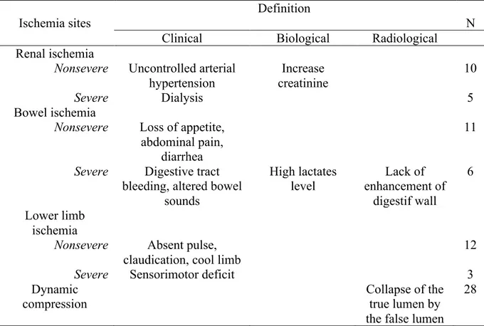
Figure 1: 71-years-old patient admitted for acute type B AD with severe bowel ischemia due to dynamic compression. Angio CT-scan on axial views (A, B), sagittal (C) and coronal reformations (D), showing thoraco-abdominal type B AD, with large primary entry tear locate in the aortic arch (arrowhead) ; compression of the true lumen (TL) by the false lumen (FL) on the abdominal level, which cause reduced flow in the abdominal arteries.
B A B C D FL FL TL FL FL TL TL
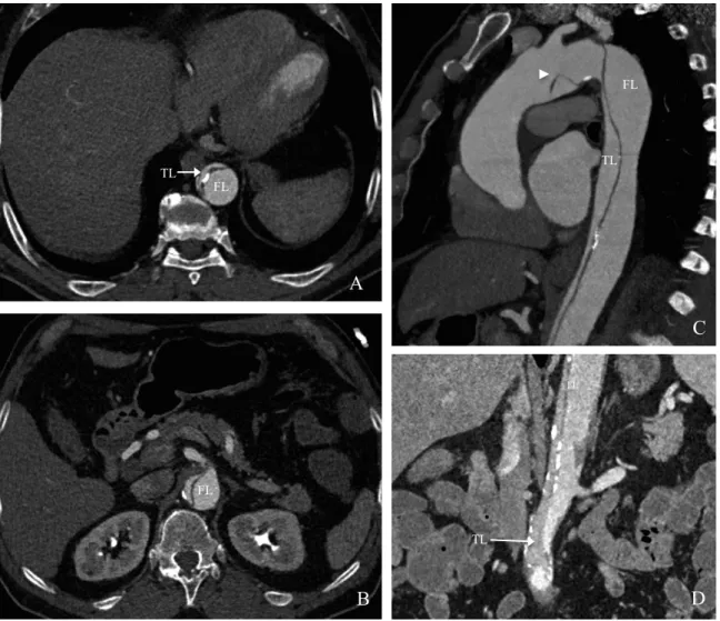
Figure 2: Aortic fenestration using the funnel technique in the same patient. A: Angiogram showing the true lumen collapse (TL). The right renal artery and the intercostal arteries originate from the true lumen (arrowsheads), and their vascularization seems compromised. B: Angiogram of the false lumen (FL) after insertion of a catheter through the intimal perforation, showing the celiac trunk, the left renal artery and the superior mesenteric artery (white arrows). C: Illustration of the funnel technique: AF using a guidewire completed by balloon angioplasty of the fenestration opening and stent grafting of the false lumen toward the true lumen. D: Final angiogram with injection in the false lumen (FL): the stentgraft (asterisk) reapplied pressure in the true lumen (TL) with opacification of both renal arteries (solid arrows), the celiac trunk (splenic and hepatic arteries) and the superior mesenteric artery (dashed arrows). E, F, G, H: Follow-up CT at D2 in axial views (E, F), sagittal reformations (G) and VRT reformations (H) showing the stentgraft in place (asterisk) within
A B C D F G H E TL FL FL TL FL TL FL TL FL TL FL TL
Another stentgraft has been placed in the superior mesenteric artery in the same procedure because of static compression (arrowhead).
If the angiographic result was inadequate, an additional subrenal fenestration or peripheral stenting of side branches was discussed.
For the following 6 months, patients received an antiplatelet treatment, except those treated with oral anticoagulants.
Definitions:
Technical success was defined as adequate flow in the true lumen at the end of the procedure, in either a single procedure (primary technical success) or two procedures (secondary technical success).
Clinical success was defined as disappearance of clinical symptoms and normalization of biological parameters at D30.
The main evaluation criterion was clinical success. The secondary evaluation criteria were mortality and complication rate at short term (30 days); onset of late complications and evolution of thoracic aorta diameter at long term.
Patients were followed up by MRI or CT at 6 months, 12 months, and then every 2 years. The mean duration of follow-up was defined as the time between the initial imaging study and the last available images. The thoracic aorta was measured on the same levels in the axial plane. Increase aortic diameter was defined by a diameter greater than 60 mm or progressing more than 4 mm/year.
Results:
Population:
From 2000 to 2013, 367 patients were admitted to our center for AD (type A n=237, type B n=130). Among patients presenting with type A, 217/237 underwent surgical aortic replacement and 20/237 died before arrival. All patients with type B AD were treated medically. Sixty-nine of 347 patients (20%) presented dynamic compression MPS, treated with either thoracic aortic stenting (n=41) or AF by FT (n=28).
Of the 28 treated by FT, 19 patients presented type A AD and underwent emergency cardiac surgery (14 aorto-aortic tubes, 4 Bentall procedures, and 1 hemiarch replacement), always done first regardless of the severity of MPS. Nine patients presented type B AD. Patients characteristics and ischemic territories are summarized in table 2.
Additional stenting for concomitant static compression was necessary in 13/28 patients: renal (n=7), superior mesenteric (n=5), and iliac (n=5).
Table 2: Population caracteristics:
Characteristic Value
Sex 28
Male 9
Female 19
Mean age (Y) 55 ± 12
Type A dissection 19 Type B dissection 9 Ischemic Territories Three sites : Renal/Mesenteric/Lower Limb 4 Two sites : 10 Renal/Mesenteric 5 Renal/Lower Limb 3 Mesenteric/Lower Limb 2 One site 15 Renal 5 Mesenteric 5 Lower Limb 5 Recanalisation:
Primary technical success was 86% (24/28), secondary technical success 97% (27/28), with no intraprocedural complications. Three patients required a second subrenal AF: one patient during the same procedure because of insufficient flow in the iliac arteries on the final angiogram, two patients later because of recurrence of their ischemic symptoms at D2 and D15.
Clinical success was achieved in 25 of 28 patients (89%). Two patients (7%) with severe ischemia (1 renal and bowel ischemia, 1 all three sites), treated with cardiac surgery and then AF at D0 died before D30: the first at D2 of
multisystem organ failure (MOF), the second of septic shock ending in death at D14.
One patient was considered as technical and clinical failure, and major complication because of worsening of a right LL ischemia at the end of the procedure, related to the onset of a thrombosis of the common femoral artery on the sheat. He was treated with embolectomy and femoro-femoral bypass resulting in good postoperative evolution.
Severe ischemia
Six out of 8 patients with severe ischemia survived. Of the 4 survivors with mesenteric ischemia, all patients evolved favorably, 3 of whom requiring bowel resection. Dialysis was reversible after AF for the 3 patients with severe renal ischemia. Of the two patients with severe LL ischemia, one of whom improved after fenestration, the second has been previously described. Table 3 summarizes the characteristics of the patients with severe ischemia.
Table 3: Treatment and evolution of patients with severe ischemia :
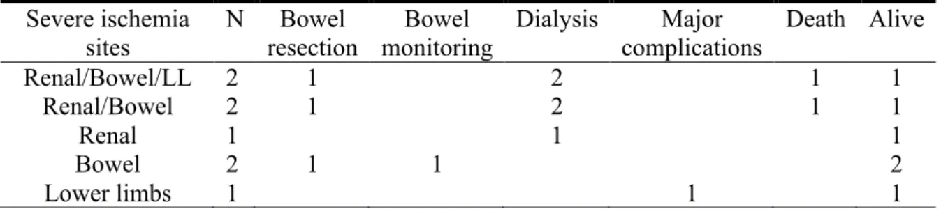
Severe ischemia sites N Bowel resection Bowel monitoring Dialysis Major complications Death Alive Renal/Bowel/LL 2 1 2 1 1 Renal/Bowel 2 1 2 1 1 Renal 1 1 1 Bowel 2 1 1 2 Lower limbs 1 1 1
Complications:
Five patients presented complications, with a major one (3.5%). Of the four minor complications, 3 were related to the puncture point (arterial thrombosis resolved with medical treatment; hematoma and infection resolved with antibiotics). The last one was an aortic stent fracture (nitinol stent) discovered on the follow-up CT at D10, with no clinical or biological repercussions and was treated by placing a second stent within the first one.
Follow-up:
During the follow-up, 3 patients had withdrawn from the study and 3 had died: Two at 1 year of follow-up, the first after surgical replacement of the descending aorta and the second of a brain tumor; the third of fissuration of a descending thoracic aorta aneurysm at 9 years of follow-up. Of the 22 patients followed up, 1 patient presented a proximal fracture of the stent detected on the 2 years follow-up CT (nitinol stent) with no consequences. No recurrence of ischemia was observed.
At a mean 55 ± 40 months of follow-up, the mean diameter of aortic isthmus was 44mm ± 16mm versus 38 ± 5mm initially, so a mean progression of 1 mm per year. Twenty-one patients (91%) had a stable aortic diameter and two patients (9%) had an increased aortic thoracic diameter.
Discussion:
The current recommended treatment for MPS caused by dynamic compression is closing the primary entry tear of DA by a covered thoracic stent grafting (13,15,20,21). However, when it is not possible, AF can resupply the true lumen at the abdominal level by creating a communication between the two lumens. Two techniques of AF have been described: fenestration with balloon angioplasty (18,19,22,23) and the scissor technique (24). Our technique is original because it enlarges the surface area of the re-entry orifice in the true lumen with an endoaortic stent between the two lumens, which increases flow and pressure in the true lumen and reduces them in the false, thus improving the efficacy of fenestration.
AF has been described in the literature as both a reliable (100% technical success) and effective technique, with clinical success between 70% and 93% at 1 month (16,19) versus 60% with surgery (20). This series also shows that AF using the FT is effective (90% clinical success).
With balloon angioplasty AF, the 30-day mortality rate is 17–25%, with an approximately 20% complication rate (18,19,23). The scissor technique has shown 30% mortality at D30 with roughly 10% complications (16). Mortality in
Patients with severe ischemia account for 26% in the series reported by Midulla et al., 22% in the Slonim et al. series, and 14% in the O’Donnell et al. series (25), with a 33% mortality rate reported in Midulla et al. and 66% in Slonim et al series. In our series, the severe ischemia rate was higher (31%), but the mortality rate lower (22%). This mortality rate with the FT therefore does not stem from the population presenting globally less severe SMP, but probably from a more effective reapplication of pressure in the true lumen. This results in the disappearance of clinical symptoms and the reversion of the acute ischemic lesions, preventing the onset of MOF. In the present series, only two patients (7%) died of MOF despite the AF, whereas this unfavorable outcome seems more frequent in the other series cited above (25% of patients with type B AD in the series reported by Midulla et al., 15% in Slonim et al.’s series).
No malpositioning problems or stent migration during grafting, injury of the aortic wall or stent thrombosis at the long term were observed. Two fractures were discovered inadvertently, both asymptomatic and involving nitinol stents (Memotherm Bard Angiomed, Wachhausstrasse, Germany). We have since decided to use only steel self-expandable Wallstents (Boston Scientific, Natick, MA, USA), with no new complications discovered to date.
Three patients underwent an additional fenestration because the first was deemed insufficient. In these patients, an 18-mm-diameter stent was selected for
a mean 30-mm-diameter aorta in the fenestration area. Finally, this stent diameter was considered too small for the size of the false lumen. Since this time, we have used stents with a larger diameter so that the re-entry orifice in the true lumen is at least two-thirds the total diameter of the aorta, thus ensuring that flow is greater than 50% between the true and false lumen. This is not always possible because maximal diameter of wallstent is 24mm.
Over the long term, decreased pressure in the false lumen seems to protect against the aneurysmal aortic evolution, since 91% of the patients (15 type A and 5 type B) presented a stable aortic diameter. As reported in the literature, the natural progression of AD is characterized by dilatation of the descending thoracic aorta in 20–50% of cases (26) and up to 83% in the segment-by-segment study of the aorta (27,28). This confirms the hypothesis postulating that blood pressure is reduced within the false lumen upon reapplication of pressure in the true lumen, thus avoiding progression toward aneurysm. Midulla et al. (9) have reported the same observation.
The small number of patients treated was the main limitation of this study. Further studies, as best comparative and multicentric, would be necessary to confirm them.
Conclusion:
AF by the FT, in patients with MPS during AD is an effective technique (clinical success: 90%) associated with low morbidity (30-day mortality: 7%, major complications : 3.5%). Over the long term, no ischemic recurrence was observed, and decreasing the applied pressure of the false lumen seems to protect from increase aortic thoracic diameter. The hemodynamic efficiency of this technique may account for a lower mortality in our series but this result has to be confirmed.
References:
1. Golledge J, Eagle KA. Acute aortic dissection. Lancet. 5 juill 2008;372(9632):55-‐‑66. 2. Asfoura JY, Vidt DG. Acute aortic dissection. Chest. mars 1991;99(3):724-‐‑9.
3. Mészáros I, Mórocz J, Szlávi J, Schmidt J, Tornóci L, Nagy L, et al. Epidemiology and clinicopathology of aortic dissection. Chest. mai 2000;117(5):1271-‐‑8.
4. Daily PO, Trueblood HW, Stinson EB, Wuerflein RD, Shumway NE. Management of acute aortic dissections. Ann Thorac Surg. sept 1970;10(3):237-‐‑47.
5. Debakey me, Henly ws, Cooley da, Morris gc jr, Crawford es, Beall ac jr. Surgical management of dissecting aneurysms of the aorta. J Thorac Cardiovasc Surg. janv
1965;49:130-‐‑49.
6. Wheat MW, Palmer RF, Bartley TD, Seelman RC. Treatment of dissecting aneurysms of the aorta without surgery. J Thorac Cardiovasc Surg. sept 1965;50:364-‐‑73.
7. Swee W, Dake MD. Endovascular management of thoracic dissections. Circulation. 18 mars 2008;117(11):1460-‐‑73.
8. Rodiere M, Thony F, Michoud M, Frandon J, Ferretti G. [Acute visceral ischaemia, early complication of the aortic syndrome: how to detect and manage it?]. Presse Médicale Paris Fr 1983. janv 2011;40(1 Pt 1):54-‐‑61.
9. Di Eusanio M, Trimarchi S, Patel HJ, Hutchison S, Suzuki T, Peterson MD, et al. Clinical presentation, management, and short-term outcome of patients with type A acute dissection complicated by mesenteric malperfusion: observations from the International Registry of Acute Aortic Dissection. J Thorac Cardiovasc Surg. févr 2013;145(2):385-‐‑90.e1. 10. Cambria RP, Brewster DC, Gertler J, Moncure AC, Gusberg R, Tilson MD, et al. Vascular complications associated with spontaneous aortic dissection. J Vasc Surg. févr 1988;7(2):199-‐‑209.
11. Williams DM, Lee DY, Hamilton BH, Marx MV, Narasimham DL, Kazanjian SN, et al. The dissected aorta: part III. Anatomy and radiologic diagnosis of branch-vessel
compromise. Radiology. avr 1997;203(1):37-‐‑44.
12. Nienaber CA, Fattori R, Lund G, Dieckmann C, Wolf W, von Kodolitsch Y, et al. Nonsurgical reconstruction of thoracic aortic dissection by stent-graft placement. N Engl J
13. Dake MD, Kato N, Mitchell RS, Semba CP, Razavi MK, Shimono T, et al.
Endovascular stent-graft placement for the treatment of acute aortic dissection. N Engl J Med. 20 mai 1999;340(20):1546-‐‑52.
14. Nienaber CA, Zannetti S, Barbieri B, Kische S, Schareck W, Rehders TC, et al. INvestigation of STEnt grafts in patients with type B Aortic Dissection: design of the INSTEAD trial--a prospective, multicenter, European randomized trial. Am Heart J. avr 2005;149(4):592-‐‑9.
15. Botsios S, Schuermann K, Maatz W, Keck N, Walterbusch G. Complicated acute type B dissections: a single-center experience with endovascular treatment. Thorac Cardiovasc Surg. août 2010;58(5):280-‐‑4.
16. Midulla M, Renaud A, Martinelli T, Koussa M, Mounier-Vehier C, Prat A, et al. Endovascular fenestration in aortic dissection with acute malperfusion syndrome: immediate and late follow-up. J Thorac Cardiovasc Surg. juill 2011;142(1):66-‐‑72.
17. Beregi J-P, Haulon S, Otal P, Thony F, Bartoli J-M, Crochet D, et al. Endovascular treatment of acute complications associated with aortic dissection: midterm results from a multicenter study. J Endovasc Ther Off J Int Soc Endovasc Spec. juin 2003;10(3):486-‐‑93. 18. Patel HJ, Williams DM, Meerkov M, Meekov M, Dasika NL, Upchurch GR Jr, et al. Long-term results of percutaneous management of malperfusion in acute type B aortic dissection: implications for thoracic aortic endovascular repair. J Thorac Cardiovasc Surg. août 2009;138(2):300-‐‑8.
19. Slonim SM, Miller DC, Mitchell RS, Semba CP, Razavi MK, Dake MD. Percutaneous balloon fenestration and stenting for life-threatening ischemic complications in patients with acute aortic dissection. J Thorac Cardiovasc Surg. juin 1999;117(6):1118-‐‑26.
20. Fattori R, Tsai TT, Myrmel T, Evangelista A, Cooper JV, Trimarchi S, et al. Complicated acute type B dissection: is surgery still the best option?: a report from the International Registry of Acute Aortic Dissection. JACC Cardiovasc Interv. août 2008;1(4):395-‐‑402.
21. Nienaber CA, Rousseau H, Eggebrecht H, Kische S, Fattori R, Rehders TC, et al. Randomized comparison of strategies for type B aortic dissection: the INvestigation of STEnt Grafts in Aortic Dissection (INSTEAD) trial. Circulation. 22 déc 2009;120(25):2519-‐‑28. 22. Kos S, Gürke L, Jacob AL. A novel fenestration technique for abdominal aortic dissection membranes using a combination of a needle re-entry catheter and the « cheese-wire » technique. Cardiovasc Intervent Radiol. déc 2011;34(6):1296-‐‑302.
23. Chavan A, Hausmann D, Dresler C, Rosenthal H, Jaeger K, Haverich A, et al.
Intravascular ultrasound-guided percutaneous fenestration of the intimal flap in the dissected aorta. Circulation. 7 oct 1997;96(7):2124-‐‑7.
24. Beregi JP, Prat A, Gaxotte V, Delomez M, McFadden EP. Endovascular treatment for dissection of the descending aorta. Lancet. 5 août 2000;356(9228):482-‐‑3.
25. O’Donnell S, Geotchues A, Beavers F, Akbari C, Lowery R, Elmassry S, et al. Endovascular management of acute aortic dissections. J Vasc Surg. nov 2011;54(5):1283-‐‑9. 26. Trimarchi S, Jonker FHW, van Bogerijen GHW, Tolenaar JL, Moll FL, Czerny M, et al. Predicting aortic enlargement in type B aortic dissection. Ann Cardiothorac Surg. mai 2014;3(3):285-‐‑91.
27. Tolenaar JL, van Keulen JW, Jonker FHW, van Herwaarden JA, Verhagen HJ, Moll FL, et al. Morphologic predictors of aortic dilatation in type B aortic dissection. J Vasc Surg. nov 2013;58(5):1220-‐‑5
28. Sueyoshi E, Sakamoto I, Hayashi K, Yamaguchi T, Imada T. Growth rate of aortic diameter in patients with type B aortic dissection during the chronic phase. Circulation. 14 sept 2004;110(11 Suppl 1):II256-‐‑61.