HAL Id: dumas-01435983
https://dumas.ccsd.cnrs.fr/dumas-01435983
Submitted on 16 Jan 2017HAL is a multi-disciplinary open access
archive for the deposit and dissemination of sci-entific research documents, whether they are pub-lished or not. The documents may come from teaching and research institutions in France or abroad, or from public or private research centers.
L’archive ouverte pluridisciplinaire HAL, est destinée au dépôt et à la diffusion de documents scientifiques de niveau recherche, publiés ou non, émanant des établissements d’enseignement et de recherche français ou étrangers, des laboratoires publics ou privés.
Efficacy and safety of laparoscopic fundoplication in lung
transplant recipients
Pauline Badrignans
To cite this version:
Pauline Badrignans. Efficacy and safety of laparoscopic fundoplication in lung transplant recipients. Human health and pathology. 2016. �dumas-01435983�
AVERTISSEMENT
Ce document est le fruit d'un long travail approuvé par le
jury de soutenance et mis à disposition de l'ensemble de la
communauté universitaire élargie.
Il n’a pas été réévalué depuis la date de soutenance.
Il est soumis à la propriété intellectuelle de l'auteur. Ceci
implique une obligation de citation et de référencement
lors de l’utilisation de ce document.
D’autre part, toute contrefaçon, plagiat, reproduction illicite
encourt une poursuite pénale.
Contact au SID de Grenoble :
UNIVERSITE
GRENOBLE ALPES
FACULTE DE MEDECINE DE GRENOBLE
Année : 2016
EFFICACITE ET TOLERANCE DE LA FUNDOPLICATURE CHEZ
LES PATIENTS TRANSPLANTES PULMONAIRES: ETUDE
RETROSPECTIVE CHEZ 25 PATIENTS DU CHU DE GRENOBLE.
THESE PRESENTEE POUR L’OBTENTION DU DOCTORAT EN
MEDECINE. DIPLOME D’ETAT
BADRIGNANS Pauline
THESE SOUTENUE PUBLIQUEMENT A LA FACULTE DE MEDECINE
DE GRENOBLE*
Le 04 mars 2016
DEVANT LE JURY COMPOSE DE
Président du jury : M. le Professeur PISON Christophe Membres
M. le Docteur CAMARA Boubou, Directeur de thèse
M. le Docteur RISSE Olivier
M. le Professeur MISMETTI Patrick
M. le Professeur CHAFFANJON Philippe
*La Faculté de Médecine de Grenoble n’entend donner aucune approbation ni improbation aux opinions émises dans les thèses ; ces opinions sont considérées comme propres à leurs auteurs.
REMERCIEMENTS
A mon président de thèse, Professeur Christophe Pison
Vous me faites l’honneur de présider ce jury.
Votre enseignement et votre disponibilité au cours de ces quatre ans d’internat m’ont permis d’apprendre mon métier;
Votre humanité m’a appris à l’aimer.
A mon directeur de thèse, Docteur Boubou Camara
Boubou, merci pour ta patience depuis mes premiers jours au 4èmeA et la prescription du doliprane, jusqu’aux derniers mois de relecture de cette thèse.
Merci pour ton soutien et pour ta confiance en tous les choix que j’ai faits pendant ces 4 ans.
Au Professeur Patrick Mismetti
Patrick, merci d’avoir accepté d’être à cette place de jury aujourd’hui. Tu m’as toujours poussée à me dépasser, sur les skis comme en médecine. Je choisis
aujourd’hui le versant clinique de ce métier qui me correspond le mieux, mais sache que j’aurai toujours une grande admiration pour ta passion de la recherche et de l’enseignement.
Au Professeur Philippe Chaffanjon
Je vous remercie de me faire l’honneur de juger mon travail.
Aux pneumologues de Grenoble, pour tout ce que vous m’avez transmis
Christel et Seb, pour votre disponibilité et vos compétences.
Anne Claire, Linda, Pr Moro Sibilot pour votre accompagnement pendant ce semestre d’onco thoracique difficile mais riche en expériences humaines.
Renaud pour ton calme et ta simplicité, pour tous les secrets du sommeil. Pr Pépin pour des visites très matinales mais très riches en enseignement. Bernard Wuyam pour avoir démystifié la VO2max.
A l’équipe des EFR
Pour mon dernier stage d’interne…
Aux pneumologues de Chambéry
Merci de me faire confiance pour la suite.
A l’équipe du 4A
Pour toutes ces heures partagées de jour comme de nuit.
Et plus particulièrement à Manu et Jordy pour mes premiers pas en VNI.
Aux inf de Chambéry
Pour vos sourires, votre dynamisme et cette ambiance de travail hors du commun!
Aux équipes de réa med et d’infectieux
A mes parents
Pour votre amour immense et votre soutien qui m’ont faite grandir et avancer. Vous avez su me transmettre le goût de l’effort et les valeurs de la famille.
Merci pour votre patience au cours de ces longues années d’études où je vous ai fait partager les angoisses et incertitudes de ma vie d’étudiante puis d’interne.
A ma sœur Marine
Je suis fière de ce que tu es en train de devenir, j’espère que tu t’épanouiras dans cette vie professionnelle pleine de cellules et de microscopes…
A ceux qui ont toujours fait un peu partie de la famille
Fred Lolo et les filles, Thierry et Martine, Sylvie, Chantal et Ded (cette thèse t’est dédiée).
Vous m’avez vue grandir et m’avez entourée chacun à votre façon, je suis heureuse que vous soyez là aujourd’hui.
A la Pneumo Team, parce qu’on ne pouvait pas rêver meilleure équipe
Louis, Elo, Léo, Rulian, Mathilde, Margaux, Totor, Arnaud, Hubert, Loïc, et les petits (Hélo, Tom, Mickael et Florent)
On est exceptionnel ou on n’est pas exceptionnel
Et encore plus aux grandes
Marie, Cec, Julie, MV
Quelle chance de vous avoir eu comme co-internes, assistantes et amies! J’espère que l’on va se suivre longtemps en pneumo et ailleurs…
Aux copains pharmaciens
Marion, Prudence, Gauthier, Momo, Bruno, Adrien, Sylvain, Mélanie (et la petite Emma)
Merci pour votre accueil dans le monde de la pharmacie et les bonnes bouffes. Et plus particulièrement à Prudence et Gauthier pour les bus, curry, tortues et cocktails sri-lankais.
Aux copains de l’internat
Olivier et Marion, George, Mirloose, Paulin, Cam, Jerem et le petit Oscar, Sandra, Mélanome
Pour les tisanes au salon, les sorties ski, les soirées internat et le petit café d’Olivier, … et à Valérie !
Plus particulièrement à Cam et Brune
Deux belles rencontres, c’est devenu très important de vous avoir à mes côtés.
Aux Bedhets
Pour les goûters du dimanche et les diners au coin du poêle, pour nous supporter de plus en plus souvent et pour tous ces projets de vie… on ne va pas partir trop loin.
Et bien sûr aux PTZ du 145
Caro, Benoit et Brune, ma famille de Grenoble.
A ceux qui ont partagé un temps mes journées de travail et sont devenus des amis
Louise, Tom-Tom, Emilie pour leur joie de vivre.
Quentin et Sam/ Tic et Tac, pour leurs baskets et les VVC sous écho.
Léa et Steph, un semestre pas comme les autres, riche en émotions et en fous rires. David, Emilie Mob-Mob, Charlotte, Nastasia: «Ne me mens pas» !! Pour cette fameuse photo paréo et pour nos soirées pizzas/bières.
A celles de toujours ou presque
Anaïs et Camille, et leurs moitiés: pour nos souvenirs d’adolescence, le solfège le trio la médaille et nos concerts, Saint Tropez, nos WE meufs et la joie de se retrouver à chaque fois.
Fanette, parce qu’on se connait par cœur, pour ces années d’insouciance mais aussi les épreuves traversées ensemble, je suis tellement heureuse que tu puisses suivre la voie qui te plait.
Morgane, pour cette connivence même à des kilomètres, pour nos fous rires sur les bancs de l’amphi, la Häagen-Dazs et les fondants au chocolat, les descentes de Tignes, nos voyages d’aventureuses et nos conversations inépuisables.
A Alice (et son Guillaume)
Pour nos soirées d’étudiantes, les repas desserts, la Sainté Lyon (j’en reviens toujours pas), la vérité si je mens. Ma biche je suis si émue que tu sois la première à te lancer dans l’aventure de la maternité…
Et bien sûr à Charles
Pour tout…
Merci pour ton calme et ta sérénité, souvent, mais encore plus devant mes tableaux Excel.
TABLE DES MATIERES
INDEX DES FIGURES ET TABLEAUX ... 12
LISTE DES ABREVIATIONS ... 13
INTRODUCTION ... 15
Efficacy and safety of laparoscopic fundoplication in lung transplant recipients ... 19
Abstract ... 20
Introduction ... 21
Materials and Methods ... 22
Results ... 25 Discussion ... 31 Acknowledgements... 36 Autorship page ... 37 BIBLIOGRAPHIE ... 39 SERMENT D’HIPPOCRATE ... 43 RESUME ... 44
INDEX DES FIGURES ET TABLEAUX
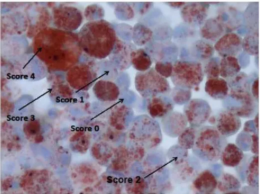
Figure 1. Lipophages scoring (intracellular lipid in BAL macrophages detected by Oil Red O staining) ... 23 Figure 2. Evolution of FEV1 from pre to post fundoplication, in L and % pred, for each
patient ... 30
Table I. Demographics of patients ... 26 Table II. Results of impedance and 24-h pH studies before surgery, n= 24 ... 28 Table III. Differences in FEV1 pre and post fundoplication (mean ± SD), for all
LISTE DES ABREVIATIONS
% pred: percentage of predicted; pourcentage de la valeur prédite BAL: bronchoalveolar lavage, LBA: lavage broncho-alvéolaire BMI: body mass index; index de masse corporelle
BOS: Bronchiolitis Obliterans Syndrome, Syndrome de Bronchiolite Oblitérante
CLAD: chronic lung allograft dysfunction; dysfonction chronique du greffon pulmonaire
COPD: chronic obstructive pulmonary disease, BPCO: bronchopneumopathie chronique obstructive
CVF: capacité vitale forcée
DLTx: double lung transplant ; transplantation double mono pulmonaire
FEV1: forced expiratory volume in 1 second, VEMS: volume expiré maximum en 1
seconde
FPI: fibrose pulmonaire idiopathique
GERD: gastro-esophageal reflux disease, RGO: reflux gastro œsophagien L: liters; litres
LI: lipid index; index lipidique
LT: lung transplant; transplanté (ou transplantation) pulmonaire
PPI: proton pump inhibitor, IPP: inhibiteurs de la pompe à protons
RAS: Restrictive Allograft Syndrome ; Syndrome Restrictif d’Allogreffe RCTs: randomized controlled trials ; étude contrôlée et randomisée SD: standard deviation; déviation standard
SIO: sphincter inférieur de l’œsophage
L’article qui fait l’objet de cette thèse est actuellement soumis à la revue Clinical
Transplantation.
Il a également été présenté:
- en poster discussion au Congrès de l’ERS à Munich le 7 septembre 2014: communication N° 272, session « Maximise lung transplant outcomes: pre and
post operatives issues ».
- en communication orale au Congrès Lung Transplantation à Paris le 25 septembre 2014: abstract N°6, session « Chronic lung allograft dysfunction ».
INTRODUCTION
Chaque année, environ 4000 greffes pulmonaires sont enregistrées auprès de l’International Society for Heart and Lung Transplantation (ISHLT). Les indications de greffe pulmonaire sont, par ordre de fréquence, la bronchopathie chronique obstructive (BPCO) et la fibrose pulmonaire idiopathique (FPI), puis la mucoviscidose, et enfin le déficit en alpha 1 antitrypsine et l’hypertension artérielle pulmonaire.
Le taux de survie après une greffe pulmonaire atteint actuellement 55% à 5 ans, ce qui est plus faible qu’après transplantation d’autres organes solides comme le rein ou le foie. Cette mortalité est notamment imputable à l’apparition fréquente d’une dysfonction chronique (1). En effet les infections (hors CMV) et la dysfonction chronique du greffon ou Chronic Lung Allograft Dysfunction (CLAD) sont les principales causes de décès dès la première année post greffe (2). La dysfonction chronique du greffon affecte environ 50% des transplantés pulmonaires après 5 ans et est responsable de plus de la moitié des décès dès 3 ans de survie (1,2). Une dysfonction chronique du greffon est suspectée lorsque le volume expiratoire maximal en une seconde (VEMS) et/ou la capacité vitale forcée (CVF) diminuent de façon prolongée (plus de 3 semaines), une fois les causes de dysfonction aigue éliminées (rejets aigus, infections, embolie pulmonaire...). Il faut alors rechercher une cause spécifique réversible à la dégradation persistante de la fonction respiratoire qui peut être liée au greffon (rejet aigu persistant, azythromycin responsive allograft dysfunction ou ARAD, sténose anastomotique…) ou non liée au greffon (pathologie pleurale, diaphragmatique…). Le cas échéant, l’évolution vers un VEMS et/ou une CVF inférieur(s) à 80% des meilleurs valeurs post greffe confirme le diagnostic de
CLAD. Il se présente alors sous la forme de 2 entités possibles: le syndrome de bronchiolite oblitérante ou bronchiolitis obliterans syndrome (BOS) et le syndrome restrictif d’allogreffe ou restrictive allograft syndrome (RAS) (1). Le RAS, décrit seulement depuis les années 2010, est le moins fréquent des deux formes de CLAD mais est associé au pronostic le plus sombre. Il se définit par une CVF inférieure à 80% des meilleures valeurs post greffe durant au moins 3 semaines et est caractérisé par un infiltrat pulmonaire interstitiel voir des signes de fibrose prédominant dans les lobes supérieurs au scanner thoracique (3). Actuellement, peu de facteurs de risque de RAS ont déjà été identifiés.
Le BOS se définit par un VEMS inférieur à 80% des meilleures valeurs post greffe durant au moins 3 semaines. Il se caractérise par une fibro-prolifération excessive dans les petites voies aériennes conduisant à une bronchiolite oblitérante responsable du trouble ventilatoire obstructif d’aggravation progressive. Le scanner thoracique peut montrer des signes de bronchiolite et un « air trapping » en expiration (1). Cette affection est médiée par des facteurs de risque allo-immuns (rejet aigu cellulaire, bronchiolite lymphocytaire, rejet humoral) ou non allo-immuns (infections respiratoires bactériennes ou fongiques, pneumopathies à CMV, reflux gastro-oesophagien (RGO)) (4,5). Il semble que la bronchiolite oblitérante ne soit pas toujours une atteinte fibrosante totalement fixée et qu’une certaine réversibilité soit possible, surtout à un stade précoce (6). Le RGO, par le biais de micro aspirations répétées, pourrait donc être un facteur favorisant réversible de BOS.
métrie anormale confirmant un RGO. Après transplantation pulmonaire le RGO atteint selon les auteurs de 50% à 70-80% des patients (4,8–10). Le rôle pathogène de l’acide gastrique est bien établi dans le cadre du RGO acide dont le traitement médicamenteux de référence repose sur les inhibiteurs de la pompe à protons (IPP). Cependant d’autres composants comme les sécrétions bilio-pancréatiques et le reflux gazeux, qui constituent le reflux non acide, peuvent également être délétères, en particulier chez les patients transplantés pulmonaires. Le diagnostic du reflux non acide se fait grâce à l’impédancemétrie (11).
Le traitement par fundoplicature laparoscopique est dans le cas général un traitement de deuxième intention du RGO, après échec d’un traitement par IPP bien conduit et si une insuffisance du sphincter inférieur de l’œsophage (SIO) est en cause (12). Chez les patients transplantés pulmonaires il n’existe pas de recommandation générale actuellement et la décision de chirurgie est individualisée. Les deux types de fundoplicature par laparoscopie sont la fundoplicature totale circulaire dite opération de Nissen et la fundoplicature partielle postérieure dite opération de Toupet enveloppant l’œsophage entre 180° et 270°. Les deux techniques semblent avoir une efficacité similaire avec, en revanche, un taux de dysphagie postopératoire et de récidives de RGO plus élevés après chirurgie de Nissen (13). Les pratiques habituelles de notre équipe consistent en un traitement probabiliste par IPP chez tous les patients transplantés, la réalisation systématique d’une pH-impédancemétrie en post greffe et une indication de fundoplicature par méthode de Toupet chez tous les patients porteurs d’un RGO, acide ou non acide.
Il a été suggéré que la présence de macrophages alvéolaires contenant des corps lipidiques phagocytés ou lipophages pouvait être un marqueur des micro-aspirations digestives dues au RGO, notamment chez les enfants présentant des
pneumopathies à répétitions ou une maladie respiratoire chronique (14). Les lipophages sont détectés par la technique de coloration à l’huile rouge ou Oil Red O stain après cyto-centrifugation de l’échantillon, préparation aux vapeurs de formol et coloration standard de May-Grünwald Giemsa pour formule. Un index est ensuite établi entre 0 et 400 par analyse de la charge lipidique de 100 lipophages consécutifs. Aucune donnée n’était disponible chez les patients transplantés pulmonaires jusqu’en 2010 où une étude monocentrique prospective sur 34 patients transplantés pulmonaires a montré une sensibilité de 82% et une spécificité de 76% entre un index de lipophages supérieur à 150 et une pH métrie anormale (15). C’est le seuil de référence actuellement retenu par notre équipe pour l’analyse des lipophages dans le LBA. Depuis, peu d’études ont étudié la corrélation entre lipophages alvéolaires et RGO ainsi que le seuil de significativité de l’index de lipophages.
L’objectif de notre travail était donc d’évaluer rétrospectivement la tolérance de la fundoplicature et son efficacité sur l’amélioration de la fonction pulmonaire chez les patients transplantés pulmonaires du CHU de Grenoble. Nous avons également étudié la spécificité des lipophages alvéolaires comme marqueurs des micro-aspirations digestives.
Efficacy and safety of laparoscopic fundoplication in lung transplant recipients
Badrignans Pauline1,2, Girard Edouard2,3, Risse Olivier2,3, Fior-Gozlan Michèle2,5, de Traversay-Leroy Caroline2,4, Quetant Sébastien1,2, Boutonnat Jean2,5, Bedouch Pierrick2,6, Roth Hubert1,2,7,8, Pison Christophe1,2,8, Camara Boubou1,2 and the Grenoble Thoracic Transplantation group*
1: Clinique Universitaire de Pneumologie, Pôle Thorax et Vaisseaux, CHU Grenoble, France
2: Université Grenoble Alpes, Grenoble, France
3: Clinique Universitaire de chirurgie digestive, CHU Grenoble, France
4 : Clinique Universitaire d’hépato-gastroentérologie, CHU Grenoble, France 5: Département d’anatomie et de cytologie pathologiques, CHU Grenoble, France 6: Pôle Pharmacie, CHU Grenoble, France
7: CRNH Rhône-Alpes, Pierre-Bénite, France 8: Inserm1055, Grenoble, France
* The Grenoble Thoracic Transplant Group, associating surgeons; anaesthetists-intensivists; physicians, research staff; E. Arnaud-Crozat, V. Bach, P.-Y.Brichon, P. Chaffanjon, O. Chavanon, A. de Lambert, J.-P. Fleury, S. Guigard, R. Hacini, K. Hireche, A. Pirvu, P. Porcu; P. Albaladejo, C. Allègre, A. Bataillard, D. Bedague, E. Briot, M. Casez-Brasseur, D. Colas, G. Dessertaine, M. Durand, G. Francony, A. Hebrard, M.R. Marino, B. Oummahan, D. Protar, D. Rehm, S. Robin, M. Rossi-Blancher; C. Augier, P. Bedouch, A. Boignard, H. Bouvaist, E. Brambilla, A. Briault, B. Camara, J. Claustre, S. Chanoine, M. Dubuc, S. Quétant, J. Maurizi, P. Pavèse, C. Pison, C. Saint-Raymond, N. Wion; C. Chérion.
Abstract
Background: Fundoplication for gastro-esophageal reflux disease (GERD) may improve lung function and survival in lung transplant (LT) recipients, but morbidity and potential benefits remain to be better reported.
Methods: Data were collected between 2002 and 2013 in 25 consecutive LT recipients who underwent fundoplication. 24 hours pH studies, forced expiratory volume in one second (FEV1) and lipid index (LI) in bronchoalveolar lavage (BAL)
were assessed before and after surgery.
Results: 25 LT recipients, 68% males, 44.9±16.6 years old had fundoplication. Fundoplication was performed for abnormal pH studies (n=14, 56%) or suspicion of lung allograft dysfunction (n=11, 44%). Before fundoplication, 20/24 (83%) patients had abnormal pH studies, 8/25 (32%) had abnormal impedance measurement and median [Q1-Q3] of LI in BAL was 9 [0-39]. Laparoscopic Toupet fundoplication was performed with three conversions to laparotomy. After surgery, all patients had normal pH studies; FEV1 increased from 2.57±0.99 L to 2.82±0.98 L (p=0.0009) and
79.2±25.6 % pred to 87.2±23.6% pred (p=0.002), median [Q1-Q3] of LI decreased from 9 to 8 [3-33] (p=0.59).
Conclusions: Laparoscopic fundoplication for GERD is safe in LT recipients and significantly improves FEV1. Roles of LI in BAL need to be further studied before and
Introduction
Lung recipients are prone to many complications such as infections, rejections, micro aspirations due to gastro-esophageal reflux disease (GERD), resulting in chronic lung allograft dysfunction (CLAD) and its primary manifestation: Bronchiolitis Obliterans Syndrome (BOS) (3,16). CLAD remains the leading cause of morbidity and death after LT, accounting for 30% of late mortality (3,16,17). BOS occurs in approximately 50% of patients who survive five years after lung transplant surgery (1,2). Many studies suggest that repeated bile acid micro aspirations due to GERD may promote early BOS development via an inflammatory process and reduce survival in lung transplant recipients (4,18,19). The presence of pre-transplant GERD seems to cause a significant decrease in respiratory function and post-transplant survival (20). GERD has a high prevalence among lung transplant recipients, is often asymptomatic (7) and results in poor lung function (8,21). After lung transplantation, many factors could contribute to increase the prevalence of GERD such as vagal damage, immunosuppression, changes in intrathoracic volume and post pneumonectomy reflux but these roles require clarification(9,22–24).
Treatment by azithromycin, a well-established option to treat (25) and prevent (26) BOS does not seem to protect lung against micro aspirations leading to long-term allograft dysfunction (27). Fundoplication, an anti-reflux surgery, consists in a partial or complete wrapping of the gastric fundus around the esophagus, and appears to improve survival and protect lung function (28,29). Several biomarkers of pulmonary aspiration can be studied by BAL. According to some studies, high lipid index in BAL could be a marker of GERD (15); for other authors it is a non-specific marker found in many respiratory diseases.
The aim of this study was to assess the safety of fundoplication and the improvement of lung function in lung transplant recipients six months after surgery. We also tested the specificity of high lipid index in BAL as a biomarker of GERD.
Materials and Methods
We conducted a single center retrospective study at the University Hospital of Grenoble. The Thoracic Transplant Group prospectively collected data from patient’s records during each medical visit. All LT recipients (mono, bi-or cardiopulmonary transplant) who had undergone an anti-reflux surgery between 2002 and 2013 were studied.
Bronchoscopy was routinely performed at one, three, six months and one year post-transplant with BAL and transbronchial biopsies. Spirometry was routinely performed every month during the first year (at every patient visit) and then spaced. The last FEV1 before fundoplication (FEV1 pre-fundoplication) and the best FEV1 obtained in
the six months following surgery (FEV1 post-fundoplication) were compared for each
patient.
Stage of BOS was determined according to ISHLT guidelines (1,30,31): BOS is defined by decline of FEV1 greater than or equal to 80% of baseline FEV1 (average of
the two best postoperative values), and all specific causes leading to decline of pulmonary function were eliminated before concluding BOS. During each endoscopy
intracellular lipid in BAL macrophages was detected by Oil Red O staining (Figure 1). A lipid index greater than 150 was considered as abnormal (15).
Figure 1. Lipophages scoring (intracellular lipid in BAL macrophages detected by Oil Red O staining)
24hours pH monitoring was systematically performed in lung transplant recipients three months post transplantation since 2008. Previously, 24 hours pH monitoring was performed for each patient with suspicion of digestive symptom of GERD, and for patients with possible pulmonary manifestations of GERD such as unexplained decline in FEV1, persistent acute rejection, abnormal LI in BAL. Esophageal
impedance was systematically performed to search for acid reflux and upper-third esophageal reflux which is often associated with respiratory manifestations of GERD.
Esophageal manometry was used to place the impedance sensor, and to make sure of the lack of esophageal motor disorder, which would be aggravated by anti-reflux surgery. Therefore, all included patients had a normal manometry. A study of gastric emptying was performed only if a disorder of gastric emptying was suspected. The percentage of reflux exposure time was measured and compared to the upper limits of normal, according to studies of Zerbib (33) and Shay (34). We systematically controlled 24-hours pH monitoring 3 months after GERD surgery and we compared pH impedance results before and after fundoplication.
Standardized laparoscopic modified Toupet fundoplication was performed for acid or non-acid reflux in lung transplant (35,36). After dissection of the hiatal pillars and distal esophageal mobilization, a posterior crural repair using non-absorbable sutures was realized. Partial fundoplication was performed by fashioning a posterior 270° wrap at the expense of the anterior fundus face, without division of the short gastric vessels. The back of the wrap was fixed to the right diaphragmatic crura with non-absorbable suture. Then, each limb of the wrap was fixed to the esophagus using non-absorbable sutures. A single surgeon performed all operations.
Continuous data are expressed as mean, standard deviation and median for normally distributed parameters, as median, first and third quartiles for non-normally distributed parameters, and as count and frequencies for qualitative parameters. McNemar’s tests were performed to compare the status (abnormal or not) of LI, pH
Ethical Considerations:
Patients were informed on the risk-benefit balance before fundoplication by chest physicians and the digestive surgeon. Participation in the database was a part of the agreement signed when patients are listed for LT as a national policy of quality control and clinical research conducted by the Agence de la BioMédecine, St Denis, France. The National Commission for Protection of Patients’ Rights and Electronic Data Recording (www.cnil.fr) approved our database and each patient listed for lung transplantation in France.
Results
We included 25 patients out of 130 lung transplanted patients over the study period. Indications for fundoplication were: abnormal 24-h pH study with or without digestive symptoms of GERD (14 patients, 56%), suspected lung allograft dysfunction (11 patients, 44%). All anti-reflux procedures were performed after lung transplantation.
Median follow-up time of these 25 patients is 1742 days [Q1-Q3: 1306-3010]. The main results are summarized in Table I.
Table I - Demographics of patients Number of patients 25 Age at transplant, y 44.9 ± 16.6 a Male, n (%) 17 (68) Type of transplant, n (%) SLTx 7 (28) DLTx 17 (68) Heart-lung 1 (4) Indication, n (%) COPD 7 (28) Pulmonary fibrosis 2 (8) Pulmonary hypertension 2 (8) Cystic fibrosis 11 (44) Bronchiectasis 3 (12)
Interval between LT and fundoplication, d 284 [191-533] b Hospital stay for fundoplication, d 2.9 [2.0-5.0] b
BMI before fundoplication, kg/m2 21.1 ± 3.8 a
FEV1, L, % pred 2.57 ± 0.99, 79.2 ± 25.6 a
All patients were treated by laparoscopic approach, except for one who was operated by laparotomy to treat incisional hernia at the same time. Three conversions to laparotomy were necessary, the first one for intestinal perforation treated by direct suture, the second one to control a short gastric vessel bleeding; and the last one for gastric wound resolved with gastric suture and covered by the wrap. Median hospital stay for fundoplication was 2.9 days [Q1-Q3: 2.0-4.9]. If patients requiring conversion to laparotomy are excluded, median hospital stay for fundoplication was 2.9 days [Q1-Q3: 1.9-3.0].
Before surgery, 15 patients had clinical symptoms of GERD, with digestive symptoms for 14 and extra-digestive symptom (cough) for one. There was a significant improvement in reflux symptoms after surgery for all patients who underwent fundoplication for digestive symptoms as these symptoms disappeared for all patients after surgery. The patient with a persistent cough after fundoplication had received a single lung transplant for pulmonary fibrosis. One patient did not undergo impedance pH study before surgery but gastrointestinal endoscopy was directly conducted because of dysphagia and showed stage 2 esophagitis. No proton pump inhibitor (PPI) therapy was begun before pH studies for two patients. For the 22 other patients PPI therapy was stopped at least 7days before pH studies, median [Q1-Q3]
time without PPI therapy before pH studies was 8 days [8-8]. Before surgery, 20 patients (83%) had pathological 24-hour pH studies and 8 patients (33%) had abnormal impedance measurement, Table II.
Table II - Results of impedance and 24-h pH studies before surgery, n= 24
Number of patients (%)
Reflux, n (%)
Upper third esophagus pathological 8 (33%)
Non acid pathological 1 (4%)
Acid pathological
Exposure time, n (%), h, mean ± SD total abnormal acid
diurnal abnormal acid nocturnal abnormal acid
20 (83%)
12 (50%), 14.2 ± 3.1 8 (33%), 12.8 ± 7.0 13 (54%), 11.6 ± 4.5
After fundoplication, no patient had abnormal pH studies in 22 out 25 patients and no patient had abnormal impedance measurement in 21 out 25 patients. Some patients did not have post-fundoplication 24-h pH impedance measurements when collecting data, due to patient refusal or for logistic reasons. Table III summarizes the difference in mean FEV1, pre and post fundoplication, for all 25 patients and for each initial
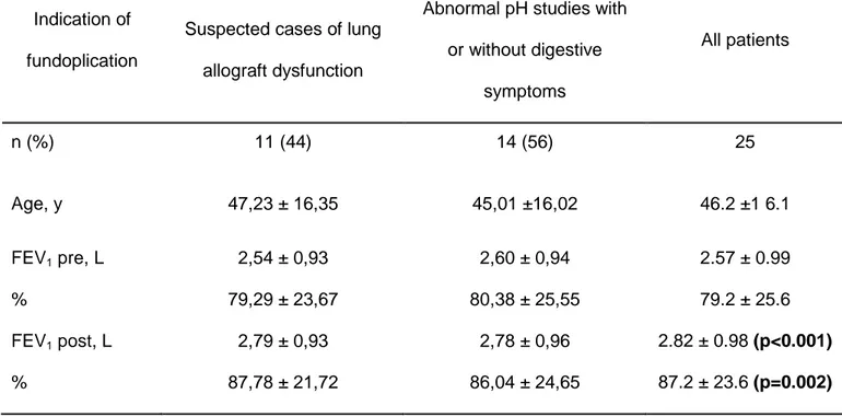
Table III - Differences in FEV1 pre and post fundoplication (mean ± SD), for all
included patients and for each indication of fundoplication
Indication of fundoplication
Suspected cases of lung allograft dysfunction
Abnormal pH studies with or without digestive symptoms All patients n (%) 11 (44) 14 (56) 25 Age, y 47,23 ± 16,35 45,01 ±16,02 46.2 ±1 6.1 FEV1 pre, L % 2,54 ± 0,93 79,29 ± 23,67 2,60 ± 0,94 80,38 ± 25,55 2.57 ± 0.99 79.2 ± 25.6 FEV1 post, L % 2,79 ± 0,93 87,78 ± 21,72 2,78 ± 0,96 86,04 ± 24,65 2.82 ± 0.98 (p<0.001) 87.2 ± 23.6 (p=0.002) Mean ± SD
As regards with the whole group of patients, FEV1 increased from 2.57±0.99 L to
2.82±0.98 L (p<0.001), and from 79.2±25.6 %pred. to 87.2±23.6 %pred. (p=0.002). For the 14 patients operated for abnormal pH studies with or without digestive symptoms of GERD, six months after surgery FEV1 had increased for 10 patients,
decreased for one and was unchanged for 3. Among the 11 patients operated for
suspected lung allograft dysfunction, six months after surgery FEV1 had increased for
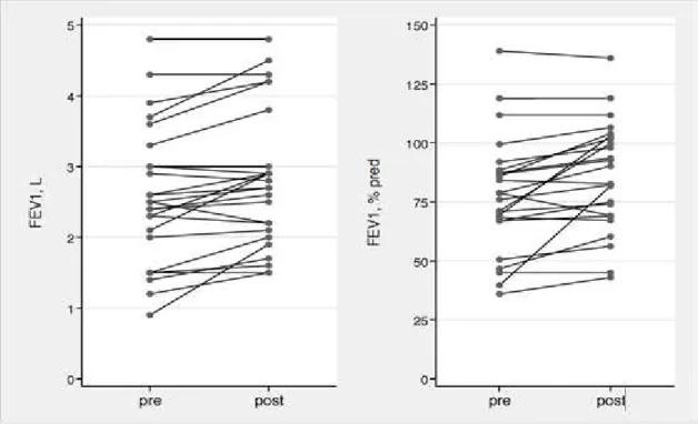
Figure 2. Evolution of FEV1 from pre to post fundoplication, in L and % pred, for each
patient
Another way to express the results is to consider the presence or absence of BOS. Before fundoplication, 7 patients had BOS and 6 months after fundoplication, BOS was persistent for 5 of these 7 patients. On latest news, 4 of these 7patients still had BOS and two new patients had BOS too.
surgery median index was 8 [Q1-Q3: 3-33], p=0.59. After surgery, LI increased for 10 patients and decreased for 9 patients.
BMI was unchanged between pre-fundoplication (21.1±0.8 kg/m²) and six months post-fundoplication (21.2±0.6 kg/m²).
There was no surgically related death or serious post-operative complication. During 6 months following surgery, one patient developed subcutaneous hematoma, six patients developed postoperative dysphagia, five patients developed spontaneously resolving diarrhea, one patient developed postoperative incisional hernia and one patient had recurrence of GERD. Two patients were re operated, one for subcutaneous hematoma and the other for incisional hernia.
Discussion
Laparoscopic fundoplication for GERD resulted in a significant increase in FEV1 with
an excellent safety profile in those frail patients.
Regarding lung function, Palmer et al. reported the first case with improvement after fundoplication for GERD as a reversible cause of allograft dysfunction in a patient who underwent lung re transplantation for BOS (37). Since then, many authors have suggested that anti reflux surgery may improve survival and lung function by decreasing bronchial inflammation, as summarized in a recent review (22). Some publications only showed a decrease in the rate of lung function decline (11,38,39), and others showed an actual improvement of FEV1, such as Davis et al. with an
improvement in FEV1 by an average of 24% six months after fundoplication, and
Hoppo and colleagues with an improvement of most recent post anti-reflux surgery FEV1 of 91% in lung transplant patients (9,28). Survival benefits were showed by
Davis et al. who demonstrated an overall one-year survival in the fundoplication group of 95% versus 78% in the overall series, and a 3 and 5 year survival of 86% and 71% in the fundoplication group as compared with 69% and 48% in the overall series (9).
Some authors also suggest that early fundoplication (not more than three months after transplantation) may improve FEV1 while late fundoplication can stabilize lung
function at best (40), with an example of development of BOS in 16% of 67 patients undergoing early fundoplication and 48% of 117 patients undergoing late fundoplication at one year post transplantation (22). Another example is a retrospective study where 100% of the reflux patients with early surgery (fundoplication at 43 ± 32 days (median 36, range 0 to 87 days)) demonstrated greater freedom from BOS than late fundoplication (surgery at 684 ± 637 days (median 447, range 106 to 2999 days)) at one and three years (29). Abbassi-Ghadi et al. (11) and Robertson et al. (38) only showed a decrease in the rate of lung function decline in their studies with a large mean interval between lung transplantation and fundoplication of 1365 ± 1381 days and 1053 ± 881 days respectively. Anyway, we demonstrated an improvement of FEV1 in our population of
a lack of power, suggesting that the optimal time for fundoplication should be early. However we acknowledge that the improvement of FEV1 could be due to natural
improvement in the first 15 months post lung transplant and that the two main limitations of all these studies including ours are their retrospective and uncontrolled designs. Conduct a case-control study with a control group of lung transplant patients with non-operated reflux was not possible. Indeed we have a too small cohort of transplant patients. RCTs are needed for further evidences of the connection between GERD and CLAD and for the role anti reflux surgery as well (41).
The presence of markers such as bile acid or pepsin in BAL seems to be an evidence of aspiration of gastric content into transplanted lungs(10,18,42,43). Lipophages are one of these potential markers and some authors have tried to establish a lipid index reflecting gastro esophageal aspirations(15,44). These studies, including a small number of patients, did not allow the precision of a reference index with different thresholds of significance. The lack of precision, the subjectivity of LI and a significant inter- and intra-observer bias probably lead to a poor reproducibility of the results (45). The lack of correlation between LI in BAL and GERD is unexpected in our study, even if the retrospective design and small number of subjects are limitations. One of the hypotheses is a not valid reference threshold of 150. Concentration of lipophages can be dependent of such factors as BAL technique, operator dependence, the time interval between last aspiration and BAL, frequency of aspirations and lipophagedetection is not standardized between centres. Further prospective studies are necessary to standardize methods of sampling and detection of lipophages, to establish a new pathological index for transplanted patients.
Regarding peri-operative safety, median hospital stay for fundoplication was 2.9 days [Q1-Q3: 2-5]. Three conversions to laparotomy were necessary for intestinal perforation, bleeding and gastric wound without postoperative complications except an increased length of hospital stay. The UK Northern Oesophago-Gastric Unit has studied laparoscopic Nissen fundoplication in 16 lung transplant patients and showed no peri-operative mortality or major complications without conversion to an open procedure, two patients developed post-operative dysphagia, the mean ± SD hospital stay was 2.6 ± 0.9 days(38). In the St Mary’s Hospital London group study, 38 lung transplant patients underwent laparoscopic Nissen fundoplication with a mean ± SD hospital stay of 2 ±1 days (range, 1–7 days). In the 30 days after surgery, no patient had a surgical complication, and one patient had persistent dysphagia, treated by dietary measures(11). Cantu et al. reported a retrospective study of 457 patients with 84% laparoscopic Nissen (360° wrap), 6% open Nissen, 5% laparoscopic Toupet (270° wrap), 4% Belsey-Mark IVs (left lateral thoracotomy through the sixth intercostal space, 270° wrap), and 2% other types of fundoplication. They reported no death during hospitalization and during 30 days after operation (29).
Therefore fundoplication seems to be relatively safe according to the literature. The laparotomy conversion rate seems high in our study but the small number of patients makes this not significant.
The Nissen technique is extensively reported while we used only Toupet technique. In the literature, there are 15 RCT comparing Nissen and Toupet (3 by laparotomy,
intervention is characterized by a shorter operating time and a better restoration of the lower oesophageal sphincter pressure. In contrast, postoperative dysphagia rate (including severe dysphagia), gas-bloat syndrome rate, postoperative endoscopic dilatation rate and re-operation rate are lower in the Toupet intervention. That is why authors conclude that Toupet fundoplication should be the technique of choice for GERD treatment and this is the reason why we have used this technique in our institution for several years.
Moreover, there were no significant differences in the intraoperative data between 68 transplanted patients and a non-transplant population of 63 patients in the study of O'Halloran et al. in 2004 (especially no intraoperative or perioperative death) but transplanted patients had an increased mean length of hospital stay (2.89 days vs 0.71 days) (47). This result is similar to that found by Abassi-Ghadi et al. with an overall mean ± SD post-operative hospital stay of 2 ± 1 days (range, 1–7 days) (11), but a slightly shorter length of stay than our study; probably because of the limited number of patients with one laparotomy who stayed 9 days and three conversions to laparotomy who stayed 7,8 and 9 days.
Our results show that laparoscopic fundoplication for GERD is safe in LT recipients and significantly improves FEV1 but optimal time for fundoplication needs to be
further defined. As lipophages are one of potential biomarkers of GERD, standardization in lipophages research is needed to specify a better index in transplanted patients.
Acknowledgements
We thank Dr. Daniel Veale, MD, FRCP, Centre de réadaptation cardio-respiratoire Dieulefit, France for reviewing our manuscript and Mrs. Christelle Fleurence, coordinator nurse of lung transplantation program.
Autorship page
Contribution of authors: 1. Conception and design 2. Data collection for the article 3. Literature search
4. Analysis and interpretation of data 5. Statistical expertise
6. Writing the article
7. Critical revision of the article 8. Coordination
9. Final approval of article
Badrignans Pauline: 2,3,4,6,8,9; Edouard Girard: 7,9; Risse Olivier: 7,9; Fior-GozlanMichèle: 2,3,4,7,9; de Taversay-Leroy Caroline: 9; QuetantSébastien: 1,3,9; Boutonnat Jean: 9; Bedouch Pierrick: 7,9; Roth Hubert: 4,5,9; Pison Christophe: 4,7,9; Camara Boubou: 1,3,4,7,8,9; The Grenoble Thoracic Transplant Group: 2.
Disclosure
BIBLIOGRAPHIE
1. Verleden GM, Raghu G, Meyer KC, Glanville AR, Corris P. A new classification system for chronic lung allograft dysfunction. J Heart Lung Transplant. 2014 Feb;33(2):127–33.
2. Christie JD, Edwards LB, Kucheryavaya AY, Benden C, Dipchand AI, Dobbels F, et al. The Registry of the International Society for Heart and Lung
Transplantation: 29th Adult Lung and Heart-Lung Transplant Report—2012. J Heart Lung Transplant. 2012 Oct;31(10):1073–86.
3. Sato M, Waddell TK, Wagnetz U, Roberts HC, Hwang DM, Haroon A, et al. Restrictive allograft syndrome (RAS): A novel form of chronic lung allograft dysfunction. J Heart Lung Transplant. 2011 Jul;30(7):735–42.
4. Blondeau K, Mertens V, Vanaudenaerde BA, Verleden GM, Van Raemdonck DE, Sifrim D, et al. Gastro-oesophageal reflux and gastric aspiration in lung transplant patients with or without chronic rejection. Eur Respir J. 2008 Apr 1;31(4):707–13.
5. Parada MT, Alba A, Sepúlveda C. Bronchiolitis Obliterans Syndrome Development in Lung Transplantation Patients. Transplant Proc. 2010 Jan;42(1):331–2.
6. Finlen Copeland CA, Snyder LD, Zaas DW, Turbyfill WJ, Davis WA, Palmer SM. Survival after Bronchiolitis Obliterans Syndrome among Bilateral Lung
Transplant Recipients. Am J Respir Crit Care Med. 2010 Sep 15;182(6):784–9. 7. D’Ovidio F, Singer LG, Hadjiliadis D, Pierre A, Waddell TK, de Perrot M, et al.
Prevalence of gastroesophageal reflux in end-stage lung disease candidates for lung transplant. Ann Thorac Surg. 2005 Oct;80(4):1254–60.
8. Hadjiliadis D, Duane Davis R, Steele MP, Messier RH, Lau CL, Eubanks SS, et al. Gastroesophageal reflux disease in lung transplant recipients. Clin
Transplant. 2003;17(4):363–8.
9. Davis RD, Lau CL, Eubanks S, Messier RH, Hadjiliadis D, Steele MP, et al. Improved lung allograft function after fundoplication in patients with
gastroesophageal reflux disease undergoing lung transplantation. J Thorac Cardiovasc Surg. 2003 Mar;125(3):533–42.
10. Stovold R, Forrest IA, Corris PA, Murphy DM, Smith JA, Decalmer S, et al. Pepsin, a Biomarker of Gastric Aspiration in Lung Allografts: A Putative Association with Rejection. Am J Respir Crit Care Med. 2007 Jun 15;175(12):1298–303.
11. Abbassi-Ghadi N, Kumar S, Cheung B, McDermott A, Knaggs A, Zacharakis E, et al. Anti-reflux surgery for lung transplant recipients in the presence of
obliterans syndrome: A study of efficacy and safety. J Heart Lung Transplant. 2013 Jun;32(6):588–95.
12. Sifrim D, Zerbib F. Diagnosis and management of patients with reflux symptoms refractory to proton pump inhibitors. Gut. 2012 Sep 1;61(9):1340–54.
13. Tian Z-C, Wang B, Shan C-X, Zhang W, Jiang D-Z, Qiu M. A Meta-Analysis of Randomized Controlled Trials to Compare Long-Term Outcomes of Nissen and Toupet Fundoplication for Gastroesophageal Reflux Disease. PloS One.
2015;10(6):e0127627.
14. Ahrens P, Noll C, Kitz R, Willigens P, Zielen S, Hofmann D.Lipid-laden alveolar macrophages (LLAM): A useful marker of silent aspiration in children.Pediatr Pulmonol.1999 Aug;28(2):83-8.
15. Hopkins PM, Kermeen F, Duhig E, Fletcher L, Gradwell J, Whitfield L, et al. Oil red O stain of alveolar macrophages is an effective screening test for
gastroesophageal reflux disease in lung transplant recipients. J Heart Lung Transplant Off Publ Int Soc Heart Transplant. 2010 Aug;29(8):859–64.
16. Belperio JA, Weigt SS, Fishbein MC, Lynch JP. Chronic lung allograft rejection: mechanisms and therapy. Proc Am Thorac Soc. 2009 Jan 15;6(1):108–21. 17. Christie JD, Edwards LB, Kucheryavaya AY, Benden C, Dobbels F, Kirk R, et al.
The Registry of the International Society for Heart and Lung Transplantation: Twenty-eighth Adult Lung and Heart-Lung Transplant Report—2011. J Heart Lung Transplant. 2011 Oct;30(10):1104–22.
18. D’Ovidio F, Mura M, Tsang M, Waddell TK, Hutcheon MA, Singer LG, et al. Bile acid aspiration and the development of bronchiolitis obliterans after lung
transplantation. J Thorac Cardiovasc Surg. 2005 May;129(5):1144–52.
19. D’Ovidio F, Mura M, Ridsdale R, Takahashi H, Waddell TK, Hutcheon M, et al. The Effect of Reflux and Bile Acid Aspiration on the Lung Allograft and Its Surfactant and Innate Immunity Molecules SP-A and SP-D. Am J Transplant. 2006 Aug;6(8):1930–8.
20. Murthy SC, Nowicki ER, Mason DP, Budev MM, Nunez AI, Thuita L, et al. Pretransplant gastroesophageal reflux compromises early outcomes after lung transplantation. J Thorac Cardiovasc Surg. 2011 Jul;142(1):47–52.e3.
24. Au J, Hawkins T, Venables C, Morritt G, Scott CD, Gascoigne AD, et al. Upper gastrointestinal dysmotility in heart-lung transplant recipients. Ann Thorac Surg. 1993 Jan;55(1):94–7.
25. Corris PA, Ryan VA, Small T, Lordan J, Fisher AJ, Meachery G, et al. A randomised controlled trial of azithromycin therapy in bronchiolitis obliterans syndrome (BOS) post lung transplantation. Thorax. 2015 May;70(5):442–50. 26. Vos R, Vanaudenaerde BM, Verleden SE, De Vleeschauwer SI,
Willems-Widyastuti A, Van Raemdonck DE, et al. A randomised controlled trial of
azithromycin to prevent chronic rejection after lung transplantation. Eur Respir J. 2011 Jan;37(1):164–72.
27. Mertens V, Blondeau K, Van Oudenhove L, Vanaudenaerde B, Vos R, Farre R, et al. Bile Acids Aspiration Reduces Survival in Lung Transplant Recipients with BOS Despite Azithromycin: Aspiration and Transplant Survival. Am J Transplant. 2011 Feb;11(2):329–35.
28. Hoppo T, Jarido V, Pennathur A, et al. ANtireflux surgery preserves lung function in patients with gastroesophageal reflux disease and end-stage lung disease before and after lung transplantation. Arch Surg. 2011 Sep
1;146(9):1041–7.
29. Cantu E, Appel JZ, Hartwig MG, Woreta H, Green C, Messier R, et al. Early Fundoplication Prevents Chronic Allograft Dysfunction in Patients with
Gastroesophageal Reflux Disease. Ann Thorac Surg. 2004 Oct;78(4):1142–51. 30. Cooper JD, Billingham M, Egan T, Hertz MI, Higenbottam T, Lynch J, et al. A
working formulation for the standardization of nomenclature and for clinical staging of chronic dysfunction in lung allografts. International Society for Heart and Lung Transplantation. J Heart Lung Transplant Off Publ Int Soc Heart Transplant. 1993 Oct;12(5):713–6.
31. Estenne M, Maurer JR, Boehler A, Egan JJ, Frost A, Hertz M, et al. Bronchiolitis obliterans syndrome 2001: an update of the diagnostic criteria. J Heart Lung Transplant. 2002;21(3):297–310.
32. Stewart S, Fishbein MC, Snell GI, Berry GJ, Boehler A, Burke MM, et al.
Revision of the 1996 working formulation for the standardization of nomenclature in the diagnosis of lung rejection. J Heart Lung Transplant Off Publ Int Soc Heart Transplant. 2007 Dec;26(12):1229–42.
33. Zerbib F, Bruley Des Varannes S, Roman S, Pouderoux P, Artigue F, Chaput U, et al. Normal values and day-to-day variability of 24-h ambulatory oesophageal impedance-pH monitoring in a Belgian-French cohort of healthy subjects. Aliment Pharmacol Ther. 2005 Nov;22(10):1011–21.
34. Shay S, Tutuian R, Sifrim D, Vela M, Wise J, Balaji N, et al. Twenty-four hour ambulatory simultaneous impedance and pH monitoring: a multicenter report of normal values from 60 healthy volunteers. Am J Gastroenterol. 2004
35. Toupet A. [Technic of esophago-gastroplasty with phrenogastropexy used in radical treatment of hiatal hernias as a supplement to Heller’s operation in cardiospasms]. Mém Académie Chir Fr. 1963 Mar 20;89:384–9.
36. Boutelier P, Jonsell G. An alternative fundoplicative maneuver for gastroesophageal reflux. Am J Surg. 1982 Feb;143(2):260–4.
37. Palmer SM, Miralles AP, Howell DN, Brazer SR, Tapson VF, Davis RD.
Gastroesophageal reflux as a reversible cause of allograft dysfunction after lung transplantation. Chest. 2000 Oct;118(4):1214–7.
38. Robertson AGN, Krishnan A, Ward C, Pearson JP, Small T, Corris PA, et al. Anti-reflux surgery in lung transplant recipients: outcomes and effects on quality of life. Eur Respir J. 2012 Mar 1;39(3):691–7.
39. Hartwig MG, Anderson DJ, Onaitis MW, Reddy S, Snyder LD, Lin SS, et al. Fundoplication After Lung Transplantation Prevents the Allograft Dysfunction Associated With Reflux. Ann Thorac Surg. 2011 Aug;92(2):462–9.
40. Burton PR, Button B, Brown W, Lee M, Roberts S, Hassen S, et al. Medium-term outcome of fundoplication after lung transplantation. Dis Esophagus. 2009 Nov;22(8):642–8.
41. Gulack BC, Meza JM, Lin SS, Hartwig MG, Davis RD. Reflux and allograft dysfunction: is there a connection? Thorac Surg Clin. 2015;25(1):97–105. 42. Fisichella PM, Davis CS, Lundberg PW, Lowery E, Burnham EL, Alex CG, et al.
The protective role of laparoscopic antireflux surgery against aspiration of pepsin after lung transplantation. Surgery. 2011 Oct;150(4):598–606.
43. Ward C, Forrest IA, Brownlee IA, Johnson GE, Murphy DM, Pearson JP, et al. Pepsin like activity in bronchoalveolar lavage fluid is suggestive of gastric aspiration in lung allografts. Thorax. 2005 Oct;60(10):872–4.
44. Adams R, Ruffin R, Campbell D. The value of the lipid-laden macrophage index in the assessment of aspiration pneumonia. Aust N Z J Med. 1997;27(5):550–3. 45. Reid-Nicholson M, Kulkarni R, Adeagbo B, Looney S, Crosby J. Interobserver
and intraobserver variability in the calculation of the lipid-laden macrophage index: Implications for its use in the evaluation of aspiration in children. Diagn Cytopathol. 2010 Dec;38(12):861–5.
SERMENT D’HIPPOCRATE
En présence des Maîtres de cette Faculté, de mes chers condisciples et devant l’effigie
d’HIPPOCRATE,
Je promets et je jure d’être fidèle aux lois de l’honneur et de la probité dans l’exercice
de la Médecine.
Je donnerai mes soins gratuitement à l’indigent et n’exigerai jamais un salaire
au-dessus de mon travail. Je ne participerai à aucun partage clandestin d’honoraires.
Admis dans l’intimité des maisons, mes yeux n’y verront pas ce qui s’y passe ; ma
langue taira les secrets qui me seront confiés et mon état ne servira pas à corrompre
les mœurs, ni à favoriser le crime.
Je ne permettrai pas que des considérations de religion, de nation, de race, de parti ou
de classe sociale viennent s’interposer entre mon devoir et mon patient.
Je garderai le respect absolu de la vie humaine.
Même sous la menace, je n’admettrai pas de faire usage de mes connaissances
médicales contre les lois de l’humanité.
Respectueux et reconnaissant envers mes Maîtres, je rendrai à leurs enfants
l’instruction que j’ai reçue de leurs pères.
Que les hommes m’accordent leur estime si je suis fidèle à mes promesses.
Que je sois couvert d’opprobre et méprisé de mes confrères si j’y manque.
RESUME
Contexte: La fundoplicature pourrait améliorer la fonction pulmonaire et la survie des patients
transplantés pulmonaires atteints de reflux gastro-œsophagien (RGO), mais la tolérance et les bénéfices de cette intervention doivent encore être précisés. Méthodes : Nous avons recueilli chez 25 transplantés pulmonaires les données suivantes, avant et après fundoplicature: pH métrie des 24 heures, volume expiratoire maximal en une seconde (VEMS), index de lipophages alvéolaires.
Résultats: 25 patients dont 68% d'hommes, âgés de 44,9 ±16,6 ans (11 mucoviscidoses, 7
bronchopneumopathies chroniques obstructives, 3 dilatations des bronches, 2 hypertensions artérielles pulmonaires et 2 fibroses pulmonaires) ont bénéficié d’une fundoplicature devant une pH métrie anormale (n=14, 56%) ou une suspicion de dysfonction du greffon (n=11, 44%). Avant l’intervention, 20/24 (83%) patients avaient une pH métrie anormale, 8/25 (32%) une impédancemétrie anormale et l’index de lipophages médian [Q1-Q3] était de 9 [0-39]. Trois conversions en laparotomie furent nécessaires, la durée médiane d’hospitalisation [Q1-Q3] fut de 2.9 [2.0-5.0] jours. Après la chirurgie, tous les patients avaient une pH métrie normalisée et une amélioration significative des symptômes digestifs du RGO ; le VEMS augmentait de 2,57±0,99 L à 2,82±0,98 L (p=0,0009) soit de 79,2±25,6% à 87,2±23,6% de la valeur prédite (p=0,002) et l’index de lipophages médian [Q1-Q3] diminuait à 8 [3-33] (p=0,59).
Conclusions: La fundoplicature est bien tolérée chez les transplantés pulmonaires et permet une
amélioration significative du VEMS. Le rôle de l’index de lipophages alvéolaire doit être mieux documenté.
ABSTRACT
Background: Fundoplication for gastro-esophageal reflux disease (GERD) may improve lung function
and survival in lung transplant (LT) recipients, but morbidity and potential benefits remain to be better reported.
Methods: Data were collected between 2002 and 2013 in 25 consecutive LT recipients who
underwent fundoplication. 24 hours pH studies, forced expiratory volume in one second (FEV1) and lipid index (LI) in bronchoalveolar lavage (BAL) were assessed before and after surgery.
Results: 25 LT recipients, 68% males, 44.9±16.6 years old (11 cystic fibrosis, 7 chronic obstructive
pulmonary disease (COPD), 3 bronchiectasis, 2 pulmonary hypertension and2 pulmonary fibrosis patients) had fundoplication. Fundoplication was performed for abnormal pH studies (n=14, 56%) or suspicion of lung allograft dysfunction (n=11, 44%). Before fundoplication, 20/24 (83%) patients had