O
pen
A
rchive
T
OULOUSE
A
rchive
O
uverte (
OATAO
)
OATAO is an open access repository that collects the work of Toulouse researchers and
makes it freely available over the web where possible.
This is an author-deposited version published in :
http://oatao.univ-toulouse.fr/
Eprints ID : 18531
To link to this article : DOI:10.1093/annonc/mdw009
URL :
https://doi.org/10.1093/annonc/mdw009
To cite this version : Bousquet, Marina and Noirot, Céline and
Accadbled, Franck and Sales de Gauzy, Jérôme and Castex,
Marie-Pierre and Brousset, Marie-Pierre and Gomez-Brouchet, Anne Whole-exome
sequencing in osteosarcoma reveals important heterogeneity of
genetic alterations. (2016) Annals of Oncology, vol. 27 (n° 4). pp.
738-744. ISSN 0923-7534
Any correspondence concerning this service should be sent to the repository
administrator:
[email protected]
Whole-exome
sequencing in osteosarcoma reveals
important
heterogeneity of genetic alterations
M.
Bousquet
1,
C. Noirot
2,
F. Accadbled
3,
J. Sales de Gauzy
3,
M. P. Castex
4,
P. Brousset
1,5*
&
A. Gomez-Brouchet
5,61
Cancer Research Center of Toulouse, INSERM U1037, laboratoire d’excellence Labex TOUCAN, Toulouse; 2
Genotoul Bioinfo, INRA, Castanet Tolosan; Departments of 3Pediatric Orthopedics; 4Pediatric Oncology, Hôpital des enfants, Toulouse; 5Department of Pathology, IUCT-Oncopole, CHU de Toulouse and Toulouse University, Toulouse;6
Pharmacology and Structural Biology Institute, CNRS UMR5089, Toulouse, France
Background:Whole-genome sequencing studies have recently shown that osteosarcomas (OSs) display high rates of
structural variation, i.e. they contain many somatic mutations and copy number alterations. TP53 and RB1 show recurrent somatic alterations in concordant studies, suggesting that they could be key players in bone oncogenesis.
Patients and methods: we carried out whole-genome sequencing of DNA from seven high-grade OS samples
matched with normal tissue from the same patients.
Results:We confirmed the presence of genetic alterations of the TP53 (including novel unreported mutations) and RB1
samples, of which only 15 have been previously reported. Interestingly, the number of mutated genes (ranging from 4 to 8) was lower in TP53mut cases compared with TP53wt cases (ranging from 14 to 45). This was also true for the mutated RB1 case. We also observed that a dedifferentiated OS harboring MDM2 amplification did not carry any other mutations.
Conclusion:This study suggests that bone oncogenesis driven by TP53 or RB1 mutations occurs on a background of relative genetic stability and that the dedifferentiated OS subtype represents a clinico-pathological entity with distinct oncogenic mechanisms and thus requires different therapeutic management.
Key words:osteosarcoma, whole-genome sequencing, TP53, MDM2
introduction
Osteosarcoma (OS) is the most common primary malignant type of bone cancer and has a worldwide incidence of approxi-mately one to three cases per million annual, with a higher in-cidence in adolescents (0.8–1.1/100 000/year for ages 15–19). The 2013 World Health organization (WHO) classification distinguishes different OS subtypes on the basis of both their location in relation to the bone cortex (central or surface OS) and their grade (low, high or intermediate [1]. High-grade OS are themselves divided into different subtypes: conventional (the most common one, 90% of all OS), telangiectasic and small-cell OS (respectively 4% and 1.5% of all OS) and the ded-ifferentiated OS [1]. This latter corresponds to the transform-ation of a low-grade OS into an OS of higher grade. Low-grade and its dedifferentiated form are defined by a simple genomic profile with episomal ring neochromosomes containing high-level amplification of MDM2 (murine double-minute type 2) and CDK4 (cyclin-dependent kinase 4) [2,3]. Microscopically, low-grade OS is characterized by a paucicellular stroma of fusi-form cells harboring minor cytonuclear atypia and well-differ-entiated bone trabeculae [1]. Dedifferentiated OS is most often composed of cells with larger atypia and mitosis than its low-grade counterparts and by the presence of an immature osteoid production. Tumor necrosis may also be present [1,3]. In the dedifferentiated form, areas of low- and high-grade tumor may or may not coexist. When the low- grade contingent is not present on biopsy specimens or surgical resection, dedifferentiated OS could be misdiagnosed with conventional OS. The diagnosis of the dedifferentiated form is made on the molecular signature (amplification of MDM2 gene) [3]. Conversely, the other high-grade OS are complex genomic sarcomas with multiple numer-ical and structural chromosomal aberrations that present no specific diagnostic signature [1].
Several heritable genetic syndromes predispose to OS: Li– Fraumeni syndrome or heritable retinoblastomas are related to germ-line mutations in the TP53 and RB1 tumor suppressor genes, respectively [4,5]. The genetic instability of OS results in recurrent amplification and DNA copy number gains at distinct chromosomal regions [1]. Recently, whole-genome sequencing studies have shown that among pediatric cancers, OS have the highest rate of structural variation, i.e. they contain many somatic mutations and copy number alterations [6,7]. A few re-current single-nucleotide variations or rere-current point muta-tions have been found [7]. These studies have confirmed the major roles of recurrent alterations of the TP53 and RB1 genes in OS (80%–90% and 10%–39%, respectively). The majority of TP53 mutations are rearrangements with breakpoints confined
to thefirst intron of the gene [7,8]. This is therefore a particu-larly unstable region that is sensitive to structural variations that occur before inactivation of TP53 and that are likely to be the initiating factors in OS development [7]. Another recent study has also confirmed the importance and prevalence of TP53 and RB1 inactivation by genomic events, which either affect the TP53 or RB1 genes directly or alter TP53/RB1-interacting genes. In this study, the authors also reported that 75% of somatic events lead to the direct or indirect inactivation of TP53 [6]. Besides TP53 and RB1, the ATRX, DLG2, RUNX2 and PTEN genes have been shown to incur recurrent somatic alterations in 30%, 30%, 52%, 18–55% and 44% of OS, respectively [7]. The ATRX gene is known to regulate telomere maintenance and could therefore have an important role in OS development [9]. Alterations in CDC27, a gene controlling the cell cycle, and the MUC4 and EI24 genes known to encode tumor suppressors, were also recently reported [10]. In addition, genome sequen-cing data have shown that the novel genetic mechanism referred to as chromothripsis (Greek; chromos for chromosome, thripsis for shattered into pieces) is involved in 33% of primary OS com-pared with 2%–3% of cancers overall [11]. Chromothripsis appears to be a cataclysmic event in which a single or in some cases, a few chromosomes are broken into many pieces and then stitched back together [11, 12]. Chromothripsis may lead to the generation of amplifications of one or more genes or to the deletion of one or more tumor suppressor genes. It may also explain the sudden onset of OSs and the complexity and hetero-geneity of the OS genome [1,11,12]. In a recent study, exome profiles in one patient showed almost 3000 somatic single-nucleotide variants (SNVs) and small indels and more than 2000 copy number variants in different chromosomes, reinfor-cing the major role of this phenomenon [10]. Kataegis (a pattern of localized hypermutation in a SNV) has also been reported in OS, although the study of Perry et al. [6] reported a higher prevalence (85% of OS) than that reported by Chen et al. [7] (50% of OS). However, this process is certainly less import-ant because it does not occur in the most recurrently mutated genes [7].
To further refine the landscape of somatic mutations in pedi-atric OS, we carried out exome sequencing of DNA from seven OS samples matched with normal tissues from the same patients (Table1). Three importantfindings arise from this work. Our results confirm: (i) the heterogeneity of genetic alterations in conventional high-grade OS by providing new unreported mutations, (ii) the dominant role of TP53 and RB1 gene inacti-vation in the development of OS, and (iii) that dedifferentiated OS is a separate entity with a different pathophysiology from that of conventional high-grade OS.
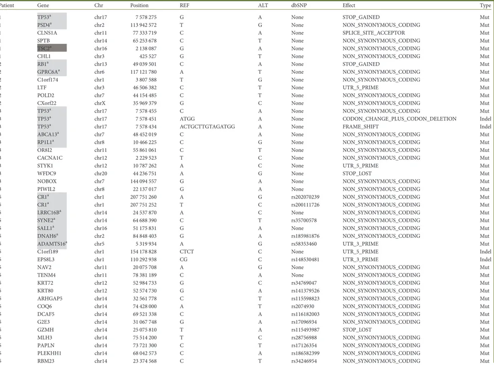
Table 1. Mutated genes detected by whole-exome sequencing
Patient Gene Chr Position REF ALT dbSNP Effect Type
1 TP53a chr17 7 578 275 G A None STOP_GAINED Mut
1 PSD4a chr2 113 942 572 T G None NON_SYNONYMOUS_CODING Mut
1 CLNS1A chr11 77 333 719 C A None SPLICE_SITE_ACCEPTOR Mut
1 SPTB chr14 65 253 678 C T None NON_SYNONYMOUS_CODING Mut
1 TSC2a chr16 2 138 087 G A None NON_SYNONYMOUS_CODING Mut
1 CHL1 chr3 425 527 G T None NON_SYNONYMOUS_CODING Mut
2 RB1a chr13 49 039 501 C A None STOP_GAINED Mut
2 GPRC6Aa chr6 117 121 780 A T None NON_SYNONYMOUS_CODING Mut
2 C1orf174 chr1 3 807 588 T G None NON_SYNONYMOUS_CODING Mut
2 LTF chr3 46 506 382 C T None UTR_5_PRIME Mut
2 POLD2 chr7 44 154 485 C T None NON_SYNONYMOUS_CODING Mut
2 CXorf22 chrX 35 969 379 G C None NON_SYNONYMOUS_CODING Mut
3 TP53a chr17 7 578 455 C A None NON_SYNONYMOUS_CODING Mut
3 TP53a chr17 7 578 451 ATGG A None CODON_CHANGE_PLUS_CODON_DELETION Indel
3 TP53a chr17 7 578 434 ACTGCTTGTAGATGG A None FRAME_SHIFT Indel
3 ABCA13a chr7 48 452 019 C A None NON_SYNONYMOUS_CODING Mut
3 RP1L1a chr8 10 466 225 C G None NON_SYNONYMOUS_CODING Mut
3 OR8I2 chr11 55 861 061 C T None NON_SYNONYMOUS_CODING Mut
3 CACNA1C chr12 2 229 523 T C None NON_SYNONYMOUS_CODING Mut
3 STYK1 chr12 10 787 262 A C None UTR_5_PRIME Mut
3 WFDC9 chr20 44 236 751 A G None STOP_LOST Mut
3 NOBOX chr7 144 094 557 G A None NON_SYNONYMOUS_CODING Mut
3 PIWIL2 chr8 22 137 017 G A None NON_SYNONYMOUS_CODING Mut
5 CR1a chr1 207 751 260 A G rs202070239 NON_SYNONYMOUS_CODING Mut
5 CR1a chr1 207 751 252 T C rs200111726 NON_SYNONYMOUS_CODING Mut
5 LRRC16Ba chr14 24 537 870 A C None NON_SYNONYMOUS_CODING Mut
5 SYNE2a chr14 64 688 390 C T rs35700578 NON_SYNONYMOUS_CODING Mut
5 SALL1a chr16 51 175 831 G A None NON_SYNONYMOUS_CODING Mut
5 DNAH6a chr2 84 848 403 G A rs185981876 NON_SYNONYMOUS_CODING Mut
5 ADAMTS16a chr5 5 319 934 A G rs58353460 UTR_3_PRIME Mut
5 C1orf189 chr1 154 178 828 CTCT C None UTR_5_PRIME Indel
5 EPS8L3 chr1 110 292 938 CG C rs148530481 UTR_3_PRIME Indel
5 NAV2 chr11 20 075 708 A G None NON_SYNONYMOUS_CODING Mut
5 TENM4 chr11 78 381 189 C A None NON_SYNONYMOUS_CODING Mut
5 KRT72 chr12 52 984 733 G C rs34769047 NON_SYNONYMOUS_CODING Mut
5 KRT80 chr12 52 574 730 G A rs141379526 NON_SYNONYMOUS_CODING Mut
5 ARHGAP5 chr14 32 561 778 C T rs115598823 NON_SYNONYMOUS_CODING Mut
5 COQ6 chr14 74 428 000 A T rs2074930 NON_SYNONYMOUS_CODING Mut
5 DCAF5 chr14 69 521 338 C A rs116182003 NON_SYNONYMOUS_CODING Mut
5 G2E3 chr14 31 067 748 G A rs17096934 NON_SYNONYMOUS_CODING Mut
5 GZMH chr14 25 075 810 T A rs115493987 STOP_LOST Mut
5 MLH3 chr14 75 514 200 T C rs28756988 NON_SYNONYMOUS_CODING Mut
5 PAPLN chr14 73 721 300 C T rs17126354 NON_SYNONYMOUS_CODING Mut
5 PLEKHH1 chr14 68 042 573 C A rs186582399 NON_SYNONYMOUS_CODING Mut
5 RNASE10 chr14 20 978 738 G T rs74037153 NON_SYNONYMOUS_CODING Mut
5 RNASE13 chr14 21 501 843 G A rs116165621 UTR_3_PRIME Mut
5 TSHR chr14 81 422 169 A G rs147137913 NON_SYNONYMOUS_CODING Mut
5 COQ9 chr16 57 493 629 G C rs61730662 NON_SYNONYMOUS_CODING Mut
5 GP2 chr16 20 330 955 G T None NON_SYNONYMOUS_CODING Mut
5 NUBP2 chr16 1 838 050 A G rs57822546 NON_SYNONYMOUS_CODING Mut
5 CHST8 chr19 34 263 855 G A None NON_SYNONYMOUS_CODING Mut
5 DHDH chr19 49 436 982 G C rs10401800 NON_SYNONYMOUS_CODING Mut
5 TMEM221 chr19 17 556 074 C T None NON_SYNONYMOUS_CODING Mut
5 PLB1 chr2 28 849 317 G A None STOP_GAINED Mut
5 SH3YL1 chr2 231 067 G C None NON_SYNONYMOUS_CODING Mut
5 SCP2D1 chr20 18 794 656 T A None NON_SYNONYMOUS_CODING Mut
5 MIRLET7BHG chr22 46 505 806 C G rs12159905 EXON Mut
5 PKDREJ chr22 46 656 242 A G rs34798212 NON_SYNONYMOUS_CODING Mut
5 TMPRSS6 chr22 37 464 655 C G rs80252000 SPLICE_SITE_DONOR Mut
5 ATR chr3 142 272 671 T C None NON_SYNONYMOUS_CODING Mut
5 MANBA chr4 103 579 032 G A rs370002189 NON_SYNONYMOUS_CODING Mut
5 MIR6082 chr4 172 107 395 C T rs28570267 EXON Mut
5 CEP120 chr5 122 714 044 T C rs61744334 NON_SYNONYMOUS_CODING Mut
5 TRDN chr6 123 714 778 C T rs35047281 NON_SYNONYMOUS_CODING Mut
5 CTNNAL1 chr9 111 735 031 G C rs16913734 NON_SYNONYMOUS_CODING Mut
5 GLDC chr9 6 606 634 C T rs28617412 NON_SYNONYMOUS_CODING Mut
5 ESX1 chrX 103 495 445 G C None NON_SYNONYMOUS_CODING Mut
6 DUSP27a chr1 167 096 814 G T None NON_SYNONYMOUS_CODING Mut
6 VPS13Da chr1 12 336 850 G C None NON_SYNONYMOUS_CODING Mut
6 EFCAB6a chr22 44 022 541 A G None NON_SYNONYMOUS_CODING Mut
6 GCSAML chr1 247 726 894 A G None NON_SYNONYMOUS_CODING Mut
6 SYNC chr1 33 161 551 C G None NON_SYNONYMOUS_CODING Mut
6 OR9Q1 chr11 57 947 475 T G None NON_SYNONYMOUS_CODING Mut
6 TRPC4 chr13 38 320 482 C A None NON_SYNONYMOUS_CODING Mut
6 PFAS chr17 8 170 715 C G None NON_SYNONYMOUS_CODING Mut
6 CATSPERD chr19 5 757 865 C A None NON_SYNONYMOUS_CODING Mut
6 DLL1 chr6 170 594 743 C T None NON_SYNONYMOUS_CODING Mut
6 DDHD2 chr8 38 109 710 C G None NON_SYNONYMOUS_CODING Mut
6 MCMDC2 chr8 67 808 447 A C None NON_SYNONYMOUS_CODING Mut
6 TAF1L chr9 32 632 563 C A None NON_SYNONYMOUS_CODING Mut
6 NRK chrX 105 189 927 A C None NON_SYNONYMOUS_CODING Mut
7 TP53a chr17 7 577 018 C T None SPLICE_SITE_DONOR Mut
7 PLCB2 chr15 40 590 865 C T None NON_SYNONYMOUS_CODING Mut
7 HID1 chr17 72 956 123 C T None NON_SYNONYMOUS_CODING Mut
7 KLB chr4 39 436 255 T A None NON_SYNONYMOUS_CODING Mut
7 GRIA1 chr5 153 181 955 G A None NON_SYNONYMOUS_CODING Mut
fragments (200–300 bp). Extracted DNA was amplified by ligation-mediated PCR (LM-PCR), then purified and hybridized to the Roche NimbleGen SeqCap EZ Exome probe. Nonhybridized fragments were washed out. Both noncaptured and captured LM-PCR products were subjected to quantitative PCR to estimate the magnitude of enrichment. Each captured library was then loaded on to a Hiseq2000 (Illumina) platform. High-throughput se-quencing for each captured library was carried out independently to ensure that each sample met the desired average fold-coverage of 50×. Raw image files were processed by Illumina base calling Software 1.7 for base calling with default parameters and the sequences of each individual were generated as 91-bp paired-end reads.
bioinformatical analysis
First, the adapter sequences in the raw data, generated from the Illumina pipeline, were removed, and low-quality reads that had too many Ns or low base quality were discarded. This step produced the‘clean data’. Second, the Burrows-Wheeler Aligner [BWA mem (0.7.9a-r786)] was used to carry out the alignment against the human reference genome Hg19. Samples were then realigned and recalibrated with GATK (v3.0). HaplotypeCaller was used to call variants using standard options and a bed of exome region. The annotation was performed with SnpEff (v3.6c) against Hg19 with the option ‘cancer’. For SNP and indels, we applied the following filters: the mutation must have not existed in cell line samples (even one read) and, for control patient samples, the mutation must have had a minimum allele frequency (MAF) below: 5% for control patient 1, 15% for control patient 2, 5% for control patient 3, 0% for control patient 4, 40% for control patient 5, 10% for control patient 6 and 10% for control patient 7. These percentages corres-pond to the putative contamination of remaining cancer cells. Only muta-tions with a depth higher than 20 were considered. Moreover, we selected mutations and indels in tumor samples with MAF >30% and MAF >20% re-spectively. Finally, for each mutation, the tumor sample MAF had to be 2.5 times higher than the MAF of the matched control sample and, for each
A C
B D
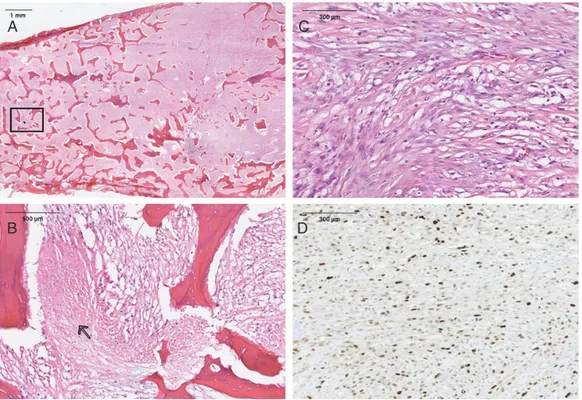
Figure 1. (A) Histological appearance of an intramedullary (central) dedifferentiated osteosarcoma (H&E). (B) Magnification of rectangle shown in A. Necrosis area (open arrow) in a dedifferentiated osteosarcoma and immature neoplastic osteoid (black arrow) (H&E). (C) Spindle cells with moderate nuclear atypia, mitosis and immature neoplastic osteoid production (H&E). (D) MDM2 nuclear immunostaining in a dedifferentiated osteosarcoma.
| Bousquet et al. Volume 27 | No. 4 | April 2016
patients
and methods
OS samples and cell lines
Seven OS samples coming from initial biopsy and seven matched control DNA samples were collected from patients registered at the ‘CRB cancer des Hôpitaux de Toulouse; BB-0033-00014’ collection. This study had approval from institu-tional and nainstitu-tional ethics committees. Thawed samples were obtained after informed consent in accordance with the Declaration of Helsinki and after au-thorization of the French ministry of higher education and research (declaration DC 2009-989; DC-2011-1388; transfer agreement AC-2008-820; AC-2011-130). Clinical and biological annotations were consistent with CNIL (Comité National Informatique et Libertés) guidelines. Samples were stored in the certified biobank of the Hôpitaux de Toulouse. Control samples corresponded to tissues taken from surgical resection that were obtained after chemotherapy treatment. The percentage of cancer cells remaining in control samples was evaluated by morphological analysis after decalcification and hematoxylin–eosin staining.
Six patients had high-grade conventional OS. Among them, five were good responders with a percentage of residual cells lower than 10% and are alive. One was a poor responder with 27% residual cells (patient 5). He died from lung metastases.
Patient 4 presented with intramedullary (central) dedifferentiated OS (Figure 1A–C) with MDM2 overexpression (Figure 1D) and amplification
(confirmed by qPCR). Due to a dedifferentiated component in the diagnostic biopsy, he received neoadjuvant chemotherapy. He was a good responder according to the surgical resection analysis and he is still alive (supplemen-tary Table S1, available at Annals of Oncology online).
Two osteoblastic cell lines (TF13 and TF15) were also used as controls [13]. DNA sample integrity was evaluated by agarose gel electrophoresis.
exome sequencing
Three micrograms of qualified genomic DNA were randomly fragmented by Covaris and then adapters were ligated to both ends of the resulting
and the entire or a portion of the DNA binding domain but they have lost their oligomerization and C-Terminal regulatory domains (Figure2). We can postulate that these mutations lead to the degradation of TP53 mRNA by the nonsense-mediated mRNA decay or that truncated TP53 act as dominant negative mutants on the TP53 wild type as previously described [14]. We also detected a STOP gained mutation (aa829) in the retino-blastoma 1 (RB1) gene in one case. This gene is also known to be frequently mutated in OS [1]. In their recent study, Chen et al. investigated recurrent somatic structural variations in pedi-atric OS and identified 2057 point mutations in 1707 genes [7]. In addition to TP53 and RB1, they observed recurrent somatic alterations of the ATRX and DLG2 genes, with the ATRX gene being one of the most frequently mutated genes. In our cohort, however, we did not observe any ATRX mutations. Thisfinding may be explained by the size of our series but also by geograph-ical variation since we would expect to detect this alteration in at least two of our cases. Alterations in CDC27, a gene controlling the cell cycle, and the MUC4 and EI24 genes, have also been re-cently reported in a single case of OS [10]. We did notfind any alteration in these genes in our patients.
However, among the 82 mutated genes described in our study, 15 were also reported by Chen et al., suggesting that these genes are recurrently mutated in OS [7]. Mutations in the TSC2 gene are our onlyfinding in common with the study by Perry et al. [6] Among the 16 genes described above, some are involved in cell cycle regulation, tumor cell division, transcrip-tion and proliferatranscrip-tion (TP53, RB1, LRRC16B, PSD4, ADAMS16, SALL1, EFCAB6), and others are involved in cytoskeletal integ-rity, cytoplasmic trafficking and energy metabolism (SYNE2, DNAH6, VPS13D, DUSP27). The CR1 gene found mutated in our study seems to have a predominant role in the immune re-sponse and TSC2 is involved in the PI3K/mTOR pathway [6] making this gene/pathway a potential therapeutic target. However, it should be mentioned that all the point mutations we observed in CR1, LRRC16B, SYNE2, SALL1, PSD4, GPRD6A, ABCA13, RP1L1, DUSP27, VPS13D, EFCAB6 and TSC2 are new mutations, distinct from those previously reported [6,7]. These findings emphasize the extreme genomic instability of OS and support the hypothesis that the processes of chromothripsis, defined by tens to hundreds of genomic rearrangements occur-ring in one-off cellular crisis and which have already been described to have a frequency of 50% in OS [6,7], is indeed a frequent event in this pathology.
Among the three patients found to have no mutations in the TP53 and RB1 genes, one ( patient 4) was a dedifferentiated OS.
TP53
protein 1–42
Patient 3
Non synonymous mutation (aa159) Deletion aa159–160
Deletion aa161–165
Patient 1
STOP gain mutation (aa192)
Patient 7 Mutation on donor splice site (aa307)
Transactivation domain SH3 DNA-binding domain 63–97 98–292 324–355 Oligomerization domain C-terminal regulatory domain 363–393
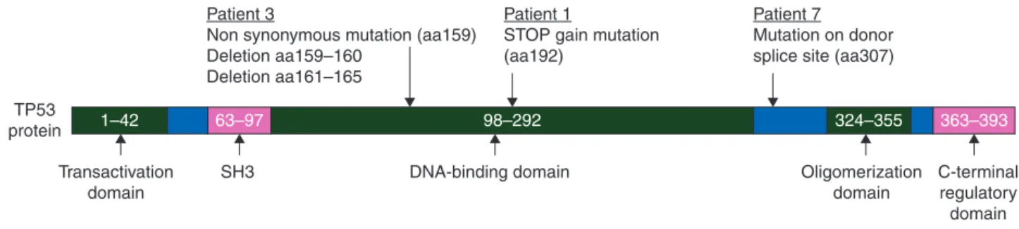
Figure 2. TP53 mutations observed in three patients.
Vlume 27 | No. 4 | April 2016 doi:10.1093/annonc/mdw009 | indel, the tumor sample MAF had to be two times higher than the MAF of
the matched control sample.
results
We identified a total of 84 point mutations and 4 deletions related to 82 different genes from our OS samples (Table 1 and supplementary data S1, available at Annals of Oncology online). None of them were recurrent mutations but, in three patients, we observed mutations affecting the TP53 gene (Figure 2). One patient contained a STOP gained mutation at amino acid number 192, and another patient had a nonsynonymous muta-tion corresponding to amino acid 159 of the TP53 protein followed by two deletions of 3 and 13 bp that leads to a frame-shift. A third patient carried a mutation located in the donor splice site of TP53 (corresponding to TP53 amino acid 307). We postulate that this latter mutation leads to the conservation of the intron, resulting in a truncated TP53 protein. Interestingly, in the dedifferentiated OS case, which carried MDM2 and CDK4 amplification, no other mutation was detected, suggesting that this tumor subtype is a distinct molecular entity (Figure 3).
discussion
In this study, we carried out whole-exome sequencing of samples from seven OS patients to try to identify new somatic mutations. We compared the mutational profile of six conven-tional high-grade OS, (comparing tumor and normal tissues from the same patient) and central dedifferentiated OS, known to have a simple genomic profile with MDM2 amplification. We then examined whether the mutational profile obtained for each patient identified common signaling pathways or biomarkers that might play some role in the pathogenesis of OS. We identi-fied a total of 84 point mutations and 4 deletions related to 82 different genes in OS samples. None of them were recurrent mutations, but in three patients we observed mutations affecting the TP53 gene (Figure 2). TP53 has already been described as one of the most mutated genes in OS [8]. Recently, Chen et al. and Perry et al. conducted whole-genome sequencing and whole-exome and RNA sequencing on 20 OS and 59 tumor/ normal sample pairs, respectively [6, 7] and identified p53 pathway alterations with a high frequency of mutations mostly in the first intron of the TP53 gene [8]. The TP53 gene muta-tions found in our study were new mutamuta-tions, not previously described in the dbSNP database. These mutations lead to a truncated form of TP53 with the TP53 transactivation domain
Interestingly, even in the dedifferentiated form, no point muta-tions were found, reinforcing the idea that this subtype of OS with MDM2 amplification is distinct from the high-grade con-ventional OS subtype. We also observed that the highest rate of somatic point mutations occurred in cases ( patients 5 and 6) that did not contain alterations in the TP53 and RB1 genes. Thesefindings suggest that TP53 mutations and, to some extent, RB1 mutations are founding events in the process of bone onco-genesis and that, in the absence of these mutations. a higher rate of genetic alteration is required. Of note, patient 5 was the only patient with a poor response to chemotherapy (27% of viable cells) and had the highest number of mutated genes (n = 45), suggesting poor response to chemotherapy, and so on; poor prognosis is associated to high level of mutations. We can also hypothesize that patients with TP 53 gene mutations may have a better response to chemotherapy.
Collectively, in line with previously reported studies our data suggest that OS is an extremely heterogeneous tumor with regard to genetic alterations (Figure3). Indeed, TP53 represents a key gene mutated in a significant number of cases. Of particu-lar interest is the wide diversity of TP53 mutations, all of them leading to inactivation of this tumor suppressor protein and supporting chromothripsis as a critical process in bone onco-genesis. Three main observations arise from our study: (i) whole-exome sequencing has allowed us to identify mutated genes that have not been previously reported in OS; (ii) dediffer-entiated OS harboring MDM2 amplifications do not carry any other mutations, strongly indicating that this subtype represents a clinico-pathological entity with distinct oncogenic mechan-isms that requires different therapeutic management (Figure3); (iii) TP53 (and RB1) inactivation is the strongest oncogenic event in OS development, requiring a limited number of sec-ondary genetic mutations (Figure3).
These data confirm that the development of targeted therapies for OS will be difficult and that in the future a personalized ap-proach is the most realistic avenue for providing patients with efficient treatment.
funding
This work was supported by the ARC foundation SL 220120605289 (MB and PB), the Institut Universitaire de France (PB), the Société Française des Cancers de l’Enfant and IUCT-cancer biobank (AGB).
disclosure
The authors have declared no conflicts of interest.
references
1. Fletcher CDM, Bridge JA, Hogendoorn PCW, Mertens M. WHO classification of tumour. In: Hogendoorn PCW, Mertens F (eds). WHO Classification of Tumours of Soft Tissue and Bone. Lyon: IARC, 2013.
2. Szymanska J, Mandahl N, Mertens F et al. Ring chromosomes in parosteal osteosarcoma contain sequences from 12q13-15: a combined cytogenetic and comparative genomic hybridization study: genes. Chromosomes Cancer 1996; 16: 31–34.
3. Dujardin F, Binh MBN, Bouvier C et al. MDM2 and CDK4 immunohistochemistry is a valuable tool in the differential diagnosis of low-grade osteosarcomas and other primaryfibro-osseous lesions of the bone. Mod Pathol 2011; 24: 624–637. 4. Kansara M, Teng MW, Smyth MJ, Thomas DM. Translational biology of
osteosarcoma. Nat Rev Cancer 2014; 14(11): 722–735.
5. Savage SA, Mirabello L, Wang Z et al. Genome-wide association study identifies two susceptibility loci for osteosarcoma. Nat Genet 2013; 45: 799–803. 6. Perry JA, Kiezun A, Tonzi P et al. Complementary genomic approaches highlight
the PI3K/mTOR pathway as a common vulnerability in osteosarcoma. Proc Natl Acad Sci USA 2014; 111: E5564–E5573.
7. Chen X, Bahrami A, Pappo A et al. Recurrent somatic structural variations contribute to tumorigenesis in pediatric osteosarcoma. Cell Rep 2014; 7: 104–112.
8. Ribi S, Baumhoer D, Lee K et al. TP53 intron 1 hotspot rearrangements are specific to sporadic osteosarcoma and can cause Li-Fraumeni syndrome. Oncotarget 2015; 6: 7727–7740.
9. Cheung NKV, Zhang J, Lu C et al. Association of age at diagnosis and genetic mutations in patients with neuroblastoma. JAMA 2012; 307: 1062–1071. 10. Reimann E, Kõks S, Ho XD et al. Whole exome sequencing of a single
osteosarcoma case—integrative analysis with whole transcriptome RNA-seq data. Hum Genomics 2014; 8: 20.
11. Stephens PJ, Greenman CD, Fu B et al. Massive genomic rearrangement acquired in a single catastrophic event during cancer development. Cell 2011; 144: 27–40. 12. Meyerson M, Pellman D. Cancer genomes evolve by pulverizing single chromosomes.
Cell. 2011;144: 9–10.
13. Cordonnier T, Langonné A, Sohier J et al. Consistent osteoblastic differentiation of human mesenchymal stem cells with bone morphogenetic protein 4 and low serum. Tissue Eng Part C Methods 2011; 17: 249–259.
14. de Vries A, Flores ER, Miranda B et al. Targeted point mutations of p53 lead to dominant-negative inhibition of wild-type p53 function. Proc Natl Acad Sci USA 2002; 99: 2948–2953. TP53 mutation Patient 1 Other mutations: n = 5 Patient 3 Other mutations: n = 8 Patient 7 Other mutations: n = 4 Patient 2 Other mutations: n = 5 Patient 5 Other mutations: n = 44 Patient 4 MDM2 amplification No recurrent mutations Other mutations: n = 0 Patient 6 Other mutations: n = 14 RB1 mutation
Figure 3. Mutational spectrum of osteosarcoma isolates distinct subgroups. Note that tumors with either TP53 or RB1 mutations display relative low number of secondary mutations compared with patients with wild-type genes. MDM2 amplification identifies a specific subgroup of tumor devoid of secondary mutations (dedifferentiated osteosarcoma).