Elsevier Editorial System(tm) for Respiratory Physiology & Neurobiology Manuscript Draft
Manuscript Number: RESPNB2818R1
Title: EFFECTS OF REFLUX LARYNGITIS ON NON-NUTRITIVE SWALLOWING IN NEWBORN LAMBS
Article Type: Original Research Article Corresponding Author: Dr Jean-Paul Praud,
Corresponding Author's Institution: University of Sherbrooke First Author: Simon Brisebois
Order of Authors: Simon Brisebois; Nathalie Samson; Pierre-Hugues Fortier; Alexandre A Doueik; Anne-Marie Carreau; Jean-Paul Praud
ABSTRACT
Reflux laryngitis in infants may be involved not only in laryngeal disorders, but also in disorders of cardiorespiratory control through its impact on laryngeal function. Our objective was to study the effect of reflux laryngitis on non-nutritive swallowing (NNS) and NNS-breathing coordination. Two groups of six newborn lambs, randomized into laryngitis and control groups, were surgically instrumented for recording states of alertness, swallowing and cardiorespiratory variables without sedation. A mild to moderate reflux laryngitis was induced in lambs from the experimental group. A significant decrease in the number of NNS bursts and apneas was observed in the laryngitis group in active sleep (p = 0.03). In addition, lower heart and respiratory rates, as well as prolonged apnea duration (p < 0.0001) were observed. No physiologically significant alterations in NNS-breathing coordination were observed in the laryngitis group. We conclude that a mild to moderate reflux laryngitis alters NNS burst frequency and autonomous control of cardiac activity and respiration in lambs.
EFFECTS OF REFLUX LARYNGITIS ON NON-NUTRITIVE SWALLOWING IN NEWBORN LAMBS
Simon Briseboisc, Nathalie Samsona, Pierre-Hugues Fortierc, Alexandre A. Doueikb,
Anne-Marie Carreaua, Jean-Paul Prauda,c
a Departments of Pediatrics and Physiology, b Department of Pathology, c Department of Surgery, ENT Division, Université de Sherbrooke, 3001 12e avenue Nord, Sherbrooke (QC), Canada J1H 5N4
Short title: Reflux laryngitis and non-nutritive swallowing
Corresponding author:
Jean-Paul Praud MD PhD
Departments of Pediatrics and Physiology Université de Sherbrooke
3001, 12e Avenue Nord, Sherbrooke (QC), Canada J1H 5N4 Email: [email protected]
Phone: 1 (819) 346-1110, ext 14851 Fax: 1 (819) 564-5215
*Manuscript
ABSTRACT
Reflux laryngitis in infants may be involved not only in laryngeal disorders, but also in disorders of cardiorespiratory control through its impact on laryngeal function. Our objective was to study the effect of reflux laryngitis on non-nutritive swallowing (NNS) and NNS-breathing coordination. Two groups of six newborn lambs, randomized into laryngitis and control groups, were surgically instrumented for recording states of alertness, swallowing and cardiorespiratory variables without sedation. A mild to moderate reflux laryngitis was induced in lambs from the experimental group. A significant decrease in the number of NNS bursts and apneas was observed in the laryngitis group in active sleep (p = 0.03). In addition, lower heart and respiratory rates, as well as prolonged apnea duration (p < 0.0001) were observed. No physiologically significant alterations in NNS-breathing coordination were observed in the laryngitis group. We conclude that a mild to moderate reflux laryngitis alters NNS burst frequency and autonomous control of cardiac activity and respiration in lambs.
Keywords: non-nutritive swallowing-breathing coordination, reflux laryngitis,
1. INTRODUCTION
Laryngopharyngeal reflux, which is very frequent in infants (Little et al., 1997; López-Alonso et al., 2006; Vandenplas et al., 1991), has been associated with several otolaryngologic manifestations including reflux laryngitis (Hawkshaw et al., 2013; Hom and Vaezi, 2013; Ulualp et al., 2007b). Previous reports have in turn ascribed cases of recurrent croup, laryngomalacia and subglottic stenosis in infants (Contencin and Narcy, 1992; Kwong et al., 2007; Rankin et al., 2013; Ulualp et al., 2007b; 2007a) as well as apneas-bradycardias in newborns (Vermeylen et al., 2005) to reflux laryngitis. Such causative link may stem from alterations in pharyngolaryngeal reflexes secondary to reflux laryngitis, as reported in adult humans, suggesting a blunted protection against laryngeal penetration and tracheal aspiration (Ulualp et al., 1998). In addition, we previously showed an enhanced respiratory inhibition during laryngeal chemoreflexes in a lamb model of reflux laryngitis (Carreau et al., 2011). In some infants, alterations of these upper airway reflexes leading to laryngeal and/or tracheal chemoreflexes could contribute to cases of apnea of prematurity, apparent life-threatening events and sudden infant death syndrome (Leiter and Böhm, 2007; Praud, 2010; Thach, 2008).
Non-nutritive swallowing (NNS) occurrence and NNS-breathing coordination are of crucial importance in the infant in order to prevent laryngeal penetration. To our knowledge, it is unknown whether reflux laryngitis alters NNS. The aim of the present study performed in our newborn ovine model was thus to test the
hypothesis that the presence of reflux laryngitis decreases NNS frequency and alters NNS-breathing coordination.
2. MATERIAL AND METHODS 2.1 Animals
Fourteen mixed-bred lambs were involved in the study. All lambs were born at term by spontaneous vaginal delivery at our local provider’s farm and housed in our animal quarters from day one or two of life until the end of the experiments. The study protocol was approved by the Ethics Committee for Animal Care and Experimentation of our institution.
2.2 Surgical Instrumentation
Aseptic surgery was performed on the third day of life as previously described (St-Hilaire et al., 2010), under isoflurane anesthesia and after premedication with ketamine, atropine and morphine. Ketoprofen was added for analgesia and repeated if needed 12 h thereafter. Gentamicin and duplocilline were injected daily throughout the experimental days. All lambs were surgically instrumented for recording swallowing activity, states of alertness and cardiorespiratory variables. Custom-built needle-electrodes were inserted into both thyroarytenoid muscles (TA; a glottal adductor) through the lateral aspect of the thyroid cartilage for electrical activity (EMG) recording. Furthermore, needle-electrodes were inserted subcutaneously for recording electrocorticogram (ECoG), eye movements (EOG) and electrocardiogram (ECG). A catheter was introduced into the left carotid artery in order to monitor arterial blood gases throughout the experiment. Leads from all electrodes were subcutaneously tunneled to exit on the back of the lambs. Correct electrode positioning was systematically verified at
necropsy. In addition, after identification of the thyroid gland, the esophagus was exposed caudally over 10 cm to visualize the right recurrent laryngeal nerve trajectory. Thereafter, a 20-Fr voice prosthesis (Blom-Singer ADVANTAGE Indwelling Voice Prosthesis, InHealth Technologies, Carpinteria, CA) was placed between the cervical esophagus and the skin to allow a one-way access to the esophagus. An 8 Fr Levine catheter was introduced through the prosthesis and pushed rostrally under direct laryngoscopic visualization, in such a way that the rostral extremity of the catheter was positioned 1-2 mm above the posterior rim of the larynx to allow laryngitis induction (see below). Finally, the caudal portion of the Levine catheter was placed around the lamb’s neck under a bandage to provide easy access.
2.3 Reflux Laryngitis Induction
Lambs were randomly assigned to either the reflux laryngitis or the control group. As reported in a previous study performed in other lambs (Carreau et al., 2011), in the reflux laryngitis group, a 2 ml solution containing 300 U/ml of pepsin (Pepsin from porcine mucosa, P7012, Sigma-Aldrich Canada Ltd, Oakville, ON, Canada) and hydrochloric acid (HCl, pH 2.0) was instilled three times daily for six consecutive days via the esophageal catheter. A final instillation was performed on the last morning prior to the recording. This acidified pepsin solution, mimicking an acid gastric reflux, has been demonstrated to be pro-inflammatory to the laryngeal mucosa (Adhami et al., 2004; Carron et al., 2001; Koufman, 1991; Ludemann et al., 1998; Roh et al., 2006; Roh and Yoon, 2006). In addition,
our solution was chosen to be representative of the gastric content of neonates, where pepsin concentration varies between 43 and 683 U/ml (Armand M, personal communication, Marseille, France). The same induction protocol was used for the control group using 2 ml of physiological saline (NaCl 0.9 %).
2.4 Recording Equipment
Instrumentation of the lamb was completed immediately before the recordings. Nasal flow was recorded with a double thermocouple wire (iron/constantan, type J; Omega Engineering, Stamford, CT) secured in an adapted dog muzzle. Respiratory inductance plethysmography was used to monitor respiratory thoraco-abdominal movements. A pulse oximeter sensor (Masimo Radical, Masimo, Irvine, CA) was attached at the base of the tail for continuous
monitoring of saturation and pulse wave. Finally, the Levine catheter was removed and replaced with a catheter-tipped pressure sensor (Microtip pressure transducer 9022K0902, Medtronic Minneapolis, MN, USA) inserted caudally into the esophagus to recognize whether laryngitis led to incomplete swallowing, i.e with no esophageal deglutition. Data from prolonged recordings (with periods of wakefulness and sleep) in freely-moving non-sedated lambs was obtained using our custom-built radiotelemetry system (Samson et al., 2011). The telemetry transmitter was connected to the electrode leads and housed in the lamb’s jacket. The raw EMG signals were rectified, integrated and averaged (moving time average = 100 ms). Polysomnographic signals were recorded on a PC, using the MP100A data acquisition system and AcqKnowledge software (version
3.7.3, Biopac Systems Inc., Santa Barbara, CA, USA). In addition, an observer was continuously present to note all events occurring during the recordings.
2.5 Design of the study
All lambs were housed with their mother in our animal quarters until the experiment. Surgical instrumentation was performed 24 hours after arrival in our animal quarters. Reflux laryngitis was induced for 6 consecutive days (postnatal days 3-8), beginning 24 hours after surgical instrumentation. Following the 6-day instillation of acidified pepsin (reflux laryngitis group) or NaCl 0.9% instillation (control group), polysomnographic recordings were performed in non-sedated lambs on postnatal day 9 between 6:00 AM and 12:00 PM. Lambs were placed in a Plexiglas chamber (1.2 m3) in which temperature (240C) and humidity (70%) was maintained constant throughout the recording. An observer was also continuously present to note all events. Finally, following completion of the recording, euthanasia was performed by an intravenous injection of 100 mg/kg of pentobarbital sodium. Correct electrode and catheter positioning was systematically verified at necropsy.
2.6 Data Analysis 2.6.1 Reflux Laryngitis
Clinical follow-up was performed daily with lambs being weighed and examined in order to detect signs of laryngitis such as chronic cough, difficulties in feeding and raucous bleat (scored on a scale of 0 to 10). Correct positioning of the
esophageal catheter was verified on the third or fourth day by X-ray imaging. Finally, histological assessment of laryngitis was performed using a scoring system initially developed for dogs, which we previously showed to be appropriate for lambs (Carreau et al., 2011; Duvareille et al., 2013; Koufman, 1991).
2.6.2 States of Alertness
Standard electrophysiological (ECoG and EOG recordings) and behavioral criteria were used to define wakefulness (W), quiet sleep (QS), active sleep (AS) and arousals (Renolleau et al., 1999). Percentage of time spent in each state of alertness was calculated to determine any alterations of sleep architecture in reflux laryngitis lambs, as well as to document whether any modification in NNS frequency or NNS frequency coordination was state-dependent.
2.6.3 Cardiorespiratory Variables
Heart and respiratory rates were calculated from the respiratory inductance plethysmography and the ECG signals during each period with a change in the state of alertness of more than 10 sec; results were first averaged in each lamb, then in each group. Any presence of bradycardia (defined by a decrease > 30% in heart rate) and desaturation (defined by a decrease in SpO2 of at least 4% or < 90%) was noted. Apneas were defined as two “missed” breaths, compared to the two preceding respiratory cycles. Apnea index (number of apneas per hour) and total duration of apneas were also tabulated. Finally, arterial blood gases and pH were measured daily throughout the experimental period.
Non-nutritive swallowing was recognized by a brief, high amplitude TA EMG burst with interruption of nasal airflow, as previously validated (Reix et al., 2003). Total NNS frequency and percentage were calculated for periods of QS, AS and quiet wakefulness. Non-nutritive swallows were then separated into isolated NNS and NNS occurring in bursts, defined as ≥ 2 NNS within 4 sec (Duvareille et al., 2007). Thereafter, using the sum signal of the respiratory inductance plethysmography and nasal airflow traces, analysis of NNS-breathing coordination was performed on all isolated swallows preceded and followed by at least two quiet breathing cycles without NNS. Finally, NNS propagation to the esophagus was assessed by identifying a brisk increase in esophageal pressure in the seconds following NNS activity.
In the sheep, a very precise NNS-breathing coordination is established prenatally (Reix et al., 2003; Reix et al., 2004; Roberge et al., 2007), likely at the level of the Kolliker-Fuse nucleus in the pons (Bonis et al., 2013). Any deviation from the normal coordination pattern represents a significant alteration in NNS-breathing relationships. NNS were thus defined depending on the respiratory cycle phase preceding and following NNS. In order to provide a complete assessment of the effect of reflux laryngitis on NNS-breathing coordination, two complementary analyses were performed on isolated NNS.
First, isolated NNS were classified as e-type NNS (preceded by and followed by expiration), ei-type NNS (at the transition from expiration to inspiration), ie-type NNS (at the transition from inspiration to expiration) or i-type NNS (preceded by and followed by inspiration) (Reix et al., 2003). NNS frequency per hour was then
calculated for each type of NNS, in each state of alertness for both groups of lambs. Furthermore, the percentage of each NNS type was calculated for each state of alertness and exposure condition.
Secondly, a quantitative assessment was performed to evaluate the timing (phase) of NNS within the breathing cycle, in an endeavor to provide a more thorough analysis of NNS-breathing coordination than that obtained by categorical assessment. This analysis has been described in detail elsewhere (McFarland and Lund, 1993). The duration from the beginning of the co-occurring breathing cycle to NNS onset is first measured. Because swallowing can perturb the co-occurring cycle, this duration measurement is expressed as a percentage of the total duration of the immediately preceding control breathing cycle. In this manner, phase is normalized to total control cycle duration. NNS-breathing phase value can range from 0% to 100%, with 0% being the beginning of inspiration of the control cycle and 100% being the end of expiration of the control cycle. Swallows occurring during a prolongation of the NNS co-occurring cycle that extend beyond the control cycle duration, including during an expiratory pause, are classified as 100% +.
2.7 Statistical analysis
Statistical analyses were performed using SAS 9.1.3 (SAS Institute Inc.) For all quantitative variables, a Shapiro-Wilk test was used to verify normality of the distributions. Since all of the latter had an asymmetric distribution, the Wilcoxon rank-sum test was used for comparative analysis of the dependent variables
between the two groups. The effect of states of alertness and NNS-breathing coordination was estimated using a generalized estimating equation model consisting of the Poisson regression model, which takes into account the correlated nature of repeated measures (GENMOD procedure). Results are expressed as the median with the upper and lower quartile. A p value of < 0.05 was considered statistically significant.
3. RESULTS 3.1 Animals
Fourteen lambs were included in the study and randomized into control and laryngitis groups. Ultimately, one lamb in each group was excluded because of unilateral vocal cord paralysis secondary to surgical instrumentation. Analyses were therefore completed in 6 lambs with laryngitis and 6 controls.
Median weight gain was identical in the two groups (150 g [100-200] vs. 160 g [100-180], p = 0.9). Although not formally measured, lambs exposed to the acidified pepsin solution demonstrated more coughing and deglutition at the time of pepsin solution instillation. Coughing was however absent during regular follow-up between the instillation periods. Raucous bleat developed mostly in the laryngitis group (raucous bleat score = 2 [1-2] vs. 0, p = 0.002).
Based upon histological criteria, a mild to moderate reflux laryngitis was induced in lambs from the laryngitis group (inflammation score = 4 [3-6] vs. 0 [0-2], p = 0.02) (Figure 1).
3.2 States of Alertness and Cardiorespiratory Variables
Recording time was 3.9 h (3.8-3.9) for the control group and 3.9 h (3.7-4.3) for the laryngitis group (p = 0.9). As reported in table 1, the presence of reflux laryngitis did not affect sleep architecture.
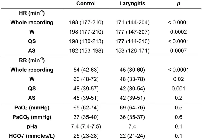
Results for cardiorespiratory variables are reported in Tables 2 and 3. A significantly lower median heart rate was observed in the laryngitis group compared to the control group, regardless of the state of alertness (p < 0.0001).
Similarly, a significantly lower respiratory rate was found in the laryngitis group in quiet wakefulness (p < 0.02) and quiet sleep (p = 0.001), as well as a longer apnea duration in quiet wakefulness (p < 0.0001) and a lower apnea index in AS (p = 0.03). While no differences were found for bradycardias associated with these apneas, a significant increase in the % of desaturation in quiet wakefulness (p = 0.02) was observed in the laryngitis group (table 3). As reported in table 2, no statistical differences were found between the two groups on day 6 with regard to arterial PCO2, PO2, pH and bicarbonates.
3.3 NNS Frequency
Results for NNS frequency are reported in figure 2. A total of 3377 NNS were recognized from polysomnographic recordings in the 12 lambs. Of these, 833 were excluded since they occurred secondarily to bleating or agitation. Therefore, 2544 NNS were included in the analysis. Regardless of exposure, NNS frequencies (total, isolated and in burst) were higher in AS than in W and QS. No differences were observed between the two groups for NNS frequency (total, isolated and burst) when all states were considered together. However, a significantly lower NNS burst frequency was found in the laryngitis group in AS (p < 0.03). Finally, 96% (95-99) of all NNS in the control group vs. 99.5% (95-100) in the laryngitis group were propagated along the esophagus (p = 0.1); no effects of state of alertness were observed.
The use of two methods of analysis, each with its own selection criteria, and with the intention to perform both analyses on the same set of NNS for comparison purposes resulted in a total of 1770 available NNS for further statistical analysis. Regardless of the state of alertness, the overall pattern of NNS-breathing coordination yielded a predominance of ie- and i-type NNS over ei- and e-types, with no statistical differences between the two groups. No effect of states of alertness was observed (figure 3). However, a statistical difference (p < 0.02) was noted using the NNS-breathing phase calculation analysis, with deglutition interrupting the breathing cycle slightly earlier in the laryngitis group (39% [26-57]) compared to control lambs (41% [26-67]).
4. DISCUSSION
4.1 Statement of Principal Findings
Results from this study, taking advantage of a unique ovine model of infant reflux laryngitis, show that the presence of mild to moderate laryngeal inflammation had a very limited effect on non-nutritive swallowing, i.e., a decrease in the number of NNS bursts in AS with no effect on NNS-breathing coordination. Of note, the AS-related decrease in NNS bursts was associated with a decrease in apnea index in AS only. Moreover, lower resting respiratory and heart rates as well as prolonged apnea duration during quiet wakefulness were clearly present in the laryngitis group. Clinical importance of our results stems from the frequent observation of reflux laryngitis in infants.
4.2 NNS Frequency
Present results in control lambs are similar to our previous results, showing a higher frequency of both isolated NNS and NNS bursts during AS (Duvareille et al., 2013; 2007; Reix et al., 2003; Samson et al., 2005). Studies from our group have also shown that certain conditions can alter NNS frequency in lambs. Indeed, an increased NNS frequency was observed with hypercapnia, while the inverse was observed with nasal application of continuous positive airway pressure or hypoxia (Duvareille et al., 2007; Samson et al., 2005). The only alteration of NNS frequency observed in the present study was a decrease in NNS burst frequency during AS. Given that AS-related NNS bursts are associated with apneas in lambs (Reix et al., 2004), the AS-related decrease in
apnea index observed with laryngitis is likely related to the decrease in NNS burst frequency. Whether these observations may have beneficial or deleterious consequences in a hypothetical infant with reflux laryngitis is unclear. On the one hand, a decreased apnea index is considered beneficial by all standards. On the other hand, a decreased number of NNS could be interpreted as limiting the pharyngeal clearance of liquids. However, an observation specific to AS-related NNS bursts is that all thyroarytenoid muscle electrical activities, which are considered NNS, are not followed by esophageal deglutition (unpublished, personal observations). We believe that certain AS-related NNS bursts in our studies may represent REM-related phasic muscle twitches, with limited efficiency as a pharyngeal clearance mechanism. If correct, this hypothesis would not support any deleterious consequence of decreased AS-related NNS bursts.
As NNS is under the control of brainstem premotor circuitry during sleep (Bonis et al., 2013; Little et al., 1997; López-Alonso et al., 2006; Miyawaki et al., 2003; Vandenplas et al., 1991), the present study does not provide additional clues as to the mechanisms (peripheral vs. central) responsible for the alteration of brainstem swallowing center function in the presence of laryngitis. Finally, the decrease in NNS burst frequency in AS raises the question of a similar impediment of nutritive swallowing bursts during bottle-feeding (Bernier et al., 2012), which may in turn decrease swallowing efficiency and promote lung aspiration. This particular aspect however remains to be studied.
4.3 NNS-Breathing Coordination
A very precise NNS-breathing coordination is required from the time of birth. While several studies have shown that coordination is largely hard-wired in the newborn brainstem at term (Kelly et al., 2008; Nixon et al., 2008), we have previously shown in lambs that some conditions can alter this coordination (Duvareille et al., 2013; 2007). In the present study, despite a comprehensive analysis of NNS-breathing coordination combining categorical assessment and NNS-breathing phase calculation, we did not find any laryngitis-induced alteration of the normal predominance of i- and ie-type NNS observed in the control group and previously reported in lambs (Duvareille et al., 2013; 2007; Reix et al., 2003; Samson et al., 2005). In addition, we believe that the only statistical difference found using NNS-breathing phase calculation (39% in control vs. 41% in laryngitis lambs) is of no physiological significance, especially when considering results obtained in control lambs (49%) in a previous study (Duvareille et al., 2013).
4.4 Cardiorespiratory Inhibition
We had already observed a decreased respiratory rate (however without any alteration in heart rate) and prolonged apnea duration in a previous study using an identical ovine model of infant reflux laryngitis (Carreau et al., 2011). The significant decrease in both heart and respiratory rates as well as prolonged apnea duration and associated desaturations observed in the present study reflect an altered autonomic control of both cardiac and respiratory activity
towards enhanced parasympathetic and/or blunted sympathetic tone. It is highly plausible that induction of laryngeal chemoreflexes by the pepsin solution thrice daily for six successive days, which corresponds to significant repeated disruption of cardiorespiratory control (Carreau et al., 2011; St-Hilaire et al., 2010), could be responsible for the observed persistent alteration in basal autonomic control. Moreover, the presence of inflammation in itself may also disrupt the autonomic control of cardiorespiratory activity. Indeed, previous studies have shown that laryngeal inflammation can inhibit respiratory centers in the brainstem via the production of IL-1B and PGE2 (Lindgren and Grögaard, 1996; Olsson et al., 2003). In addition, an increase in vagal tone can arise through the cholinergic anti-inflammatory pathway (Tracey, 2007; 2002), which is likely present in our experimental setting. Finally, the decreased respiratory rate and prolonged apnea duration might be secondary to the pain associated with local inflammation of the laryngopharyngeal region. However, we believe this latter explanation to be unlikely, given that the degree of laryngitis was rather limited, and an enhanced sympathetic tone with increased heart rate would be expected in such cases.
4.5 Study limitations
4.5.1 Species differences for NNS-breathing coordination
In contrast to humans, NNS occurs predominantly during inspiration. The latter is not specific to the sheep and has previously been reported in other species, including the dog (Kawasaki M et al., 1964) and the rabbit (McFarland and Lund,
1993). Several explanations have been put forward to account for this difference between humans and the above species, including the difference in upper airway anatomy (especially the superposition of the epiglottis and soft palate, which separates the nasal breathing route from the oral feeding route) and different standing posture (quadruped vs. biped) (McFarland and Lund, 1993). Regardless of the explanation, we nonetheless maintain that differences with humans do not represent a major limitation of our studies. Irrespective of the pattern of NNS-breathing coordination, we are interested in knowing whether a given condition (laryngitis in the present instance) can alter this coordination or not.
4.5.2 Reflux laryngitis model
Our results were obtained in a newborn animal model of reflux laryngitis induced by an acidified pepsin solution. Previous results have shown that acid refluxes are present in infants, even in newborns, and suggest they are responsible for pathological consequences, including laryngitis (Cresi et al., 2013; Jadcherla et al., 2011; López-Alonso et al., 2006; Shin et al., 2012; Vermeylen et al., 2005). However, our results may not apply to reflux laryngitis secondary to weakly acid reflux, which are especially prominent in newborns.
Our histological score showed mild to moderate inflammation of the larynx. While results from the present study, as well as from our previous study on laryngeal chemoreflexes using the same model (Carreau et al., 2011), show that even this level of inflammation can alter both NNS and cardiorespiratory activity, these results may not apply to severe laryngeal inflammation.
Although the present study has sufficient statistical power to clearly confirm the presence of an altered autonomic control of cardiac and respiratory activity in our laryngitis ovine model, it only shows a limited effect on NNS and NNS-breathing coordination. This latter finding suggests that, aside from the effect on NNS bursts in AS, laryngitis has no big effect on NNS and NNS-coordination. However, we cannot dismiss a more subtle effect of laryngitis on NNS and NNS-coordination, which might have been observed with a larger number of lambs in each experimental group. Unfortunately, to our knowledge, there are no currently-available tools (software, mathematical methods or scientific article) that allow a retrospective calculation of statistical power in the case of generalized estimating equation models (statistical method used for NNS and NNS-coordination).
4.6 Conclusion
Our results suggest that a mild to moderate reflux laryngitis is associated with a decrease in NNS burst frequency and apneas in active sleep in newborn lambs, without any significant alterations of NNS-breathing coordination. Moreover, this level of reflux laryngitis is associated with a decrease in baseline heart and respiratory rate, suggesting an altered autonomic control of cardiorespiratory activity. Further studies are necessary to assess the impact of reflux laryngitis on nutritive swallowing.
ACKNOWLEDGEMENTS
The authors wish to acknowledge the gracious donation of Blom-Singer Advantage tracheoesophageal voice prostheses by InHealth Technologies, Carpinteria, CA - USA. The study was supported by the Canadian Institutes of Health Research and by the Canada Research Chair on Neonatal Respiratory Physiology allocated to J-P Praud. Both S. Brisebois and A.M. Carreau were supported by a training scholarship from the Fonds de la recherche en santé du Québec. J-P Praud is a member of the Clinical Research Center Etienne-Le Bel, Sherbrooke University Hospital.
REFERENCES
Adhami, T., Goldblum, J.R., Richter, J.E., Vaezi, M.F., 2004. The role of gastric and duodenal agents in laryngeal injury: an experimental canine model. Am J Gastroenterol 99, 2098–2106.
Bernier, A., Catelin, C., Ahmed, M.A.H., Samson, N., Bonneau, P., Praud, J.-P., 2012. Effects of nasal continuous positive-airway pressure on nutritive swallowing in lambs. J Appl Physiol 112, 1984–1991.
Bonis, J.M., Neumueller, S.E., Krause, K.L., Pan, L.G., Hodges, M.R., Forster, H.V., 2013. Contributions of the Kölliker-Fuse nucleus to coordination of breathing and swallowing. Respir Physiol Neurobiol 189, 10-21.
Carreau, A.-M., Patural, H., Samson, N., Doueik, A.A., Hamon, J., Fortier, P.-H., Praud, J.-P., 2011. Effects of simulated reflux laryngitis on laryngeal chemoreflexes in newborn lambs. J Appl Physiol 111, 400–406.
Carron, J.D., Greinwald, J.H., Oberman, J.P., Werner, A.L., Derkay, C.S., 2001. Simulated reflux and laryngotracheal reconstruction: a rabbit model. Arch Otolaryngol Head Neck Surg 127, 576–580.
Contencin, P., Narcy, P., 1992. Gastropharyngeal reflux in infants and children. A pharyngeal pH monitoring study. Arch Otolaryngol Head Neck Surg 118, 1028–1030.
Cresi, F., Locatelli, E., Marinaccio, C., Grasso, G., Coscia, A., Bertino, E., 2013. Prognostic values of multichannel intraluminal impedance and pH monitoring in newborns with symptoms of gastroesophageal reflux disease. J Pediatr 162, 770–775.
Duvareille, C., St-Hilaire, M., Samson, N., Bakirtzian, P., Brisebois, S., Boheimier, M., Djeddi, D.-D., Doueik, A.A., Praud, J.-P., 2013. Effects of postnatal environmental tobacco smoke on non-nutritive swallowing-breathing coordination in newborn lambs. Respir Physiol Neurobiol 185, 446– 453.
Duvareille, C., Lafrance, M., Samson, N., St-Hilaire, M., Pladys, P., Micheau, P., Bournival, V., Langlois, C., Praud, J.-P., 2007. Effects of hypoxia and hypercapnia on nonnutritive swallowing in newborn lambs. J Appl Physiol 103, 1180–1188.
Hawkshaw, M.J., Pebdani, P., Sataloff, R.T., 2013. Reflux laryngitis: an update, 2009-2012. J Voice 27, 486–494.
Hom, C., Vaezi, M.F., 2013. Extraesophageal manifestations of gastroesophageal reflux disease. Gastroenterol. Clin. North Am. 42, 71–91. Jadcherla, S.R., Peng, J., Chan, C.Y., Moore, R., Wei, L., Fernandez, S., DI
Lorenzo, C., 2011. Significance of gastroesophageal refluxate in relation to physical, chemical, and spatiotemporal characteristics in symptomatic intensive care unit neonates. Pediatr Res 70, 192–198.
Kawasaki, M., Ogura, J.H., Takenouchi, S., 1964. Neurophysiologic observations of normal deglutition. I. Its relationship to the respiratory cycle. Laryngoscope. 74, 1747-65.
Kelly, B.N., Huckabee, M.-L., Frampton, C.M.A., Jones, R.D., 2008. Arousal has no effect on non-nutritive breathing-swallowing coordination during the first year of human life. Int. J. Dev. Neurosci. 26, 385–390.
Koufman, J.A., 1991. The otolaryngologic manifestations of gastroesophageal reflux disease (GERD): a clinical investigation of 225 patients using ambulatory 24-hour pH monitoring and an experimental investigation of the role of acid and pepsin in the development of laryngeal injury. Laryngoscope 101, 1–78.
Kwong, K., Hoa, M., Coticchia, J.M., 2007. Recurrent croup presentation, diagnosis, and management. Am J Otolaryngol 28, 401–407.
Leiter, J.C., Böhm, I., 2007. Mechanisms of pathogenesis in the Sudden Infant Death Syndrome. Respir Physiol Neurobiol 159, 127–138.
Lindgren, C., Grögaard, J., 1996. Reflex apnoea response and inflammatory mediators in infants with respiratory tract infection. Acta Paediatr 85, 798– 803.
Little, J.P., Matthews, B.L., Glock, M.S., Koufman, J.A., Reboussin, D.M., Loughlin, C.J., McGuirt, W.F., 1997. Extraesophageal pediatric reflux: 24-hour double-probe pH monitoring of 222 children. Ann Otol Rhinol Laryngol Suppl 169, 1–16.
López-Alonso, M., Moya, M.J., Cabo, J.A., Ribas, J., del Carmen Macías, M., Silny, J., Sifrim, D., 2006. Twenty-four-hour esophageal impedance-pH monitoring in healthy preterm neonates: rate and characteristics of acid, weakly acidic, and weakly alkaline gastroesophageal reflux. PEDIATRICS 118, e299–308.
Ludemann, J.P., Manoukian, J., Shaw, K., Bernard, C., Davis, M., al-Jubab, A., 1998. Effects of simulated gastroesophageal reflux on the untraumatized
rabbit larynx. J Otolaryngol 27, 127–131.
McFarland, D.H., Lund, J.P., 1993. An investigation of the coupling between respiration, mastication, and swallowing in the awake rabbit. J Neurophysiol 69, 95–108.
Miyawaki, S., Tanimoto, Y., Araki, Y., Katayama, A., Fujii, A., Takano-Yamamoto, T., 2003. Association between nocturnal bruxism and gastroesophageal reflux. Sleep 26, 888–892.
Nixon, G.M., Charbonneau, I., Kermack, A.S., Brouillette, R.T., McFarland, D.H., 2008. Respiratory-swallowing interactions during sleep in premature infants at term. Respir Physiol Neurobiol 160, 76–82.
Olsson, A., Kayhan, G., Lagercrantz, H., Herlenius, E., 2003. IL-1 beta depresses respiration and anoxic survival via a prostaglandin-dependent pathway in neonatal rats. Pediatr Res 54, 326–331.
Praud, J.-P., 2010. Upper airway reflexes in response to gastric reflux. Paediatr Respir Rev 11, 208–212.
Rankin, I., Wang, S.M., Waters, A., Clement, W.A., Kubba, H., 2013. The management of recurrent croup in children. J Laryngol Otol 127, 494–500. Reix, P., Arsenault, J., Langlois, C., Niyonsenga, T., Praud, J.-P., 2004.
Nonnutritive swallowing and respiration relationships in preterm lambs. J Appl Physiol 97, 1283–1290.
Reix, P., Fortier, P.-H., Niyonsenga, T., Arsenault, J., Létourneau, P., Praud, J.-P., 2003. Non-nutritive swallowing and respiration coordination in full-term newborn lambs. Respir Physiol Neurobiol 134, 209–218.
Renolleau, S., Letourneau, P., Niyonsenga, T., Praud, J.-P., Gagné, B., 1999. Thyroarytenoid muscle electrical activity during spontaneous apneas in preterm lambs. Am J Respir Crit Care Med 159, 1396–1404.
Roberge, S., Samson, N., Dorion, S., Dorion, D., Praud, J.P., 2007. Non-nutritive swallowing and respiration coordination among states of alertness in adult sheep. J Otolaryngol 36, 140-147.
Roh, J.-L., Lee, Y.-W., Park, H.T., 2006. Effect of acid, pepsin, and bile acid on the stenotic progression of traumatized subglottis. Am J Gastroenterol 101, 1186–1192.
Roh, J.-L., Yoon, Y.-H., 2006. Effect of acid and pepsin on glottic wound healing: a simulated reflux model. Arch Otolaryngol Head Neck Surg 132, 995–1000. Samson, N., Dumont, S., Specq, M.-L., Praud, J.-P., 2011. Radio telemetry
devices to monitor breathing in non-sedated animals. Respir Physiol Neurobiol 179, 111–118.
Samson, N., St-Hilaire, M., Nsegbe, E., Reix, P., Moreau-Bussière, F., Praud, J.-P., 2005. Effect of nasal continuous or intermittent positive airway pressure on nonnutritive swallowing in the newborn lamb. J Appl Physiol 99, 1636– 1642.
Shin, M.S., Shim, J.O., Moon, J.S., Kim, H.S., Ko, J.S., Choi, J.-H., Seo, J.K., 2012. Impedance-pH monitoring and conventional pH monitoring are complementary methods to detect association between gastroesophageal reflux and apnea-related symptoms in preterm infants and neonates. J. Matern. Fetal. Neonatal. Med. 25, 2406–2410.
St-Hilaire, M., Duvareille, C., Avoine, O., Carreau, A.-M., Samson, N., Micheau, P., Doueik, A., Praud, J.-P., 2010. Effects of postnatal smoke exposure on laryngeal chemoreflexes in newborn lambs. J Appl Physiol 109, 1820–1826. Thach, B.T., 2008. Some aspects of clinical relevance in the maturation of
respiratory control in infants. J Appl Physiol 104, 1828–1834.
Tracey, K.J., 2007. Physiology and immunology of the cholinergic antiinflammatory pathway. J. Clin. Invest. 117, 289–296.
Tracey, K.J., 2002. The inflammatory reflex. Nature 420, 853–859.
Ulualp, S.O., Rodriguez, S., Cunningham, S., Shen, J., 2007a. Pharyngeal pH monitoring in infants with laryngitis. Otolaryngol Head Neck Surg 137, 776– 779.
Ulualp, S.O., Rodriguez, S., Holmes-Wright, C.N., 2007b. Flexible laryngoscopy-guided pharyngeal pH monitoring in infants. Laryngoscope 117, 577–580. Ulualp, S.O., Toohill, R.J., Kern, M., Shaker, R., 1998. Pharyngo-UES contractile
reflex in patients with posterior laryngitis. Laryngoscope 108, 1354–1357. Vandenplas, Y., Goyvaerts, H., Helven, R., Sacre, L., 1991. Gastroesophageal
reflux, as measured by 24-hour pH monitoring, in 509 healthy infants screened for risk of sudden infant death syndrome. PEDIATRICS 88, 834– 840.
Vermeylen, D., Franco, P., Hennequin, Y., Pardou, A., Brugmans, M., Simon, P., Hassid, S., 2005. Laryngeal oedema in neonatal apnoea and bradycardia syndrome (a pilot study). Early Hum Dev 81, 361–367.
FIGURE LEGENDS
Figure 1: Hematoxylin and eosin staining of the laryngeal mucosa (20x) illustrating a mild to moderate stromal (arrow 2) and intraepithelial (arrow 1) inflammation in a lamb from the laryngitis group (left) compared to a control lamb (right).
Figure 2: Effect of reflux laryngitis on the frequency of total non-nutritive swallowing (NNS), isolated NNS and NNS bursts in control lambs (white boxes) and laryngitis lambs (grey boxes) in each state of alertness (*, p < 0.05). Regardless of exposure, NNS frequencies (total, isolated and burst) were higher in AS than in W and QS. A lower NNS burst frequency was found in AS for the laryngitis group compared to controls (p < 0.03). No other differences were noted.
Figure 3: Effect of reflux laryngitis on NNS-breathing coordination, isolated NNS and NNS bursts in control lambs (upper panel) and laryngitis lambs (lower panel) (n = 6 for each group) in each state of alertness. Measured NNS types include: i-type NNS, NNS beginning and ending in inspiration; e-i-type NNS, NNS beginning and ending in expiration; ie-type NNS, NNS beginning in inspiration and ending in expiration; ei-type NNS, NNS beginning in expiration and ending in inspiration. Results are expressed in percentage of the total number of NNS. Regardless of the state of alertness, similar patterns of NNS-breathing coordination were
observed, with ie- and i-type NNS being the most frequent and e-type the least frequent in both control and laryngitis groups.
Table 1: Effect of reflux laryngitis on sleep architecture
Control Laryngitis p
W (%) 42 (37-44) 45 (42-48) 0.4
QS (%) 52 (49-57) 45 (41-48) 0.1
AS (%) 7 (6-9) 9 (4-17) 0.5
Values are medians (upper and lower quartile) and are expressed in percentage of total recording time. W, wakefulness; QS, quiet sleep; AS, active sleep.
Table 2: Effects of reflux laryngitis on cardiorespiratory variables Control Laryngitis p HR (min-1) Whole recording 198 (177-210) 171 (144-204) < 0.0001 W 198 (177-210) 177 (147-207) 0.0002 QS 198 (180-213) 177 (144-210) < 0.0001 AS 182 (153-198) 153 (126-171) 0.0007 RR (min-1) Whole recording 54 (42-63) 45 (30-60) < 0.0001 W 60 (48-72) 48 (33-78) 0.02 QS 48 (39-57) 42 (30-54) 0.001 AS 45 (39-51) 42 (39-51) 0.2 PaO2 (mmHg) 65 (62-74) 69 (64-76) 0.5 PaCO2 (mmHg) 37 (35-40) 36 (35-37) 0.6 pHa 7.4 (7.4-7.5) 7.4 0.1 HCO3- (mmoles/L) 26 (23-28) 22 (21-24) 0.1
Results for heart and respiratory rates are expressed as medians (upper and lower quartile), first for the whole recording, then for each state of alertness (W, QS and AS) individually. HR, heart rate; RR, respiratory rate; see table 1 for other abbreviations.
Table 3: Effects of reflux laryngitis on apneas and associated bradycardias and desaturations Control Laryngitis ρ Apnea index (h-1) 6 (4-11) 7(6-9) 0.7 W 11 (8-21) 12 (10-15) 0.7 QS 2 (1-3) 3 (1-6) 0.5 AS 8 (7-8) 4 (3-6) 0.03 Apnea duration (s) 6.1 (3.6-9.7) 8.4 (6.0-10.7) < 0.0001 W 5.8 (3.3-8.9) 8.1 (5.8-10.7) < 0.0001 QS 10.4 (6.0-11.9) 9.4 (7.9-11.6) 0.5 AS 6.3 (4.9-10.9) 8.4 (5.1-9.7) 0.7 Bradycardia index (h-1) 3 (1-7) 1 (0-4) 0.6 W 7 (4-13) 3 (1-6) 0.2 QS 1(1-3) 3 (1-3) 0.5 AS 2(2-2) - - Bradycardia duration (s) 4.4 (1.7-7.5) 4.3 (2.4-6.9) 0.8 W 3.7 (1.6-6.6) 4.0 (2.2-6.9) 0.7 QS 7.1 (1.8-8.7) 4.4 (2.4-6.5) 0.7 AS 9.0 (9.0-9.0) - - Desaturation index (h-1) 0 (0-3) 4 (2-5) 0.08 W 1 (1-1) 4 (2-8) 0.2 QS 1 (0-1) 1 (1-5) 0.08 AS 11 (4-19) 5 (3-6) 0.4 Desaturation (%) 5.6 (4.5-7.6) 7.0 (5.9-9.9) 0.03 W 4.8 (4.4-6.5) 6.9 (5.9-9.9) 0.02 QS 4.7 (4.4-6.5) 7.8 (5.0-11.7) 0.1 AS 7.0 (5.3-10.0) 5.3 (4.8-5.8) 0.2
Results are expressed as medians (upper and lower quartile) for the whole recording, then for each state of alertness (W, QS and AS) individually. See table 1 for abbreviations.
Figure 1
Figure 2
Figure 3