M E E T I N G A B S T R A C T S
Open Access
39th International Symposium on Intensive
Care and Emergency Medicine
Brussels, Belgium, 19-22 March 2019
Published: 19 March 2019Accepted abstracts for 39th
International Symposium on Intensive
Care and Emergency Medicine
P001
Prognostic value of a genetic polymorphism of AQP5 in sepsis depends on a source of infection
V Pisarev1, A Chumachenko1, I Tyurin2, R Cherpakov2, A Tutelyan3 1
Federal Research and Clinical Center of Intensive Care Medicine and Rehabilitology, V.A.Negovsky Institute of General Reanimatology, Moscow, Russia;2V.M.Buiyanov City Clinical Hospital, Anesthesia-Reanimatology Department, Moscow, Russia;3Central Institute of Epidemiology, Moscow, Russia
Critical Care 2019, 23(Suppl 2):P001
Introduction: The purpose of the study was to determine whether the preferential localization of the infection and age affect the prog-nostic value of the genetic marker AQP5 (1364A/C, rs3759129) in out-come prediction in sepsis patients. Studies by Adamzik and colleagues have demonstrated that aquaporin AQP5 polymorphism (1364A/C, rs3759129) associates with increased 30-day survival in sepsis patients presumably due to increased gene expression that enhance the leukocyte migration. To increase the informative value of the prediction and decrease the cost, it might be crucial to deter-mine at a pre-test level the subset of patients who might benefit most from the prognostic genotyping.
Methods: Sepsis and septic shock were defined in patients according to SEPSIS-3 (2016) recommendations. Study groups (n=152) included ICU patients with abdominal sepsis (AS, including pancreatitits, peri-tonitis, cholecystitis, appendicitis; n=98) and sepsis patients with other sources of infections. AQP5 polymorphism was studied by ana-lyzing PCR products in a 2% agarose gel using a AQP5 1364A/C spe-cific tetra primer set. Data were analyzed by Kaplan-Meyer plot and Fisher test, and odds ratios were calculated.
Results: Distribution of alleles (A and C) and genotypes (AA, CA and CC) AQP5 1364A/C in patients with sepsis or sepsis subgroups (sepsis with no septic shock and sepsis shock patients) versus control group (healthy volunteers) did not differ. Although there was a trend to preferential survival of sepsis patients with genotype C AQP5 despite the source of infection, only patients with AQP5 CC or CA genotype and abdominal sepsis (Sepsis-3), or a subgroup of the same AQP5 genotype experiencing septic shock, demonstrated increased 30-day survival versus AA homozygotic patients (P<0.002).
Conclusions: The informative value of detecting the AQP5 CC or CA genotype for prognosis of 30-day survival versus AA homozygotic pa-tients is increased only in abdominal sepsis papa-tients.
P002
Depressed expression of FCER1A gene is associated with increased mortality in infected surgical patients
R Almansa1, C Andrés2, M Martín-Fernández3, S Montero4, C Jambrina5, C Doncel6, J Sánchez-Crespo5, M Heredia-Rodríguez7, J Rico4, C González8, E Sánchez-Barrado5, M Lorenzo-López7, S Martín4, L Muñoz-Bellvis8, M Vaquero5, E Tamayo7, C Aldecoa4, J Bermejo-Martín6
1Hospital Clínico Universitario de Valladolid/IECSCYL, BioSepsis (Group of Biomedical Research in Sepsis), Valladolid, Spain;2Hospital Clínico Universitario de Valladolid, Clinical Analysis Service, Valladolid, Spain; 3
Hospital Clínico Universitario de Valladolid/IECSCYL, BioSepsis (Group for Biomedical Research in Sepsis), Valladolid, Spain;4Hospital Universitario Rio Hortega, Anesthesiology and Reanimation Service, Valladolid, Spain;5Hospital Clínico Universitario de Salamanca, Anesthesiology and Reanimation Service, Salamanca, Spain;6Hospital Clínico Universitario de Valladolid/IECSCYL, BioSepsis (Group for Biomedical Research in Sepsis), Valladolid, Spain;7Hospital Clínico Universitario de Valladolid, Anesthesiology and Reanimation Service, Valladolid, Spain;8Hospital Clínico Universitario de Salamanca, Department of General and Gastrointestinal Surgery, Salamanca, Spain Critical Care 2019, 23(Suppl 2):P002
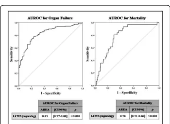
Introduction: Increasing evidence supports a central role for “im-munosuppression” in sepsis. It is necessary to develop biomarkers of immune dysfunction that could help to identify patients at risk of poor outcomes [1]. The decreased expression of human leucocyte antigen (HLA)-DRA is proposed as a major feature of immunodepres-sion and its persistent decrease is associated with mortality in sepsis [2]. In a previous study, we evidenced that FCER1A (Fc Fragment Of IgE Receptor Ia) is the gene showing the lowest expression levels of the en-tire transcriptome in sepsis [3]. Here we studied the association be-tween FCER1A expression and mortality in infected surgical patients. Methods: FCER1A and HLA-DRA expression levels were quantified by droplet digital PCR in blood of 257 infected surgical patients. 26 pa-tients died within 28 days (10.11%). Spearman test was used to evaluate the association between gene expression and the Sequen-tial Organ Failure Assessment (SOFA) score. Areas under Receiver Op-erating Curves (AUROC) were used to determine the gene expression cut-off values predicting mortality. Kaplan-Meier survival curves were obtained and differences in survival between groups were evaluated using the Log rank test. Cox regression was employed to assess mor-tality risk at 28 days.
Results: Gene expression levels of FCER1A and HLA-DRA correlated inversely with patients’ severity (r: -0.5 p<0.001; r: -0.3, p<0.001 re-spectively). Both genes showed significant AUROCs to predict sur-vival, but FCER1A showed the best accuracy (Fig. 1). Patients with
© The Author(s). 2019 Open Access This article is distributed under the terms of the Creative Commons Attribution 4.0 International License (http://creativecommons.org/licenses/by/4.0/), which permits unrestricted use, distribution, and reproduction in any medium, provided you give appropriate credit to the original author(s) and the source, provide a link to the Creative Commons license, and indicate if changes were made. The Creative Commons Public Domain Dedication waiver (http://creativecommons.org/publicdomain/zero/1.0/) applies to the data made available in this article, unless otherwise stated.
low levels of FCER1A or HLA-DRA had an increased risk of mortality and died 3 days earlier than non survivors with higher expression levels of these genes (Fig. 2, Table 1-2).
Conclusions: Depressed FCER1A gene expression is associated with severity and increased mortality in surgical patients with infection. References
1 Hotchkiss R et al. Lancet Infect Dis 13(3): 260–268, 2013 2 Cazalis MA et al. Crit Care 10;17(6):R287, 2013 3 Almansa R et al. J Infect 70(5):445-56, 2015
P003
Genetic markers of nosocomial pneumonia, acute respiratory failure and renal insufficiency in critically ill patients
M Khadzhieva1, O Belopolskaya1, T Smelaya2, A Kuzovlev2, L Salnikova3 1N.I. Vavilov Institute of General Genetics, Russian Academy of Sciences, Moscow, Russia;2Federal Research and Clinical Center of Intensive Care Medicine and Rehabilitology, Moscow, Russia;3Federal Research and Clinical Center of Intensive Care Medicine and Rehabilitology, N.I. Vavilov Institute of General Genetics, Russian Academy of Sciences, Moscow, Russia
Critical Care 2019, 23(Suppl 2):P003
Introduction: Severe pulmonary and renal conditions such as acute respiratory distress syndrome (ARDS), respiratory failure, and deterior-ation in kidney function often occur in patients with nosocomial pneumonia (NP). The emergence and course of infection is genetic-ally determined, hence host genetic landscape may influence an abil-ity to resist infection.
Methods: Variants for genotyping were selected using the PheWAS Catalog which presents genotypic data for 13835 Caucasian patients, 1358 phenotypes and 3144 single nucleotide polymorphisms (SNPs) with P < 0.05 [1]. SNPs with the lowest P-values for phenotypes with both, respiratory and renal manifestations were selected: intergenic variants rs7130588 and rs4980785, rs347344 (EDIL3) and rs2470893 (CYP1A1). CYP1A1 gene was associated with pneumonia and ARDS in our previous investigations, so we included in our analysis three sites of CYP1A1 gene (rs2606345, rs4646903 and rs1048943) studied on a smaller sample. Genotyping was performed on 7 sites for a sample Table 1 (abstract P002). Predictive capacity of FCER1A gene expression
cut-off for 28-day mortality in surgical patients with infection. (COX regression)
Hazard Ratio 95% CI P
Age 1.05 (1.00-1.09) 0.038
Diabetes 1.98 (0.85-4.62) 0.112
Respiratory focus 1.63 (0.66-4.01) 0.289
OOP FCER1A: 17.4 copies/ng RNA 4.18 (1.52-11.47) 0.005
Table 2 (abstract P002). Predictive capacity of HLA-DRA gene expression cut-off for 28 day mortality in surgical patients with infection. (COX regression)
Hazard Ratio 95% CI P
Age 1.05 (1.00-1.10) 0.011
Diabetes 1.90 (0.85-4.31) 0.120
Respiratory focus 2.0 (0.86-4.64) 0.108
OOP HLA-DRA: 2744 copies/ng RNA 2.36 (1.01-5.53) 0.048
Fig. 1 (abstract P002). AUROCs for differential diagnosis of mortality in surgical patients with infection
Fig. 2 (abstract P002). Kaplan-Meier survival curves. Kaplan-Meier survival curves were established after stratification based on calculated thresholds (optimal operating points of FCER1A (A) and HLA-DRA (B) expression levels)
of resuscitation patients with or without NP and other pulmonary complications (n = 354 and n = 216, respectively).
Results: Allele rs2606345-G of the CYP1A1 gene was protective against ARDS and an increase in creatinine level (Fig. 1). The rs7130588-G allele was associated with lung complications and with the development of severe respiratory insufficiency (Fig. 2).
Conclusions: The SNPs rs2606345 and rs7130588 can influence the aggravation of pulmonary and renal symptoms through genetically mediated response to infection.
Reference
1. Denny JC et al. Nat Biotechnol 31: 1102–1110, 2013.
P004
Mesenchymal stem cells regulate LPS–stimulated macrophages polarization balance by paracrine transforming growth factor beta F Liu
School of Medicine, Zhongda Hospital, Southeast University, Department of Critical Care Medicine, Nanjing, Jiangsu, China
Critical Care 2019, 23(Suppl 2):P004
Introduction: An uncontrolled inflammatory response plays a major role in the sepsis related organ dysfunction. Mesenchymal stem cells(MSCs) can improve survival of sepsis experimental models by modulating the inflammatory response. Macrophages have been considered as important immune effector cells and their polarization imbalance aggravates the disordered inflammation reaction. The pro-ject aims to identify the effects of MSCs on macrophages polarization against dysregulated inflammatory response.
Methods: RAW264.7 cells were plated in the lower chambers of transwell system in the presence or absence of Lipopolysaccharide (LPS). Then, MSCs were seeded in the upper chambers and incuba-tion for different time. Finally, transforming growth factor beta (TGF-β) receptor (TGF-βR) inhibitor was added in transwell system. The
phenotype of RAW264.7 cells were analyzed by flow cytometry, the levels of inflammatory cytokines were detected by Enzyme-linked im-munosorbent assay (ELISA).
Results: Our data showed that LPS increased the level of interleukin (IL)-6 in RAW264.7 cells (p<0.001) (Fig. 1). In line with IL-6 expression, LPS induced the expression of M1 macrophage (p<0.001). Moreover, LPS stimulated RAW264.7 cells co-culture with MSCs in transwell sys-tem, MSCs inhibited the expression of IL-6 and M1 macrophages, while increased M2 macrophages (p<0.001). Compared with LPS group, the concentration of TGF-Β was obviously increased in MSCs treatment groups (p<0.001), furthermore, there were no significantly difference between MSCs directed and indicted groups. More signifi-cantly, TGF-βR inhibitor abolished the impact of MSCs on LPS stimu-lated RAW264.7 cells (p<0.001) (Fig. 2).
Conclusions: MSCs polarized M1 macrophages into M2 macro-phages and decreased pro-inflammatory cytokine levels by para-crining TGF-β.
Fig. 1 (abstract P004). Lipopolysaccharide (LPS) promoted RAW 264.7 M1 polarization. (a): LPS 500 ng/ml stimulated RAW264.7 for 12, 24, 48 and 72 hours, the level of IL-6 significantly increased in a time-dependent manner. (b, c): Flow cytometry showed LPS enhanced the expression of M1 macrophages at all time points. *** P<0.001 compared with control group
Fig. 1 (abstract P003). Protective effect of the rs2606345-G allele (CYP1A1 gene) on the risk of ARDS development (left) and an increase of serum creatinine level on the 14th day after hospitalization (right)
Fig. 2 (abstract P003). Association of the rs7130588-G allele with lung complications (LC) (NP, ARDS, pleurisy, abscess, etc.) (left) and with the development of severe respiratory failure (right)
Fig. 2 (abstract P004). TGF-β secreted by MSCs regulated the M1 to M2 in LPS-Stimulated RAW264.7. LPS stimulated RAW264.7 co-culture with MSCs in transwell system for 24, 48 and 72 hours. (a): LPS increased the level of IL-6, whereas MSCs inhibited expression of IL-6. (b, c): MSCs reduced M1 macrophages while increased M2 macrophages in LPS stimulated RAW264.7. (d): The concentration of TGF-β was obviously increased in MSCs directed or in-directed group. (e, f, g): LPS increased IL-6 and M1 macrophages, and MSCs inhibited the IL-6 and M1 macrophages while increased M2 macrophages. TGF-βR inhibitor reversed the effect of MSCs on LPS-stimulated RAW264.7. *P<0.05, **P<0.01, *** P<0.001 compared with control group. # # # P<0.001 compared with LPS stimulated group. &&& P<0.001 compared with MSCs treatment group
P005
Chronomics in ICU: effect of timing of septic shock onset on circadian rhythm profiles of melatonin and cortisol E Sertaridou1, I Chouvarda2, K Arvanitidis3, E Filidou4, G Kolios3, I Pnevmatikos1
1University Hospital of Alexandroupolis, Intensive Care Unit,
Alexandroupolis, Greece;2Aristotle University of Thessaloniki, Faculty of Medicine, Thessaloniki, Greece;3Democritus University of Thrace, Faculty of Medicine, Alexandroupolis, Greece;4Democritus University of Thrace, Faculty of Mrdicine, Alexandroupolis, Greece
Critical Care 2019, 23(Suppl 2):P005
Introduction: Circadian rhythmicity of melatonin and cortisol has been found to be affected by sepsis in both experimental and clinical studies. Methods: In this study, we evaluated the potential effect of septic shock on circadian rhythms of urinary excreted aMT6s, a melatonin’s metabolite and cortisol in 26 patients, divided into two groups: Group A (N=15) included subjects with septic shock upon admission to the ICU and Group B (N= 11) included patients who developed septic shock during ICU stay. Urine samples were collected every 4 h over a 24-h period, whereas data were available during entry and be-fore discharge from the ICU in Group A and during entry, septic shock and before exit from the ICU in Group B. Circadian analysis was performed leading to the estimation of mesor (mean value), amplitude of the oscillation and acrophase (phase shift of maximum values in hours).
Results: Circadian markers of aMT6s and cortisol exhibited inverse changes, both within and between groups, since amplitude of aMT6s was reduced in entry in relation to exit (437.2±309.2 vs 674.1±657.6 ng/4h, p<0.05), whereas amplitude of cortisol was increased upon admission compared to exit (13.3±31 ng/4h vs 8.7±21.2 ng/4h p<0.05), in Group A. Furthermore, in Group B, mesor of aMT6s was increased during septic shock (2492.2± 1709.1 ng/4h) compared to both entry (895.4±715.5 ng/4h) and exit (1308.6± 1214.4 ng/4h, p<0.05 for all comparisons). However, cortisol’s mean values were re-duced during septic shock (10±5.3 ng/4h) compared to both entry (30±57.9 ng/4h) and exit (14.4±20.7 ng/4h, p<0.05 for all compari-sons) and correlated with higher APACHE II score and longer ICU and hospital stay (p<0.05 for all comparisons).
Conclusions: Septic shock induced inverse changes of aMT6s and cortisol circadian rhythm profiles, depending on timing of onset.
P006
Investigation of the relationship between organ damage, microcirculatory dysfunction and reactive oxygen intermediate formation in experimental sepsis
J Kaszaki, A Rutai, R Fejes, S Tallósy, M Poles, D Érces, M Boros, A Szabó Universitiy of Szeged, Institute of Surgical Research, Szeged, Hungary Critical Care 2019, 23(Suppl 2):P006
Introduction: Sepsis is dysregulated response to an infection, which can lead to progressive microcirculatory dysfunction, release of react-ive oxygen intermediates (ROI) and life-threatening organ dysfunc-tion. Our aim was to investigate the relationship between organ damage - characterized by the Sequential Organ Failure Assessment (SOFA) scores, microcirculatory failure and ROI production, in a large animal model of experimental sepsis.
Methods: Fecal peritonitis was induced in anesthetized minipigs (n=28; 0.5g/kg autfeces containing 5-9 x10⁶CFU bacteria i.p.), control animals (n=9) received sterile saline i.p. Invasive hemodynamic moni-toring and blood gas analyses were performed between 16-24 hrs, the signs for failure of circulatory, respiratory and urinary systems were evaluated in accordance with the SOFA score. The microcircula-tory perfusion rate in the sublingual region was measured by orthog-onal polarization spectral imaging technique (Cytoscan A/R). The leukocyte-origin ROI production was determined by lucigenine (mostly O2-.) and luminol-based (H2O2) chemiluminescence methods.
Results: Between 16-24 hrs after induction the SOFA score indicated moderate organ failure in 19 animals (M: 1.9; 25p: 1.5, 75p: 2.9) and the change was statistically significantly higher in 9 pigs, suggesting severe organ dysfunction (M: 4.1; 25p: 3.5, 75p: 5.2). The microcircula-tion was significantly deteriorated in all cases, independently of SOFA score data. The H2O2production was significantly lower in septic ani-mals as compared to controls, while the lucigenine enhanced ROI production correlated with the SOFA score-indicated moderate and severe organ dysfunction.
Conclusions: Sublingual microcirculatory parameters are not correlat-ing with the severity of SOFA score-indicated organ dysfunction in abdominal sepsis. The measurement of ROI production of the whole blood seems to be better biomarker for the detection of the progres-sion of events from moderate to severe organ damages.
Grant supports: NKFIH K116689; EFOP-3.6.2-16-2017-00006
P007
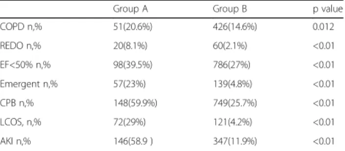
Increased rate of mechanical ventilation in septic patients with left ventricular dysfunction
A Newsome, S Smith, T Jones
The University of Georgia College of Pharmacy, Pharmacy, North Augusta, United States
Critical Care 2019, 23(Suppl 2):P007
Introduction: The purpose of this study was to characterize differ-ences in sepsis management in patients with and without left ven-tricular (LV) dysfunction. Septic patients with LV dysfunction have higher mortality, and limited guidance exists for sepsis management of patients with LV dysfunction. The possibility exists that the corner-stones of sepsis management may contribute to these poor outcomes.
Methods: A retrospective chart review was conducted from May 2016 -January 2018 at two centers. Adult patients who had a diagnosis of sepsis, were treated with vasopressors for > 3 hours, and had an echo-cardiogram within 12 months were included. Patients were divided into two groups: reduced ejection fraction (EF) of < 40% and preserved EF defined as EF≥40%. Information about patient outcomes and sepsis management were collected. The primary outcome was the need for mechanical ventilation (MV). Categorical and continuous data were ana-lyzed using the Chi-Squared and Mann-Whitney U tests, respectively. The IRB has approved this project.
Results: A total of 37 patients with EF < 40% and 42 patients with EF ≥40% were included. No significant differences in fluid management, vasoactive agent maximum rate or duration, or steroid use were ob-served. Net fluid balance between low and preserved EF was positive 4.6 liters vs. 5.1 liters (p = 0.814), respectively. The number of pa-tients that needed MV was higher in the low EF cohort (86% vs. 57%, p = 0.004), and this cohort had fewer MV-free days (20, IQR 0-25 vs. 24 (IQR 0 -28), p=0.064.
Conclusions: No significant differences were observed with regard to sepsis management, reflecting current guidelines. The significantly increased need for MV is a provocative result. A potential mechanism is the inability of a patient with reduced LV dysfunction to maintain appropriate cardiac and respiratory function in the face of fluid over-load. Prospective analysis of the role of fluid balance in septic pa-tients with LV dysfunction is warranted.
P008
Biomarkers of myocardial injury and cytokine plasma levels in septic patients
M Assuncao1, FR Machado2, MK Brunialti3, O Rigato4, R Salomao3 1
Hospital Israelita Albert Einstein, Department of Critical Care, Sao Paulo, Brazil;2Federal University of Sao Paulo, Department of Anesthesiology, Pain and Intensive Care, Sao Paulo, Brazil;3Federal University of Sao Paulo, Division of Infections Diseases, Sao Paulo, Brazil;4Hospital Sirio Libanes, Department of Critical Care, Sao Paulo, Brazil
Introduction: The relationship between myocardial injury and systemic inflammation in sepsis response is not well understood [1]. It´s pro-posed to evaluate the association between myocardial injury bio-markers, high-sensitive troponin T (hs-cTnT) and N-terminal pro-brain natriuretic peptide (NT-ProBNP), with inflammatory mediators (6, IL-1Β , IL-8, IL-10, IL-12 / IL-23p40, IL17A, IL- 21 and TNF-α ) and bio-markers, C protein reactive (CPR) and procalcitonin (PCT), in septic patients
Methods: This was a prospective cohort study performed in three in-tensive care units, from September 2007 to September 2010
enrol-ling patients with sepsis (infection associated with organ
dysfunction), and septic shock (hypotension refractory by fluids infu-sion requiring vasopressor). Blood samples were collected up to 48h after the development of first organ dysfunction (D0) and on the 7th day after inclusion in the study (D7)
Results: Ninety-five patients were enrolled, with median age 64 years (interquatile?48–78), APACHE II: median 19 (14-22), SOFA: median 8 (5-10); 24.2% were admitted in ICU with sepsis and 75.8% with septic shock. Hospital mortality was 34.7%. In D0, NT-ProBNP correlated with IL-8 (r = 0.495, p <0.001) and IL-10 (r = 0.471, p <0.001). In D7, hs-cTnT and NT-ProBNP correlated with PCT (r = 0.446, p < 0.001 and r = 0.495, p < 0.001; respectively). NT-ProBNP D0 was higher in non-survivors than in non-survivors on mortality in seventh day (p = 0.029) and in-hospital mortality (p = 0.030). hs-cTnT D7 (p = 0.030) and NT-ProBNP D7 (p <0.001) were significantly higher in non-survivors on in-hospital mortality. NT-ProBNP D7 (OR 9.28; IC95% 2.05-41.94, p=0,004) and hs-cTnT D7 (OR 10,93; IC95% 2.139– 55.795, p=0,04) were independently associated with in-hospital mortality
Conclusions: NT-ProBNP plasma levels at D0 correlated with IL-8 and IL-10, and both NT-ProBNP and hs-cTnT at D7 correlated with PCT. In addition, NT-ProBNP has been shown to be an important predictor of mortality
Reference
1. Landesberg G et al. Chest. 2015;148:93-102.
P009
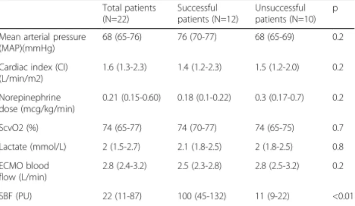
Repeated measures of heparin-binding protein correlate with mean arterial pressure and systemic vascular resistance index in septic shock: a pilot study on biomarker kinetics from a Swedish intensive care unit
J Tverring1, N Nielsen2, F Kahn3, A Linder3, P Åkesson3 1
Division of Infection Medicine, BMC, B14, Faculty of Medicine, Department of Clinical Sciences, Lund, Sweden;2Division of Anesthesiology and Intensive Care, Department of Clinical Sciences, Lund, Sweden;3Division of Infection Medicine, BMC, B14, Lund, Sweden Critical Care 2019, 23(Suppl 2):P009
Introduction: Heparin-binding protein (HBP) acts proinflammatory on immune cells and induces vascular leakage through cytoskel-etal rearrangement and cell contraction in the endothelium and is a promising novel prognostic biomarker in sepsis and septic shock. However, studies on repeated measures of HBP are lack-ing. Our objective was to describe the kinetics of plasma HBP
during septic shock and correlate it to hemodynamic
parameters.
Methods: We included patients with septic shock (sepsis-3) on ad-mission to Helsingborg hospital’s intensive care unit (ICU) during September 2016 to February 2018. Patients were sampled from ICU admission and every 4 hours for 72 hours or until death or ICU discharge. The plasma samples were analyzed for HBP and converted using the natural log (lnHBP) for normality. lnHBP was then evaluated against mean arterial pressure (MAP) as primary analysis and against systemic vascular resistance index (SVRI) as a secondary analysis, using mixed-effects linear regression models, treating patient id as a random intercept and adjusting for hemodynamic parameters.
Results: A total of 22 patients were included with median age 67 years, 9 females (41%), 7 surgical admissions (32%), median SOFA-score 12 points on day one and 6 deaths from all causes
within 90 days (27%). Plasma HBP ranged from 0 to 932 ng/ml with a median of 47 ng/ml (lnHBP range 1.6 to 6.8, median: 3.9). An increase lnHBP was significantly associated with a decrease in MAP (Coef. -2.58 mmHg, 95% CI: -0.62 to -4.55, p=0.010, n=22), when adjusting for heart rate (HR), noradrenaline (NA), vasopres-sin (VP), dobutamine (DBT) and levosimendan (LS). In a secondary subgroup analysis, an increase in lnHBP was also significantly as-sociated with a decrease in SVRI (Coef. -94.2 dyne*s*cm-5*m-2, 95% CI: -1.3 to -187.1, p=0.047, n=13), when adjusting for MAP, HR, NA, VP, DBT, LS and cardiac index.
Conclusions: Repeated measures of plasma HBP during septic shock were correlated with important hemodynamic parameters in this small pilot study.
P010
Mid-regional pro-adrenomedullin (MR-proADM) as early mortality predictor in septic shock
V Lovati, F Marsigli, E Pierucci
Azienda Ospedaliero Universitaria Policlinico Sant´Orsola Malpighi, Dipartimento di Scienze Mediche e Chirurgiche, Bologna, Italy Critical Care 2019, 23(Suppl 2):P010
Introduction: Mid-regional pro-Adrenomedullin (MR-proADM)
comes from the synthesis of the hormone adrenomedullin (ADM), which is overexpressed during inflammation and progression from sepsis to septic shock. Thus, MR-proADM can be a useful biomarker for the clinical management of septic patients [1]. The aim of our study was to understand the ability of MR-proADM to predict 30-day (30-d) mortality and to find a correlation between MR-proADM and Sequential Organ Failure Assessment (SOFA) score in the first 24 hours from Intensive Care Unit (ICU) admission.
Methods: We evaluated 28 consecutive septic shock patients ac-cording to 2016 Sepsis III definitions. Clinical data from the med-ical records included demographics, comorbidities, laboratories, microbiology and biomarker levels. Whole blood samples for bio-marker profiling were collected at 24, 72 and 120 hours from ICU admission. MR-proADM measurement was detected in EDTA plasma using a sandwich immunoassay by TRACE® (Time Resolved Amplified Cryptate Emission) technology (Kryptor Thermo Fischer Scientific BRAHMS).
Results: Overall 30-d mortality rate was 50.0%. MR-proADM [odds ra-tio (OR) = 1.195], SOFA score (OR = 2.174) and Lactate (Lac) levels (OR = 1.956) in the first 24 hours were associated with 30-d mortality in univariate logistic analysis (P value < 0.05, Table 1). 30-d mortality rate was not associated with procalcitonin (PCT) levels (OR = 1.002). Further linear regression analysis showed significant correlation be-tween MR-proADM and SOFA score at 24 hours from ICU admission (P value<0.001, Fig. 1, Table 2).
Conclusions: MR-proADM demonstrated superior accuracy to pre-dict 30-d mortality compared to PCT levels and is directly linked to SOFA score at 24 hours from admission. MR-proADM may aid early identification of poor prognosis septic patients who could benefit a more intensive management.
Reference
1. Andaluz-Ojeda D. et al. Ann Intensive Care 7, 15 (2017)
Table 1 (abstract P010). Univariate logistic analysis between 30-day mortality, MR-proADM, SOFA score, lactate levels, PCT levels
Odds Ratio Standard Error P value 95% Confidence Intervals
MR-proADM 1.195 0.102 0.037 1.011 - 1.413
SOFA score 2.174 0.693 0.015 1.164 - 4.061
Lactate 1.956 0.652 0.044 1.018 - 3.760
P011
Pre-sepsin as diagnostic and prognostic marker in sepsis: prospective evaluation by test and confirmation cohorts N Melachroinopoulos1, S Pouriki2, A Prekates3, K Toutouzas4, C Mathas5, E Giamarellos-Bourboulis6
1National and Kapodistrian University of Athens, Athens, Greece; 2
Hippokrateion General Hospital, Athens, Greece;3Tzaneion General Hospital, Intensive Care Unit, Piraeus, Greece;4National and Kapodistrian University of Athens, 1st Department of Propedeutic Surgery, Athens, Greece;5Konstantopouleion General Hospital, Intensive Care Unit, Athens, Greece;6National and Kapodistrian University of Athens, 4th Department of Internal Medicine, Athens, Greece
Critical Care 2019, 23(Suppl 2):P011
Introduction: Biomarkers have not yet been studied in prospective studies using Sepsis-3. The diagnostic and prognostic validity of pre-sepsin (soluble CD14) was studied in both one test and one confirm-ation cohort.
Methods: The test cohort was the prospective clinical study INTELLIGENCE-1 (ClinicalTrials.gov NCT03306186) enrolling patients with documented infections and at least one qSOFA sign. The con-firmation cohort was the prospective clinical study INTELLIGENCE-2 (ClinicalTrials.gov NCT03306186) with patients admitted in the emer-gencies with at least one qSOFA sign. Blood samples were collected within the first 24 hours of the presence of the qSOFA criteria and pre-sepsin was measured in plasma using the PATHFAST assay. Pa-tients were classified as sepsis and non-sepsis using Sepsis-3 defini-tions; 28-day mortality was recorded.
Results: In the test cohort, 62 patients were classified as non-sepsis and 111 as sepsis. Using ROC curve analysis, it was found that the best trade-off between sensitivity and specificity was provided at 350 pg/ml. The odds ratio for sepsis with presepsin above 350 pg/ml was 4.04 (p<0.0001) providing diagnostic sensitivity 80.2%. In logistic
regression analysis, it was found that Charlson’s comorbidity index more than 2, history of type 2 diabetes mellitus and of chronic ob-structive pulmonary disease and presepsin more than 350 pg/ml were the only variables independently associated with sepsis. Presep-sin above 350 pg/ml was associated with sensitivity 91.5% for 28-day mortality. The odds ratio for mortality with presepsin above 350 pg/ ml was 6.84 (p: 0.001). In the confirmation cohort, 59 patients were enrolled. The sensitivity of presepsin above 350 pg/ml for the diag-nosis of sepsis was 85.7% and for the prediction of 28-day mortality 100%.
Conclusions: Using a test and confirmation cohort approach, presep-sin above 350 pg/ml was proved a valuable indicator for the diagno-sis of sepdiagno-sis and outcome prognodiagno-sis among the most severe patients with one qSOFA sign.
P012
Toll-like receptors as biomarkers of sepsis in the emergency department
C Graham, LY Leung, R Lo, YK Leung, K Hung
The Chinese University of Hong Kong, Accident and Emergency Medicine Academic Uint, Hong Kong, China
Critical Care 2019, 23(Suppl 2):P012
Introduction: We aimed to investigate circulating TLRs gene signa-tures in Emergency Department (ED) patients at high risk of develop-ing sepsis. Sepsis is “life-threatening organ dysfunction due to dysregulated host responses to infection”. Toll-like receptors (TLRs) are proteins that play a key role in the immune system’s response to infection. Thus, TLRs may act as early markers to identify patients at high risk of sepsis.
Methods: This is single-center, prospective study conducted in the ED of Prince of Wales Hospital, Hong Kong (July-September 2017). Patients presented with suspected infection were recruited. Blood samples were collected and buffy coat TLR mRNA levels were mea-sured by real-time polymerase chain reaction (PCR). Beta-2-microglobulin (B2M) was used as a control gene.
Results: Among 67 patients recruited (median age 69 years, IQR: 56-84; 46.3% male), we analyzed TLR gene signatures in 21 infection pa-tients and 13 sepsis papa-tients. We recruited 10 gout papa-tients and 10 healthy controls (HC). Median buffy coat TLR-3 mRNA levels were lower in sepsis patients compared with infection, gout and HC groups (0.26 vs 1.67 vs 1.15 vs 1.25 ng/ng B2M, p<0.05). Higher TLR-7 levels were found in infection patients than in the gout and HC groups (0.46 vs 0.28 vs 0.30 ng/ng B2M, p<0.05), whereas lower TLR-9 levels were found in sepsis than infection and HC groups (0.015 vs 0.034 vs 0.025 ng/ng B2M, p<0.05). Receiver operator curve analysis of TLR-3, -7 & -9 for discriminating sepsis and non-sepsis patients, the areas under the curve (AUC) were 0.82, 0.61 and 0.68 respect-ively. The combination of TLR-3, -7 and -9 demonstrated the largest AUC: 0.94.
Conclusions: TLRs mRNA signatures in buffy coat vary among differ-ent pathological conditions and have the potdiffer-ential to be an early marker to identify patients at high risk of development of sepsis. Combinations of TLR-3, -7 and -9 could further improve the diagnos-tic potential of the prediction of sepsis development.
P013
Evaluation of cell-free DNA (cfDNA) as predictor of mortality and severity in hospitalized septic patients
L Maia, P Frizera Vassallo, R Caldeira Machado Berger, V Garrone Barauna, V M. Curty, D Zaniqueli
UFES, Physiology, Vitória, Brazil Critical Care 2019, 23(Suppl 2):P013
Introduction: Study of the expression of cell free DNA (cfDNA) in the search for new biomarkers for infection, sepsis and septic shock. Methods: The population studied was all patients included in the sepsis protocol from March 2017 to January 2018, hospitalized pa-tients of a federal public hospital. Plasma samples were collected for Table 2 (abstract P010). Linear regression analysis between
MR-proADM and SOFA score related to Fig. 1
Coeff. Standard Error P value 95% Confidence Intervals
0.321 0.073 <0.001 0.170 - 0.472
Fig. 1 (abstract P010). Linear regression analysis between MR-proADM and SOFA score at 24h from ICU admission: SOFA score = Coeff. x MR-proADM + Const. Coeff. = 0.3211 L/nmol Const. = 8.5158
quantification of cfDNA, which after centrifugation were stored at -80 ° C and then thawed and analyzed by fluorescence using a Varioskan Flash fluorometer). CfDNA values were expressed as ng/mL. The pa-tients were divided into 2 groups: Infection and sepsis/septic shock. We analyzed mortality, Sequential Organ Failure Assessment Score (SOFA score), qSOFA (quick SOFA), comorbidities, cfDNA and labora-tory parameters of 111 patients.
Results: Among the 111 patients, 28% were classified as infection and 72% sepsis/septic shock. Overall lethality was 33%, infection 9.7%, and sepsis/septic shock 42.5% (p<0.001). The mean of cfDNA, SOFA and lactate was higher according to the classification of infec-tion and sepsis/septic shock: CfDNA (159.4±117.3 and 282.7±358.6, p=0.006), SOFA (1.9±2.1 and 6.6±4.3, p<0.001), QSOFA (positive in 25% and 75%, lactate (1.6±0.8 and 3.8±3.5, p<0.001). We analyzed leukocytes, creatinine, CRP (C reactive protein), INR (International Normalized Ratio), as predictors of severity and only CRP showed no association with disease severity (P=0.84). Levels of cfDNA and qSOFA showed worse prognostic utility as a predictor of sepsis / septic shock when compared to lactate and SOFA: OR 1.00 (95% CI 0.41-2.45), p=0.98 for cfDNA, OR 2.4 (95% CI 1.37-4.21), p=0.002 for SOFA and OR 2.00 (95% CI 0.94-4.28), p=0.072 for lactate. Negelkerke R Square was 0,633 for cfDNA. In addition, area under the curve for cfDNA mortality was 0.60 (95% CI 0.46-0.73) and SOFA 0.81 CI 95% 0.19-0.91).
Conclusions: Our study suggests that cfDNA and qSOFA have worse prognostic accuracy when compared to lactate and SOFA, variables already used in clinical practice and easily measured.
P014
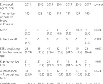
LCN2 expression correlates with organ failure in surgical patients with infection
M Martín-fernández1, R Almansa1, S Montero2, J Almeida Cristo-Barbosa3, A Ortega1, A Hernández Valero3, E Gómez Sánchez4, E Gómez Pesquera4, J Rico-Feijoo2, MC Esteban-Velasco5, JM Calvo-Vecino3, M Vaquero3, C Aldecoa2, E Tamayo4, J Bermejo-Martín1
1Hospital Clínico Universitario de Valladolid/IECSCYL, BioSepsis (Group for Biomedical Research in Sepsis), Valladolid, Spain;2Hospital Universitario Río Hortega, Anesthesiology and Reanimation Service, Valladolid, Spain;3Hospital Universitario de Salamanca, Anesthesiology and Reanimation Service, Salamanca, Spain;4Hospital Clínico Universitario de Valladolid, Anesthesiology and Reanimation Service, Valladolid, Spain;5Hospital Universitario de Salamanca, Department of General and Gastrointestinal Surgery, Salamanca, Spain
Critical Care 2019, 23(Suppl 2):P014
Introduction: The aim of this study is to develop a“molecular equiva-lent” to Sequential Organ Failure Assessment (SOFA) score, which could identify organ failure in an easier, faster and more objective manner, based on the evaluation of Lipocalin-2 (LCN2/NGAL) expression levels by using droplet digital PCR (ddPCR). Sepsis has been classically defined as the exuberant, harmful, pro-inflammatory response to infection. This concept is changing [1] and the presence of a life-threatening organ dysfunction caused by a dysregulated host response to infection is now considered a central event in the pathogenesis of sepsis [2]. Methods: LCN2 expression levels were quantified by ddPCR in blood of a total of 257 surgical patients with a diagnosis of infection. Spear-man analysis was used to evaluate if LCN2 correlated in a significant manner with SOFA score. Area under the receiver operating curve (AUROC) analysis and multivariate regression analysis were employed to test the ability of LCN2 to identify organ failure and mortality risk. Results: Spearman analysis showed that there was a positive, signifi-cant correlation between LCN2 expression levels and SOFA score (Fig. 1). AUROCs analysis showed that LCN2 presents a good diagnos-tic accuracy to detect organ failure and mortality risk (Fig 2). In the multivariate regression analysis, patients showing LCN2 expression levels over the Optimal Operating Points (OOPs) identified in the AUROCs showed a higher risk of developing organ failure (Table 1) and a higher mortality risk (Table 2).
Conclusions: Quantifying LCN2 expression levels by ddPCR is a promising approach to improve organ failure detection and mortality risk in surgical patients with infection.
References
1. Bermejo-Martin JF et al. J Infect 72: 525–536, 2016. 2. Singer M et al. JAMA 315: 801–810, 2016.
Table 1 (abstract P014). Multivariate analysis for evaluating the risk of organ failure based on the LCN2 expression levels. Adjusting variables were [age], [chronic cardiac disease], [cancer], [immunosuppression], [hypertension], [chronic respiratory disease], [chronic renal failure], [respiratory focus], [abdomen focus]
OR Multivariate analysis p
[CI 95%]
LCN2 (copies/ng) Ln 2.20 [1.61-3.02] < 0.001
LCN2 OOP (638 copies/ng) 11.13 [4.59-26.96] < 0.001
Table 2 (abstract P014). Multivariate analysis for evaluating the risk of mortality based on the LCN2 expression levels. Adjusting variables were [age], [chronic renal failure], [diabetes], [respiratory focus]
OR Multivariate analysis p
[CI 95%]
LCN2 (copies/ng) Ln 1.85 [1.36-2.53] < 0.001
LCN2 OOP (3458 copies/ng) 9.03 [3.23-25.21] < 0.001
Fig. 1 (abstract P014). Dot plot showing the correlation between LCN2 expression levels and SOFA score in surgical patients with infection
Fig. 2 (abstract P014). AUROCs for differential diagnosis of organ failure and mortality in surgical patients with infection
P015
Prospective validation of an 18-mRNA score for diagnosis of infection in critically ill patients
TE Sweeney1, AR Moore2, J Roque2, B Shaller2, T Asuni2, JE Levitt3, JG Wilson2, P Khatri4, M Remmel1, D Rawling1, O Liesenfeld1, AJ Rogers2 1
Inflammatix, Burlingame, United States;2Stanford University, Medicine, Palo Alto, United States;3Stanford University, Levitt, Palo Alto, United States;4Stanford University, Immunity, Transplantation and Infections, Palo Alto, United States
Critical Care 2019, 23(Suppl 2):P015
Introduction: There is an urgent need for improved diagnostics for acute infections to help physicians decide whether to treat with anti-biotics. We previously described an 18-host-mRNA diagnostic consist-ing of an 11-mRNA Sepsis MetaScore (SMS) to determine the presence of an infection, and a 7-mRNA bacterial-viral score (BVS) to discriminate between bacterial and viral sources [1,2].
Methods: We collected PAXgene™ RNA blood from 165 patients en-rolled in the Stanford ICU Biobank from 2016-2017. Infection status was adjudicated by Stanford physicians post-hoc using clinical data from the electronic medical record. Low-RNA samples were removed, and target mRNAs were quantitated using NanoString nCounter™ and difference-of-geometric-mean mRNA scores were calculated by Inflammatix [2], blinded to clinical phenotyping. Primary outcome was performance of the two scores in correctly diagnosing physician-adjudicated bacterial or viral infection. Secondary outcome was com-parison to procalcitonin (PCT), which was drawn only according to treating physician preference.
Results: Of 165 patients, physicians adjudicated patients as: 29 non-infected, 102 infected (71 bacterial, 14 viral, 2 fungal, 15 mixed infec-tions), and 33 with uncertain status. The SMS had an AUROC of 0.83 for separating infection of any type from noninfected status. The BVS had an AUROC of 0.95 for separating bacterial from viral infection. Both SMS and BVS were substantially better than PCT across all adju-dicated patients and in matched pairs (PCT AUROC 0.7 for any infec-tion vs noninfected; PCT AUROC 0.82 for bacterial vs. viral; Fig. 1). When used together, the SMS and BVS were able to separate pa-tients based on infection status (Fig. 2).
Conclusions: We prospectively validated an 18-mRNA host-response diagnostic module in a blinded, independent study, confirming high accuracy for the presence and type of infection in a critically ill population.
References
1) Sweeney TE et al, Sci Transl Med, 287ra71, 2015 2) Sweeney TE et al, Sci Transl Med, 346ra91, 2016
P016
Low IgG and IgM levels do not predict mortality in cancer patients with septic shock
G Oliveira, C Park, J Almeida, M Mourão, S Rizk, J Fukushima, L Hajjar Instituto do Cancer do Estado de São Paulo, Intensive care unit, São Paulo, Brazil
Critical Care 2019, 23(Suppl 2):P016
Introduction: Sepsis is an inflammatory state due to an exacerbated immune response against infection. In cancer patients, sepsis pre-sents a 10-fold higher mortality than in general population and leads to longer intensive care unit (ICU) and hospital lengths of stay. It has been shown that reduced levels of circulating immunoglobulins (Ig) might be a surrogate marker of unfavorable outcome in sepsis [1]. The aim of this study was to evaluate the association between Ig levels in plasma and 60-day mortality rate in cancer patients with septic shock.
Methods: From December 2017 to November 2018, we conducted a prospective study in the intensive care unit (ICU) of Cancer In-stitute of State of Sao Paulo, an 84-bed ICU linked to University of Sao Paulo. Patients≥18 years old with cancer and septic shock were enrolled. Descriptive statistics were computed for demo-graphic and outcome variables. Laboratory data and Ig levels were collected at ICU admission and at days 1, 2 and 3. A multi-variate analysis was performed to evaluate predictors of 60-day mortality.
Results: A total of 190 patients were included in the study. The 30-day and 60-day mortality were 40.5% and 45.3%, respectively. No significant differences in IgM and IgG levels were observed between survivors and non-survivors. In both groups, the median IgM levels were low and the median IgG levels were normal. In the multivariate analysis for 60-day mortality, a favorable status performance measured by the Eastern Cooperative Oncology Group (ECOG) was associated with better survival; metastatic dis-ease, higher Sequential Organ Failure Assessment (SOFA) score at admission and higher levels of initial lactate were associated with increased mortality.
Conclusions: Low levels of serum endogenous immunoglobulins are not predictors of 60-day mortality in cancer patients with septic shock.
Reference
1. Bermejo-Martín JF et al. J Intern Med. 276(4):404-12, 2014. Fig. 1 (abstract P015). ROC curves of the infection-diagnostic
Sepsis MetaScore (SMS) and the bacterial-vs-viral BVS score, shown both for all patients, and in head-to-head comparison
with procalcitonin
Fig. 2 (abstract P015). Plotting patients according to transcriptomic scores in two dimensions (x-axis, SMS; y-axis, BVS) shows accurate discrimination by adjudicated infection type
P017
Measurement of plasticity (deformability) of neutrophils and monocytes provides a rapid and early indicator of sepsis R Sheybani1, M Hem1, A Shah1, M Samoszuk1, H Omran1, T Caffery2, C Thomas2, H Tse1, H O’Neal2
1Cytovale, San Francisco, United States;2Louisiana State University Health Sciences Center, Baton Rouge, Louisiana, United States Critical Care 2019, 23(Suppl 2):P017
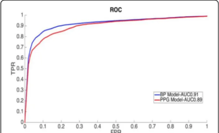
Introduction: Cytovale has developed a rapid biophysical assay of the host immune response which can serve as a rapid and reliable indicator of sepsis. Neutrophils and monocytes undergo characteristic structural and morphologic changes in response to infection. One type of re-sponse is the generation of neutrophil extracellular traps (NETs), these have been proposed as potential mediators for widespread tissue dam-age. During NETosis there is a fundamental reorganization of a cell’s chromatin structure– a signal that we have shown is sensitively mea-sured by the Cytovale cytometer. We hypothesized that quantification of plasticity (deformability) of leukocytes in the peripheral blood pro-vides an early indicator of sepsis. The Cytovale assay uses microfluidic cytometry to measure the plasticity of up to 100,000 white blood cells from EDTA-anticoagulated, peripherally-collected whole blood and pro-vides a result in 5 minutes.
Methods: In two prospective studies conducted in two academic medical centers in Baton Rouge, LA, the Cytovale test was performed on peripheral blood samples obtained from 500 patients who pre-sented to the emergency department with signs or symptoms sug-gestive of infection. The two studies included high acuity patients (400 patient study) and low acuity patients (100 patient study). An adjudicated reference diagnosis of sepsis or no sepsis was estab-lished for each subject, using consensus definitions, by review of the complete medical records.
Results: The Receiver Operator Curve (ROC) performance of the Cyto-vale assay for both studies demonstrated an Area Under the Curve (AUC) greater than 0.85 (Fig. 1).
Conclusions: Measurement of neutrophil and monocyte plasticity by a novel assay provides an accurate and rapid indication of sepsis in patients who present to an emergency room with signs or symptoms of infection.
P018
Plasma hepatocyte growth factor in sepsis and its association with mortality: a prospective observational study
F Peng, C Liang, W Chang, Q Sun, J Xie, H Qiu, Y Yang
Zhongda Hospital, School of Medicine, Southeast University, Department of Critical Care Medicine, Nanjing, China
Critical Care 2019, 23(Suppl 2):P018
Introduction: Sepsis and septic shock are commonly associated with endothelial cell injury. Hepatocyte growth factor (HGF) is a multifunc-tional protein involved in endothelial cell injury and plays a pivotal role in sepsis. This study assesses its correlation with relevant endo-thelial cell injury parameters and prognostic value in patients with sepsis.
Methods: A prospective, observational cohort study was conducted in patients with sepsis admitted to the department of critical care medicine at the Zhongda Hospital from November 2017 to March 2018. The plasma HGF level was collected on the first 24h after ad-mission (day 1) and day 3, then was measured by enzyme-linked im-munosorbent assay. The primary endpoint was defined as all-cause 28-day mortality. Furthermore, we analyzed the correlation of HGF with relevant endothelial cell injury markers.
Results: Eighty-six patients admitted with sepsis were included. HGF levels of non-survivors were elevated upon day 1 (1940.62 ± 74.66pg/mL vs. 1635.61 ± 47.49pg/mL; p = 0.002) and day 3 (1824.82 ± 137.52pg/mL vs. 1309.77 ± 83.49pg/mL; p = 0.001) compared with that in survivors, and showed a strong correlation with von Willeb-rand factor (r = 0.45, p <0.0001), lactate (r = 0.35, p = 0.0011), pul-monary vascular permeability index (r = 0.38, p = 0.0241), first 24 h fluid administration (r = 0.38, p <0.0001) and sequential organ failure assessment score (r = 0.40, p = 0.0001) (Fig. 1). Plasma levels were able to discriminate prognostic significantly on day 1(AUC: 0.72, 95%CI: 0.60-0.84) and day 3 (AUC: 0.77, 95%CI: 0.63-0.91) (Fig. 2). Conclusions: HGF levels are associated with sepsis and are correlated with established markers of endothelial cell injury. Elevated HGF level in sepsis patients is a predictor of mortality.
Fig. 1 (abstract P018). Correlation between HGF levels with markers of endothelial cell injury and SOFA
Fig. 1 (abstract P017). ROC performance of Cytovale test
Fig. 2 (abstract P018). Prognostic value of HGF level for sepsis patients
P019
Calprotectin, a powerful biomarker for the diagnosis of bacterial infections
A Havelka1, P Venge2, A Larsson2 1
Department of Molecular Medicine and Surgery, Karolinska Institute, Stockholm, Sweden;2Department of Medical Sciences, Uppsala University, Uppsala, Sweden
Critical Care 2019, 23(Suppl 2):P019
Introduction: The rapidly growing problem with antibiotic sistance has resulted in demand for more specific and re-stricted use of antibiotics. Biomarkers which can diagnose an infection in early stage and distinguish between bacterial and viral infections could possibly reduce the use of antibiotics. Calprotectin is one of the most abundant proteins in the cyto-sol of neutrophil granulocytes and is released upon activation of neutrophils. The aim of this study was to investigate the performance of calprotectin as a marker for bacterial infection and its possibility to distinguish between bacterial and viral infections.
Methods: The study group consisted of 432 subjects including 144 healthy, noninfected, control patients and 288 patients with confirmed etiology of their infections, 185 patients with bacterial infection, 54 with viral infection, 26 with mycoplasma infection, and 23 with a bacterial infection as a secondary in-fection to influenza. Calprotectin was measured in serum sam-ples with a particle enhanced turbidimetric assay (Gentian AS, Norway). Heparin Binding Protein (HBP) and Procalcitonin were analyzed by sandwich immunoassays (Hycult Biotech and Thermo Fisher Scientific).
Results: Performance of Calprotectin in the diagnosis of bacter-ial infections as well as in distinguishing between bacterbacter-ial and viral infections was compared to performance of Procalci-tonin and HBP. Calprotectin was superior in diagnosis of bac-terial infections as well as in differentiating bacbac-terial from viral infections. Results are presented in Table 1. Interestingly, cal-protectin was the only biomarker with ability to distinguish between mycoplasma and viral infections.
Conclusions: Calprotectin is a promising biomarker for diagno-sis of bacterial infections. Our results indicate that Calprotectin is superior to Procalcitonin and HBP in diagnosis of bacterial infections and in differentiation between bacterial and viral in-fections including mycoplasma inin-fections.
P020
A personalized randomized trial of validation and restoration of immune dysfunction in severe infections and sepsis: PROVIDE N Antonakos1, I Tsangaris2, D Markopoulou3, N Rovina4, A Prekates5, E Antoniadou6, V Theodorou7, A Stefos8, G Vlachogianni9, E Giamarellos-Bourboulis1, G Dimopoulos2, M Netea10
1
National and Kapodistrian University of Athens, 4th Department of Internal Medicine, Athens, Greece;2National and Kapodistrian University of Athens, 2nd Department of Critical Care Medicine, Athens, Greece; 3KAT General Hospital, Intensive Care Unit, Athens, Greece;4National and Kapodistrian University of Athens, Intensive Care Unit 1st Department of Respiratory Medicine, Athens, Greece;5Tzaneion General Hospital, Intensive Care Unit, Piraeus, Greece;6G.Gennimatas General Hospital, Intensive Care Unit, Thessaloniki, Greece;7Dimocriteion University of Thrace, Intensive Care Unit, Alexandroupolis, Greece;8University of Thessaly, Department of Internal Medicine, Larissa, Greece;9Aghios DImitrios General Hospital, Intensive Care Unit, Thessaloniki, Greece; 10Radboud University, Department of Internal Medicine, Nijmegen, Netherlands
Critical Care 2019, 23(Suppl 2):P020
Introduction: Based on the recent post-hoc analysis of previous trials of the efficacy of anakinra in patients with macrophage-like activation syndrome (MALS), PROVIDE (ClinicalTrials.gov registra-tion NCT03332225) is the first double-blind, double-dummy on-going trial aiming to the impact of immunotherapy according to personalized needs.
Methods: Adult patients with septic shock by the Sepsis-3 classifi-cation due to lung infection or primary bacteremia or acute chol-angitis are screened using two consecutive measurements of ferritin and of HLA-DR/CD14 co-expression for MALS (ferritin above 4,420 ng/ml) or immunosuppression (HLA-DR/CD14 less than 30%) and randomized into immunotherapy with either ana-kinra (targeting MALS) or recombinant IFNγ (targeting immuno-suppression) and into placebo treatment. Main exclusion criteria are primary and secondary immunodeficiencies and solid and hematologic malignancies.
Results: 101 patients have been screened so far. Most common in-fections are community-acquired pneumonia (41.6%), hospital-acquired pneumonia (26.7%) and primary bacteremia (12.9%). Mean +/- SD SOFA score is 12.6 +/- 2.9 and Charlson’s comorbidity index 5.21 +/- 2.44; 25 patients have MALS (24.8%); two immunosuppres-sion (2%); the majority remain unclassified for immune state. Conclusions: Current screening suggests greater frequency of MALS than recognized so far in a setting of septic shock due to lung infection or primary bacteremia or acute cholangitis.
P021
Development of an algorithm to predict mortality in patients with sepsis and coagulopathy
D Hoppensteadt1, A Walborn2, M Rondina3, J Fareed1
1Loyola University Medical center, Pathology, Maywood, United States; 2
Loyola University Medical center, Pharmacology, Maywood, United States;3University of Utah and the GRECC, Internal Medicine and the Molecular Medicine Program, Salt Lake City, United States
Critical Care 2019, 23(Suppl 2):P021
Introduction: Sepsis is a systemic response to infection which in-volves inflammation, infection response, hemostatic dysregulation, endothelial dysfunction, and platelet activation. The purpose of this Table 1 (abstract P019). Diagnostic performance of the studied
biomarkers
Biomarker in group comparison AUROC (95% CI) Specificity (%) Sensitivity (%) Healthy vs bacteria
Calprotectin cut-off 1.19 mg/L 0.950 93.0 87.8
HBP cut-off 5.7μgL 0.888 85.4 76.0
PCT cut-off 0.08μg/L 0.883 94.4 78.4
AUROC (95% CI)
Bacteria vs virus Calprotectin HBP PCT
study was to develop an equation incorporating biomarker levels at ICU admission to predict mortality in patients with sepsis, to test the hypothesis that using a combination of biomarkers of multiple sys-tems would improve predictive value.
Methods: Plasma samples were collected from 103 patients with sep-sis at the time of ICU admission. Biomarker levels were measured using commercially available, ELISA methods. Clinical data, including the ISTH DIC score, SOFA score, and APACHE II score were also col-lected. 28-day mortality was used as the primary endpoint. Stepwise linear regression modeling was performed to generate a predictive equation for mortality.
Results: Differences in biomarker levels between survivors were quantified and using the Mann-Whitney test and the area under the receiver operating curve (AUC) was used to describe predictive abil-ity. Significant differences (p<0.05) were observed between survivors and non-survivors for PAI-1 (AUC=0.70), procalcitonin (AUC=0.77), HMGB-1 (AUC=0.67), IL-6 (AUC=0.70), IL-8 (AUC=0.70), protein C (AUC=0.71), Angiopoietin-2 (AUC=0.76), endocan (AUC=0.58), and platelet factor 4 (AUC=0.70). A predictive equation for mortality was generated using stepwise linear regression modeling. This model in-corporated procalcitonin, VEGF, the IL-6:IL-10 ratio, endocan, and PF4, and demonstrated a better predictive value for patient outcome than any individual biomarker (AUC=0.87).
Conclusions: The use of a mathematical modeling approach resulted in the development of a predictive equation for sepsis-associated mortality with performance than any individual biomarker or clinical scoring system. Furthermore, this equation incorporated biomarkers representative of multiple physiological systems that are involved in the pathogenesis of sepsis.
P022
The effects of biomarker clearances as markers of improvement of severity in abdominal septic shock during blood purification T Taniguchi1, K Sato2, M Okajima2
1
Kanazawa University, Anesthesiology and Intensive Care Medicine, Kanazawa, Japan;2Kanazawa University Hospital, Intensive Care Unit, Kanazawa, Japan
Critical Care 2019, 23(Suppl 2):P022
Introduction: Recently the clearances of biomarkers (BM) such as pro-calcitonin (PCT) and presepsin (p-SEP), appear to be better indicators than single cutoff values to diagnose septic complications or predict outcomes. Moreover, blood purification (BF) such as endotoxin absorp-tion therapy (PMX) and continuous renal replacement therapy (CRRT) has been carried out for abdominal septic shock (ASS). However, there are few studies about BM clearances in ASS during BF. Therefore, the present study retrospectively evaluated the effects of BM clearances on improvements of severity in critical patients with ASS during BF. Methods: Thirty-three patients (M/F 21/12, mean age 69 years) were entered. Septic shock was defined in sepsis-3 criteria. PMX was under-gone twice and CRRT was underunder-gone for 5 days. BM levels were mea-sured for 5 days after ICU admitted. Moreover, SOFA scores were measured for 5 days after ICU admitted. BM clearances were deter-mined at the entering ICU and 1, 3, and 5 days after ICU admitted. The improvements of severity were determined the differences of SOFA scores after ICU admitted. Primary outcome is the correlation between BM clearances and the improvement of severity. Secondary outcomes are the changes of BM after ICU admitted, and mortality in ICU. Results: Two of 33 patients died after ICU admission. PCT and lactate levels were improved 5 days after ICU admitted (61.1 to 10.8 ng/mL; p<0.05, 4.2 to 1.1 mmol/L; p<0.05). CRP and p-SEP levels were not improved. SOFA scores decreased (13.4 to 6.5; p<0.05) at 5 days after ICU admitted. There were significantly correlations between PCT and lactate clearances and the improvement of severity (Y=4.02 +0.11X; R2=0.07, p=0.0102, Y=2.75 +0.03X; R2=0.226, p<0.0001). There were not significantly correlations between CRP and p-SEP clearances with the improvement of severity.
Conclusions: The present study showed that PCT and lactate clear-ances significantly correlated the improvement of severity in critical patients with ASS during BF.
P023
Decreased thrombin generation potential is associated with increased thrombin generation markers in sepsis associated coagulopathy
J Fareed1, F Siddiqui1, R Laddu1, D Hoppensteadt1, M Rondina2, E Brailovsky3
1Loyola University Medical Center, Pathology, Maywood, United States; 2
University of Utah School of Medicine, Department of Internal Medicine, Salt Lake City, United States;3Loyola University Medical Center, Cardiology, Maywood, United States
Critical Care 2019, 23(Suppl 2):P023
Introduction: Sepsis associated coagulopathy (SAC) is commonly seen in patients which leads to dysfunctional hemostasis. The pur-pose of this study is to determine the thrombin generation potential of baseline blood samples obtained from SAC patients and demon-strate their relevance to thrombin generation markers.
Methods: Baseline citrated blood samples were prospectively col-lected from 49 patients with SAC at the University of Utah clinic. Citrated normal controls (n=50) were obtained from George King Bio-medical (Overland Park, KS). Thrombin generation studies were car-ried out using a flourogenic substrate method. TAT and F1.2 were measured using ELISA methods (Seimens, Indianapolis, IN). Func-tional antithrombin levels were measured using a chromogenic sub-strate method.
Results: The peak thrombin levels were lower (82 ± 40nM) in the DIC patients in comparison to higher levels observed in the normal plasma (133 ± 10nM). The AUC was lower (561 ± 280) in the DIC group in comparison to the normals (624 ± 18). The DIC group showed much longer lag time (4.1 ± 2.1) in comparison to the nor-mal group (2.1 ± 2.2). Wide variations in the results were observed in these parameters in the DIC group. The F1.2 levels in the DIC group were much higher (570±48 pmol) in comparison to the normal (210 ± 25 pmol). The TAT levels also increased in the DIC group (27.9 ± 5.1 ng/ml) in comparison to the normal (2.8 ± 0.8 ng/ml). The func-tional antithrombin levels were decreased in the DIC group (64 ± 11%).
Conclusions: These results validate that thrombin generation such as F1.2 and TAT are elevated in patients with DIC. However thrombin generation parameters are significantly decreased in this group in comparison to normals. This may be due to the consumption of pro-thrombin due to the activation of the coagulation system. The de-creased functional AT levels observed in the DIC group are due to the formation of the complex between generated thrombin and antithrombin.
P024
Relationship of markers of inflammation, infection, and endothelial function to mortality and severity of coagulopathy in patients with sepsis-associated DIC
D Hoppensteadt1, A Walborn2, M Rondina3, J Fareed1 1
Loyola University Medical center, Pathology, Maywood, United States; 2Loyola University Medical center, Pharmacology, Maywood, United States;3University of Utah School of Medicine, Internal medicine, Salt Lake City, United States
Critical Care 2019, 23(Suppl 2):P024
Introduction: Sepsis-associated disseminated intravascular coagula-tion (DIC) is a complex clinical scenario involving derangement of many processes, including hemostasis. Assessment of markers includ-ing inflammation, endothelial function, and endogenous anticoagu-lants may provide insight into DIC pathophysiology and lead to improved methods for assessment of patient condition and response to treatment.
Methods: Citrated plasma samples were collected from 102 patients with sepsis and suspected DIC at ICU admission and on days 4 and 8. DIC score was determined using the ISTH scoring algorithm (e.g. platelet count, PT/INR, fibrinogen and D-Dimer). CD 40 Ligand (CD40L), Plasminogen inhibitor 1 (PAI-1), nucleosomes, Procalcitonin (PCT), Microparticle tissue factor (MP-TF) and Prothrombin 1.2 (F1.2)
were measured using commercially available ELISA kits. Protein C ac-tivity was measured using a clot-based assay. Interleukin 6 (IL-6), Interleukin 8 (IL-8), Interleukin 10 (IL-10), Tumor necrosis factor alpha (TNFα), and Monocyte chemoattractant protein (MCP-1) were mea-sured using biochip technology.
Results: Significant differences in levels of Protein C (p=0.009), PCT (p=0.0005), IL-6 (p=0.019), IL-8 (p=0.0149), PAI-1 (p=0.015), were ob-served between survivors and non-survivors. Significant variation of Protein C (p=0.002), nucleosomes (p=0.05), PCT (p<0.0001), IL-6 (p=0.001), IL-8 (p=0.003), IL-10 (p=0.011), TNFα (p=0.021) and MCP-1 (p=0.021) were observed based on severity of DIC score.
Conclusions: Markers from multiple systems perturbed in DIC were associated with mortality, suggesting that while these systems may not be routinely evaluated in the normal course of patient care, dys-function of these systems contributes significantly to mortality. In addition, numerous inflammatory cytokines showed an association with DIC score. This suggests that the measurement of additional markers in sepsis-associated DIC may be of value in the prediction of mortality and may be helpful in guiding treatment for these patients.
P025
Usefulness of plasminogen activator inhibitor-1 (PAI-1) as a predictive marker for identification of sepsis-induced DIC J Maruyama, K Hoshino, Y Irie, S Miyagawa, R Hokama, M Koie, M Nakashio, Y Kawano, T Kitamura, H Ishikura
Fukuoka University Hospital, Department of Emergency and Critical Care Medicine, Fukuoka, Japan
Critical Care 2019, 23(Suppl 2):P025
Introduction: There is a crosstalk between inflammation and coagu-lation and disseminated intravascular coagucoagu-lation (DIC) especially sepsis-induced DIC is a one of the most significant causes of mortal-ity in intensive care units. Meanwhile, there are various types of DIC and sepsis-induced DIC is a type of suppressed fibrinolysis DIC. In this study, we aimed to identify coagulation/fibrinolysis markers useful for discriminating whether sepsis-induced DIC or not.
Methods: This is a single-center retrospective observational study of 233 patients with DIC according to the Japanese Association for Acute Medicine (JAAM) DIC criteria (JAAM DIC score≥4) from July 2017 to June 2018. We divided DIC patients into sepsis and non-sepsis using Sepsis-3 diagnosed criteria and univariate and multivari-ate logistic regression analyses were performed to identify an
inde-pendent predictive marker of sepsis-induced DIC among
coagulation/fibrinolysis markers on ICU admission.
Results: Sepsis-induced DIC (S-DIC) group (n=62) was significantly higher DIC score and SOFA score rather than non-sepsis-induced DIC (NS-DIC) group (n=171) [DIC score; 5 (5-7) vs. 4 (4-5), P<0.01. SOFA score; 11 (9-14) vs. 6 (4-10), P<0.01.].
About coagulation/fibrinolysis markers, S-DIC group was significantly lower the FDP, D-dimer, and PIC rather than NS-DIC group (P<0.01), and higher the PAI-1 (P<0.01). Moreover, PAI-1 was identified as one of the independent predictive markers of sepsis-induced DIC by multivariate logistic regression.
Conclusions: Recently, we reported that PAI-1 was a useful predictive marker of mortality in sepsis. From this study we suspected that PAI-1 is a useful marker for discriminating sepsis-induced DIC. Further-more, we confirmed that sepsis-induced DIC was a type of DIC with suppressed fibrinolysis. Therefore, we recommend measuring PAI-1 against sepsis and sepsis-induced DIC patients.
P026
Does EAA value reflect severity of condition in patients admitted to intensive care unit?
T Ikeda1, S Ono1, S Suda1, T Nagura1, M Tomino2, M Sugi2, Z Wajima2 1
Tokyo Medical University, Hachioji Medical University, Division of Critical Care Medicine, Tokyo, Japan;2Tokyo Medical University, Hachioji Medical University, Department of Anesthesiology, Tokyo, Japan
Critical Care 2019, 23(Suppl 2):P026
Introduction: The Endotoxin Activity Assay (EAA) is a rapid immuno-diagnostic test based on chemiluminescence. It was approved by the FDA in 2003 as a diagnostic reagent for risk assessment of severe sepsis in the ICU. Ascertaining endotoxin levels in the bloodstream is important in targeting patients and determining the appropriate tim-ing for initiation of treatment. It has high sensitivity and specificity for endotoxin, and is considered to be useful in predicting clinical symptoms and determining prognosis. The usefulness of the EAA has yet to be fully clarified.
Methods: A total of 142 patients admitted to the ICU between Janu-ary 2014 and June 2018 with suspected sepsis or sepsis were en-rolled. The EAA was conducted within 24 hr after admission. Patient characteristics were determined, together with levels of IL-6, procalci-tonin, presepsin, and PaO2/FiO2. Thereafter, the patients were classi-fied into 5 groups depending on their EAA value: 1) < 0.2; 2) from≤ 0.2 to < 0.4; 3) from≤ 0.4 to < 0.6; 4) from ≤ 0.6 to < 0.9; and 5) ≤0.9). The transition of various markers was also examined. The Spearman rank correlation, Wilcoxon rank sum test, and a non-repeated ANOVA were used for the statistical analysis. A P-value of < 0.05 was considered statistically significant.
Results: The EAA values showed a positive correlation with both the APACHE II (r=0.48) and SOFA scores (r=0.56)(P<0.01), although that with the latter was stronger. A significant correlation was also observed with levels of procalcitonin (r=0.45) and presepsin (r=0.51). The EAA showed a high value (P<0.01) only in patients showing a positive result for blood culture; no other marker showed a significant difference between those showing a positive or negative result. At 28 days, significantly higher values were observed in the non-survival group (P<0.05) in values for the EAA and other markers.
Conclusions: EAA value tended to correlate with disease severity (APACHEII and SOFA scores) in patients admitted to the ICU.
P027
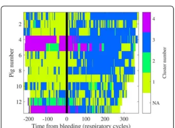
Intraperitoneal microdialysis detects early peritonitis caused by intraabdominal bacterial infection in a pig model
S Hødnebø, S Pischke, A Barratt-Due, E Lindholm, T Tønnessen Oslo University Hospital, Division of Emergencies and Intensive Care, Cardiothoracic clinic, Oslo, Norway
Critical Care 2019, 23(Suppl 2):P027
Introduction: Common complications following abdominal sur-gery are intestinal leaks, with subsequent abdominal sepsis. Early diagnosis is important to allow early intervention. The current clinical methods are insufficient for early detection. We hypothesized that intraperitoneal microdialysis allows detection of peritonitis prior to changes in standard clinical parameters in a pig model.
Methods: Bacterial peritonitis was induced in 5 pigs by bowel perforation and intraperitoneal fecal instillation, one pig under-went sham surgery. Intraperitoneal microdialysis catheters were placed in each abdominal quadrant. The observation time was 10 hours.
Results: In peritonitis pigs the intraperitoneal lactate increased during the first two hours and remained elevated throughout the observation time (Table 1), whereas the arterial lactate remained within reference range (<1.6 mM). Intraperitoneal glucose de-creased significantly. Hemodynamics were hardly influenced dur-ing the first two hours, and decreased thereafter. Sham surgery did not influence in any of the parameters.
Conclusions: A rapid and pronounced increase in intraperitoneal lactate and decrease in intraperitoneal glucose was observed after instillation of intraabdominal feces. Systemic lactate increase was absent, and the hemodynamic response was delayed. Post-operative intraperitoneal microdialysis is applicable in detecting peritonitis earlier than standard clinical monitoring and should be evaluated in a clinical study in order to explore if early interven-tion based on MD data will reduce ICU length of stay, morbidity and mortality.