1
UNIVERSITE DE MONTPELLIER
FACULTE DE MEDECINE MONTPELLIER-NIMES
THESE
Pour obtenir le titre de
DOCTEUR EN MEDECINE
Présentée et soutenue publiquement
Par
Léo-Paul SECCO
le 14 septembre 2020
TITRE
Lésions cutanées vasculaires acrales au cours de la pandémie Covid-19 :
caractéristiques cliniques, anatomopathologiques, immunologiques et
virologiques
Directeur de thèse : Professeur Didier Bessis
JURY
Président :
Professeur Thierry Vincent
Assesseurs :
Professeur Didier Bessis Docteur Edouard Tuaillon Docteur Luc Durand
3
Remerciements
Il ne me semble point trivial de concéder tout d'abord ce travail à ma famille, et en particulier mes parents, qui ont toujours su, avec un élégant altruisme, une clémente fermeté, et en se gardant de toute influence passionnée envers moi, à la fois m'abandonner et guider la maîtrise de mes choix, parfois contre les mœurs. De cette libération d'esprit, j'ai pu en tirer la plus grande sagacité. Autrement dit, je les remercie pour avoir fait de la proposition suivante mon fil conducteur : "les hommes se trompent quand ils se croient libres ; cette opinion consiste en cela seul qu'ils sont conscients de leurs actions et ignorants des causes par lesquelles ils sont déterminés". Par là-même, je suis reconnaissant de mes professeurs qui m'ont transmis les clés de cette démonstration par l'amour et la science, et qui n'ont eu de cesse de me prouver qu'il n'existait pas de science sans littérature, tout comme "Deus sive Natura". De merveilleuses applications de ces démonstrations ont été bâties avec mes amours, mes amis, mes camarades d'études, mes collègues de travail. Je m'émeus de la singularité de ces infinis souvenirs avec eux, qui par leur beauté, pallient à mon esprit parfois rattrapé par ses passions faibles et mon humeur taciturne. Ma mère le voulut ainsi : Léo, pour la force ; Paul, pour la faiblesse.
Bien sûr, je dédie entièrement la conception, l'élaboration et la réflexion de ce travail passionnant à Didier. Je suis vivement reconnaissant de la qualité, du dévouement et des brillantes idées d'Edouard. De même, mon cher Luc m'a tant appris et aidé dans ce travail. Enfin, je suis honoré de soumettre ce manuscrit au jugement de Thierry Vincent qui y a porté un intérêt certain, et que j'estime grandement.
Il va sans dire que mes remerciements suivants se dirigent à l'ensemble des patients qui sont au cœur même de cette étude. Je suis pleinement admiratif de leur disponibilité et de leur temps qu'ils ont pu m'accorder.
Ma reconnaissance éternelle à tous mes collègues et aux équipes qui ont participé à cette aventure humaine et administrative.
Je remercie mes deux amis et correcteurs, qui ont empreint de leur amour le travail scientifique suivant.
17
Sommaire
Introduction 18
Prolegomena 20
Background 22
Material and methods 24
Results 27 Discussion 30 Bibliographie 34 Annexes 40 Commentaire 74 Serment 76
Introduction
Les acrosyndromes vasculaires constituent un groupe d’anomalies vasculaires des extrémités, la plupart du temps les mains ou les pieds. Ces anomalies, permanentes ou transitoires, peuvent être déclenchées par plusieurs facteurs comme le froid.
Les engelures constituent un acrosyndrome vasculaire fréquent chez les sujets jeunes, observées pendant les mois froids et humides où la température moyenne journalière n’excède pas douze degrés. Elles se présentent typiquement par des papules oedémateuses, prurigineuses et peu douloureuses, érythémateuses et cyaniques de la face latérale des doigts et des orteils. Ces lésions persistent au moins une journée et guérissent spontanément en quelques semaines, mais tendent à récidiver les années ultérieures. Un autre acrosyndrome est souvent associé aux engelures. Bien que banales, le mécanisme des engelures est mal compris, d’où le nom d’engelure idiopathique. Les hypothèses physiopathologiques reposent sur l’existence d’un vasospasme anormal, une instabilité du tonus neurovasculaire et de la thermorégulation. Une hypoxie locale secondaire à ces phénomènes provoquerait une réaction inflammatoire secondaire. Parfois, les engelures sont liées à une maladie auto-immune sous-jacente, une néoplasie, ou une infection virale.
Le SARS-CoV-2 (Severe Acute Respiratory Syndrome-Coronavirus-2) isolé pour la première fois en Chine en 2019, typiquement responsable de pneumopathies sévères, s’est progressivement diffusé à l’échelle mondiale, donnant lieu à la pandémie de Covid-19 (Coronavirus disease-19). Plusieurs organes peuvent également être lésés par le biais notamment d’une atteinte vasculaire.
Une grande variété de manifestations cutanées a été décrite dans la littérature au cours de cette pandémie : éruption varioliforme, urticaire diffuse, livédos, éruptions cutanées non spécifiques, et des lésions de pseudo-engelures.
Ces lésions de pseudo-engelures ont été rapportées par des cliniciens de pays différents à partir du début du mois d’avril 2020. L’hypothèse d’un lien entre ce type de manifestation cutanée et l’infection à SARS-CoV-2 a été émise sur plusieurs arguments : caractéristiques cliniques différentes des engelures idiopathiques, absence d’exposition au froid du fait de mesures sanitaires restreignant les déplacements en extérieur de populations et de leur survenue à une période de l’année inhabituelle,
19 symptômes infectieux suggérant une infection à SARS-CoV-2 dans les jours ou semaines précédant les symptômes cutanés.
Par ailleurs, les interféronopathies de type I récemment individualisées comme le lupus-engelures familial ou le SAVI (STING-associated vasculopathy with onset on infancy) sont caractérisées par une production excessive d’interféron de type I, secondaire à une mutation d’un gène codant pour une protéine impliquée dans cette voie de signalisation. Cette production anormale d’interféron peut être détectée par la mesure du score interféron. Ce groupe de maladies présente des caractéristiques sémiologiques analogues à celles des lésions à type d’engelures rapportées pendant la pandémie de Covid-19. Notamment, le SAVI associe des lésions cutanées acrales et pulmonaires, avec des stigmates biologiques d’auto-immunité.
Or, l’on sait que l’un des mécanismes de l’immunité antivirale repose sur la voie de l’interféron de type I, dont la sécrétion est augmentée en cas d’infection virale.
L’hypothèse de cet exposé est que les lésions cutanées à type d’engelure rapportées dans la littérature au cours de la pandémie à Covid-19 sont des lésions singulières et originales, qui présentent des similitudes cliniques avec les interféronopathies de type I car elles sont en lien avec une infection à SARS-CoV-2, qui induit une activation de la voie de l’interféron. Les caractéristiques cliniques, histologiques, immunologiques et virologiques de trente patients ont été étudiées prospectivement afin d’étayer cette hypothèse.
Prolegomena
SARS-CoV-2 (Severe Acute Respiratory Syndrome Coronavirus 2) is part of the family-coronavirus, mainly reported with single-strand RNA (Ribonucleic Acid), nucleocapsid protein (N), envelop protein (E), membrane protein (M), spike (S) glycoprotein1. S protein forms the crown-like structure on the outer surface of the virus, and attaches to the human angiotensin converting enzyme-2 (hACE2) receptor with high affinity. The hACE2 receptor is expressed by many epithelial cells, particularly type-2 pneumocytes, by which SARS-CoV-2 enters into the host cell2,3,4. Uncoating and release of genomic RNA in the cytoplasm of host cell is followed by replication, transcription, assembling processes. Virions are finally released either by fusing with the plasma membrane or by the mechanism of exocytosis.
Pathogenesis behind SARS-CoV-2 is not fully understood, but several studies accordingly find the virus can: (i) downregulate ACE2 receptors expression and increase production of angiotensin-2 (AT2) in the lung, leading to pulmonary vascular permeability and lung injury5 ; (ii) escape to antiviral response by limiting type I and type III interferon (IFN-I and IFN-III) pathways6,7 ; (iii) elicit a strong chemotactic and inflammatory response called “cytokine storm syndrome”8, and activate complement pathway, both promoting coagulopathy9. Taken together, these features can lead in some genetically predisposed individuals with comorbidites such as diabetes, liver failure, kidney failure, to acute respiratory distress syndrome (SARS), disseminated intravascular coagulation, and multiorgan failure, with subsequent morbidity and mortality10.
Clinical symptoms of SARS-CoV-2 infection onset are about a week after contamination, but differ from age and immune system of the person11. Among these symptoms, the most commons are fever, cough, fatigue, sputum. Sometimes, patients describe headache, muscle weakness, breathlessness, sore throat, anosmia, agueusia. Many other organs may be involved, secondary to cytokine storm syndrome. On the other hand, many people who develop SARS-CoV-2 infection remain asymptomatic.
Diagnosis at the beginning of the symptoms can be performed by nucleic acid amplification test through a nasal swab using a real-time fluorescence polymerase chain reaction (RT-PCR)12. However, if the viral load in the nasal swab is low, sensitivity decreases. Therefore, when high suspicion of SARS-CoV-2 infection despite negative RT-PCR performed on nasopharyngeal swab, computed-tomography (CT) imaging is
21 recommended in patients with moderate-to-severe clinical features13. Indeed, chest CT can show multiple ground-glass opacities in the lungs, early in coronavirus disease (Covid-19), since the first days of the respiratory symptoms14. Later, to assess previous symptoms were linked to Covid-19 if diagnostic testing were not performed or negative, many serological tests have been developed, and some validated by sanitary authorities15. They have high sensitivity (96%) and specificity (98%) when performed 21 to 35 days after the onset of infectious symptoms suggesting Covid-19, but there were no studies exclusively in asymptomatic patients16.
Treatment of SARS-CoV-2 infection remains challenging17. On one hand, pathophysiology remains unclear, and each individual develop a specific clinical course, depending on the immune system of the individual and the timing of immune response against the virus. On the other hand, no specific medical therapy or vaccine is available. Some treatments yet available have been studied (remdesivir, chloroquine, hydroxychloroquine, protease inhibitors) with disparate results, depending on the timing of the drug administration and the severities of the cases of Covid-19.
Regarding on the lack of efficacy treatment and the very high transmission between humans, preventive measures against transmission of the SARS-CoV-2 in order to prevent his spreading have been proposed by the World Health Organization (WHO)18: social distancing, masks, regular cleaning of hands. Such measures were mandatory in many countries over the world, because health care systems were overwhelmed and subsequent Covid-19 pandemic a public health problem.
Background
During Covid-19 pandemic, many dermatological manifestations have been reported, but their link with the SARS-CoV-2 is unclear, because of the failing of demonstrating markers of SARS-CoV-2 infection. Some authors have proposed different classifications of the cutaneous signs, based on semiology. One of the most described is “chilblain-like lesions”, also called “pseudochilblains”18, or “Covid toes”19, with more than 1200 patients (Table 1).
Chilblain-like lesions concerned young people (mean age: 19 years-old), with few or no past medical history nor drug intake. Nine per cent reported history of chilblains and Raynaud’s phenomenon. Dermatological presentation was quite homogeneous, with erythematous macules and papules, sometimes purpuric or edematous. Lesions were located on the feet (82,3%), hands (17,7%), or both (12,6%). Associated symptoms were pruritus (38,6%), burning (61,9%), pain (63,7%). The mean duration of the skin symptoms was 18,3 days, and healed spontaneously without any treatment in the majority of the cases. 41% of the cases reported infectious symptoms suggesting SARS-CoV-2 infection. The mean time between the onset of infectious symptoms and the onset of the skin lesions was 8 days. A close contact with a person who had proven Covid-19 was reported in 8,7% of the cases. Majority of the patients were not admitted in hospital and mortality rate was low. Pathological examination on skin biopsy was done in 9,3%, showing superficial and deep lymphocytic infiltrate with purpura (79,6%), papillary edema (72,6%), and vascular abnormalities like endothelial cell swelling (77,1%) or thrombi (21,8%). Direct immunofluorescence on skin biopsy was performed in 47 cases and showed vascular C3 deposits in 22 cases. Virological testing noted positive RT-PCR for SARS-CoV-2 performed on nasopharyngeal swab in 3,6% of the cases, and positive serology in 6,3% of the cases. Abnormal blood testing results were: D-dimer elevation (5,2%), low fibrinogen concentration in 3 cases, positive antinuclear antibodies in 8 cases, and one case with weak level of anticardiolipin immunoglobulin (Ig) G antibody. No perturbation of classic complement pathway, liver enzymes, serum creatinine, lactate deshydrogenase was observed.
Some authors proposed that chilblain-like lesions during Covid-19 pandemic were secondary to a robust IFN-I response20. Moreover, this hypothesis is supported by clinical similarities with type I interferonopathies, a group of Mendelian disorders characterized by an up-regulation of type I interferon21. Indeed, IFN-I is a crucial
23 pathway for antiviral immunity22. The physiological response to virus infection is generally initiated at the cellular level following replication. After the virus entry, the infected cells detect the presence of the virus replication through any one of a number of pattern recognition receptors (PRRs). PRRs sense aberrant RNA structures of the virus, and downstream interferon regulator factors (IRFs) and nuclear factor kB (NF-kB). These results in the launch of two general antiviral programs: (i) induction of type I and III interferon and subsequent upregulation of IFN-stimulated genes (ISGs); (ii) recruitment of leukocytes and chemokine secretion12. Evaluation of these ISGs in the blood is possible with a validated ISG score, based on the mean expression of six ISGs defining type I interferon signature23. The SARS-CoV-2 is sensitive to IFN, but develops several mechanisms to evade from IFN-I pathway, consistent with reduced ISGs expression, low plasma level of interferon alpha-2 (IFN-a2) in critical Covid-19 patients24. Conversely, critical patients have high cytokine and chemokine-related genes expression, with the induction of a unique response to IFN-II6.
Diagnosis of SARS-CoV-2 infection may be challenging because of the lack of sensibility of RT-PCR and serological blood testing, despite clinical symptoms consistent with Covid-19. Therefore, searching for mucous antibodies may increase sensibility. Some patients with conventional RT-PCR performed on nasopharyngeal swab and blood serological testing, symptomatic or not, showed antibodies against SARS-CoV-2 in salivary, nasal fluid of tears25,26,27.
Material and methods
Study design and patients
The study design is a monocentric, descriptive, prospective study of patients presenting vascular acral symptoms or lesions during the Covid-19 pandemic. A total of 30 patients were enrolled in this study. Patients were screened and recruited from April to July 2020 at the Department of Dermatology, in Montpellier Hospital Center, France. A dermatologist diagnosed vascular acral injury. Oral and written informed consent was given to each patient or their legal guardians for children. The Institutional Review Board and Center of Biological Resources of Montpellier University Hospital both approved this study.
Clinical evaluation
A standardized case-report form was created, collecting data on demographic characteristics of the patients, medical history including personal and familial autoimmune diseases, medications, infectious symptoms that may suggest a SARS-CoV-2 infection in the past weeks or months.
Clinical characteristics of cutaneous symptoms and lesions were dated and described using elementary lesions, topography, and subjective functional signs (pruritus, pain, burning).
Photographs of cutaneous lesions were taken for each patient at the end of the clinical evaluation, with written signed consent.
Study procedures
After clinical evaluation, patients included in the study underwent: (i) testing for SARS-CoV-2 on nasopharyngeal swab and saliva collection, (ii) skin biopsy in 26 patients, (iii) blood testing for hematological parameters, SARS-CoV-2 serology, and serum interferon level.
Pathological analyses
Two pathologists read hematoxylin-eosin-stained, Periodic Acid Schiff and Alcian Blue sections of formalin-fixed paraffin-embedded skin biopsies of 26 patients. Histopathological features assessed were: (i) epidermal changes (spongiosis, necrotic keratinocytes, basal vacuolar changes, thickening of the basement membrane,
25 parakeratosis), (ii) papillary edema, (iii) presence of lymphocytic infiltrate and its characteristics (density, lichenoid pattern, superficial or deep location in the dermis), (iv) existence of vessel alterations (fibrin deposition, vascular ectasia, thrombi, mural lymphocytes, thickening of the vessel wall, endothelial cell swelling), (v) extravasation of erythrocytes, (vi) mucin deposition pattern (dermal or around eccrine sweat glands). Direct immunofluorescence on frozen skin biopsies was performed in six patients using human anti-IgG, anti-IgM, anti-IgA, anti-C3c antibodies.
The study of the expression of plasmacytoid dendritic cells was carried out using the anti-CD123 antibody. This immunohistochemistry was performed on skin biopsies using Ventana BenchMark Ultra, with a 3,3'-diaminobenzidine chromogen.
An anti-SARS-Cov Nucleoprotein IgG rabbit antibody (Sino Biological) assessed immunostains for SARS-Cov-2, with a 1:1000 concentration on paraffin-embedded tissue. This immunohistochemistry was performed on skin biopsies using Ventana BenchMark Ultra, with a 3,3'-diaminobenzidine chromogen and OptiView Amplification Kit to increase the staining intensity.
Laboratory testing
Real-time reverse transcriptase polymerase chain reaction (RT-PCR) against SARS-CoV-2 was performed on nasopharyngeal swab in 23 patients, and on skin lesions in 2 patients, using a validated test by the National Reference Center of viruses of respiratory infections in France.
Several methods assessed blood testing for SARS-CoV-2 serology: (i) semi-quantitative indirect enzyme-linked immunosorbent assay (ELISA) targeting IgG antibodies directed against SARS-CoV-2 nucleocapsid (ID Screen, IDvet, Montpellier, France), (ii) semi-quantitative ELISA targeting IgA antibodies of SARS-CoV-2 (IDvet), (iii) chemiluminescent microparticle immunoassay (CMIA) intended for the quantitative detection of IgG antibodies directed against the nucleocapsid protein of SARS-CoV-2 (Abbott Laboratories, USA), (iii) enzyme immunoassay (EIA) intended for the qualitative detection of total antibodies (IgG/IgA/IgM) to SARS-CoV-2 in human serum (IMMY, USA), (iv) ELISA for the determination of IgA against SARS-CoV-2 using the S1 domain of the spike protein (Euroimmun, Germany).
Patients also underwent investigations to screen for autoimmune diseases or coagulopathy: blood cell count, C-reactive protein, serum creatinine, cryoglobulins, cryofibrinogen, cold agglutinins, antineutrophil cytoplasm antibodies (ANCA), antinuclear, anti-desoxyribonucleic acid (DNA), ribonucleic protein, myositis,
antiphospholipid, anticardiolipin antibodies, rheumatoid factor, serum protein electrophoresis, D-dimers, prothrombin time, fibrinogen, activated partial thromboplastin time.
Interferon signature
Interferon score assessment was performed by Immunology Laboratory in Hospices Civils de Lyon, France. RNA was extracted from whole blood contained in Paxgene tubes (Kit PreAnalytix, Qiagen, SW) and quantified by spectrophotometry assay (Nanodrop 2000, Thermo Scientific, MA, USA). RNA integrity was then evaluated by Agilent RNA microarray (Agilent Technologies, Santa Clara, CA, USA). mRNA quantification of 6 ISGs (interferon alpha inducible protein 27 (IFI27), interferon induced protein 44 like (IFI44L), Interferon Induced Protein With Tetratricopeptide Repeats 1 (IFIT1), ISG15 Ubiquitin Like Modifier (ISG15), Radical-S-Adenosyl Methionine Domain Containing 2 (RSAD2), Sialic Acid Binding Ig Like Lectin 1 (SIGLEC1) and three housekeeping genes (Actin Beta (ACTB), Hypoxanthine Phosphoribosyltransferase 1 (HPRT1), RNA Polymerase II Subunit A (POLR2A)), was performed using nanostring technology. Data standardization was obtained using the geometric mean of internal control and housekeeping genes count number. The relative expression was determined for each normalized ISG expression divided by the median normalized expression of each ISG from a control group. The median of these 6 ISG relative expression was used to calculate the IFN score. The threshold was defined as the mean of control group IFN score + 2 standard derivations corresponding to a score of 2,3 (area under the curve of the ROC curve of 0,856; sensitivity of 89,4%, negative predictive value 95,9%; specificity 72,0%).
27
Results
Clinical characteristics
Fourteen males and sixteen females were studied (Tables 2 and S2). 10/30 reported previous acrosyndrome history, either chilblain, acrocyanosis or acrorhigosis. 24/30 presented infectious symptoms suggesting a SARS-CoV-2 infection. These symptoms occurred before or at the same time than the onset of skin lesions in all cases. Skin lesions could affect hands, feet or both, but feet were more involved than hands. Although most of the patients presented at consultation with macules and papules resembling chilblains (Figures 1 and 5), it did not represent the predominant clinical feature in several cases. Indeed, some patients only developed acrosyndrome (Figures 2a and 2b), without permanent lesion, such as Raynaud’s phenomenon (7/30) or acrocyanosis (12/30). Permanent or transient skin lesions were associated with symptoms in 24/30 cases.
Finally, two different patterns of skin lesions were observed: (i) paroxysmal vasomotor signs, and (ii) permanent vascular lesions. Both were associated in some patients.
Raynaud’s phenomenon involved parts of fingers or toes, without cold triggering (Figure 2b). Ears, nose and nipples were not affected. Associated symptoms were not significant. The cyanic and erythematous phases were absent. Acrocyanosis was reported as de novo, or worsening. It was reinforced in the periungueal nail fold. In some patients, wrists or ankles were affected. No patient described associated pain. Cyanic erythema was a vascular erythema, livid, not infiltrated or purpuric, but sometimes with a skin coloration time up to twenty seconds. When standing up, typical semiological presentation of BASCULE syndrome was observed, but without any ichty or feeling of heavy legs complain (Figure 3).
On the other hand, chilblain-like lesions could develop on normal, cyanic, erythematous or livid skin. Vesiculo-bullous lesions were situated on the dorsal or lateral parts of the fingers or the toes, and in two patients on the ankle. One patient displayed maculo-papules and nodules of the limbs associated with chilblain-like lesions. Three unusual features of chilblains were noted: (i) necrotic evolution, sometimes extensive (Figure 4), (ii) purpuric areas on the palmar face of the toes, and (iii) duration of the lesions (at least one month for all cases, more than 4 months for 6 patients) (Table S1).
Pathological features
Standard pathological examination exhibited chilblain-like features in 23 cases (Table 3 and S3): (i) few epidermal changes apart from some isolated necrotic keratinocytes and mild spongiosis, (ii) superficial and deep lymphocytic infiltrate around vessels and eccrine glands (Figure 6), (iii) perieccrine mucin deposition. Sometimes, a lichenoid pattern of the lymphocytic infiltrated was observed. Dermal papillar edema was mild or absent, except for some patients it was prominent. Three patients did not show similar findings: two showed only nonspecific telangiectatic vessels with a slight lymphocytic infiltrate around them and perieccrine mucin deposits insufficient to conclude to a chilblain-like aspect, maybe because of the time between the onset of skin symptoms and skin biopsy performing (37 days and 3 months). The third patient had a too shallow skin biopsy.
Two features were unusual for chilblain-like pattern: (i) vascular damage stigmata in several cases like nuclear dusting (15/26), endothelial cell swelling (16/26), thrombi (10/26), purpura (16/26) (Figure 7) and (ii) vascular deposits of C3 inside the vessel walls in 4/8 cases on direct skin immunofluorescence. However, frank necrosis in the lymphocytic infiltrate was never observed.
Study of CD123 pattern expression within the lymphocytic infiltrate showed a few up to a moderate number of marked cells, without any particular disposition (Figure 8). However, all of the 21 tested cases displayed at least few positive CD123 cells.
Anti-SARS-CoV antibody showed endothelial and epithelial sweat glands discrete microgranular cytoplasmic staining in 18/21 cases (Figure 9). In two cases, it was not possible to conclude due to the exhaustion of the biopsy material. The last case remained negative.
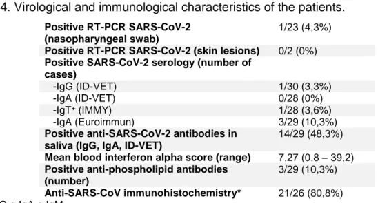
SARS-CoV-2 testing results
Virological testing hardly highlights viral particles of SARS-CoV-2 (Table 4 and S4), with 0/2 positive RT-PCR performed on skin lesions, and 1/23 RT-PCR performed on nasopharyngeal swab. In the same way, SARS-CoV-2 serology remains negative for all patients with IgG, except for the only patient with positive RT-PCR (Table 5). Notably, the patient with positive RT-PCR during infectious symptoms on March 30th 2020 had a first positive serology 25 days after the first infectious symptoms, and subsequent negative serologies. Three patients had transient blood IgA against SARS-CoV-2. Otherwise, an increased number of patients presented positive immunoglobulins
29 (IgA) against SARS-CoV-2 in saliva samples (14/29) (Table 6). Over an interval of two months, among the patients who had two saliva samples, one patient had an IgA antibody level above the detection threshold at the first sample and below for the second one, whereas a positivity of the second sample was observed for four patients who had a previous negative saliva sample two months earlier.
Immunological findings
Relevant immunological data on standard blood testings were (Table 7 and S5): (i) mild cytopenias, lymphopenia (3/29), thrombopenia (2/29), and neutropenia (3/29), (ii) elevated D-dimer (> 500 ng/mL) for 2/29 patients, (iii) C3 diminution in 1 patients, (iv) positive antinuclear antibodies in 12/28 cases, without any specificity, and significant values in 4 patients.
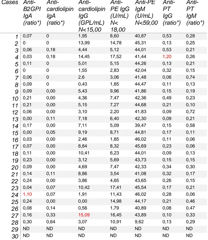
Antimyositis antibodies were performed on 26 patients and show only weak positivity for anti-NXP2 for one patient, and remained negative on a second blood testing. Anti-DNA antibodies, rheumatoid factor were negative. Hemostasis evaluation showed 2/29 cases with elevated activated partial thromboplastin time, one case with positive antiprothrombin IgG antibodies, one case with positive anti-beta-2-glycoprotein-1 IgA antibodies, and one patient with anticardiolipin IgG antibodies (Table 8).
Concerning the type I interferon pathway study (Table S1 and S4), 17/27 patients presented at least one increased interferon score value (threshold: 2,3), some with high ones (> 10 for 3/27 patients). Kinetics of the values if checking was performed showed either increased or decreased values: case number 3 with a 13,5 initial interferon score increased at 24,9 22 days later and 32,9 44 days after the first blood testing; on the other hand case number 6 show reverse biological pattern with initial interferon score at 39,2, then 6,5 eleven days after, and finally 2,5 forty-six days after the first one. 10/27 patients did not had an interferon signature in the blood. 1/3 cases had differential interferon score between lesional and non lesional skin on biopsy. These biopsies were performed: one month after the first skin symptoms for the patient with differential interferon scoring; 14 and 5 days after the first skin symptoms for the two others.
Discussion
The detailed analysis of these 30 cases confirms the initial hypothesis based on the clinical impression of skin lesions of vascular origin, as evidenced by purpuric areas and the necrosis of the most severe cases. This can be related to number of superficial and deep thrombosis observed in small skin vessels in pathological examination, as well as nuclear dusting in the lymphocytic infiltrate. Although further immunohistochemical studies would have been desirable to confirm this, nuclear dust seems to come from necrosis of endothelial cells on the observation of several cases of thrombosed vessels with destroyed walls, surrounded by a sparse and unaltered lymphoid infiltrate.
Vascular lesions are localized on the distal parts of the fingers and toes in almost all cases, suggesting a circumscribed selective pathophysiological mechanism of small-wall vessels alteration. Indeed, no sign of systemic organ injury was detected in none of the patients, neither by blood testings or urine sediment. Lacking of coagulopathy was evidenced too. These isolated acral vascular lesions then oppose the vascular lesions observed in the course of severe Covid-19, in patients hospitalized in intensive care unit, with severe pulmonary and renal damage, sometimes with thrombosis of large-caliber vessels, and abnormal hemostasis evaluation28. To note, however, one case has been reported of acral vascular skin lesions associated with retinal vasculitis29.
Why mostly feet and hands are involved remains unclear, although a unique case of auricle perniosis was reported30. Most authors who reported similar cases have compared these lesions to chilblains because they mostly affected young to middle-age adults, with low body mass index31. Yet, pathophysiology of this acrosyndrome is poorly understood, idiopathic32. The most commonly accepted hypothesis is an abnormal reaction to cold with neurovascular instability. However, cases of chilblain-like lesions during Covid-19 pandemic did not occur in the cooler months, and were clustered in several families (2 families in this study). A clear difference and unusual finding for idiopathic chilblain is the discrepancy between this study and all of the previous studies in the mean duration of symptoms (9 weeks versus 18 days), which could be explained by the follow-up of four months, although the majority of the authors reported rapid and complete self-healing. Perhaps these acral vascular lesions and chilblains share a common pathophysiological mechanism leading to clinically visualized lesions, but the triggering factor is different.
31 While some authors, for lack of other explanation, have proposed a link with containment33, others have hypothesized a type I-IFN response21. This study argues in favor of his role, on the one hand because of the clinical similarities with type I interferonopathies22, on the other hand with the proof of an interferon signature in 17/27 cases. This interferon atmosphere is consistent with presence of CD123 cells in skin biopsies. Another study compared Covid-19 associated chilblains with chilblain lupus erythematosus34. Authors did not find any significant difference on interferon markers (MxA, CD123) expression on immunohistochemistry. Conversely, there were significant more red blood cell extravasation, lymphocytic vasculitis, and less lichenoid alterations in epidemic chilblain group than in chilblain lupus erythematosus one. All of these previous elements emphasize involvement of interferon pathway. In the absence of type I interferonopathy, since these vascular lesions are acquired, the presence of an interferon blood signature is suggestive of an ongoing viral infection. The other arguments were previous or current non-specific symptoms suggesting viral infection in 24/30 patients, mild cytopenias in 6/29 patients, and the absence of significant elevation of C-reactive protein. Carrying out Epstein-Barr, Parvovirus B19, Human Immunodeficiency, Hepatitis B and C viruses was non contributive.
Concerning the SARS-CoV-2 testings, the results of this study agree with those published previously, with low rate of positive RT-PCR or blood serology. However, the high rate of detection of antibodies against SARS-CoV-2 in the saliva in this study is not reported in the literature to date. Indeed, few data are available on the immune mucosal response in SARS-CoV-2 infection25,26,27,35. There is a humoral response against S glycoprotein, with IgG, IgA and IgM. A study performed on healthcare professionals exposed to SARS-CoV-2, negative with RT-PCR performed on nasopharyngeal swab and asymptomatic, demonstrates combined detection serum and saliva increases probability of detection of individuals who have developed humoral response against SARS-CoV-2. Correlation between the humoral blood and saliva response is contradictory in two studies, but both evidenced some patients were negative with blood IgG and IgA whereas they had IgA in the mucous secretions, and their level was inversely correlated with age. In moderate or mild Covid-19, mucosal antibodies are positive at low levels, with a transient peak of a few days. Forwarded hypothesis is that local mucous plasma cells and memory B cells are capable of reactivating local production of immunoglobulins in case of subsequent contact with the virus. Hence the absence of marked infectious symptoms and the possible under-detection of certain proportion of patients who potentially developed salivary immunoglobulins, due to a
heterogeneous time between the onset of symptoms and the moment of saliva sampling. Interestingly, patients with the highest interferon score in the blood did not show any anti-SARS-CoV-2 antibody in blood or saliva.
The proportion of individuals with blood or saliva antibodies in this study compared to the general population is in favor of a link between acral skin lesions and SARS-CoV-236. The corollary is to know whether it is rather viro-induced lesions, or a para-viral immunological reaction triggered by SARS-CoV-2. Immunohistochemical staining in endothelial cells and the eccrine epithelium with anti-SARS-CoV antibody in 18/21 cases as previously reported, is consistent with presence of viral particles in these so infected cells, and at least in favor of the first hypothesis. This result should be interpreted with caution in this study for several reasons: (i) unknown specificity of this antibody for SARS-CoV-2. Yet, SARS-CoV has 90% sequence homology with the S2 subunit of N-protein37; (ii) another study with a control group of chilblain before Covid-19 pandemic did not find immunohistochemical detection with an anti-SARS-CoV-2 antibody38; (iii) sensitivity of the antibody due to polyclonal IgG against N-protein used in this study with possible false-positive results; (iv) weak signal detection intensity related to a defect in the development and optimization of the antibody.
It is likely that SARS-CoV-2 infects endothelial cells and eccrine epithelial cells through the ACE2 receptor39. Triggering of IFN pathway leads to necrosis of infected cell. Then, apoptotic bodies or the virus itself activate complement pathway and coagulation pathway which is closely linked, provoking little vessels thrombosis, mirroring what is observed in thrombotic microangiopathies40,41. The role of the complement pathway is illustrated by the demonstration of vascular C3 deposits in 4/8 cases in this study, and 22/47 cases in the literature. Furthermore, C5b9 deposits in the vessels were observed in one reported case42. This activation seems to be local, as few abnormal C3 or C4 levels in peripheral blood was measured. Like systemic lupus erythematosus, the expression of apoptotic bodies from endothelial cells damaged via the complement pathway could account for the high level of antinuclear antibodies observed43.
During the course of severe Covid-19, screening for antiphospholipid antibodies was studied because of high rate of thrombotic events44. Despite high frequency and wide variety of detected antibodies, their significance remains not elucidated, not knowing if it is an epiphenomenon that can occur during the course of many infections, or participate to Covid-19 related coagulopathy in severe cases. Positivity for these antibodies in 3/28 patients was not associated with clinical signs of thrombosis,
33 although further investigations were not performed. Likewise, these antibodies could represent stigmata secondary to viral infection, transient, without pathological significance45, as antinuclear antibodies found in 12/28 cases. For this, a new sample is necessary to judge the persistence or not, and the qualification of a possible secondary antiphospholipid syndrome.
This study therefore makes it possible to demonstrate that these acral skin lesions that occurred during the Covid-19 pandemic have a vascular substrate, with the involvement of IFN-I pathway. Biphasic evolution of severe cases of Covid-19, both inhibition of the IFN-I by the virus and sensitivity by the virus to IFN highlights the compartmentalization of the immune response, function of the timing in which IFN is secreted46. Complement pathway also appears to play a central role in the genesis of the vascular injury. The prospective follow-up of the patients made it possible to highlight a prolonged duration of the evolution of the symptoms, contrasting with no other organ injury. Although a set of arguments previously exposed converge towards a link between skin lesions and viral infection, the design of this study, with in particular the absence of a control group, the limited number of patients, and the descriptive nature, do not allow to affirm causality. Further investigations are warranted and open the way to the study of the neutralizing and protective character of antibodies47, the role of mucous humoral response in Covid-19, as well as the long-term follow-up of infected patients in order to determine whether the biological stigmas of autoimmunity give rise to the development of real autoimmune diseases48,49.
Bibliographie
1. Samudrala PK, Kumar P, Choudhary K, Thakur N, Wadekar GS, Dayaramani R, et al. Virology, pathogenesis, diagnosis and in-line treatment of COVID-19. Eur J Pharmacol. 2020 Jul 16;173375.
2. Radzikowska U, Ding M, Tan G, Zhakparov D, Peng Y, Wawrzyniak P, et al. Distribution of ACE2, CD147, CD26 and other SARS-CoV-2 associated molecules in tissues and immune cells in health and in asthma, COPD, obesity, hypertension, and COVID-19 risk factors. Allergy. 2020 Jun 4;
3. Xue X, Mi Z, Wang Z, Pang Z, Liu H, Zhang F. High Expression of ACE2 on Keratinocytes Reveals Skin as a Potential Target for SARS-CoV-2. J Invest Dermatol. 2020 May 23;
4. Zou X, Chen K, Zou J, Han P, Hao J, Han Z. Single-cell RNA-seq data analysis on the receptor ACE2 expression reveals the potential risk of different human organs vulnerable to 2019-nCoV infection. Front Med. 2020 Apr;14(2):185–92.
5. Zhou Z, Ren L, Zhang L, Zhong J, Xiao Y, Jia Z, et al. Heightened Innate Immune Responses in the Respiratory Tract of COVID-19 Patients. Cell Host Microbe. 2020 10;27(6):883-890.e2.
6. Park A, Iwasaki A. Type I and Type III Interferons - Induction, Signaling, Evasion, and Application to Combat COVID-19. Cell Host Microbe. 2020 10;27(6):870–8.
7. Li J-Y, Liao C-H, Wang Q, Tan Y-J, Luo R, Qiu Y, et al. The ORF6, ORF8 and nucleocapsid proteins of SARS-CoV-2 inhibit type I interferon signaling pathway. Virus Res. 2020 Jun 23;286:198074.
8. Yang D, Chu H, Hou Y, Chai Y, Shuai H, Lee AC-Y, et al. Attenuated Interferon and Proinflammatory Response in SARS-CoV-2-Infected Human Dendritic Cells Is Associated With Viral Antagonism of STAT1 Phosphorylation. J Infect Dis. 2020 04;222(5):734–45.
35 9. Lo MW, Kemper C, Woodruff TM. COVID-19: Complement, Coagulation, and Collateral Damage. J Immunol. 2020 Jul 22;
10. Rod JE, Oviedo-Trespalacios O, Cortes-Ramirez J. A brief-review of the risk factors for covid-19 severity. Rev Saude Publica. 2020;54:60.
11. Kim G-U, Kim M-J, Ra SH, Lee J, Bae S, Jung J, et al. Clinical characteristics of asymptomatic and symptomatic patients with mild COVID-19. Clin Microbiol Infect. 2020 Jul;26(7):948.e1-948.e3.
12. Younes N, Al-Sadeq DW, Al-Jighefee H, Younes S, Al-Jamal O, Daas HI, et al. Challenges in Laboratory Diagnosis of the Novel Coronavirus SARS-CoV-2. Viruses. 2020 26;12(6).
13. Rubin GD, Ryerson CJ, Haramati LB, Sverzellati N, Kanne JP, Raoof S, et al. The Role of Chest Imaging in Patient Management During the COVID-19 Pandemic: A Multinational Consensus Statement From the Fleischner Society. Chest. 2020;158(1):106–16.
14. Ye Z, Zhang Y, Wang Y, Huang Z, Song B. Chest CT manifestations of new coronavirus disease 2019 (COVID-19): a pictorial review. Eur Radiol. 2020 Aug;30(8):4381–9.
15. Plateforme COVID-19 [Internet]. [cited 2020 Aug 17]. Available from: https://covid-19.sante.gouv.fr/tests
16. Deeks JJ, Dinnes J, Takwoingi Y, Davenport C, Spijker R, Taylor-Phillips S, et al. Antibody tests for identification of current and past infection with SARS-CoV-2. Cochrane Database Syst Rev. 2020 25;6:CD013652.
17. Jean S-S, Lee P-I, Hsueh P-R. Treatment options for COVID-19: The reality and challenges. J Microbiol Immunol Infect. 2020 Jun;53(3):436–43.
18. Water, sanitation, hygiene, and waste management for SARS-CoV-2, the virus that causes COVID-19 [Internet]. [cited 2020 Aug 17]. Available from: https://www.who.int/publications-detail-redirect/water-sanitation-hygiene-and-waste-management-for-the-covid-19-virus-interim-guidance
19. Kanitakis J, Lesort C, Danset M, Jullien D. Chilblain-like acral lesions during the COVID-19 pandemic (“COVID toes”): Histologic, immunofluorescence, and immunohistochemical study of 17 cases. J Am Acad Dermatol. 2020 Jun 2;
20. Caselli D, Chironna M, Loconsole D, Nigri L, Mazzotta F, Bonamonte D, et al. No evidence of SARS-CoV-2 infection by polymerase chain reaction or serology in children with pseudo-chilblain. Br J Dermatol. 2020 Jul 1;
21. Kolivras A, Dehavay F, Delplace D, Feoli F, Meiers I, Milone L, et al. Coronavirus (COVID-19) infection-induced chilblains: A case report with histopathologic findings. JAAD Case Rep. 2020 Apr 18;
22. Munoz J, Marque M, Dandurand M, Meunier L, Crow Y-J, Bessis D. [Type I interferonopathies]. Ann Dermatol Venereol. 2015 Nov;142(11):653–63.
23. Pescarmona R, Belot A, Villard M, Besson L, Lopez J, Mosnier I, et al. Comparison of RT-qPCR and Nanostring in the measurement of blood interferon response for the diagnosis of type I interferonopathies. Cytokine. 2019;113:446–52.
24. Yuen C-K, Lam J-Y, Wong W-M, Mak L-F, Wang X, Chu H, et al. SARS-CoV-2 nsp13, nsp14, nsp15 and orf6 function as potent interferon antagonists. Emerg Microbes Infect. 2020 Dec;9(1):1418–28.
25. Randad PR, Pisanic N, Kruczynski K, Manabe YC, Thomas D, Pekosz A, et al. COVID-19 serology at population scale: SARS-CoV-2-specific antibody responses in saliva. medRxiv. 2020 May 26;
26. Faustini SE, Jossi SE, Perez-Toledo M, Shields A, Allen JD, Watanabe Y, et al. Detection of antibodies to the SARS-CoV-2 spike glycoprotein in both serum and saliva enhances detection of infection. medRxiv. 2020 Jun 18;
37 27. Cervia C, Nilsson J, Zurbuchen Y, Valaperti A, Schreiner J, Wolfensberger A, et al. Systemic and mucosal antibody secretion specific to SARS-CoV-2 during mild versus severe COVID-19. bioRxiv. 2020 May 23;2020.05.21.108308.
28. Zhang Y, Cao W, Xiao M, Li YJ, Yang Y, Zhao J, et al. [Clinical and coagulation characteristics of 7 patients with critical COVID-2019 pneumonia and acro-ischemia]. Zhonghua Xue Ye Xue Za Zhi. 2020 Mar 28;41(0):E006.
29. Quintana-Castanedo L, Feito-Rodríguez M, Fernández-Alcalde C, Granados-Fernández M, Montero-Vega D, Mayor-Ibarguren A, et al. Concurrent chilblains and retinal vasculitis in a child with COVID-19. J Eur Acad Dermatol Venereol. 2020 Jul 2;
30. Proietti I, Tolino E, Bernardini N, Mambrin A, Balduzzi V, Marchesiello A, et al. Auricle perniosis as a manifestation of Covid-19 infection. Dermatol Ther. 2020 Jul 27;e14089.
31. Herman A, Peeters C, Verroken A, Tromme I, Tennstedt D, Marot L, et al. Evaluation of Chilblains as a Manifestation of the COVID-19 Pandemic. JAMA Dermatol. 2020 Jun 25;
32. Nyssen A, Benhadou F, Magnée M, André J, Koopmansch C, Wautrecht J-C. Chilblains. VASA. 2020 Mar;49(2):133–40.
33. Neri I, Virdi A, Corsini I, Guglielmo A, Lazzarotto T, Gabrielli L, et al. Major cluster of paediatric “true” primary chilblains during the COVID-19 pandemic: a consequence of lifestyle changes due to lockdown. J Eur Acad Dermatol Venereol. 2020 Jun 13;
34. Battesti G, El Khalifa J, Abdelhedi N, Ferre V, Bouscarat F, Picard-Dahan C, et al. New insights in COVID-19-associated chilblains: a comparative study with chilblain lupus erythematosus. J Am Acad Dermatol. 2020 Jul 2;
35. Haymond A, Mueller C, Steinberg H, Hodge KA, Lehman CW, Lin S-C, et al. Clinical Utility of a Highly Sensitive Lateral Flow Immunoassay as determined by Titer Analysis
for the Detection of anti-SARS-CoV-2 Antibodies at the Point-of-Care. medRxiv. 2020 Aug 2;
36. Coronavirus : chiffres clés et évolution de la COVID-19 en France et dans le Monde [Internet]. [cited 2020 Aug 17]. Available from: /dossiers/coronavirus-covid-19/coronavirus-chiffres-cles-et-evolution-de-la-covid-19-en-france-et-dans-le-monde
37. Okba NMA, Müller MA, Li W, Wang C, GeurtsvanKessel CH, Corman VM, et al. Severe Acute Respiratory Syndrome Coronavirus 2-Specific Antibody Responses in Coronavirus Disease Patients. Emerging Infect Dis. 2020 Jul;26(7):1478–88.
38. Ko CJ, Harigopal M, Damsky W, Gehlhausen JR, Bosenberg M, Patrignelli R, et al. Perniosis during the COVID-19 pandemic: Negative Anti-SARS-CoV-2 Immunohistochemistry in Six Patients and Comparison to Perniosis Before the Emergence of SARS-CoV-2. J Cutan Pathol. 2020 Aug 3;
39. Ziegler CGK, Allon SJ, Nyquist SK, Mbano IM, Miao VN, Tzouanas CN, et al. SARS-CoV-2 Receptor ACE2 Is an Interferon-Stimulated Gene in Human Airway Epithelial Cells and Is Detected in Specific Cell Subsets across Tissues. Cell. 2020 28;181(5):1016-1035.e19.
40. Yan B, Freiwald T, Chauss D, Wang L, West E, Bibby J, et al. SARS-CoV2 drives JAK1/2-dependent local and systemic complement hyper-activation. Res Sq. 2020 Jun 9;
41. Roncati L, Ligabue G, Fabbiani L, Malagoli C, Gallo G, Lusenti B, et al. Type 3 hypersensitivity in COVID-19 vasculitis. Clin Immunol. 2020;217:108487.
42. Magro C, Mulvey JJ, Berlin D, Nuovo G, Salvatore S, Harp J, et al. Complement associated microvascular injury and thrombosis in the pathogenesis of severe COVID-19 infection: A report of five cases. Transl Res. 2020;220:1–13.
43. Bouts YM, Wolthuis DFGJ, Dirkx MFM, Pieterse E, Simons EMF, Boekel AMV, et al. Apoptosis and NET formation in the pathogenesis of SLE. Autoimmunity. 2012 Dec 1;45(8):597–601.
39 44. Amezcua-Guerra LM, Rojas-Velasco G, Brianza-Padilla M, Vázquez-Rangel A, Márquez-Velasco R, Baranda-Tovar F, et al. Presence of antiphospholipid antibodies in COVID-19: case series study. Ann Rheum Dis. 2020 Aug 4;
45. Mendoza-Pinto C, García-Carrasco M, Cervera R. Role of Infectious Diseases in the Antiphospholipid Syndrome (Including Its Catastrophic Variant). Curr Rheumatol Rep. 2018 Aug 20;20(10):62.
46. Jamilloux Y, Henry T, Belot A, Viel S, Fauter M, El Jammal T, et al. Should we stimulate or suppress immune responses in COVID-19? Cytokine and anti-cytokine interventions. Autoimmun Rev. 2020 Jul;19(7):102567.
47. Zeng C, Evans JP, Pearson R, Qu P, Zheng Y-M, Robinson RT, et al. Neutralizing antibody against SARS-CoV-2 spike in COVID-19 patients, health care workers and convalescent plasma donors: a cohort study using a rapid and sensitive high-throughput neutralization assay. medRxiv. 2020 Aug 4;
48. Lucchese G, Flöel A. SARS-CoV-2 and Guillain-Barré syndrome: molecular mimicry with human heat shock proteins as potential pathogenic mechanism. Cell Stress Chaperones. 2020 Jul 29;
49. Marino Gammazza A, Légaré S, Lo Bosco G, Fucarino A, Angileri F, Conway de Macario E, et al. Human molecular chaperones share with SARS-CoV-2 antigenic epitopes potentially capable of eliciting autoimmunity against endothelial cells: possible role of molecular mimicry in COVID-19. Cell Stress Chaperones. 2020 Aug 4;
Annexes
Figures
Figure 1. Permanent vascular macules and papules of the distal part of the limbs.
(A) Case 10. (B) Case 18. (C) Case 20. (D) Case 16. Red, blue or purple macules and papules of the toes and fingers. These lesions are well-circumscribed in cases 10 and 16, and more diffuse with cyanic, livid coloration of the skin around these lesions in cases 18 and 20.
41
Figure 2a. Transient vascular acrosyndromes.
(A), (B) Cases 21 and 23 showing diffuse acrocyanosis of the hand involving the wrist in B, with periungueal and periarticular reinforcement. There was neither associated
hyperhidrosis nor pain, but extremities were cold. This acrocyanosis was more or less pronounced, depending on the position of the hands, and could disappear.
(C) Case 17. Raynaud's phenomenon of the fourth and fifth fingers of both hands, with cold extremities, associated erythematous and cyanic erythema of the others fingers, without individualisable skin lesion.
Figure 2b. Transient vascular acrosyndromes. Case 18. Patient presenting
background of acrocyanosis without any evident lesion (left photograph), and then spontaneous white discoloration of parts of the fingers during examination (right photograph).
43
Figure 3. BASCULE (Bier anemic spots, cyanosis with urticaria-like eruption) syndrome presentation. (A) Case 27. (B) Case 4. After a 30-second standing, a
particular vascular rash consists of three colors: white, red, blue. This was reversible to the elongated position. Case 27 presented associated distal persistent macules and papules, sometimes purpuric and necrotic.
Figure 4. Purpuric and necrotic acral lesions.
(A) Case 6 presenting with purpuric macules of the distal part of the toes, surrounded by white discoloration mimicking Raynaud's phenomenon.
(B) Case 13 showing macules and papules of the fingers with Raynaud's phenomenon and ulcerated, crusted, necrotic painful lesions.
(C) Case 26 showing macules and papules of the dorsal part of the fingers.
(D) Case 30: this patient complained with sudden onset of distal purpuric subungueal lesions, with no other skin or systemic symptoms.
45
Figure 5. Dermoscopic view of the purpuric lesions. (A) Case 18. (B), (C) Case 20.
(D) Case 21. Red-to-purple areas, more or less circumscribed, with small red dots and white areas within the macules, suggesting purpura, inflammation, ischemia and focal thrombosis.
Figure 6. Typical histological features of acral vascular lesions. Same patient
(case 8). On the left (hematoxilin-eosin, x 40 magnification), skin biopsy of a finger shows normal epidermis with few papillary edema and a moderate lymphoid infiltrate cuffing superficial and deep vessels, without perieccrine reinforcement. On the right (hematoxilin-eosin, x 200 magnification), the details of the lymphocytic infiltrate show monotonous and small monocytes and lymphocytes, around vessels with proeminent endothelium.
47
Figure 7. Histological vascular injury on skin biopsies.
(A) Case 2. Lymphocytic infiltrate and extravasated red blood cells in papillar dermis, with no distinguishable wall of the vessels, nuclear dusting. Hematoxilin eosin,
magnification x 200.
(B) Case 24. Altered wall of a vessel in the reticular dermis, surrounded by a dense lymphocytic infiltrate, some nuclear dusting, endothelial alteration and purpura. Hematoxilin-eosin, magnification x 200.
Figure 8. Immunohistochemical staining with anti-CD123 antibody.
(A) Case 14, magnification x 40. Lymphocytic infiltrate is moderate to dense. Marked cells are mainly located in the papillary dermis, or sometimes inside the epidermis (this case displayed an interface dermatitis on hematoxilin-eosin staining), whereas they are not majority in the infiltrate around deeper dermal vessels.
(B) Case 24, magnification x 40. Scattered cells are marked with anti-CD123 antibody around capillaries in the superior dermis. Vessels and epidermis are not altered. On the contrary, lymphocytic infiltrate around deeper vessels in the dermis is mild and most of the cells display a positivity with anti-CD123.
49
Figure 9. Immunohistochemical staining with anti-SARS-CoV antibody. (A) Case
12 (magnification x 200). (B) Case 22 (magnification x 800). Slight microgranular staining inside the cytoplasm of endothelial cells and eccrine epithelial cells.
51 Table 1. Epidemiological, clinical, pathological, biological, virological data of acral vascular skin lesions occuring during COVID-19 pandemic in the previous reported studies or cases report
References Gianotti et al.1 Herman et al.2 Kanitakis et al.3 Le Cleach et al.4 Caselli et al.5 Piccolo et al.6 Galvan Casas et al.7 Rizzoli et al.8 Aguirre et al.9 Troccoli et al.10 Locatelli et al.11
Baseline characteristics
-Number of patients 14 31 17 311 38 63 71 12 74 1 1
-Mean age (range), years 18 (13-39) 22 (6-72) 32 (15-63) 25,7 (18-38,3) 13,5 (7-18) 14 (12-16) 32,5 13,5 (9-19) 14,5 (3-100) 13 16
-Sex ratio (male/female) (6/8) (12/19) (11/6) (129/182) (22/16) 30/33 (23/48) 0,5 (4/8) (42/32) 1/0 1/0
-Personal autoimmune disease NA 2/31 0/17 6/311 2/38 6/63 ND ND ND ND ND -Familial autoimmune disease NA ND ND NA ND ND ND ND ND ND ND -Other comorbidities 0/14 7/31 ND ND 1/38 3/63 ND ND ND 0/1 ND -Current smoker NA 1/31 ND ND ND ND 7/71 ND ND ND ND -Cold exposure 0/14 ND ND ND ND ND ND ND ND ND 0/1 -Hospitalized patients 0/14 0/31 NA NA ND NA 9/71 ND ND 0/1 ND -Outpatients 14/14 31/31 NA NA ND NA 62/71 ND ND 1/1 ND -Drug intake 0/14 3/31 ND NA ND ND ND ND ND 0/1 ND -History of chilblains ND 9/31 ND 32/311 ND ND 1/71 ND ND ND 0/1 -History of acrosyndrome ND 4/31 ND 31/311 ND ND ND ND ND ND 0/1 -BMI (kg/m2) ND 19,13 (15,57 - 33,56) ND NA ND ND ND ND ND ND ND Dermatological presentation
-Macules Yes, NA Yes, NA Yes, NA NA NA NA ND ND NA 1/1 1/1
-Papules Yes, NA Yes, NA Yes, NA NA Yes, NA NA ND ND 76,4% 1/1 1/1
-Purpura NA NA NA NA NA NA ND ND 40,54% 1/1 NA
-Vesicles Yes, NA Yes, NA NA NA Yes, NA NA ND ND NA 1/1 NA
-Bullae Yes, NA Yes, NA NA 22/245 Yes, NA NA ND ND NA 1/1 NA
-Necrosis NA Yes, NA Yes, NA NA NA NA ND ND NA NA NA
-Digital swelling Yes, NA NA Yes, NA NA NA NA ND ND NA NA 1/1
-Livedo NA NA NA NA NA NA ND ND NA NA NA
-Acrocyanosis NA NA NA NA NA NA ND ND NA NA NA
-Involvement of feet 8/14 1/31 15/17 236/311 NA, Mostly 85,7% ND 10/12 95,94% 1/1 1/1
-Involvement of hands 4/14 27/31 2/17 36/311 NA, Few cases 6% ND 3/12 8,1% 0/1 1/1
-Involvement of both feet and hands
2/14 3/31 NA 37/311 NA 7% ND 1/12 4,05% 0/1 1/1
-Involvement of limbs/head/trunk
NA NA NA ND NA NA ND ND NA NA 0/1
-Mean duration of the skin symptoms 2-4 weeks 13,1 days (3-28 days) ND "resolved spontaneously within a few weeks" 25 (3-88 days) NA but 14% relapsing course, 6,3% fugacious, 79,4% stable
12,7 days ND ND Around 10 days Several weeks (> 20 days) -Associated symptoms Pruritus Burning Pain 3/14, mild NA NA Yes, NA ND NA ND ND 21/71 8/71 23/71 ND 32,4% (24) 27% (20) ND 1/1 1/1 1/1 0/1 0/1 0/1 Infectious symptoms 3/14 20/31 5/17 151/311 8/38 NA NA NA NA 1/1 1/1 -Fever 3/14 1/31 NA NA 6/38 4,8% (3) 44/71 ND 33,33% (25) 1/1 NA -Cough 3/14 7/31 NA NA NA 7,9% (respiratory symptoms) (5) 37/71 ND 52,38% (39) ND NA -Rhinitis ND 12/31 ND NA NA ND ND ND ND ND NA -Dyspnea ND 6/31 ND NA NA ND 18/25 ND 9,52% (7) ND NA -Fatigue ND 4/31 NA NA NA ND 37/71 ND 28,57% (asthenia + myalgias) (21) ND NA -Myalgias ND ND ND NA NA ND ND ND 1/1 NA -Diarrhea ND 7/31 ND NA 2/38 11,1% (GI symptoms) (7) 17/71 (diarrhea + vomiting) ND 19% (diarrhea, nausea, vomiting) (14) ND 1/1 -Headache ND ND ND NA NA ND 27/71 ND ND 1/1 NA -Anosmia/agueusia ND ND ND NA NA ND 13/71 ND 4,76% (3) ND 1/1
-Close contact with COVID-19 person Not proved Proved 0/14 0/14 ND 3/31 (possible or confirmed) NA NA 6/17 NA 2/17 ND ND 10/63 8/63 2/63 ND 8/12 6/12 2/12 24,32% referred close contact with confirmed or clinically diagnosed COVID-19 (18/74) 1/1 Yes (sister, mother) 1/1 0/1 1/1 0/1 1/1 (mother)
Mean time onset infectious symptoms and skin symptoms (range) ND ND ND 10,5 days (1-16), data available for 148 patients. In 23 cases, skin preceded infectious symptoms ND ND All : Before 5/71 Same time 24/71 After 42/71 Confirmed cases : Before 1 Same time 15 After 13 ND 16,15 days (skin after infectious symptoms for 66,7%) 2 days (infectious after skin symptoms) 3 days (infectious symptoms before skin)
Treatment for skin
-None 14/14 NA ND NA ND ND 33/71 ND ND NA ND -Hydroxychloroquine 0/14 NA ND NA ND ND 6/71 ND ND NA ND -Dermocorticoids 0/14 NA ND NA ND ND ND ND ND NA ND -Other 0/14 NA ND NA ND ND -Paracetamol 32/71 -NSAIDs 11/71 -Lopinavir/ritonavir : 3/71 -Tocilizumab : 2/71 -Corticoids : 1/71 -Azithromycin : 3/71 ND ND "Oral macrolide and topical therapy" ND Histological features 4/14 22/31 17/17 17/17 ND ND ND ND ND ND 1/1 -Spongiosis NA NA 2/17 NA ND ND ND ND ND ND NA -Necrotic keratinocytes 1/4 NA 7/17 17/17 ND ND ND ND ND ND NA
-Basal vacuolar changes NA 4/22 3/17 17/17 ND ND ND ND ND ND NA
-Thickening of the basement membrane ND NA NA NA ND ND ND ND ND ND NA -Parakeratosis NA NA 12/17 NA ND ND ND ND ND ND NA -Papillary edema 1/4 NA 13/17 NA ND ND ND ND ND ND 1/1 -Superficial lymphocytic infiltrate 2/4 NA NA 17/17 ND ND ND ND ND ND 1/1 -Deep lymphocytic infiltrate 2/4 NA NA 17/17 ND ND ND ND ND ND 1/1 -Density of the lymphocytic infiltrate Light Mild Marked 0/4 2/4 2/4 NA 5/17 10/17 2/17 NA ND ND ND ND ND ND NA -Pattern of lymphocytic infiltrate Perivascular Perieccrine 4/4 NA 22/22 17/22 17/17 8/17 NA ND ND ND ND ND ND 1/1 1/1
-Type of infiltrate Prevalence of cytotoxic CD8+ lymphocytes NA -Lymphocytes: 13/17 -Ly + eo: 4/17 NA ND ND ND ND ND ND lymphocytic -Nuclear dusting ND NA NA NA ND ND ND ND ND ND NA -Lichenoid infiltrate/interface dermatitis ND NA NA NA ND ND ND ND ND ND NA -Extravasation of erythrocytes 1/4 NA 14/17 NA ND ND ND ND ND ND NA -Fibrin deposition NA NA 5/17 NA ND ND ND ND ND ND NA -Vascular ectasia NA NA NA NA ND ND ND ND ND ND NA
-Thickening of the vessel walls
2/4 NA NA NA ND ND ND ND ND ND NA
-Thrombi "some cases" 6/22 3/17 NA, "some cases" ND ND ND ND ND ND NA
-Endothelial cell sweling 2/4 NA 11/17 NA ND ND ND ND ND ND "no sign of
endothelial damage" -Mural lymphocytes NA NA NA NA ND ND ND ND ND ND NA -Perieccrine mucin deposition ND ND 7/17 NA ND ND ND ND ND ND ND -Deposits on direct immunofluorescence on skin lesions (type of deposits) ND 7/15 -IgG : 0/15, NS -C3 : 7/15, NS -IgM : 4/15, NS -IgA : 1/15, NS -C1q : 0/15 14/17 : perivascular -IgM : 9/17 -IgA : 5/17 -C3 : 5/17 NA ND ND ND ND ND ND ND
53
-Other features "Many eosinophils involving the secretory portion of the sweat glands" -"lymphocytic vasculitis": 6/22
NA ND ND ND ND A skin biopsy was
performed : lymphocytic perivascular and perieccrine infiltrate without vascular occlusion nor vascular thrombi. DIF negative ND ND Virological characteristics -Positive Sars-CoV-2 RT-PCR performed on nasopharyngeal swab 0/3 0/31 0/17 7/121 0/38 2/11 NA 0/12 1/11 0/1 1/1 -Positive Sars-CoV-2 RT-PCR performed on rectal swab 0/2 ND ND ND ND ND ND ND ND ND ND -Positive Sars-CoV-2 RT-PCR performed on skin lesions ND 0/21 0/3 ND ND ND ND ND ND ND ND
-Positive Sars-CoV-2 blood test serology
ND 0/31 (IgG, IgM) 0/17 5/75 (IgG) 0/38 (IgG, IgA, IgM)
2/6 NA 1/12 (IgG) ND ND ND
-Positive anti-Sars-CoV-2 salivary antibody testing
ND ND ND ND ND ND ND ND ND ND ND
Blood testing data ND
-Thrombopenia 0/14 No, NA No, NA 0/57 ND ND ND ND ND ND NA
-Anemia 0/14 No, NA No, NA 0/57 ND ND ND ND ND ND NA
-Neutropenia 0/14 No, NA No, NA 0/57 ND ND ND ND ND ND NA
-Lymphopenia 0/14 0/31 No, NA 0/57 ND ND ND ND ND ND NA
-Eosinopenia 0/14 0/31 No, NA 0/57 ND ND ND ND ND ND ND
-High C-reactive protein (> 5 mg/L) 0/14 1/26 ND 0/20 ND ND ND ND ND ND ND -High aPTT ND ND ND NA ND ND ND ND ND ND ND -D-dimer elevation (> 500 ng/mL) 0/14 3/28 No, NA ND ND ND ND ND ND ND ND -Fibrinogen abnormality ND ND ND NA ND ND ND ND ND ND ND -High LDH 0/14 ND ND NA ND ND ND ND ND ND ND -Low PT ND No, NA ND NA ND ND ND ND ND ND ND -Positive antinuclear antibodies (> 1/160)
ND 1/28 No, NA 4/NA 1/NA ND ND ND ND ND NA
-Positive native ADN antibodies
ND ND ND 1/NA (low titer) ND ND ND ND ND ND ND
-Complement abnormality CH50 C3 C4 ND No, NA No, NA 0/14 ND ND ND ND ND ND ND -Antimyositis antibodies ND ND ND ND ND ND ND ND ND ND ND
-ANCA ND No, NA ND 1/NA ND ND ND ND ND ND ND
-Cryoglobulin ND No, NA No, NA 0/NA ND ND ND ND ND ND NA
-Cryofibrinogen ND ND ND 0/30 ND ND ND ND ND ND ND
-Cold agglutinins ND No, NA ND 0/34 ND ND ND ND ND ND ND
-EBV serology 0/14 ND ND NA ND ND ND 0/3 ND ND ND -Parvovirus serology 0/14 ND ND 0/57 ND ND ND 0/2 ND ND ND -CMV serology 0/14 ND ND NA ND ND ND 0/3 ND ND ND -Coxsackie serology 0/14 ND ND 0/57 ND ND ND 0/2 ND ND ND -HIV serology ND ND ND NA ND ND ND ND ND ND ND -HCV serology ND ND ND NA ND ND ND ND ND ND ND -HBV serology ND ND ND NA ND ND ND ND ND ND ND
-Liver laboratory testing abnormality
ND No, NA ND NA ND ND ND ND ND ND ND
-Abnormal blood creatinin ND ND ND NA ND ND ND ND ND ND ND
-Rheumatoid factor ND No, NA ND NA ND ND ND ND ND ND ND
-APL antibodies Lupus anticoagulant Anti-B2GPI (IgG, IgM) Anticardiolipin ND No, NA No, NA No, NA NA NA NA
1/NA low IgG