HAL Id: hal-02001421
https://hal.insa-toulouse.fr/hal-02001421
Submitted on 31 Jan 2019HAL is a multi-disciplinary open access archive for the deposit and dissemination of sci-entific research documents, whether they are pub-lished or not. The documents may come from teaching and research institutions in France or
L’archive ouverte pluridisciplinaire HAL, est destinée au dépôt et à la diffusion de documents scientifiques de niveau recherche, publiés ou non, émanant des établissements d’enseignement et de recherche français ou étrangers, des laboratoires
Evaluation of microbial proliferation on cementitious
materials exposed to biogas systems
Célestine Voegel, Nadège Durban, Alexandra Bertron, Yann Landon,
Benjamin Erable
To cite this version:
Célestine Voegel, Nadège Durban, Alexandra Bertron, Yann Landon, Benjamin Erable. Evaluation of microbial proliferation on cementitious materials exposed to biogas systems. Environmental Tech-nology, Taylor & Francis: STM, Behavioural Science and Public Health Titles, 2019, pp.1-11. �hal-02001421�
Evaluation of microbial proliferation on cementitious materials exposed to
biogas systems
Célestine Voegel(1,2), Nadège Durban(1,2), Alexandra Bertron(1)*, Yann Landon(3), and Benjamin
Erable(2)*
(1) LMDC, Université de Toulouse, UPS, INSA Toulouse, France (2) LGC, Université de Toulouse, CNRS, INPT, UPS, Toulouse, France
(3) ICA, Université de Toulouse, UPS/INSA/Mines Albi/ISAE, UMR CNRS 5312, Toulouse, France *Corresponding authors
ORCID: Bertron A.: 6144-6810, Erable B.: 5332-9622, Landon Y.: 0000-0002-3822-2421
Abstract
Understanding the interactions between biofilm and cementitious materials in biogas production systems is an essential step toward the development of durable concrete for this expanding sector. Although the action of the liquid phase medium on the material has been the subject of several research studies, the possible impact of the material’s properties on biofilm formation and composition has been little investigated, if at all. The aim of this paper is to evaluate the characteristics of the biofilm according to the surface properties of the materials. Four cementitious materials with different chemical and mineralogical compositions, and various topological surface characteristics (pastes of CEM I, CEM III/C and CAC, and CEM I paste treated with oxalic acid) were exposed to the liquid phase of a fermenting biowaste for 10 weeks. The steps of biofilm formation were observed using SEM. Even though all the cementitious material surfaces were intensely colonized at the end of the experiments, the establishment of the biofilm seems to have been delayed on the oxalate-treated CEM I and on CAC coupons. Roughness and surface pH effects were not of prime importance for the biofilm development. The analysis of bacterial population diversity using 16S rDNA sequencing showed a less diversified microbial flora in the biofilm than in the reaction medium.
Keywords: biofilm, cementitious material, proliferation, fermenting biowaste, surface.
1. Introduction
The anaerobic digestion process enables agricultural, agro-industrial and household organic waste to be treated and recycled as biogas (methane, etc.), which is used as green energy to produce heat and electricity in cogeneration technologies. European policy currently supports the development of biogas systems to ensure the transition towards greener energy systems and the associated financial support has made the biogas sector a potentially attractive organic-waste recovery industry. In the dynamic biogas market, concrete has established itself as a suitable construction material thanks to its economic interest, its airtightness and its high thermal inertia [1,2].
However, in contact with biowaste, concrete structures are subject to deteriorations at every stage of biogas production because of the chemical compounds excreted by the microorganisms (volatile fatty acids, NH4+, CO2, etc. [2–4] and because the biological compounds form biofilms
at the concrete surface and may create very aggressive local conditions [3,5]. The impact of these deteriorations on biogas plants may be both economic (loss of productivity, repair costs, etc.) and environmental (leaks of polluting effluents to the environment). In a context of significant expansion of the biogas industry, the sustainable development of the sector requires a better understanding of the interactions between concrete and the microorganisms in order to improve the durability of structures in these environments. The interaction phenomenology comprises (i) the reactions between the biochemical compounds in the medium and the cementitious matrix, leading to the alteration of the latter (biodeterioration) and (ii) the possible impact of the material on the biofilm structure and activity at the surface of the material (which can condition the aggressiveness of the medium towards the cementitious matrix). Some studies have focused on the first aspect of these interaction phenomena [3,6,7] but, to our knowledge, no study has investigated the second aspect so far, even though it may help in the design of durable materials in biogas systems.
This paper aims to evaluate the biofilm characteristics as a function of the surface properties of the materials. Four cementitious materials with very different chemical and mineralogical compositions (hardened pastes of CEM I, CEM III/C and CAC, and hardened CEM I paste surface-treated with oxalic acid) were exposed to the liquid phase of a fermenting biowaste. The initial steps of biofilm formation were investigated using SEM observations and DNA analyses. The influence of surface characteristics, i.e. chemical and mineralogical composition, surface pH and topology (quantified by focus variation 3D optical microscope), on the biofilm formation are discussed.
2. Methods
2.1 Materials
2.1.1 Types of materials: main chemical and mineralogical features
Four types of materials with various mineralogical and chemical compositions were prepared: (i) cement pastes made of ordinary Portland cement (noted CEM I), considered as the reference material, and materials with favourable behaviour when exposed to aggressive aqueous environments, i.e. cement pastes made of (ii) slag cement (CEM III/C), and (iii) calcium aluminate cement (CAC), and (iv) a CEM I paste, the surface of which was treated with oxalic acid (noted CEM I-Ox) [8]. Twenty-four hours after pouring in PVC cylindrical moulds (25 mm high, 75 mm in diameter), pastes of CEM III/C, CAC, and non-treated and treated CEM I, were demoulded and maintained in sealed plastic bags for 4 weeks (endogenous curing). At the end of the 4-week cure, 5-mm thick slices were sawn in the cylinders. Some CEM I paste slices were then immersed in 0.28 M oxalic acid solutions (C2H2O4) with a pH of
1 for 7 days (the other specimens were kept in sealed bags). At the end of this immersion period, XRD analyses and SEM observations on the surface of the treated specimens showed the precipitation of whewellite, or calcium oxalate dihydrate [8,9]. The layer was adherent to the surface and continuous: it completely covered the specimen. Table 1 shows the main mineralogical phases identified on the surface of the different specimens (XRD analyses).
Table 1: Main mineralogical phases of cement paste specimens
Cement paste Main mineralogical phases of sound specimen surfaces CEM I C-S-H, Ca(OH)2, ettringite, CxAHy, C3S, C2S
CEM III/C C-(A)-S-H, Ca(OH)2, ettringite, C3S, slag, merwinite, calcite
CAC C3AH6, AH3, C2AS, CA
CEM I-Ox Calcium oxalate hydrate (whewellite)
Prior to the immersion experiment (5 weeks after pouring), the surface pH of the specimens was between 12 and 13 for CEM I, CEM III and CAC pastes and was 1 for CEM I-Ox specimens.
2.1.3 Preparation of surfaces and initial surface topology measurement
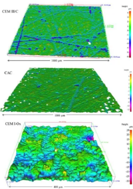
After the 5 weeks of endogenous curing, the surfaces of CEM I, CEM III and CAC cement paste slices were dry polished with a SiC polishing disc (ESCIL, ref. P800, i.e. 22 μm, 2 min polishing per surface), to remove roughness and orderly patterned striations made by the diamond saw blade. In contrast, the surface of CEM I-Ox pastes was kept as it was in order to preserve the Ca-oxalate precipitate layer. With such preparation, cement paste surfaces presented variable topology in all directions, without any repetitive pattern. A directional measurement of roughness (typically evaluated by a criterion such as Ra) is insufficient and non-representative of the surface topology [10]. Surface topology was thus measured using a focus variation 3D optical microscope (Alicona InfiniteFocus SL). Surface areas of 1000 x1000 µm and 400 x 400 µm were acquired with magnifications of x20 and x50 and coded on 4 Mpixels. Lateral and axial resolutions were 2.9 µm and 16 nm, respectively. Data processing was performed according to standard ISO 25178-2. Surfaces were balanced but they were not filtered, in order to provide complete primary profiles. The surface condition parameters selected to characterize surface topology were Sa (mean arithmetic height), Ssk (skewness, surface asymmetric factor) and Sku (Kurtosis – surface flattening factor). The Sa value is related to the scale of the surface texture while Ssk and Sku represent characteristics of the surface height distribution. The skewness value, Ssk, represents the proportion of negative and positive points in the profile, as it can take negative or positive values. Ssk=0 is the reference value for equilibrated Gaussian distributions. Negatively skewed surfaces are predominantly peak biased, whereas positively skewed surfaces are predominantly made of peaks. Kurtosis, Sku, gives an idea of the abruptness of the surface. A Gaussian distribution is characterized by a kurtosis value of 3. Higher values correspond to leptokurtic surfaces, typical of spiky surfaces, whereas kurtosis lower than 3 is typical of non-abrupt platykurtic surfaces.
2.2 Exposure to fermenting biowaste
The composition of a synthetic biowaste representative of the proportions of organic domestic waste was provided by IRSTEA, Antony (France). The composition is given in [3]. The biowaste was homogenized by blending for 10 min at 20 °C. It was then inoculated in order to initiate the anaerobic digestion process [11]. The inoculum was a sludge sampled from a municipal wastewater treatment plant in Toulouse (France). The organic loads, expressed as chemical oxygen demands (COD), were 50 g L-1 for the substrate (biowaste) and 20 g L-1 for
the inoculum (sludge). Proper establishment of the anaerobic digestion process depends on the inoculum/substrate (biowaste) quantity ratio [12]. The inoculation was carried out with an optimal ratio of 1 g COD(inoculum)/g COD(biowaste) as already reported in a previous work
[3,13]. Therefore, biowaste was diluted in inoculum to reach this ratio. The cement paste was immersed in anaerobic bioreactors (usable volume: 500 mL) at 37 °C in a thermostatically controlled incubator during the complete biowaste digestion (see [13]). The solid/liquid ratio (cement paste surface area/inoculated biowaste volume) was approximately 224 cm2 L-1. Several 5-week digestion cycles were run and the biowaste was renewed at the end of each cycle.
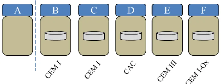
A reference bioreactor without cement paste specimens (reactor A) was also run. Two replicate reactors containing CEM I paste were noted reactors B and C. Reactors containing CAC, CEM III and CEM I-Ox were noted D, E and F, respectively. (Figure 1).
Figure 1 : Biowaste fermentation bioreactors
2.3 Observation of biofilm morphology
The biofilm on the surface of the specimen was observed by SEM-FEG (JEOL 7100F TTLS combined with EDX XMax, 5 kW) after 5 weeks (one cycle) and 10 weeks (2 cycles) of immersion in the biowaste. Cement paste specimens were sawn carefully to preserve the biofilm developed at their surfaces. Before SEM observations, the biofilms on the cement pastes were chemically fixed and then dehydrated according to the method presented in [3,13].
2.4 Bacterial community analysis
The bacterial population diversity was analysed using 16S rDNA sequencing to investigate the diversity of bacteria, notably involved in the hydrolysis, acidogenesis and acetogenesis reactions of the anaerobic digestion. Other microorganisms, such as archaea, also involved in the biowaste fermentation, were not investigated here. After 4 weeks of biowaste fermentation, supernatant was collected in the 5 bioreactors containing the cement paste specimens (reactors B and C: CEM I, reactor D: CAC, reactor E: CEMII/C, reactor F: CEMI-Ox) and in the control (reactor A, without cement paste). The biofilm bound to the cement paste surface after two cycles of biowaste fermentation (about 8 weeks of exposure of the cement paste to the biowaste) was collected by sonication in 2 mL of physiological water (3 min at 80 W)[14]. Then, the samples were sent to the Research and Testing Laboratory (RTL, Texas, USA), where the DNA was extracted prior to PCR and sequencing with the bacterial primers 28F (5′- GAG TTT GAT YMT GGC TC -3′) and 519R (5′- GWA TTA CCG CGG CKG CTG -3′) according to RTL protocols (www.researchandtesting.com). Subsequent data analyses with the DNA quality, DNA sequence alignment, clustering in operational taxonomic units [15] and assignment were also performed by RTL according to their protocol. In order to confirm or to clarify some assignations, the representative DNA sequence of the OTU was compared with the Blast database [16].
3. RESULTS
3.1 Initial surface topology of the cement pastes
Sa values were of the same order of magnitude for CEM I, CEM III and CAC surfaces (1.10-1.62 µm) but the value was much higher for CEM I-Ox ones (5 µm) (Table 2). Some studies have investigated the impact of mortar surface roughness on fungal colonization [17,18] and have found the Sa parameter to be between several tens of nm and several tens of µm. However, surface texture was not characterized, notably through skewness or kurtosis. Skewness and kurtosis can be easily interpreted so as to relate the characteristics of a surface with its functional performance [19]. For instance, medical, food and antifatigue components require low kurtosis surfaces, which are related to the unscratched surfaces desired for those applications.
CAC, CEM III and CEM I surfaces were relatively plane and presented some striations. These were deeper for CEM I and CEM III pastes than for CAC although the surface preparation was the same for the three pastes. Negative Ssk values (predominant presence of striation) and high values of Sku (the streaks were deep and steep) were obtained for the CEM III/C and CEM I pastes. The lower Sku value for the CAC paste indicates that the streaks were less marked. The surface topology of the CEM I-Ox paste was different from that of the others. It had as many peaks as hollows (Ssk close to 0), which were less marked (Sku weaker and close to 3). This typical roughness was due to the calcium oxalate crystals precipitated on the surface, the size of which oscillated around ten micrometres.
Table 2: Roughness parameters: Sa (mean arithmetic height [20]), Ssk (surface asymmetric factor) and Sku (surface flattening factor) of cement paste surfaces
Roughness parameter CEM I CEM III/C CAC CEM I + Ox
Sa (µm) 1.10 ± 0.29 1.62 ± 0.37 1.36 ± 0.56 5.54 ± 0.84 Ssk -2.63 ± 0.85 -3.49 ± 1.12 -1.05 ± 0.39 -0.3 ± 0.19 Sku 32.07 ± 13 29.88 ± 9.82 6.09 ± 2.49 3.42 ± 0.96
In the literature, surfaces with an Sa < 0.8 μm are theoretically considered as "hygienic", i.e. not very sensitive to microbial contamination [20]. In contrast, surfaces with Sa > 0.8 μm are subject to microbial adhesion.
This criterion is consistent with the hypothesis that topographies having scales that correspond to the microbial cell size are more amenable to colonization [20]. Sa values were of a few micrometres, except for CEM I-Ox paste. According to this criterion, the surfaces tested here were susceptible to colonization by microorganisms. In addition, bacteria are more able to colonize pits than peaks [21]. Here, the surfaces not treated with oxalic acid showed hollows (Ssk <0) and also hollows with extreme depths corresponding to streaks due to abrasion (Sku much higher than 3). The abundance of hollows on the surfaces as well as the order of magnitude of Sa for CEM I, CEM III / C and CAC pastes thus seemed to correspond to a surface roughness favourable to microbial proliferation. Conversely, the surface texture of the CEM I-Ox paste (Ssk close to 0 and Sku close to 3) seemed less favourable to microbial growth, despite its high Sa value.
Figure 2: 3D representation of surface topology analysed by focus variation 3D optical measurements of CAC, CEM III/C (similar to CEM I) (X20) and CEM I-Ox (X50) pastes
3.2 Microscopic observation of biofilms
A coupon was collected in each reactor after 5 and 10 weeks of immersion (1st and 2nd digestion cycles) to observe biofilms formed on its surface. If the adhesiveness to the surface was not quantified, the biofilms formed during this experiment were adherent to the surfaces, i.e. not simply deposited or sedimented. They were indeed submitted to several rinsing operations, first when the coupons were collected from the bioreactor, and then during the chemical biofilm dehydration protocol (necessary to their observation with SEM). All what remained on the cement paste surface was thus strongly adhered.
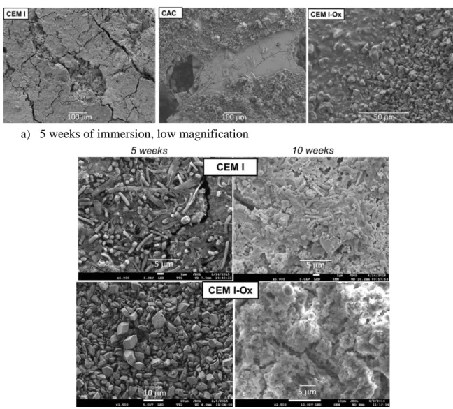
A uniform, dense biofilm was observed on the surfaces of the CEM I and CEM III C coupons for both sampling times. It seemed to consist mainly of bacilli but single or double cocci were also observed (Figure 3). Microorganisms were surrounded by a rather dense extracellular matrix, in particular on the CEM III paste surface.
After 5 weeks of immersion, few microorganisms were observed on the surface of the CAC coupons, and those present were essentially bacilli. After 10 weeks, the surface structures had
evolved with the appearance of inorganic S- and K-rich precipitates (maybe K2SO4 crystals)
according to the EDX analyses (data not shown). CAC being poor in these elements, they are likely to have come from the biowaste. Plates of biofilms with a surface area of about 40 μm were observed here and there, spaced a hundred micrometres apart. Cocci were observed in these clusters.
a) 5 weeks of immersion, low magnification
b) Comparison of 5 weeks and 10 weeks of immersion, high magnification
Figure 3: SEM observation of biofilms on cement paste after 5 weeks of immersion in the biowaste Unlike those of untreated CEM I pastes, the surface of the CEM I-Ox coupon showed no microbial colonization after 5 weeks of immersion in the biowaste; calcium oxalate precipitates were intact. After 10 weeks of immersion, as for untreated CEM I, the entire surface of the coupon was colonized by a thick microbial carpet with the presence of bacteria having different morphologies (cocci, bacilli, etc.), but with a less dense extracellular matrix than on CEM I and CEM III coupons.
From these observations, it may be concluded that surface treatment with oxalic acid has a retarding effect on microbial colonization. This effect had not yet been reported in the literature.
Microbial colonization also appeared to be slowed down or locally inhibited on the CAC coupon, with a slower, less dense and non-uniform surface coverage.
3.2 Microbial diversity in the supernatant and in the biofilm
After 4 weeks of biowaste fermentation, the microbial diversity of the supernatant and the biofilm anchored to the cement paste surface were analysed for the six fermentation conditions tested for each sample collected in the liquid phase of the biowaste and in the biofilm. The DNA sequences having at least 97% of similarity were grouped in Operational Taxonomic Units (OTU). Table 3 gives the total number of OTU and of sequences, together with the corresponding diversity indexes, which were calculated from this information (Shannon and Simpson indexes). The number of sequences produced from a sample (supernatant or biofilm) was between 27032 and 145241 and the number of operational taxonomic units (OTUs) identified per sample was between 288 and 1108.
The OTU number, which can be considered as the number of species, was of the same order of magnitude, either in the supernatant or in the biofilm, for reactors containing CEM I coupons. However, based on the lower Shannon and Simpson indices for biofilm samples, the biofilm seemed to be less rich and probably less diverse than the supernatant. The same trend was observed for the experiments carried out with CEM I-Ox and CEM III coupons, with Shannon indices between 2.7 and 1.6 and Simpson indices between 0.8 and 0.62. In these experiments, the number of OTU was 2 to 5 times lower in the biofilm than in the supernatant despite a comparable number of sequences analysed for CEM I-Ox between the biofilm and the supernatant. For the medium with CAC, although the number of OTUs was larger in the biofilm than in the supernatant, the diversity and richness were comparable. The selection pressure in the biofilm vs. supernatant was lower than for the other reactors.
Table 3: Groups of DNA sequences in terms of total number of OTU, and diversity indexes of analyses (Shannon and Simpson indexes)
Sample Number of sequences OTU97 Shannon [21] Simpson index [22] B iow as te Med A (control) 103800 986 4.02 0.94 Med B (CEM-I) 54141 657 3.54 0.92 Med C (CEM-I) 68963 672 3.12 0.87 Med D (CAC) 49545 798 3.86 0.94 Med E (CEM-III/C) 90385 1068 3.73 0.92 Med F (CEM-I-Ox) 43636 799 3.98 0.94 B iof il m CEM I (B) 55788 535 2.66 0.72 CEM I (C) 145241 694 2.63 0.81 CAC 84575 1108 4.05 0.91 CEM III/C 27032 288 1.65 0.62 CEM I-Ox 39393 420 2.70 0.80
In the supernatant and in the biofilm, for the four conditions of cement paste composition tested, three phyla dominated the microbial community: Firmicutes, Proteobacteria and Bacteroidetes (Figure 4). These three phyla are commonly found in the microbial communities of the anaerobic digester process [24–26].
Firmicutes and Bacteroidetes are often involved in lipid protein and polysaccharide hydrolysis [27]. Proteobacteria are involved in glucose, butyrate, propionate and acetate degradation during hydrolysis and acetogenesis [25].
The microbial community of reactor B (CEM I) supernatant was 44% dominated by Proteobacteria while reactor C supernatant, containing the same type of material, was 66% dominated by Bacteroidetes. The supernatants containing the other types of materials were dominated by two phyla, Bacteroidetes and Proteobacteria for the CAC reactor, Bacteroidetes and Firmicutes for CEM III and CEM-I-Ox reactors. It seems difficult to establish a correlation between the type of materials present in the environment and the dominance of a phylum.
Regarding the biofilm microbial community, it was generally dominated mainly by one phylum: Proteobacteria at 60% for CAC and CEM I-Ox coupons, Firmicutes at 90% and 70% for CEM I B and CEM III coupons, respectively. It was not necessarily the dominant population in the supernatant that was dominant within the biofilm on the surface of cement pastes, except for the biofilm of CEM I C, dominated at 60% by Bacteroidetes. The microbial community of this coupon at the phylum level was very similar to that of the supernatant. Except for this coupon, Bacteroidetes were very little represented in the biofilms. It is possible that microorganisms of this phylum are adapted or less competitive than other organisms in the microbial colonization of cementitious materials whatever their composition.
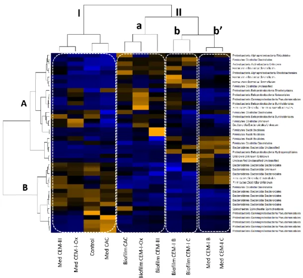
Figure 4: Pyrosequencing analysis of gene coding for RNAr 16S: bacterial phyla in relative percentage of total population after 3 weeks of anaerobic digestion (liquid media and biofilms on cement pastes) A clustered heat mapping was performed by R package (http://bioconductor.org/biocLite.R) [28] and the hierarchical clustering was determined for both dimensions of the heat maps using Ward’s method and correlation distances [29]. This statistical analysis was used to compare the
taxonomic compositions between the supernatant and the biofilm, according their abundance, for the different reactors. Only the OTUs with an abundance > 2% were considered. According to the heat-map analysis, the samples clustered into two major groups named I and II (Figure 5).
Figure 5: Colour-scaled clustered heat map. OTUs with abundance> 2% – black: abundance in the average range on the line – yellow orange: abundance > average on the line – blue: abundance <
average
Group I brought together the samples from the supernatants of the control reactor and the reactors containing CEM III, CEM I-Ox and CAC cement paste specimens. The dominant species inside this group were Pseudomonas sp., Spirochaetales sp., Bacteriodales sp. and Clostridiales sp., which formed cluster B.
Group II was subdivided into three other groups. Group II.a included the sample from biofilm collected over the cement paste surface of CAC, CEM I-Ox and CEM III. Bacillales sp. from the Firmicutes phylum dominated the CEM III microbial community, whereas the communities
from biofilm collected on CEM I-Ox and CAC specimens were dominated by several species of Proteobacteria, such as Pseudomonadales sp., Burkholderiales sp., Neisseriales sp., Rhodocyclales sp. and Rhizobiales sp. Subgroups II.b and II.b’ corresponded to reactors containing CEM I specimens (reactors A and B). The profile similarities were notable between biofilms collected on CEM I coupons (subgroup II.b on the heat map) and other biofilms (subgroup II.a). The microbial community of the biofilm (subgroup II.b) and the supernatant (subgroup II.b’) had very close compositions with profiles rich in Hydrogenophilic sp., Clostridiales sp. and Bacillales sp. The species and orders dominating the microbial communities of other supernatants (cluster B on the left) were less represented on the profiles corresponding to the supernatant collected in CEM I reactor. Generally, the species and orders predominantly present in the different profiles, e.g. Bacteroidales and Clostridiales, were the major orders involved in anaerobic digestion [25].
4. DISCUSSION
4.1 Biofilm establishment
Roughness and surface pH are among the essential factors that theoretically condition microbial growth on the surface of cementitious materials. The optimum pH values for the microorganisms involved in anaerobic digestion lie between 4 and 8. A priori, the alkaline conditions (pH 12-13) of cementitious materials do not offer favourable conditions for the growth of microorganisms in the particular context of anaerobic digestion. The surface roughness of the cement pastes used here, measured by optical profilometry, was of the order of 1-5 μm for the Sa criterion. The scale of this roughness seemed to be conducive to microbial adhesion and then growth. Surfaces where the sizes of asperities or topographical structures (valleys, holes or scratches) and the roughness parameters were of the order of magnitude of the size of the microorganisms that promoted the initial adhesion of bacteria, a precursory step to biofilm formation [30–32].
The asperities or topographical structures were not the only parameters influencing the adhesion of the first cells; surface conditioning also played a significant role. Surface conditioning by the indirect action of the microorganisms (conditioning film formation, chemical interactions of solutes with substratum surfaces, etc.) has been shown to promote the adhesion of the first microorganisms and then biofilm anchoring on all types of surface as soon as the water and the necessary nutrients are available [33]. In addition, it can be assumed that, in the first stages of the immersion, the surface pH of all cement pastes was modified by the prior deterioration of the material (decalcification and dissolution of portlandite related to AGV and NH4+) or by the carbonation of the material because of the CO2 metabolized by the bacteria
near the surface [34,35]. The microbial richness involved in anaerobic digestion as well as the diversity of the biodegradable material are also conductive to biofilm formation. In some anaerobic digesters, supports (zeolite, biochar, etc.) have been added to promote the development of immobilized microorganisms in the form of biofilm, as in the case of anaerobic fixed bed or fluidized bed processes [36,37]. The presence of a solid support, such as straw, also suitable for biofilm formation, has led to significantly improved biogas production [38,39]. The formation of a biofilm promotes proximity of the microorganisms and could facilitate syntrophic associations between the different microbial families responsible for the degradation stages of organic matter in anaerobic digestion [40] and also encourage cross-feeding, where some microorganisms take advantage of the metabolites produced by other microorganisms. In
addition to being conducive to the development of a biofilm, the presence of a solid support can have other advantages for microbial activities, such as: (i) providing a mineral structure that can trap toxic compounds [37] and (ii) releasing some mineral components (Ca2+, Mg2+, Na+,
etc.) by leaching [41].
Despite having roughness similar to other pastes, CAC surfaces were less colonized: the biofilm was thinner and the distribution at the surface was very heterogeneous compared to those on CEM I and CEM III. Apart from a possibly different surface conditioning, the CAC chemical composition was significantly different from CEM I and CEM III, in particular by the high Al content (50% by mass) compared to Ca (40% by mass). In contrast, CEM I and CEM III/C contained mainly Ca (respectively about 60% and 40%) and lower Al content (about 4% and 10%, respectively). These significant differences in chemical composition may have been responsible for limited proliferation at the surface of CAC paste. The study by Herisson et al. [42] on calcium aluminate cements provides arguments in this direction to justify the performance of CACs with respect to biodeterioration in sewage networks. The authors explain the better durability of CACs by the abundance of aluminium in the binder, which has "bacteriostatic" properties [42]. The bacteriostatic properties of a compound inhibit microbial growth without being lethal to bacterial cells. Buvignier et al. [43] recently investigated the impact of Al (in the form of AlCl3) on bacteria and showed that the bacteriostatic impact of Al
was only temporary and that, with time, bacteria became acclimatized to high Al concentrations. The microorganisms seem to have adapted to the chemical composition of this material since, although observed later on CAC than on other materials, microbial proliferation was established on all the surfaces after 10 weeks of exposure.
Colonization of the surface covered with Ca-oxalate showed a radical change from one deadline to the next. After 5 weeks of immersion, no microbial growth was present on this type of paste. Then, colonization observed after 10 weeks was as intense as on a conventional CEM I paste. Three hypotheses can be proposed to account for this: (i) microbial growth may have occurred later on the surface of the oxalate salt, (ii) the oxalate on the surface may have been dissolved before microbial colonization took place, or (iii) some microorganisms may have made Ca-oxalate bioavailable and degraded it [44–46]. Regarding the second hypothesis, in pure water at 30 °C (pH 7), Ca-oxalate monohydrate is characterized by a solubility (6.7 10-3
g.100 cm-3) that is very low in comparison with that of Ca-acetate (34.7 g.100 cm -3). Nevertheless, the medium may have contained compounds, e.g. citrates, capable of dissolving Ca-oxalate [47,48].
4.2 Heterogeneity between the planktonic bacterial populations (media) and the biofilm populations on the surface of the cement pastes
The microbial community on the surface of cement pastes was less diverse and less rich than that of the medium of anaerobic digestion (supernatant). The microbial populations were more dominated in the biofilm.
The microbial community profiles of biofilms (clustered in Group II.a. Figure 5) were different from those of the supernatants (clustered in Group I, Figure 4) for the CAC, CEM III and CEM I-Ox reactors. A different distribution of microbial communities between the supernatant and the biofilm in the same reactor or in the same medium has already been reported in the literature. For example, microbial proliferation on the surface of steel in marine environments shows
similar characteristics [49,50]. The phylum Bacteroidetes was poorly represented in the biofilm on the surface of cementitious materials, whereas it was present in the reaction medium (Figure 3). This difference between the medium and the biofilm in terms of microbial flora might reflect a real dissociation between the composition of the medium and the composition of the biofilm at the surface of the material (Figure 3, Figure 4). Celikkol-Aydin et al. [51] characterized a marine biofilm dominated by the phyla Cyanobacteria, Bacteroidetes and Proteobacteria while the seawater in which it was formed was dominated by the phyla firmicutes and proteobacteria [51]. In this study, the steel surface was a favourable environment for the enrichment of Cyanobacteria and Bacteroidetes.
In the presence of CEM I, the selection pressure between the supernatant and the biofilm was less strong. The microbial profiles were clustered in the same group (II), although subdivided into two subgroups, II.b and II.b’. It is interesting to note that the profiles of the microbial community of the 2 reactors containing CEM I were close to the profiles obtained on the biofilms rather than being close to those obtained in the supernatants. CEM I cementitious materials are chemically less stable than the other materials tested, such as CEM III materials, which contain slag [52,53]. It is possible that CEM I, unlike other materials, had a higher leaching rate and that the medium was enriched with cation, in particular calcium. This could explain why the selection pressure between biofilm and supernatant was lower for reactors containing CEM I. Therefore, the nature of the material may have an impact on the microbial community profile on its surface (in the form of biofilm). The chemical composition of the supernatant according to the type of immersed material may also influence the microbial communities in the supernatant according to the compounds that are released by the materials. However, these results must be taken with caution because, within the biofilms themselves, the microbial distribution is not uniform over the upper and lower layers [54]. The structure of a multi-species biofilm is made up of strata and Rochex et al. [55] showed a structure comprising a thicker upper layer, composed of a dominant and weakly cohesive species, and a thinner, but highly cohesive, basal layer with greater microbial diversity than the upper layer [55]. The cohesion of the biofilm is heterogeneous according to the strata and increases with the depth of the biofilm [56]. Despite all the precautions taken to recover as much biofilm as possible, it is possible that not everything was detached from the surface of the cement paste.
5. CONCLUSION
All the surfaces of cementitious materials tested, made of CEM I (ordinary Portland cement), CEM III/C (blast furnace slag cement), CAC (calcium aluminate cement), and CEM I treated with oxalic acid, were intensely colonized at the end of the experiment. However, it should be noted that the treatment with oxalic acid seemed to delay the establishment of the biofilm. Microbial colonization also appears to have been slowed down or locally inhibited on the CAC coupon, which showed a slower, less dense and non-uniform surface coverage. The effects roughness and surface pH do not appear to be of prime importance for biofilm development.
The results of the microbial population analyses showed a less diversified microbial flora in the biofilm than in the reaction medium
ACKNOWLEDGEMENTS
The authors gratefully thank the PRES Université de Toulouse and the Midi-Pyrénées Region for their financial support, and IRSTEA, Antony, for the biowaste production protocol.
REFERENCES
[1] Bertron A. Understanding interactions between cementitious materials and microorganisms: a key to sustainable and safe concrete structures in various contexts. Mater. Struct. 2014;47:1787–1806.
[2] Bertron A, Lavigne MP, Patapy C, et al. Biodeterioration of concrete in agricultural, agro-food and biogas plants: state of the art and challenges. RILEM Tech. Lett. 2017;2:83–89.
[3] Voegel C, Bertron A, Erable B. Mechanisms of cementitious material deterioration in biogas digester. Sci. Total Environ. 2016;571:892–901.
[4] Giroudon M, Lavigne MP, Patapy C, et al. Biodeterioration mechanisms and kinetics of SCM and aluminate based cements and AAM in the liquid phase of an anaerobic digestion. MATEC Web Conf. 2018;199:2003.
[5] Magniont C, Coutand M, Bertron A, et al. A new test method to assess the bacterial deterioration of cementitious materials. Cem. Concr. Res. 2011;41:429–438.
[6] Giroudon M, Peyre Lavigne M, Patapy C, et al. Biodeterioration mechanisms and durability of SCM and aluminate based cements and AAM in the liquid phase of an anaerobic digestion system. Proc. RILEM 253-MCI Conf. Toulouse: Bertron A, Jonkers H; 2018.
[7] Koenig A, Dehn F. Main considerations for the determination and evaluation of the acid resistance of cementitious materials. Mater. Struct. 2016;49:1693–1703.
[8] Voegel C, Bertron A, Erable B, et al. Chemical treatment with oxalic acid to improve the durability of cement-based materials in acidic environment. Proc. 13th Int. Conf. Durab. Build. Mater. Compon. RILEM Proceedings. Sao Paulo: Marco Quattrone, Vanderley M. John; 2014. p. 670–678.
[9] Larreur-Cayol S, Bertron A, Escadeillas G. Degradation of cement-based materials by various organic acids in agro-industrial waste-waters. Cem. Concr. Res. 2011;41:882– 892.
[10] Landon Y, Cherif M. Characterization of the Surface Quality of Holes Drilled in CFRP Laminates. Adv. Mater. Res. 2013;698:107–116.
[11] Neves L, Oliveira R, Alves MM. Influence of inoculum activity on the bio-methanization of a kitchen waste under different waste/inoculum ratios. Process Biochem. 2004;39:2019–2024.
[12] Elbeshbishy E, Nakhla G, Hafez H. Biochemical methane potential (BMP) of food waste and primary sludge: Influence of inoculum pre-incubation and inoculum source. Bioresour. Technol. 2012;110:18–25.
[13] Voegel C, Bertron A, Erable B. Biodeterioration of cementitious materials in biogas digester. Matér. Tech. 2015;103:202.
[14] Blanchet E, Desmond E, Erable B, et al. Comparison of synthetic medium and wastewater used as dilution medium to design scalable microbial anodes: Application to food waste treatment. Bioresour. Technol. 2015;185:106–115.
[15] Edgar RC. UPARSE: highly accurate OTU sequences from microbial amplicon reads. Nat. Methods. 2013;10:996–998.
[16] Zhang Z, Schwartz S, Wagner L, et al. A greedy algorithm for aligning DNA sequences. J. Comput. Biol. J. Comput. Mol. Cell Biol. 2000;7:203–214.
[17] Tran TH, Govin A, Guyonnet R, et al. Influence of the intrinsic characteristics of mortars on biofouling by Klebsormidium flaccidum. Int. Biodeterior. Biodegrad. 2012;70:31–39. [18] Manso S, Calvo-Torras MÁ, De Belie N, et al. Evaluation of natural colonisation of cementitious materials: Effect of bioreceptivity and environmental conditions. Sci. Total Environ. 2015;512–513:444–453.
[19] Stout KJ, Davis EJ. Surface topography of cylinder bores — the relationship between manufacture, characterization and function. Wear. 1984;95:111–125.
[20] Flint SH, Brooks JD, Bremer PJ. Properties of the stainless steel substrate, influencing the adhesion of thermo-resistant streptococci. J. Food Eng. 2000;43:235–242.
[21] Mitik‐Dineva N, Wang J, Mocanasu RC, et al. Impact of nano‐topography on bacterial attachment. Biotechnol. J. 2008;3:536–544.
[22] Shannon E. A mathematical theory of communication. Bell Syst. Tech. J. 1948;27:379-423-656.
[23] Simpson E. Measurement of diversity. Nature. 1949;163:688.
[24] Nelson MC, Morrison M, Yu Z. A meta-analysis of the microbial diversity observed in anaerobic digesters. Bioresour. Technol. 2011;102:3730–3739.
[25] Lee S-H, Kang H-J, Lee YH, et al. Monitoring bacterial community structure and variability in time scale in full-scale anaerobic digesters. J. Environ. Monit. 2012;14:1893–1905.
[26] De Vrieze J, Saunders AM, He Y, et al. Ammonia and temperature determine potential clustering in the anaerobic digestion microbiome. Water Res. 2015;75:312–323.
[27] Zhang T, Shao M-F, Ye L. 454 Pyrosequencing reveals bacterial diversity of activated sludge from 14 sewage treatment plants. ISME J. 2012;6:1137–1147.
[28] Warnes GR, Bolker B, Bonebakker L, et al. gplots: Various R Programming Tools for Plotting Data [Internet]. 2016. Available from: https://CRAN.R-project.org/package=gplots.
[29] Bize A, Cardona L, Desmond-Le Quemener E, et al. Shotgun metaproteomic profiling of biomimetic anaerobic digestion processes treating sewage sludge. Proteomics. 2015;15:3532–3543.
[30] Characklis WG, Marchall KC. Biofilms: a basis for an interdisciplinary approach. Biofilms. A Wiley-Interscience Publication, John Wiley & Sons. New York: Characklis WG, Marshall KC,; 1990.
[31] Epstein AK, Hong D, Kim P, et al. Biofilm attachment reduction on bioinspired, dynamic, micro-wrinkling surfaces. New J. Phys. 2013;15:95018.
[32] Lu N, Zhang W, Weng Y, et al. Fabrication of PDMS surfaces with micro patterns and the effect of pattern sizes on bacteria adhesion. Food Control. 2016;68:344–351.
[33] Dang H, Lovell CR. Microbial Surface Colonization and Biofilm Development in Marine Environments. Microbiol. Mol. Biol. Rev. 2016;80:91–138.
[34] Leemann A, Lothenbach B, Hoffmann C. Biologically induced concrete deterioration in a wastewater treatment plant assessed by combining microstructural analysis with thermodynamic modeling. Cem. Concr. Res. 2010;40:1157–1164.
[35] Giovannacci D, Leclaire C, Horgnies M, et al. Algal colonization kinetics on roofing and façade tiles: Influence of physical parameters. Constr. Build. Mater. 2013;48:670–676. [36] Poirier S, Madigou C, Bouchez T, et al. Improving anaerobic digestion with support
media: Mitigation of ammonia inhibition and effect on microbial communities. Bioresour. Technol. 2017;235:229–239.
[37] Montalvo S, Guerrero L, Borja R, et al. Application of natural zeolites in anaerobic digestion processes: A review. Appl. Clay Sci. 2012;58:125–133.
[38] Andersson J, Björnsson L. Evaluation of straw as a biofilm carrier in the methanogenic stage of two-stage anaerobic digestion of crop residues. Bioresour. Technol. 2002;85:51– 56.
[39] Bengelsdorf FR, Gabris C, Michel L, et al. Syntrophic microbial communities on straw as biofilm carrier increase the methane yield of a biowaste-digesting biogas reactor. Bioeng. 2015 Vol 2 Pages 264-276 [Internet]. 2015 [cited 2018 May 24]; Available from: http://www.aimspress.com/article/10.3934/bioeng.2015.3.264.
[40] Biodégradations et métabolismes. Les bactéries pour les technologies de l’environnement - Jean Pelmont [Internet]. [cited 2018 May 24]. Available from: https://www.decitre.fr/livres/biodegradations-et-metabolismes-9782868837455.html.
[41] Rafrafi Y, Durban N, Bertron A, et al. Use of a continuous-flow bioreactor to evaluate nitrate reduction rate of Halomonas desiderata in cementitious environment relevant to nuclear waste deep repository. Biochem. Eng. J. 2017;125:161–170.
[42] Herisson J, van Hullebusch ED, Moletta-Denat M, et al. Toward an accelerated biodeterioration test to understand the behavior of Portland and calcium aluminate cementitious materials in sewer networks. Int. Biodeterior. Biodegrad. 2013;84:236–243. [43] Buvignier A, Peyre-Lavigne M, Patapy C, et al. Understanding the resistance of calcium aluminate cements in sewer environments: role of soluble aluminium on the SOB activity and role of the biofilm in the degradation. Proc. 14th Int. Conf. Durab. Build. Mater. Compon. Ghent; 2017.
[44] Cailleau G, Mota M, Bindschedler S, et al. Detection of active oxalate–carbonate pathway ecosystems in the Amazon Basin: Global implications of a natural potential C sink. CATENA. 2014;116:132–141.
[45] Martin G, Guggiari M, Bravo D, et al. Fungi, bacteria and soil pH: the oxalate-carbonate pathway as a model for metabolic interaction. Environ. Microbiol. 2012;14:2960–2970. [46] Hervé V, Junier T, Bindschedler S, et al. Diversity and ecology of oxalotrophic bacteria.
World J. Microbiol. Biotechnol. 2016;32:28.
[47] Tracy CR, Pearle MS. Update on the medical management of stone disease. Curr. Opin. Urol. 2009;19:200.
[48] Chutipongtanate S, Chaiyarit S, Thongboonkerd V. Citrate, not phosphate, can dissolve calcium oxalate monohydrate crystals and detach these crystals from renal tubular cells. Eur. J. Pharmacol. 2012;689:219–225.
[49] Salta M, Wharton JA, Blache Y, et al. Marine biofilms on artificial surfaces: structure and dynamics. Environ. Microbiol. 15:2879–2893.
[50] Pepe-Ranney C, Hall EK. The effect of carbon subsidies on marine planktonic niche partitioning and recruitment during biofilm assembly. Front. Microbiol. [Internet]. 2015
[cited 2018 May 24];6. Available from:
https://www.ncbi.nlm.nih.gov/pmc/articles/PMC4500991/.
[51] Celikkol-Aydin S, Gaylarde CC, Lee T, et al. 16S rRNA gene profiling of planktonic and biofilm microbial populations in the Gulf of Guinea using Illumina NGS. Mar. Environ. Res. 2016;122:105–112.
[52] Bertron A, Duchesne J, Escadeillas G. Accelerated tests of hardened cement pastes alteration by organic acids: analysis of the pH effect. Cem. Concr. Res. 2005;35:155– 166.
[53] Beddoe RE. Modelling acid attack on concrete: Part II. A computer model. Cem. Concr. Res. 2016;88:20–35.
[54] Zhang TC, Bishop PL. Structure, Activity and Composition of Biofilms. Water Sci. Technol. 1994;29:335–344.
[55] Rochex A, Massé A, Escudié R, et al. Influence of abrasion on biofilm detachment: evidence for stratification of the biofilm. J. Ind. Microbiol. Biotechnol. 2009;36:467– 470.
[56] Derlon N, Massé A, Escudié R, et al. Stratification in the cohesion of biofilms grown under various environmental conditions. Water Res. 2008;42:2102–2110.
![Table 2: Roughness parameters: Sa (mean arithmetic height [20]), Ssk (surface asymmetric factor) and Sku (surface flattening factor) of cement paste surfaces](https://thumb-eu.123doks.com/thumbv2/123doknet/12231721.318442/6.892.117.781.608.712/roughness-parameters-arithmetic-surface-asymmetric-surface-flattening-surfaces.webp)