Pépite | Etudes spatio-temporelle de la lésion de la moelle épinière par des approches OMICs et physiologique
234
0
0
Texte intégral
(2) Thèse de Stéphanie Devaux, Lille 1, 2016. ” The future belongs to those who believe in the beauty of their dreams.” ―. © 2016 Tous droits réservés.. Eleanor Roosevelt. lilliad.univ-lille.fr.
(3) Thèse de Stéphanie Devaux, Lille 1, 2016. Acknowledgment I would like to express a sincere thank you to everyone who has contributed to my work: MVDr. Dasa Cizkova DrSc- my thesis adviser at the University of Veterinary Medicine and Pharmacy, in Kosice, Institute of Neurobiology, SAS- for having welcomed me to her institute, for her support in this co-supervised thesis, for her valuable guidance, enthusiasm and motivation, and for giving me the opportunity to learn from her expertise. Pr. Michel Salzet- my thesis adviser at the Université de Lille1- for his continued support, for always being open, for all of his precious ideas, for guiding me through all these years of research. Pr. Isabelle Fournier for her advice and her skills in proteomics. My thesis reporters, Pr. Stephen McMahon and Pr. Joost Verhaagen for having kindly accepted to evaluate my work. My thesis examiners, Pr. Serge Nataf, Pr Régis Bordet, Ass. Pr. Bhide Manges, PhD Ass. Pr. Norbert Zilka, DrSc, for accepting to judge my work. Université de Lille 1, Conseil regional Nord-Pas-de-Calais and University of Veterinary Medicine and Pharmacy for giving me access to resources and funding for my PhD thesis, and also the Ecole Doctorale Biologie Santé de Lille. Members of the laboratory in Lille for their availability and their assistance, Annie, Benoit, Maxence, Jean Pascal, Julien, Françoise, Christophe, Christelle, Céline, Pierre Eric, Franck, Jacopo, Lucie and especially to Jusal for his precious help with cryostat, “Patience is a virtue”. Students or ex-students from Lille 1 laboratory, Antonella, Marie, Dounia, Philippe, Khalil, Adriana, Tanina, Vivian, Tony for keeping a cheerful atmosphere. Members of Slovak laboratory, Marika, Lucia, Eva, Miriam for the warm welcome in Kosice, their precious advice, their sympathy, and especially to Juraj for his time and his patience when dealing with surgery, confocal microscopy and all “critical steps”. My parents and my sister for their unfailing support and their encouragement throughout my way “C-C-C”, for always being there, as well as my family and my friends for listening to me when I needed it most. Kris for taking time to read and correct my manuscript. And finally to Nicolas, for being present every day and for giving me strong motivation each step of the way.. © 2016 Tous droits réservés.. lilliad.univ-lille.fr.
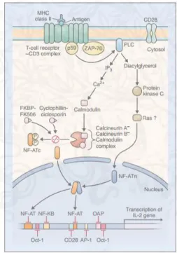
(4) Thèse de Stéphanie Devaux, Lille 1, 2016. Content ACKNOWLEDGMENT ........................................................................................................................ 3 PUBLICATIONS ................................................................................................................................. 6 LIST OF FIGURES ............................................................................................................................... 8 ABBREVIATIONS ............................................................................................................................. 11 INTRODUCTION .............................................................................................................................. 13 1.. SPINAL CORD ORGANIZATION ............................................................................................... 17. 2.. MODEL OF INJURY ................................................................................................................. 18. 3.. PATHOPHYSIOLOGY .............................................................................................................. 22. 3.1.. PRIMARY INJURY ....................................................................................................................... 22. 3.2.. SECONDARY INJURY ................................................................................................................... 23. 4.. IMMUNE RESPONSE AFTER SCI .............................................................................................. 24. 4.1.. INNATE IMMUNE SYSTEM ........................................................................................................... 25 4.1.1.. Neutrophils recruitment .................................................................................................. 25. 4.1.2.. Microglia cells ................................................................................................................. 26. 4.1.3.. Astrocytes........................................................................................................................ 28. 4.2.. RELATION BETWEEN GLIAL CELLS AND NEURONS .............................................................................. 29 4.2.1.. Cellular interactions ........................................................................................................ 30. 4.2.2.. Molecular interactions .................................................................................................... 30. 4.3. 5.. T CELLS AND INNATE IMMUNE CELLS ............................................................................................. 33 TREATMENTS FOR SPINAL CORD INJURY................................................................................ 34. 5.1.. MYELIN NEURITE GROWTH INHIBITORS .......................................................................................... 35. 5.2.. PROTEOGLYCAN INHIBITOR ......................................................................................................... 38. 5.3.. CELLULAR THERAPY ................................................................................................................... 39 5.3.1.. Autologous Schwann cell transplantation ...................................................................... 39. 5.3.2.. Olfactory ensheathing cells (OECs) ................................................................................. 40. 5.3.3.. Human embryonic stem cells, induced pluripotent stem cells ........................................ 41. 5.3.4.. Neural stem cells ............................................................................................................. 41. 5.3.5.. Bone marrow stromal cells (BMSCs) ............................................................................... 42. 5.4.. © 2016 Tous droits réservés.. PHARMACOTHERAPY ................................................................................................................. 43 5.4.1.. Minocycline ..................................................................................................................... 43. 5.4.2.. Withanoside IV ................................................................................................................ 44. 5.4.3.. Neuroimmunophilin ligand ............................................................................................. 44. lilliad.univ-lille.fr.
(5) Thèse de Stéphanie Devaux, Lille 1, 2016. 5.4.4. 6. 6.1.. Estrogen .......................................................................................................................... 45. PROTEOMICS ........................................................................................................................ 47 PROTEOMIC APPROACH .............................................................................................................. 47. PART 1: SPATIAL AND TEMPORTAL ANALYSES AFTER SPINAL CORD INJURY..................................... 50 CHAPTER 1: SPATIAL AND TEMPORAL ANALYSES AFTER SPINAL CORD INJURY .......................................................... 50 Article 1: Alterations of protein composition along the rostro-caudal axis after spinal cord injury: proteomic, in vitro and in vivo analyses ...................................................................................................... 53 Article 2: Proteomic analysis of the spatio-temporal based molecular kinetics of acute spinal cord injury identifies a time- and segment-specific window for effective tissue repair....................................... 69 CONCLUSION CHAPTER 1 & 2 ................................................................................................................ 100 IMMUNOGLOBULINS ................................................................................................................... 100 MEMO1-RHOA-DIAPH1 signaling pathway ................................................................................. 105 PART 2: MODULATION OF THE INFLAMMATION AND IMPROVEMENT OF THE REGENERATION PROCESS AND SPINAL CORD PLASTICITY: IN VITRO AND IN VIVO STUDIES .................................................. 119 CHAPTER 2: PROPERTIES OF FACTORS RELEASED BY BONE MARROW STROMAL CELLS TO MODULATE MICROGLIA CELLS . 119 Article 3: Modulation properties of factors released by bone marrow stromal cells on activated microglia: an in vitro study ........................................................................................................................ 122 CONCLUSION CHAPTER 2 ...................................................................................................................... 138 CHAPTER 3: ALGINATE BIOMATERIAL COUPLED WITH GROWTH FACTORS TO PROMOTE IN VIVO REGENERATION .......... 140 Article 4: Delivery of Alginate Scaffold Releasing Two Trophic Factors for Spinal Cord Injury Repair ................................................................................................................................................................... 144 CONCLUSION CHAPTER 3 ...................................................................................................................... 164 DISCUSSION ................................................................................................................................. 166 REFERENCES ................................................................................................................................. 169 ANNEX ......................................................................................................................................... 194. © 2016 Tous droits réservés.. lilliad.univ-lille.fr.
(6) Thèse de Stéphanie Devaux, Lille 1, 2016. Publications Accepted Publications Cizkova D., Le Marrec-Croq F., Franck J., Slovinska L., Grulova I., Devaux S., Lefebvre C., Fournier I., Salzet M. Alterations of protein composition along the rostro-caudal axis after spinal cord injury: proteomic, in vitro and in vivo analyses. Fontiers in Cell. Neurosci. 2014, 8(105):1-15 Devaux S.*, Cizkova D.*, Le Marrec-Croq F., Franck J., Slovinska L., Rosocha J., Spakova T., Lefebvre C., Fournier I., Salzet M. In vitro modulation properties of bone marrow stromal cells on BV2 microglia after spinal cord injury conditioned media activation. Scientific Reports 2014, 4: 7514. * co-first authors Slovinska L., Szekiova E., Blaško J., Devaux S., Salzet M., Cizkova D. Comparison of dynamic behavior and maturation of neural multipotent cells derived from different spinal cord developmental stages: an in vitro study. Acta Neurobiol Exp (Wars) 2015; 75(1):107-14 Grulova I., Slovinska L., Blaško J., Devaux S., Wisztorski M., Salzet M., Fournier I., Kryukov O., Cohen S., Cizkova D. Functional improvement of injured spinal cord following injection of alginate scaffold releasing two trophic factors. Scientific Reports 2015, 5:13702 Devaux S.*, Cizkova D.*, Quanico J., Franck J., Nataf S., Pays L., Hauberg-Lotte L., Mass P., Jobart J.H., Kobeissy F., Mériaux C., Wisztorski M., Slovinska L., Blasko J., Cigankova V., Fournier I., Salzet M. Proteomic analysis of the spatio-temporal based molecular kinetics of acute spinal cord injury identifies a time- and segment-specific window for effective tissue repair. Molecular and Cellular Proteomics (in Press). * co-first authors. Proceedings Devaux S., Cizkova D., Slovinska L., Blasko J., Nagyova M., Lefebvre C., Fournier I., Salzet M. Spatio-temporal proteins study of rat spinal cord injury and glial cells involvement. GLIA (2015) 63, E414-E415 Salzet M., Devaux S., Cizkova D., Quanico J., Franck J., Lotte L.H., Maass P., Wisztorski M., Slovinska L., Blasko J., Fournier I. Spatial and temporal 3D MSI and proteomic studies of rat spinal cord injury: Evidence of caudal segment for possible therapy target. Journal of Biotechnology (2015), 208, S10. Adam A., Salzet M., Devaux S. Profiling of Lipids Expression Along the Rostral and Caudal Axis After Spinal Cord Injury: Matrix Assisted Laser Desorption/Ionization Approach. The FASEB (2015) Journal 29 (1 Supplement), 715.21. Oral communication Devaux S, Cizkova D, Le Marrec-Croq F, Franck J, Slovinska L, Grulova I, Lefebvre C, Fournier I, Salzet M. Proteomic analyses along the rostro-caudal axis of injured spinal cord. EURON, 12-13th september 2013, Liège, Belgium Devaux S, Cizkova D, Le Marrec-Croq F, Franck J, Slovinska L, Lefebvre C, Fournier I, Salzet M. Proteomic analyses along the rostro-caudal axis of injured spinal cord. Young club SFSM : Société Française de spectrométrie de masse 24-28th March 2014, Dieppe, France. 6 © 2016 Tous droits réservés.. lilliad.univ-lille.fr.
(7) Thèse de Stéphanie Devaux, Lille 1, 2016. Devaux S, Cizkova D, Le Marrec-Croq F, Franck J, Slovinska L, Lefebvre C, Fournier I, Salzet M. Traumatic injury spinal cord- Temporal and spatial change in proteome. Young club SFEAP : Société Française d’électrophorèse et d’analyse protéomique 14-16th May 2014, Toulouse, France. Devaux S, Cizkova D, Wisztorski M, Slovinska L, Blasko J, Fournier I, Salzet M. Spatial and temporal MSI and proteomic studies of rat spinal cord injury: evidence of caudal segment for possible therapy target. 14th HUPO, 27-30th September 2015, Vancouver, Canada.. Poster Devaux S, Cizkova D, Le Marrec-Croq F, Franck J, Slovinska L, Grulova I, Lefebvre C, Fournier I, Salzet M. Proteomic analyses along the rostro-caudal axis of injured spinal cord. 7th International Symposium on experimental and clinical neurobiology, June 2013, Kosice, Slovakia Devaux S, Cizkova D, Le Marrec-Croq F, Franck J, Slovinska L, Blasko J, Nagyova M, Lefebvre C, Fournier I, Salzet M. Alterations of protein composition along the rostro-caudal axis after spinal cord injury: proteomic analyses. HUPO, 13th Annual World Congress of the Human Proteome Organization, Madrid, Spain, 5-8 October 2014 (Prize, Poster Award HUPO 2014) Devaux S, Cigánkova V, Slovinska L, Blasko J, Lefebvre C, Fournier I, Salzet M, Cizkova D. Proteomic analysis of spinal cord injury: chemokines and neurotrophic factors. XVI. Celostátní conference biologické psychiatrie v Luhačovicích, 3-6th June 2015 Luhacovice, Czech Republic Devaux S, Cizkova D, Slovinska L, Blasko J, Nagyova M, Lefebvre C, Fournier I, Salzet M. Spatiotemporal proteins study of rat spinal cord injury and glial cells involvement. Glia, XII European Meeting on Glial cells in Health and Disease, 15-18th July 2015, Bilbao, Spain. Devaux S, Cizkova D, Wisztorski M, Slovinska L, Blasko J, Fournier I, Salzet M. Spatial and temporal MSI and proteomic studies of rat spinal cord injury: evidence of caudal segment for possible therapy target. 14th HUPO, 27-30th September 2015, Vancouver, Canada. Devaux S, Cizkova D, Slovinska L, Blasko J, Fournier I, Salzet M. Spatio-temporal proteins study of rat spinal cord injury and immune cells involvement. FENS Featured Regional Meeting 2015, 7-10th October 2015, Thessaloniki, Greece. Devaux S, Blasko J, Slovinska L, Cigankova V, Cizkova D, Salzet M. Immunomodulatroy properties of bone marrow stromal cells on activated microglia for spinal cord injury therapy. 10th FENS Forum of Neuroscience, 2-6th July 2016, Copenhagen, Denmark.. Teaching 2015-2016: 64 hours practical courses in animal biology. Students supervision Amna Adam, Bachelor degree, Internship 2 months 2014 Zahra Laou, Master degree, Internship 1 month 2016 Delphine Mansy, Master degree, Internship 4 months 2016. 7 © 2016 Tous droits réservés.. lilliad.univ-lille.fr.
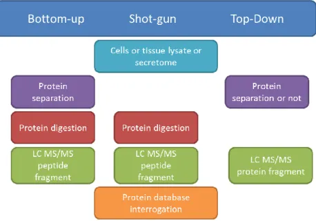
(8) Thèse de Stéphanie Devaux, Lille 1, 2016. List of Figures FIGURE 1: CONSEQUENCES OF THE LOCALIZATION OF THE LESION (ADAPTED MYHEALTH.ALBERTA.CA). ........................................ 17 FIGURE 2: A. CLIP COMPRESSION. B. AN INTRAOPERATIVE PICTURE SHOWING A CLIP COMPRESSING THE SPINAL CORD AT T2 TO CREATE THE INJURY (POON ET AL. 2007). ........................................................................................................................ 20. FIGURE 3: BALLOON COMPRESSION MODEL. A. A PHOTOGRAPH REPRESENTING THE SIZE OF THE T9 VERTEBRA AND CATHETERS INFLATED WITH DIFFERENT VOLUMES OF SALINE 10, 15 AND 20µL RESPECTIVELY. B. LOCOMOTOR FUNCTION RECOVERY AFTER. SCI GRADED ON AN EXPANDED SCALE (BBB SCORE). 21 IS NORMAL LOCOMOTOR FUNCTION, 0 REPRESENTS NO LOCOMOTOR FUNCTION. (ADAPTED FROM VANICKY ET AL. 2001) ................................................................................................ 20. FIGURE 4: SCHEMA OF EVENTS FOLLOWING SPINAL CORD INJURY ......................................................................................... 22 FIGURE 5: TIME RECRUITMENT OF MICROGLIA, NEUTROPHILS, MONOCYTES/MACROPHAGES AND LYMPHOCYTES AFTER SCI (NEIRINCKX ET AL. 2014). .................................................................................................................................................. 25. FIGURE 6: MICROGLIA ARE THE ONLY HEMATOPOIETIC CELLS FOUND IN THE PARENCHYMA OF THE CNS (RANSOHOFF AND CARDONA 2010)............................................................................................................................................................ 26 FIGURE 7: MACROPHAGE POLARIZATION AFTER SCI. AT THE EARLY STAGE MACROPHAGES ARE PREDOMINANTLY M1 WITH THE PRESENCE OF PRO-INFLAMMATORY MOLECULES AND M1 WILL RELEASE PRO-INFLAMMATORY FACTORS. AT THE LATER STAGE THE RATIO M1/M2 EVOLVES TO HAVE AN ANTI-INFLAMMATORY RESPONSE AND THEN CYTOTOXIC EFFECTS (ADAPTED FROM. DAVID & KRONER 2011). .................................................................................................................................. 28 FIGURE 8: CX3CR1 RECEPTOR IN MICROGLIA AND ITS LIGAND CX3CL1 EXPRESSED BY A NEURON. A) FRACTALKINE COMPOSITION AND THE PROTEO CLEAVAGE BY ADAM10/17. B) DIFFERENT EFFECTS OF THE CLEAVAGE OF CX3CL1 ON MICROGLIA ACTIVITY. (WOLF ET AL. 2013)......................................................................................................................................... 32 FIGURE 9: RECRUITMENT OF INNATE IMMUNE CELLS AND ADAPTIVE IMMUNE CELLS AFTER SCI AND THE PROGRESSION TO THE CHRONIC PHASE (PHILLIP G. POPOVICH AND JONES 2003). ................................................................................................... 33. FIGURE 10: SEVERAL PARAMETERS ARE INVOLVED IN SPINAL CORD REPAIR............................................................................. 35 FIGURE 11: MYELIN NEURITE GROWTH INHIBITORS NOGO-A, MAG AND OMGP AND THEIR DIFFERENT RECEPTORS NGR, P75. INHIBITION OF NOGO-A IS POSSIBLE BY THE DIFFERENT PROCESSES IN RED (MARTIN E SCHWAB 2004). ............................ 36 FIGURE 12: THE MULTI-POTENTIALITY OF MSCS (UCCELLI ET AL. 2008). ............................................................................. 42 FIGURE 13: FK506-FKBP COMPLEX INHIBITS NF-AT PATHWAY ELSEVIER IMAGE 29832........................................................ 45 FIGURE 14: ANTI-INFLAMMATORY ROLE OF ESTROGEN AND ER AGONISTS IN CNS DISORDERS. INJURIES OF CNS INDUCE ACTIVATION OF MICROGLIA AND ASTROCYTES WHICH LEADS TO NEUROINFLAMMATION WITH THE RELEASE OF CYTOKINES AND CHEMOKINES PRO-INFLAMMATORY. ESTROGEN AND ER AGONISTS INDUCE ANTI-INFLAMMATORY EFFECTS AND THE ACTIVATION OF. TH1/TH17 TO BLOCK NEUROINFLAMMATION (CHAKRABARTI ET AL. 2014). ................................................................ 46 FIGURE 15: PROTEOMIC STRATEGIES: THE BOTTOM-UP, THE SHOT-GUN AND THE TOP-DOWN APPROACHES ................................. 48 FIGURE 16: INFLAMMATION EXPANSION IN TIME DEPENDENT MANNER. BLUE CIRCLE CORRESPONDS TO THE INFLAMMATION SITE 10 MIN POST INJURY. THE RED CIRCLE CORRESPONDS TO THE SPREADING OF THE INFLAMMATION 24 HOURS AFTER SCI. ......... 101. FIGURE 17: COMPARISON OF PROTEINS IDENTIFIED 24 HOURS AFTER INJURY. VENN DIAGRAM SHOWS THE COMMON AND THE EXCLUSIVE PROTEINS FOR THESE 5 CONDITIONS. 1681 PROTEINS ARE IN COMMON AND 114, 83, 121, 69 AND 126 EXCLUSIVES FOR R2, R1, L, C1 AND C2 RESPECTIVELY. .......................................................................................... 101. 8 © 2016 Tous droits réservés.. lilliad.univ-lille.fr.
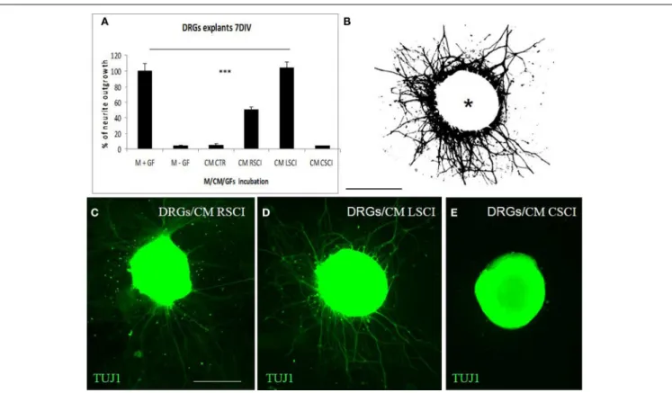
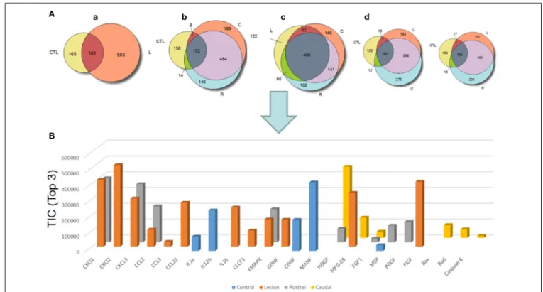
(9) Thèse de Stéphanie Devaux, Lille 1, 2016. FIGURE 18: HEAT MAP FROM THE HIERARCHICAL CLUSTERING OF CONDITIONED MEDIA 24 HOURS AFTER INJURY. PROTEIN UNDEREXPRESSION IS COLORED IN GREEN AND OVER-EXPRESSION IS IN RED. TABLE FOCUS ON THE CLUSTER SHOWN IN WHITE IN THE HEAT MAP, IN WHICH IG AND COMPLEMENT FACTORS ARE EXPRESSED. ...................................................................... 103. FIGURE 19: QUANTIFICATION OF SYNAPTOPHYSIN (SYN) AT THE LESION SITE (A) AND ROSTRAL-CAUDAL SEGMENTS (E) SHOWED SIGNIFICANT DECREASE OF SYN AFTER INJURY, WHILE RHOAI + FK506 TREATMENT INCREASED SYN EXPRESSION SIGNIFICANTLY AT LESION, BUT NOT IN ROSTRAL OR CAUDAL SEGMENTS (E), *P<0.05, ** P<0.001, *** P<0.0001 ONE-WAY ANOVA.. REPRESENTATIVE IMAGES OF SYNAPTOPHYSIN IMMUNOREACTIVITY (SYN, GREEN) REVEALED INTENSELY STAINED SYNAPTIC VESICLES – PUNCTATE STRUCTURES WITHIN THE SPINAL CORD- LESION SITE IN CONTROL (B) AND TREATED GROUP (D), NOT IN. SCI (C). CONFOCAL IMAGINES WITH DOUBLE LABELING OF GAP-43 (RED) AND SYN(GREEN) ANTIBODIES, CONFIRMED ENHANCED GROWTH OF AXONS WITH DENSE SYNAPTIC VESICLES DISTRIBUTION AFTER RHOA INHIBITOR + FK506 TREATMENT. (G). THE IN VIVO EXPERIMENTS DID NOT REVEALED SIGNIFICANT DIFFERENCES BETWEEN SCI AND TREATED RHOAI+ FK506 GROUPS IN GAP-43 IMMUNOREACTIVITY (I), OUTLINING GROWING AXONS WITHIN DAMAGED DORSAL AND LATERAL WHITE MATTER TRACTS (HA, HB). NOTE, HIGH NUMBER OF GAP-43 AXONS PENETRATING THE LESION SITE. SCALE BAR =25µM. BBB SCORE STUDY OF RAT WITH INJURY (SCI) AND AFTER TREATMENT WITH RHOAI + FK506 AT 0, 7, 14, 21, 28, 35, 42 AND 49 DAYS POST INJURY, REVEALS THAT WITH TREATMENT BBB SCORE REACHED TO 5, 14 DAYS AFTER SCI WHEREAS WITHOUT TREATMENT RATS NEED AROUND 30 DAYS TO GET SCORE AT 5. HOWEVER, THE SCORE DOES NOT INCREASE AFTER 14 DAYS (J).. ................................................................................................................................................................... 107 FIGURE 20: ND7/23 DRG CELL LINE CULTURED FOR 24 HOURS IN 1/3 OF SCI-CM ROSTRAL (R1), LESION (L) OR CAUDAL (C1) OR DMEM AS CONTROL, THEN AN ADDITION OR NOT OF RHOA INHIBITOR WAS APPLIED FOR 24 HOURS (A). QUANTIFICATION OF NEURITE OUTGROWTH BY IMAGEJ DEMONSTRATES THE EFFECT OF ROAI ON NEURITE OUTGROWTH (B) (ONE WAY ANOVA FOLLOWED BY TUKEY-KRAMER TEST *P<0.05, **P<0.01, ***P<0.001, NS= NON-SIGNIFICANT). ARROWS INDICATE THE NEURITE OUTGROWTH ..................................................................................................................................... 108. FIGURE 21: HEAT MAP OF PROTEINS FROM THE SECRETOME AFTER DIFFERENT STIMULATION OF ND7/23 DRG CELL LINE. CONTROL (DMEM) OR LESION (L), ROSTRAL (R1) OR CAUDAL (C1) CONDITIONED MEDIA FROM SPINAL CORD 3 DAYS AFTER INJURY WERE USED TO STIMULATE THE CELLS WITH OR WITHOUT STIMULATION OF RHOA INHIBITOR 24 HOURS AFTER CM STIMULATION (A).. ZOOM OF THE CLUSTER SHOWING A DIFFERENCE BETWEEN SCI-CM MEDIA STIMULATION WITH LESION CM AND PROTEINS NAME EXPRESSED IN THIS CLUSTER (B).. ............................................................................................................... 109. FIGURE 22: IMMUNOGLOBULINS IN SPINAL CORD INJURY TISSUE FROM ROSTRAL, LESION AND CAUDAL SEGMENTS AFTER 3, 7 AND 10 DAYS POST INJURY. EXPRESSION OF HEAVY CHAIN OF IGG (A) AND LIGHT CHAIN EXPRESSION (B) WERE ASSESSED BY WESTERN BLOT. HIGHER EXPRESSION OF IGGS HEAVY OR LIGHT CHAIN WAS OBSERVED AT 3 DAYS POST SCI IN THE LESION SITE.. ........ 111. FIGURE 23: HEAT MAP OF PROTEINS SECRETED BY ND7/23 CELL LINE AFTER STIMULATION WITH OR WITHOUT RHOAI. CONTROL IS CELL IN DMEM WITH OR WITHOUT RHOAI, CELLS STIMULATED WITH CONDITIONED MEDIA FROM SCI AND RHOAI IS NAMED. RHO AND WITHOUT RHOAI IS CALLED (NT). VENN DIAGRAM WITH THE TOTAL PROTEINS IDENTIFIED SHOW THE PRESENCE OF EXCLUSIVE AND COMMON PROTEINS FOR EACH CONDITIONS (A). HEAT MAP OF SECRETED PROTEINS FROM CONTROL VERSUS. SCI-CM STIMULATION WITH OR WITHOUT ROAI TREATMENT (RHO VS NT) HAVE SHOWN DISTINCT CLUSTERS. ................. 112 FIGURE 24: VENN DIAGRAM OF PROTEINS IDENTIFIED FROM THE CELLULAR PROTEIN EXTRACT (PE) OF ND7/23 CELL LINE. COMPARISON OF CONTROL (DMEM) WITH OR WITHOUT RHOAI AND PROTEIN EXTRACT FROM CELLS TREATED WITH SCI-CM WITH (RHOAI) OR NOT TREATED (NT). ............................................................................................................... 115. 9 © 2016 Tous droits réservés.. lilliad.univ-lille.fr.
(10) Thèse de Stéphanie Devaux, Lille 1, 2016. FIGURE 25: EVIDENCE FOR PROTEIN-PROTEIN INTERACTION NETWORK OF THE ENRICHED TRANSCRIPTION FACTORS CONSTRUCTED BY STRING. ...................................................................................................................................................... 116. 10 © 2016 Tous droits réservés.. lilliad.univ-lille.fr.
(11) Thèse de Stéphanie Devaux, Lille 1, 2016. Abbreviations ALG: alginate. IFN: interferon. ASC: adipocyte stem cell. IGF: insulin growth factor. BBB score: Basso, Beattie and Bresnahan. Ig: immunoglobulin. BBB: blood-brain-barrier. IL-: interleukin. BDNF: brain-derived neurotrophic factor. iPSC: induced pluripotent stem cells. BMSC: bone marrow stromal cell. KS: keratan sulfate. C5: cervical 5. LFA-1: Lymphocyte antigen 1. function-associated. CD: cluster of differentiation LPS: lipopolysaccharide ChABC: chondroitinase ABC MAC-1: macrophage-1 antigen CINC-1: cytokine-induced neutrophil chemoattractant 1. MAG: myelin associated glycoprotein. CNS: central nervous system. MALDI: matrix laser desorption ionization. CNTF: ciliary neurotrophic factor. MBP: myelin basic protein. CSF: cerebrospinal fluid. MHC: major histocompatibility complex. CSPG: chondroitin sulfate proteoglycans. MMPs: matrix metalloprotinases. DRG: dorsal root ganglia. MS: mass spectrometry. EGF: epidermal growth factor. MSC: mesenchymal stem sell. ER: estrogen receptor. MV: microvesicle. ESI: electrospray ionization. NCAM: Neural Cell Adhesion Molecule=CD56. FasL: Fas ligand. NG2: neuron-glial antigen 2. FcRβ : Fc receptor β chain. NGF: nerve growth factor. FGF-2: fibroblast growth factor 2. NO: nitrite oxide. GAG: glycosaminoglycan. Nogo-66 receptor: NgR. G-CSF: granulocyte-colony stimulating factor. NPC: neural progenitor cell. GDNF: Glial cell-derived neurotrophic factor. OEC: Olfactory ensheathing cell. GF: growth factor. OMgp: oligodendrocyte myelin glycoprotein. hESC: human embryonic stem cell. PNS: peripheral nervous system. HMGB1: high–mobility group box 1. RAG: recombinant activating gene. ICAM-1: Intercellular Adhesion Molecule 1 RAGE: Receptor for Advanced Glycation Endproducts. 11 © 2016 Tous droits réservés.. lilliad.univ-lille.fr.
(12) Thèse de Stéphanie Devaux, Lille 1, 2016. RhoAi: RhoA inhibitor ROCK: Rho kinase SAL: saline SCI: spinal cord injury T12: thoracic 12 TF: transcription factor TGF β: transforming growth factor β1 TIMPs: metalloproteinase inhibitor. 12 © 2016 Tous droits réservés.. lilliad.univ-lille.fr.
(13) Thèse de Stéphanie Devaux, Lille 1, 2016. Introduction Spinal cord injury (SCI) is one of the major traumas of the central nervous system (CNS). Traumatic SCI can lead to paralysis with complete or partial loss of neurological function below the injury site. The overall impact of SCI on both the patient and the society depends on a range of factors, including: the age of patient, the extent of the injury, the availability and timing of appropriate health care, services and the environment in which the person lives (physical, social, economic and attitudinal). Symptomatology treatment and physiotherapy care of patients with spinal cord injury requires high financial costs not only for the patient and his or her family but also for society. Statistics indicate that the U.S. has an annual growth of 11000 patients, which represents approximately 1.3 million people with a spinal cord injury. The economic costs of health care of a 25 years old patients are around $3 mil U.S. In the European Union, there are about five hundred thousand people affected by SCI. The incidence of SCI in France is roughly 20 cases per million inhabitants per year or 934 cases per year (Albert and Ravaud 2005). In Europe the most commonly injured age group is 16-34 years olds (43%), followed by 31-45 years olds (28%). Traumatic SCI can be the result of several different causes such as traffic accidents, falls, violence and sports injuries. SCI can be induced by non-traumatic causes involving communicable diseases such as tuberculosis and human immunodeficiency virus (HIV), and non-communicable diseases such as osteoarthritis leading to spinal stenosis, cardiovascular disease, nutritional deficiencies, neural tube defects, vitamin B12 deficiency and complication of medical care. The neurological outcomes depend on the range of damaged neuronal populations at the injury site, the level of disconnection of ascending and descending neuronal pathways, the secondary damage (edema, inflammation, ischemia) and the activation of regenerative processes (endogenous production of trophic factors, revascularization). Currently there is no effective therapy available for patients with SCI. One of the approved clinical treatments for SCI is the administration of methylprednisolone in order to modulate inflammation. However, high-doses of this drug are often associated with severe immunosuppression and side effects, such as pulmonary or urinary tract infections. The central. 13. © 2016 Tous droits réservés.. lilliad.univ-lille.fr.
(14) Thèse de Stéphanie Devaux, Lille 1, 2016. nervous system has a limited regenerative capacity due to the inflammatory response, inhibitory molecules and scar tissue. Different molecules and therapies have been used by many laboratories to preserve healthy tissue, to stimulate and reactivate spared tissue and to promote neuronal survival and axonal outgrowth. Injury to the spinal cord leads to an acute inflammatory response (Phillip G Popovich, Wei, and Stokes 1997). In parallel, immune response regulates the inflammation by the production of anti-inflammatory molecules (Riegger et al. 2007). Immune response is primordial to the preservation of tissue homeostasis. Acute phase (hours to days after injury) is directly linked to the trauma and results in neurodegeneration along with cell death at the lesion site. After primary trauma, cellular and molecular inflammatory cascades are initiated causing activation of resident microglia and astrocytes, as well as infiltration of innate immune cells such as lymphocytes, monocytes. The local release of cytokines and chemokines by microglia, macrophages and neurons induces a particular environment that can be either neurotoxic or neurotrophic. During acute phase, macrophages phagocyte cell debris and glial scar protect healthy tissue. Chronic inflammatory processes (weeks after trauma) lead to aberrant tissue remodeling and organ dysfunction. Among different mono-therapies, more complex-cellular therapy has several advantages targeting multiple aims: to bridge cavities or cysts, to replace dead cells, and to create a favorable environment allowing axonal regeneration (Rowland et al. 2008; Thuret, Moon, and Gage 2006). The transplantation of peripheral nerves, olfactory nervous system cells, embryonic CNS tissue, embryonic stem cells, adult stem cells (MSCs, NPCs), or activated macrophages (Thuret, Moon, and Gage 2006) has been studied to provide a solution to these aims. Molecular therapy is used to protect neurons from secondary process, to promote axon growth and to enhance conduction. Different types of molecules are used such as erythropoietin and minocycline (neuroprotective effect), growth factors (BDNF, GDNF, NGF, NT-3), chondroitinase ABC to eliminate chondroitin sulfate proteoglycans (CSPG) with the major component NG2 which inhibits the regeneration of damaged axons (Bradbury et al. 2002), and nogo-A is one of several neurite growth inhibitory molecules. Thereby, Nogo neutralizing 14. © 2016 Tous droits réservés.. lilliad.univ-lille.fr.
(15) Thèse de Stéphanie Devaux, Lille 1, 2016. antibodies or blockers of the post-receptors components RhoA are used to improve longdistance axon regeneration and sprouting (Martin E Schwab 2004). Rho pathway is important to control the neuronal response after CNS injury. A drug called cethrin that blocks activation of Rho is actually in phase I/IIa of clinical trials (Fehlings et al. 2011). However, among all these molecules and therapies currently used to ameliorate neuroprotection or neurite outgrowth, or to reduce inflammation, none of them allows a total understanding of the inflammation mechanism in the entire spinal cord to target specific segment at the appropriate time for SCI treatment. A lack of understanding of the molecular cross-talk occurring between cells at the lesion site and in the adjacent segments needs to be investigated. Such a study could give molecular targets taking into account both spatial and temporal data. Such an investigation could be performed by a proteomic approach which can be connected to cellular and physiological studies as well as to a global regeneration-activated gene (RAG) investigation. Mass spectrometry (MS) plays a central role among proteomic approaches. Several developments allow fast identification and relative quantification through label free quantification methods to thousands of proteins, allowing to identify such proteins of lower abundance as cytokines and chemokines (Meissner et al. 2013). MS is highly used in neuroscience to discover biomarker candidates and also to study the differential expressions of proteins at any given time in a proteome and they are then compared with the pattern of healthy ones (Singh et al. 2012).. Hypothesis In the current biomedical research, it is important to understand the mechanisms of immune response, molecular and biological processes following injury to improve regeneration of nerve tissue, to promote axonal growth and to replace damaged nerve cells to create favorable conditions for regeneration. Therefore, it is crucial to the understanding of the physiopathology that occurs after SCI to use the strength of mass spectrometry in order to understand the microenvironment along the entire spinal cord throughout different stages of the injury evolution. It is necessary to find a new treatment that would better control: i) the second process involving inflammation leading to the expansion of the central lesion which affects the rostro-caudal spinal segments that have not been damaged by the primary lesion 15. © 2016 Tous droits réservés.. lilliad.univ-lille.fr.
(16) Thèse de Stéphanie Devaux, Lille 1, 2016. ii) the formation of the glial scar at the lesion representing a chemical and mechanical barrier for a number of molecules involved in axonal regrowth iii) the production of trophic factors, both spatially and temporally to stimulate axonal growth, plasticity, and demyelination causing irreparable damage.. Objectives My PhD thesis was divided into 2 main parts. The first objective of my PhD thesis focuses on the understanding of the biological process occurring after a spinal cord injury through a spatial and temporal point of view during the first stage of the injury. Spatial and temporal analyses along the rostro-caudal axis were performed by mass spectrometry without preconditions. Based on in vitro and proteomics data, factors with immuno-modulatory efficiency on primary microglia and microglia cell line were characterized. Activation of microglial cells is carried out in the presence of the conditioned medium of the spinal cord to recreate the in vivo microenvironment. The switch M1 / M2 of microglial cells was studied to determine whether the phenotype would be pro or anti-inflammatory according to the position of the lesion site The second part aims to promote the regeneration in vitro using cell therapy, followed by in vivo stimulation of the regeneration process and spinal cord plasticity. Bone marrow derived- mesenchymal stromal cells are known for their immuno-modulatory effects after injury. In this context, we analyze the BMSCs secreted factors by mass spectrometry to identify the molecules involved in the modulation of the microglia. In vitro studies on primary microglia and BV2 cell line confirmed the high modulatory potential on inflammation and microglia polarization. Then the in vivo stimulation of the regeneration process and spinal cord plasticity will be studied with the aim to increase the number of axons going through the lesion site which are able to create connections between each other. In vivo, the injection of alginate biomaterial, directly into the lesion site fills the cavity to produce a scaffold for the growth of neurons and axons. Alginate coupled with trophic growth factors (GF) such as Epidermal Growth Factor (EGF) and Fibroblast Growth Factor 2 (FGF2) significantly enhanced the sparing of spinal cord tissue and increased the number of surviving neurons.. 16. © 2016 Tous droits réservés.. lilliad.univ-lille.fr.
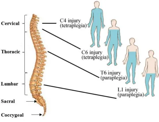
(17) Thèse de Stéphanie Devaux, Lille 1, 2016. 1. Spinal cord organization The spinal cord is part of the central nervous system which controls the voluntary movement of the limbs and trunk, and which receives sensory information from these regions. The spinal cord is located in the vertebral canal. In humans, the spinal cord is approximately 1cm thick and around 42 cm long and occupies the upper two-thirds of the vertebral canal. The spinal cord is cylindrical but also slightly flattened dorso-ventrally. The human spinal cord is composed of 31 segments in total: 8 cervical segments, 12 thoracic segments, 5 lumbar segments, 5 sacral segments and 1 coccygeal segment. Conversely, in rats, the spinal cord is made up of 34 segments: 8 cervical, 13 thoracic, 6 lumbar, 4 sacral, 3 coccygeal. The consequences of spinal cord injuries such as tetraplegia and paraplegia are dependent on the location of the trauma (Figure 1).. Figure 1: Consequences of the localization of the lesion (adapted myhealth.alberta.ca).. The spinal nerves run along the entire spinal cord- in humans there are 31 pairs of spinal nerves as supposed to 34 pairs in rats. The spinal nerves transmit sensory information from the target organs to the central nervous system, and send motor commands from the central nervous 17. © 2016 Tous droits réservés.. lilliad.univ-lille.fr.
(18) Thèse de Stéphanie Devaux, Lille 1, 2016. system to muscles and target organs. Each spinal nerve is attached by a ventral and a dorsal root. One root is formed by six to eight rootlets. The ventral rootlets are made up of axons of motor neurons and the dorsal rootlets are made up of the axons of the sensory neurons. Each dorsal root bears an ovoid swelling named dorsal root ganglion (DRG) that contains primary sensory neurons. These neurons have pseudo-unipolar morphology with a single short axon that divides into a peripheral and central branch. The central branch enters the spinal cord via the dorsal roots and carries information from the cell body of the DRG neuron to the spinal cord. The peripheral branch conveys sensory information from the body to the DRG neuron. The spinal cord is composed of gray and white matter. A transverse section shows that the gray matter is arranged in the form of a butterfly which is surrounded by the white matter. The white matter is composed of longitudinally running axons and glial cells. The gray matter is made up of neuronal cell bodies, dendrites, axons and glial cells. The neurons are mostly multipolar, but vary greatly in size in different laminae. The laminae were first described by Rexed in 1954 in the cat. Afterwards, these were used to describe cyto architectonic boundaries in the spinal cord in rats by Molander in 1984. Ten laminae are described and these are organized in a series of layers from dorsal to ventral axis. The spinal cord is enclosed in the meninges and consists of three layers: the dura mater, the intermediate arachnoid mater and the pia mater. These membranes and the cerebrospinal fluid provide a protection and nourishment for the spinal cord and spinal nerve roots.. 2. Model of injury SCI can result from three main causes which lead to tissue damage: compression caused by spinal discs or bone material pressing against the cord, destruction from direct trauma, and reduction in blood flow from the initial damage (ischemia). The injuries can be divided in two main categories: complete and incomplete. According to the American Association of Spinal Cord Injury Nurses (AASCIN), complete SCI refers to no preservation of motor and/or sensory function that exists more than three segments below the level of injury, whereas as incomplete SCI refers to some preservation of motor or/and sensory function existing more than three segments below the level of the injury. Complete transection is clinically rare. Most injured spinal cords maintain some tissue continuity across the area of injury to create an environment more tractable for regeneration. Several animal models have been developed with the aim to 18. © 2016 Tous droits réservés.. lilliad.univ-lille.fr.
(19) Thèse de Stéphanie Devaux, Lille 1, 2016. reproduce the complete or incomplete SCI in order to understand the anatomical and biological consequences. The rat model is widely used because the studies are inexpensive, the incidence of post-surgical infections are rare and rats are easy to care. Mouse models are also largely studied. Larger mammals such as dogs and cats are rarely used since they require expensive after care and housing, and due to the fact that stringent ethical restrictions are in place. Several animal models are used in research to mimic clinical human SCI. The majority of human SCIs are due to motor vehicle accidents, falls and sport injuries, and involve a sudden compression of the spinal cord, usually as a result of vertebral column damage that allows bone or disc material to intrude on the spinal canal space. Compression injuries occur as a secondary consequence of injury or as a result of tumor growth. The laminectomy allows the exposition of the spinal cord by removing the bone. Complete transection injury reflects clinical symptoms of complete SCI patients. This model is performed after laminectomy with spring scissors. It offers the advantage of complete damage to a given tract at a defined place and time. The complete transection model, when performed accurately, excludes the possibility that axonal sparing results in improved functional outcomes. However most of the traumas after injuries are not complete transections of the spinal cord. Clip compression is processed with calibrated clip to exert a specific force- 50g for a severe response and 35g for a moderate response (Bruce, Oatway, and Weaver 2002) (Figure 2) but this method does not truly mimic the injury seen in humans.. 19. © 2016 Tous droits réservés.. lilliad.univ-lille.fr.
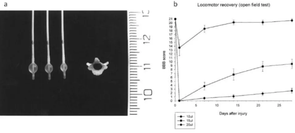
(20) Thèse de Stéphanie Devaux, Lille 1, 2016. Figure 2: a. Clip compression. b. An intraoperative picture showing a clip compressing the spinal cord at T2 to create the injury (Poon et al. 2007).. The balloon compression model was first described by Tarlov in 1953 in dogs (Tarlov, Klinger, and Vitale 1953). The method uses different volumes to inflate the balloon and different compression durations in order to induce different grades of damage (Figure 3). After laminectomy, a 2-French Fogarty catheter is inserted into the dorsal epidural space through a small hole made in the T10 vertebral arch, which is then advanced cranially to the T8-9 spinal level and inflated for 5 min. A volume of 15µL of saline produced complete paraplegia followed by gradual recovery.. Figure 3: Balloon compression model. a. A photograph representing the size of the T9 vertebra and catheters inflated with different volumes of saline 10, 15 and 20µL respectively. b. Locomotor function recovery after SCI graded on an expanded scale (BBB score). 21 is normal locomotor function, 0 represents no locomotor function. (adapted from Vanicky et al. 2001). 20. © 2016 Tous droits réservés.. lilliad.univ-lille.fr.
(21) Thèse de Stéphanie Devaux, Lille 1, 2016. Computer controlled contusion consists of an animal trap that reproducibly delivers a defined weight to the exposed spinal cord with a computer monitoring the dynamics impact (Stokes, Noyes, and Behrmann 1992). This method is reproducible but the equipment is expensive. The transaction model mimics a complete spinal transaction (rare in clinic) which is performed by using spring scissors following laminectomy, producing a laceration of the cord (Nakae et al. 2011). The behavioral patterns are correlated with the severity of the injury and are evaluated in animals by using the BBB score, which is an open field locomotor activity test that was originally developed at the Ohio State University (Basso, Beattie, and Bresnahan 1995). The scale ranges from 0 to 21 with 0 indicating complete paraplegia and 21 indicating physiological locomotor movement (Table 1). BBB score. Observations. 0 1 2 3 4 5 6 7 8 9. No movement Slight moveent of 1-2 joints Extensive movement of 1 joint (+ slight momvement of another) Extensive movement of 2 joints Slight movement of all 3 joints Slight movement of 2 joints AND extensive movement of 1 Extensive movement of 2 joints AND slight movement of 1 Extensive movement of all 3 joints Sweeping OR plantar placement (No weight support) Plantar placement (weight support in stance only) OR weight supporting dorsal stepping. 10 11 12 13 14. Occasional steps, No coordination Frequent-consistent steps, no coordination Frequent-consistent steps, occasional coordination Frequent-consistent steps, frequent coordination Consistent steps, consistent coordination, paw position rotated OR occasional stepping. 15. Consistent steps, consistent coordination, No or occasional toe clearance. 16. Consistent steps, consistent coordination, frequent toe clearance, paw rotated at lift off. 17. Consistent steps, consistent coordination, frequent toe clearance, paw is predominantly parallel Consistent steps, consistent coordination, consistent toe clearance, paw is parallel at initial contact, and rotated at lift off Consistent steps, consistent coordination, consistent toe clearance, paw is parallel at initial contact and at lift off tail is down part or all time Tail consistently up, trunk instability Trunk stability. 18 19 20 21. Movements only. Plantar placement. Coordination. Paw position, Toe clearance. Tail, trunk stability. Table 1: The 21-point Basso, Beattie, Bresnahan locomotor rating scale (Adapted from Bassso et al. 1995).. 21. © 2016 Tous droits réservés.. lilliad.univ-lille.fr.
(22) Thèse de Stéphanie Devaux, Lille 1, 2016. The BBB score evaluates the movements of the hind limb of an animal after injury to express a state of recovery. A method for gait analysis called catwalk is used to evaluate the paw position of the animal after injury and the improvement after treatment (Hamers et al. 2001). Each front and hind paw of animal that makes contact with the glass floor is automatically detected. The software registers footprints and calculates a wide range of parameters relative to the individual footprints (stand, print length), the relative positions of footprints (stride length, base of support), the time-based relationship between footprints (cadence), toe spread and paw angle.. 3. Pathophysiology Spinal cord injury is one of the major injuries of the central nervous system. The pathological mechanisms consist of two phases; the primary and the secondary phase (Figure 4). The most common form of acute SCI is a compressive type of injury in which displaced inter-vertebral discs and ligaments exert force on the cord causing immediate traumatic injury and often sustained compression (Sekhon and Fehlings 2001).. Figure 4: Schema of events following spinal cord injury. 3.1. Primary injury Primary injury is the result of the mechanical trauma which produces traction and compression forces. Primary mechanisms rarely transect or fully disrupt the anatomical continuity of the cord. Blood vessels are directly damaged and the axons are disrupted. Axons are present to traverse the lesion site with a demyelinated long tract axon. A few minutes after injury, micro-hemorrhages start to develop in the central grey matter and spread out radially 22. © 2016 Tous droits réservés.. lilliad.univ-lille.fr.
(23) Thèse de Stéphanie Devaux, Lille 1, 2016. and axially over the next hours. The swelling of the spinal cord is observed at the injury site and it occupies the entire space of the spinal canal. Ischemia occurs when cord swelling exceeds venous blood pressure. Microglia cells are the first glial cells to be activated following injury, in less than two hours (Figure 4). These pathological events, such as damages to axons, cells and blood vessels, leads to the release of toxic chemicals and factors that attack intact neighboring cells within the early acute phase (after 48 hours).. 3.2. Secondary injury Secondary. injury. refers. to. the. molecular. and. cellular. cascade. causing. neuroinflammation (Donnelly and Popovich 2008), immune associated neurotoxicity which contributes to axonal and neuronal necrosis, edema, glutamate mediated excitotoxicity, ROS production and vascular dysfunction (Figure 4). The secondary phase results in tissue destruction whereas neurological deficits are present immediately following injury. Resident microglia respond immediately to the injury (Watanabe et al. 1999). Between 2 and 7 days most of the cells in the lesion are infiltrating macrophages (Phillip G Popovich, Wei, and Stokes 1997; Donnelly and Popovich 2008). This period of macrophages infiltration corresponds to the ascending sensory axon retraction. This process is not permanent, because when axons lost macrophages contact after retraction, they were able to extend (Horn et al. 2008). Reactive astrocytes deposited proteoglycans (CSPGs), including neurocan and phosphacan, are major factors that inhibit regeneration by forming the glia scar within hours after injury. The glial scar serves to compact inflammatory cells to demarcate the injury site from the healthy tissue (Faulkner et al. 2004) and re-seal the blood brain barrier after the injury. Astrocytes become hypertrophic after moving away from the center of the lesion and dramatically upregulate the production of inhibitory chondroitin sulfate proteoglycans (CSPGs) that prevent regeneration (Silver and Miller 2004). But the glial scar seems to also have beneficial functions for stabilizing fragile CNS tissue by separating the injury from healthy tissue and preventing a cascading wave of uncontrolled tissue damage (Faulkner et al. 2004). Scar tissue together with CSPGs and NG2 are known for their inhibitory effect on axonal growth (Fidler et al. 1999; McKeon, R J et al. 1991; Smith-thomas et al. 1994). The degradation. 23. © 2016 Tous droits réservés.. lilliad.univ-lille.fr.
(24) Thèse de Stéphanie Devaux, Lille 1, 2016. of CSPGs by using inhibitors or specific enzymes shows an increase in axonal growth and regeneration (Caggiano et al. 2005; Smith-thomas et al. 1995; Tom and Houlé 2008). After injury, neurons are exposed to a microenvironment that contains toxic factors, which results in neuronal loss and degeneration. Astrocytes have an important scavenging function, for example the regulation of excessive levels of glutamate. CSPGs produced by activated astrocytes in the lesion area create a diffusion barrier for molecules that are harmful to the healthy tissue and attenuate the spread of neurotoxicity (Roitbak and Syková 1999; Vorísek et al. 2002). Astrocytes provide trophic support at the injury site (White, Yin, and Jakeman 2008). Glial scar could be crucial for neuronal survival by filling the gaps in the lesion area, creating a scaffold for the vascularization network. Astrocytes are known to have a direct effect on the intensity of blood flow (Parri and Crunelli 2003). Neuroinflammation is one of mechanisms involved after injury. An immune response is important to protect the organism but immune activity in the CNS could have deleterious effects by accelerating tissue damage. In the CNS, immune cells acquire diverse phenotypes depending on the microenvironment. Among glial cells, microglia cells, first to be recruited, are able to play a bifunctional role by secreting toxic factors and contributing to tissue damage, but they are also able to release neuroprotective and neurotrophic molecules to allow tissue repair (Aloisi 2001).. 4. Immune response after SCI The central nervous system has a highly specialized immune-modulatory microenvironment to protect itself from immune-mediated inflammation. The blood-brainbarrier (BBB) is a physiological and anatomical element to balance peripheral immune cells and molecules entry. Immune response in the CNS is mediated by resident microglia and astrocytes, which are innate immune cells without direct counterparts in the periphery. However, microglia and astrocytes are able to engage a cross-talk with CNS-infiltrating T cells, neurons and other components of the innate immune system.. 24. © 2016 Tous droits réservés.. lilliad.univ-lille.fr.
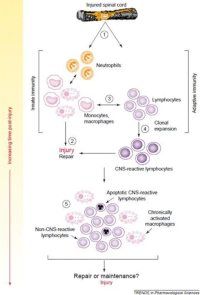
(25) Thèse de Stéphanie Devaux, Lille 1, 2016. Figure 5: Time recruitment of microglia, neutrophils, monocytes/macrophages and lymphocytes after SCI (Neirinckx et al. 2014).. Inflammatory responses are the central point in the physiopathologic processes of acute and chronic phases of SCI. Neutrophils are the first inflammatory actors to invade the injury site with the peak appearing at 24h post injury. Circulating monocytes/macrophages are then recruited peaking at 7 days post-injury, with lymphocytes progressively invading the lesion epicenter thereafter. (Figure 5).. 4.1. Innate immune system After SCI, cells need restorative support, which is provided by inflammatory responses. However, excessive or chronic inflammation can become harmful. The micro-environment present at the lesion site confers this balance. This injured environment is composed of proinflammatory cytokines as tumor necrosis factor α (TNFα), interleukins IL-1 and Il-6 at the early phase of the inflammation but also some anti-inflammatory molecules are released as transforming growth factor β1 (TGFβ), IL-10. 4.1.1. Neutrophils recruitment Neutrophils are considered the first inflammatory cells to arrive at the site of injury with a peak at 24 hours after injury (Means and Anderson 1983). They are rapidly mobilized from the bone marrow in response to signals from pro-inflammatory CXC (CXCL8) family chemokines, IL-8 and cytokine-induced neutrophil chemo-attractant 1 (CINC-1) to mediate pleiotropic functions in the immune-inflammatory response (Tonai et al. 2001). Neutrophils adhere to post-capillary venules 6-12 hours post SCI and by 24h they migrate into the lesion site to phagocytose debris (Taoka et al. 1997). Neutrophils generate their 25. © 2016 Tous droits réservés.. lilliad.univ-lille.fr.
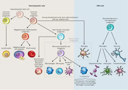
(26) Thèse de Stéphanie Devaux, Lille 1, 2016. own cytokines after stimulation by pro-inflammatory mediators and produce proteases via the NF-kB translocation pathway. Phagocytic activity can induce NF-kB activation (McDonald and Cassatella 1997). Matrix metalloproteases (MMPs) as MMP-9 and cytokines TNFα, IL-1, IL-8 and TGF-β are these mediators (Cassatella 1995). 4.1.2. Microglia cells Through the production of inflammatory factors, microglia cells represent the archetypal cells. Microglia are a unique myeloid cell population, derived from the yolk sac during a narrow time window before vascularization or definitive hematopoiesis in the embryo (Figure 6). Microglia cells, in the CNS parenchyma, are sustained by proliferation of resident progenitors, independent of blood cells. Rio-Hortega was the first to use the term microglia in 1917. He observed that these cells were not stationary and were able to consume cellular debris. They scan the microenvironment with their branched processes to phagocyte cell and myelin debris. Microglia are small glial cells present in the brain and spinal cord. They have a small soma, little perinuclear cytoplasm and fine, branched processes.. Figure 6: Microglia are the only hematopoietic cells found in the parenchyma of the CNS (Ransohoff and Cardona 2010).. Resident microglia cells express myeloid-monocytic markers such as Fc receptorscluster of differentiation (CD32 and CD64), complement receptors CD11b and CD11c 26. © 2016 Tous droits réservés.. lilliad.univ-lille.fr.
(27) Thèse de Stéphanie Devaux, Lille 1, 2016. integrins, major histocompatibility complex MHC class I and II, and CD45 (Tambuyzer, Ponsaerts, and Nouwen 2009). After injury, the resident ramified microglia morphologically transform into cells with retracted processes and enlarged cell bodies, and increase in number at the affected site. Microglia cells with this particular shape are generally referred to as “activated microglia or reactive microglia”. Even if macrophages and microglia have different origins, microglia are related to resident tissue macrophages. Microglia response following pathological stimuli is characterized by an accumulation at the lesion site and the release of various bioactive molecules. Two categories of molecules are released- some are cytotoxic or pro-inflammatory, and others may aid survival and regeneration. Resident monocytes are the first cell types to respond after injury within 1-2 hours, which starts the initial acute inflammatory response accompanied by an expression of TNFα and IL-1 (M1 phenotype). This leads to the recruitment of other immune cells. M1 macrophages promote phagocytosis. 8 hours after of the injury, the production of proinflammatory cytokines is terminated, thus promoting the differentiation of macrophages into an anti-inflammatory M2 phenotype with the expression of arginase 1 and a mannose receptor (CD206). M2 macrophages promote angiogenesis and matrix remodeling while suppressing destructive immunity (Sica et al. 2006). The ratio M1/M2 varied in terms of microenvironment (Figure 7).. 27. © 2016 Tous droits réservés.. lilliad.univ-lille.fr.
(28) Thèse de Stéphanie Devaux, Lille 1, 2016. Figure 7: Macrophage polarization after SCI. At the early stage macrophages are predominantly M1 with the presence of pro-inflammatory molecules and M1 will release pro-inflammatory factors. At the later stage the ratio M1/M2 evolves to have an anti-inflammatory response and then cytotoxic effects (adapted from David & Kroner 2011).. 4.1.3. Astrocytes Reactive astrocytes are a prominent feature of the cellular response to SCI. They allow for an exchange in gene expression, hypertrophy, process extension and cell division (Eddleston and Mucke 1993). Reactive astrocytes are involved in scar tissue formation which prevents axonal regeneration (Rudge and Silver 1990). Therefore, they are often considered detrimental to the functional outcome after SCI. However, the action of reactive astrocytes is conserved throughout the vertebrate evolution (Larner et al. 1995), suggesting a beneficial role. Astrocytes are able to clear glutamate and potassium ions from the extracellular space, representing a potential energy source and providing trophic support. All these factors are crucial for the survival of neurons located at the margins of the lesion site and are colocalized with the scar. Growth factors sucg as insulin growth factors (IGFs), nerve growth factor (NGF), brain-derived neurotrophic growth factor (BDNF) and neurotophin-3 (NT-3), as well metabolites including glucose and nutrients are produced by astrocytes to support the survival of the cells (Schwartz and Nishiyama 1994). It was shown that astrocytes can directly protect neurons from nitric oxide toxicity through a glutathione-dependent mechanism (Y. Chen et al. 2001). Although astrocytes produce CSPGs in the lesion area, this molecule creates a diffusion 28. © 2016 Tous droits réservés.. lilliad.univ-lille.fr.
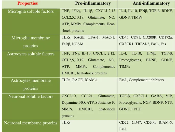
(29) Thèse de Stéphanie Devaux, Lille 1, 2016. barrier for molecules that are potentially harmful to the spared tissue (Roitbak and Syková 1999) and thereby attenuates the spread of neurotoxicity and prevents excitatory amino acidinduced neuronal death (Sato et al. 2008).. 4.2. Relation between glial cells and neurons The communication between the neural and the immune system is obvious. The communication between the two is mediated by soluble factors such as neurotransmitters, neuromodulators, neuropeptides or through cell-cell contact by neuroimmune regulatory molecules that can reduce or inhibit any exacerbated inflammatory response (Tian et al. 2012). Glial cells, such as microglia, astrocytes and neurons can express pro- and anti-inflammatory molecules to modulate the injured environment and these molecules play a major role in their communication (Table 2). Properties Microglia soluble factors. Pro-inflammatory. Anti-inflammatory. TNF, IFNγ, IL-1β, CXCL1,2,12. IL-4, IL-10, IFNβ, TGF-β, BDNF,. CCL2,5,10,19, Glutamate, NO,. GDNF, TIMPs. ATP, MMPs, Complements, Heatshock proteins. Microglia membrane proteins Astrocytes soluble factors. TLRs, RAGE, LFA-1, MAC-1,. CD45, CD91, CD200R, CD172a,. FcRβ, NCAM. CX3CR1, TREM-2, FasL, Fas. TNF, IFNγ, IL-1β, CXCL1, 2,12,. IL-4,. IL-10,. IFNβ,. TGF-β,. CCL2,5,10,19, Glutamate, NO,. Proteoglycans,. BDNF,. GDNF,. ATP,. TIMPs. MMPs,. Complements,. HMGB1, heat-shock proteins. Astrocytes membrane. TLRs, RAGE, ICAM-1. FasL, Complement inhibitors. proteins Neuronal soluble factors. Glutamate,. TGF-β, CX3CL1, GABA, VIP,. Dopamine, NO, ATP, Substance-P,. Proteoglycans, NGF, BDNF, NT3,. MMPs,. GDNF, CNTF. CXCL10,. CCL21,. HMGB1,. heat-shock. proteins. Neuronal membrane proteins TLRs. CD22, CD47, CD200, ICAM-5, FasL. Table 2: Soluble factors and membrane proteins expressed by microglia, astrocytes and neurons and their pro- or anti-inflammatory properties (adapted Tian et al., 2012).. 29. © 2016 Tous droits réservés.. lilliad.univ-lille.fr.
(30) Thèse de Stéphanie Devaux, Lille 1, 2016. 4.2.1. Cellular interactions Neurons possess several membrane molecules to control local immune functions by targeting local immune cells such as microglia, astrocytes and peripheral immune cells, which are present in the CNS. Neurons might indirectly suppress T-cell activation by restricting antigen, presenting properties of glial cells, directly suppressing T-cell activation, favoring a Th2 profile or promoting apoptosis of activated microglia and T cells (Tian et al. 2012). The microglia activation may be beneficial, deleterious or neutral. Neurons express cell surface glycoproteins (CD22, CD47, CD200, NCAM) (Table 1) to prevent microglia activation. A relationship between the nervous and the immune system has been studied this past decade. Indeed, glial cells (microglia and astrocytes) not only serve supportive and nutritive roles for neurons, but serve also to defend the CNS. An excessive and prolonged glial cells activation results in more severe and chronic neuronal damage, which leads to neuroinflammation and neurodegeneration. The interaction in healthy CNS between neurons and astrocytes allows regulation of blood supply control, neurotransmission, synaptic formation and plasticity and metabolic support. In healthy CNS, neurons-microglia communication allows for debris clearance and immune surveillance. After a CNS injury, the balance is broken which gives way to excitotoxicity, glial scar and inflammatory damage. 4.2.2. Molecular interactions The neuronal cell adhesion molecule (NCAM/CD56) is expressed on the surface of neurons, astrocytes and microglia (Chang, Hudson, Wilson, Liu, et al. 2000; Chang, Hudson, Wilson, Haddon, et al. 2000). NCAM is involved in cell-cell adhesion, synaptic plasticity, neurite outgrowth and others processes. The astrocytes-neuronal interactions via NCAM lead to the modulation of glial scar formation by the inhibition of astrocyte proliferation (Krushel et al. 1998). Furthermore, NCAM modulate microglial activation by decreasing TNFα and nitric oxide (NO) after glial stimulation with lipopolysaccharides (LPS) (Chang, Hudson, Wilson, Liu, et al. 2000; Chang, Hudson, Wilson, Haddon, et al. 2000).. 30. © 2016 Tous droits réservés.. lilliad.univ-lille.fr.
(31) Thèse de Stéphanie Devaux, Lille 1, 2016. CD200 (OX2) is an important molecule which contributes to the anti-inflammatory and regulatory environment (Table 2). CD200 expression has been detected in oligodendrocytes, reactive astrocytes and neurons, but not in astrocytes or microglia (Koning et al. 2009). Microglia possess CD200 receptors (CD200R), thus neuronal CD200 down-modulates the activation state of perivascular macrophages and microglia (Hoek et al. 2000). Defects in the CD200-CD200R pathway play a critical role in neurodegenerative disease development as Multiple sclerosis and Parkinson’s disease (Koning et al. 2007; S. Zhang et al. 2011). The expression of chemokine CX3CL1 (fractalkine) and its receptor CX3CR1 is limited to neurons and microglia (Hughes et al. 2002). Microglia are characterized by a prominent expression of the chemokine receptor CX3CR1. CX3CR1 is a conventional Gαi-coupled seven transmembrane receptor. CX3CL1, the ligand of CX3CR1, is synthesized by a transmembrane protein with the CX3C chemokine domain displayed on an extended highly glycosylated, mucin-like stalk (Bazan et al. 1997). Under inflammatory processes, proteolytic cleavage of CX3CL1 occurs by disintegrin-like metalloproteinase ADAM 10 or 17 (Hundhausen et al. 2003; Garton et al. 2001) (Figure 8). CX3CR1 receptor expression has also been demonstrated for an NK cell subset and certain T cell populations (Imai et al. 1997). Cocultures of neuron-microglia with CX3CL1 exposure reduced inflammatory neuronal death in vitro (Mizuno et al. 2003). A higher level of microglia activity was observed in mice with a CX3CR1 deficiency in three models of brain diseases, and this was also accompanied by increased neuronal vulnerability (Cardona et al. 2006). These studies have shown that in normal mice, neurotoxic microglia activity is suppressed by CX3CL1-CX3CR1 signaling.. 31. © 2016 Tous droits réservés.. lilliad.univ-lille.fr.
(32) Thèse de Stéphanie Devaux, Lille 1, 2016. Figure 8: CX3CR1 receptor in microglia and its ligand CX3CL1 expressed by a neuron. A) Fractalkine composition and the proteo cleavage by ADAM10/17. B) Different effects of the cleavage of CX3CL1 on microglia activity (Wolf et al. 2013).. Neurons are able to control microglia with two types of signals; “On” or “Off” (Biber et al. 2007) (Table 3). Off signals (TGF-β, CD22, CX3CL1, neurotransmitters, CD20) are found in healthy conditions to maintain homeostasis and also restrict microglia activities under inflammatory conditions to prevent damage to healthy tissue. Conversely, “On” signals (CCL21, CXCL10, MMP3 (from apoptotic neurons) are produced by damaged and impaired neurons to activate microglia (pro- or anti-inflammatory). Neuron signals. Released signals TGF-β,. Off signals. CD22,. Membrane signals. CX3CL1, CD200,. neurotransmitters,. CD22,. CD47,. NGF, CX3CL1?. BDNF, NT-3 On signals. CCL21,. CXCL10,. ATP, TREM2 ligand. UTP, Glutamate, MMP3. Table 3 : Neuron-mediated Off and On signals to modulate microglia activity (Adapted from Biber et al., 2007).. 32. © 2016 Tous droits réservés.. lilliad.univ-lille.fr.
(33) Thèse de Stéphanie Devaux, Lille 1, 2016. 4.3. T cells and innate immune cells Lymphocytes represent the adaptive cellular arm of the immune system (Figure 9). Lymphocytic infiltration of the injury site occurs during the first week post-injury and is maintained chronically (Beck et al. 2010). Some lymphocytes can react with CNS proteins and the activation of CNS–reactive T cells leads to the demyelinating disorder in multiple sclerosis (Martin, McFarland, and McFarlin 1992). Popovich in 1996 demonstrated that CNS-reactive T cells are activated in SCI (P G Popovich, Stokes, and Whitacre 1996).. Figure 9: Recruitment of innate immune cells and adaptive immune cells after SCI and the progression to the chronic phase (Phillip G. Popovich and Jones 2003).. 33. © 2016 Tous droits réservés.. lilliad.univ-lille.fr.
(34) Thèse de Stéphanie Devaux, Lille 1, 2016. Lymphocytes respond to myelin protein after SCI and may contribute to post-traumatic secondary damages. However, there is increasing evidence that autoreactive T-lymphocytes may also convey neuroprotection and promote functional recovery after a CNS injury. Myelin basic protein (MBP)-reactive T cells are increased in SCI and stroke patients, which provides evidence of an association between CNS trauma and the activation of CNS reactive T cells (Kil et al. 1999). In order to explain the effects of MBP-reactive T cells, Lewis rats and transgenic mice enriched in MBP-reactive T cells were used. These studies demonstrated that CNS-reactive T cells can exacerbate axonal injury, demyelination and functional loss after SCI (P G Popovich, Stokes, and Whitacre 1996; T. B. Jones et al. 2002). T-cells reactions against CNS proteins exacerbate acute tissue damage while simultaneously triggering the induction of chronic immunoregulatory networks. Surprisingly, autoimmune cells originally considered as pathogenic in autoimmune diseases were shown to be neuroprotective after CNS trauma. In contrast, several studies have suggested that T cells were beneficial to disease progression and survival after amyotrophic lateral sclerosis (Chiu et al. 2008; Beers et al. 2008). The presence of CD4+ T cells provides supportive neuroprotection by modulating the trophic/cytotoxic balance of glia. These contradictory results may be explained by the distinct roles of each Tlymphocyte subset.. 5. Treatments for spinal cord injury Spinal cord injuries induce several complications; patients suffer from motor and sensory impairments leading to complications in their lives. Over the last two decades, numerous clinical trials have been fully evaluated to restore damaged spinal cords. Despite broad research on animals, only few studies lead to pharmacological therapies due to the side effects that occur, or to the lack of regeneration or functional recovery. After SCI, many molecules are involved in motor and sensory dysfunction. Therefore, therapy should target several points, such as neuroprotection, plasticity, regeneration and replacement of lost cells (Figure 10).. 34. © 2016 Tous droits réservés.. lilliad.univ-lille.fr.
(35) Thèse de Stéphanie Devaux, Lille 1, 2016. Figure 10: Several parameters are involved in spinal cord repair. Several molecules are studied for their various properties, such as neuroregeneration and the sprouting of spinal tracts (BDNF, GDNF, NT-3), remyelination (Withanoside IV, 4Aminipyridine), antagonization of inhibitory factors from myelin or glial scar (Cethrin, Chondroitinase ABC, Y27632), protection of neuronal death, inhibition of inflammatory responses in the chronic phase (Minocycline, Estrogen, MPSS), plasticity: rewiring of propriospinal interneurons, and cells and tissue transplantation (OECs, BMSCs, Peripheral nerve cells, hNSCs, nerve grafts).. 5.1. Myelin neurite growth inhibitors Axonal regeneration is one of the important key factors after SCI. Three criteria bring out the crucial role of myelin-associated neurite growth inhibitors in preventing CNS regeneration. The depletion of oligodendrocytes and myelin enhances the regeneration of descending tracts in the differentiated spinal cord of rats (M E Schwab and Bartholdi 1996). Antibodies against Nogo-A enhance regenerative sprouting and long distance elongation (Schnell and Schwab 1990). Finally, the autoimmunization of rats with myelin or spinal cord homogenates allows for regenerative sprouting and axonal growth after SCI (D. W. Huang et al. 1999). Nogo (Martin E Schwab 2004), myelin associated glycoprotein (MAG, (McKerracher et al. 1994)) and oligodendrocyte myelin glycoprotein (OMgp, (K. C. Wang et al. 2002)) are all neurite growth inhibitory components found in white matter of the CNS. Nogo-A is 35. © 2016 Tous droits réservés.. lilliad.univ-lille.fr.
(36) Thèse de Stéphanie Devaux, Lille 1, 2016. principally expressed by CNS oligodendrocytes, whereas Nogo-B is expressed in the central and peripheral nervous system and other peripheral tissues and Nogo-C is found in muscle. By binding to an axonal Nogo-66 receptor (NgR) protein, Nogo, MAG and OMgp cause the collapse of axonal growth cones and stop axonal extension (McKerracher et al. 1994; M. S. Chen, Huber, van der Haar, et al. 2000; K. C. Wang et al. 2002). Diverse studies have used peptides that competitively inhibit Nogo-66 interaction with NgR (S. Li and Strittmatter 2003), anti-Nogo-A antibodies (Schnell and Schwab 1990) and soluble ectodomain fragments of the NgR (Fournier et al. 2002). P-75, the receptor for nerve growth factor (NGF), may act as a signal transducing subunit. Nogo-A can interact with different receptors such as nogo-A receptors, NgR, p75 and is similar for MAG and OMgp (Figure 11). Both calcium and RhoA/ Rho kinase (ROCK) pathway are linked with Nogo (Bandtlow et al. 1993; Fournier, Takizawa, and Strittmatter 2003). The inhibition of one of these components has been shown to prevent myelin and Nogo-A induced growth cone collapse and growth inhibition (Niederost et al. 2002).. Figure 11: Myelin neurite growth inhibitors Nogo-A, MAG and OMgp and their different receptors NgR, p75. Inhibition of Nogo-A is possible by the different processes in red (Martin E Schwab 2004).. The inhibition effects of Nogo can be prevented by using antibodies against Nogo-A, Nogo gene deletions, soluble NgR fragments, NgR blocking peptides, inhibition of Rho-A, 36. © 2016 Tous droits réservés.. lilliad.univ-lille.fr.
Figure
+7
Documents relatifs
4 -Marie Camille Débourg, Joël Clavelin et Olivier Perrier, pratique de marketing,2. Berti édition Alger, 2 ème édition, 2004,
Episodic shocks appear in both series — a 35 year high in woodcock numbers in the exceptionally cold winter of 1962/1963, and the 1929 stock market crash.. Neither series
State transfer faults may also be masked, though to mask a state transfer fault in some local transition t of M i it is necessary for M to be in a global state that leads to one or
Le concept dénotatif défini est très particulier en ce que le groupe nominal correspondant peut souvent être remplacé par un nom propre de l'entité qu'il dénote, ce
Nos résultats ont montré que l’expression de Prdm12 dans la partie ventrale de la moelle épinière est dépendante de l’acide rétinoïque et du facteur de
Le cours de sécurité informatique et cryptologie des systèmes d‟information se veut pour objectif primordial de donner aux étudiants ayant participés des techniques
• Fixation de ces mêmes neurotransmetteurs sur la membrane post synaptique provocant ainsi sa dépolarisation et par conséquent le transfert de l’ influx nerveux...
To further investigate the direct transmission of antimicrobial immune effectors into eggs from primed mothers, we conducted a RNA interference approach aimed at comparing
