Université de Montréal
Study of the Role of the p16INK4a Gene in Tumor Progression and Tissue
Regeneration/Function Following Exposure to Ionizing Radiation.
Par Lina Palacio
Département de Pharmacologie, Université de Montréal Faculté de Médecine
Thèse présentée à la Faculté de Médecine en vue de l’obtention du grade de Docteur en Pharmacologie
Décembre 2017
© Lina Palacio, 2017 Université de Montréal
Département de Pharmacologie, Faculté de Médecine
_______________________________________________________________
Cette thèse intitulée:
Study of the Role of the p16INK4a Gene in Tumor Progression and Tissue
Regeneration/Function Following Exposure to Ionizing Radiation.
Présentée par: Lina Palacio
A été évaluée par un jury composé des personnes suivantes : Dr Noel Raynal Président-rapporteur Dr Christian Beauséjour Ddirecteur de recherche Dr Gerardo Ferbeyre Membre du jury Dr David Dankort Examinateur externe Dr Francis Rodier Représentante du doyen
III
Résumé
La sénescence est un important mécanisme cellulaire qui prévient la tumorigenèse et se caractérise par un arrêt permanent du cycle cellulaire orchestré principalement par les inhibiteurs des cycline-kinases dépendantes (i.e p16INK4a).
La sénescence est une caractéristique importante du vieillissement, mais un déséquilibre dans son induction peut être délétère pour la régénération tissulaire et paradoxalement pour la progression tumorale. L'irradiation (IR) est couramment utilisée comme approche thérapeutique dans le cancer. Chez les enfants survivants du cancer, l’exposition à l’irradiation et à la chimiothérapie entrainent le développement d’importants effets secondaires, lesquels sont associés à une forme de vieillissement prématuré. La formation de cellules sénescentes, en inhibant la prolifération tissulaire et en sécrétant des cytokines proinflammatoires, pourrait être en être responsable. Notre groupe a précédemment démontré que le gène p16INK4a est augmenté de manière tardive (environ 8 semaines) suite à une
exposition à l’irradiation. Il n'a pas encore été étudié si cette expression retardée survient en réponse aux dommages causés par l'irradiation sur l’homéostasie tissulaire ou à titre de mécanismes de suppression tumorale. Un objectif de cette thèse visait donc à déterminer s’il était possible de moduler/inhiber l’expression de
p16INK4a dans le but d’accroitre la régénération tissulaire sans nécessairement
accroitre les risques d’incidence du cancer. En effet, ceci pourrait être possible dans la mesure ou la sénescence induite par p16INK4a est également irréversible
IV
in vivo. Nos résultats ont démontré que l’inhibition de l’expression de p16INKa (suite
à l’administration de tamoxifen chez les souris p16L/LCre), induit à la fois une augmentation de la régénération tissulaire mais malheureusement également une augmentation de l’incidence du cancer. Nous voulions également connaitre l’impact de l’accumulation de ces cellules sénescentes sur les tissus, plus spécifiquement sur la fonction des cellules immunitaires de la rate. Nous avons démontré que des altérations (dépendantes de p16INK4a) au sein du
microenvironnement splénique pouvaient altérer les fonctions intrinsèques des macrophages, des cellules dendritiques et des lymphocytes T. En outre, l'élimination systémique des cellules p16INK4a positives (modèle de sourie
p16-3MR) a conduit à une restauration partielle de la fonction de ces cellules immunitaires. La combinaison de ces données nous permet de mieux comprendre le rôle et la fonction du gène p16INK4a dans le processus de sénescence induite
par l’irradiation. Nos résultats suggèrent qu’il est envisageable d’utiliser des agents pharmacologiques tels que des composés sénolytiques, capables d’induire l’apoptose chez les cellules sénescentes spécifiquement, afin de potentiellement diminuer les effets du vieillissement prématuré induit par la sénescence cellulaire chez les survivants du cancer.
Mots-clés: Rayonnement ionisant, sénescence, microenvironnement splénique,
cellules stromales, INK4a / ARF, fonction hématopoïétique, phagocytose, prolifération cellulaire, activité b-gal, prolifération des cellules T.
V
Abstract
Senescence is an important cellular mechanism that prevents tumorigenesis and is characterized by a permanent cell cycle arrest orchestrated by cyclin-dependent kinases inhibitors (i.e p16INK4a). Senescence is an important hallmark of aging and unbalanced levels of senescence is considered deleterious for tissue regeneration, and paradoxically for tumor progression. Irradiation (IR) is commonly used therapeutic approach in cancer treatment. Together with surgery and chemotherapy, it has helped to increase the life expectancy of patients and, in some cases, leads to complete remission. However, long-after therapy, children who survive cancer encounter alterations in the integrity of tissues/organs associated with premature aging. The accumulation of senescent cells may be responsible for this accelerated aging by limiting tissue proliferation and secreting pro-inflammatory cytokines. Our group has previously demonstrated that the p16INK4a gene is increased in a delayed manner (approximately 8 weeks) following
exposure to IR. It has not yet been investigated whether this delayed expression occurs in response to IR-induce damage of tissue homeostasis or as tumor suppression mechanisms. One objective of this thesis was to determine whether it was possible to modulate / inhibit the expression of p16INK4a in order to increase
tissue regeneration without necessarily increasing the risk of cancer incidence.
Indeed, this may be possible since p16INK4a-induced senescence is also
irreversible in vivo. Our results demonstrated that the inhibition of p16INK4a
expression in conditional-p16INK4a null mice, induces both an increase in tissue
VI
wanted to know the impact of the accumulation of these senescent cells on the tissues, more specifically on the function of the immune cells in the spleen. We have demonstrated that alterations (p16INK4a-dependent) within the splenic
microenvironment can alter the intrinsic functions of macrophages, dendritic cells and T cells. In addition, the systemic elimination of p16INK4a positive cells (mouse
model p16-3MR) has led to a partial restoration of the function of these immune cells. The combination of these data allows us to better understand the role and function of the p16INK4a gene in the irradiation-induced senescence process. Our
results suggest that it is conceivable to use pharmacological agents such as senolytic compounds, capable of inducing apoptosis in senescent cells specifically, in order to potentially reduce the effects of premature aging induced by cellular senescence in cancer survivors.
Keywords: ionizing radiation, senescence, splenic microenvironment, stromal cells, INK4a/ARF, p16INK4a, hematopoietic function, phagocytosis, cell proliferation,
VII TABLE OF CONTENTS
RÉSUMÉ ... III ABSTRACT ... V I. LIST OF TABLES ... X II. LIST OF FIGURES ... XI III. LIST OF ABBREVIATIONS ... XIII
CHAPTER 1: INTRODUCTION ... 1
1.1 SENESCENCE AS A HALLMARK OF AGING ... 1
1.2 SENESCENCE PHENOTYPE ... 3
1.2.1 Cell cycle arrest ... 3
1.2.2 Altered morphology ... 5
1.2.3 Senescence-associated β-galactosidase activity ... 5
1.3 EPIGENETIC STRESS OF SENESCENT CELLS ... 6
1.3.1 SAHF a hallmark of senescence ... 6
1.3.2 The nuclear Lamina and senescence ... 8
1.3.3 The Lamina A and B ... 9
1.3.4 The senescence-associated secretory phenotype ...10
1.3.5 Regulation of SASP expression ...10
1.3.6 Cell-autonomous reinforcement of senescence ...12
1.3.7 Non-cell-autonomous activity of SASP ...12
1.4 MOLECULAR PATHWAYS LEADING TO SENESCENCE ... 14
1.4.1 DNA damage response: sending a SOS to repair, die or senesce ...14
1.4.2 p19ARF/p53 pathway ...16
1.4.3 p53 activation ...16
1.4.4 p53 and apoptosis ...17
1.4.5 p53/p19ARF and senescence ...17
1.4.6 p16INK4a/Rb pathway regulation ...18
1.4.7 The INK4a/ARF/INK4b locus ...18
1.4.8 Transcriptional regulation of INK4a/ARF/INK4b locus ...19
1.4.8.1 Transcriptional regulation of p19ARF promoter ...19
1.4.8.2 Regulatory DNA-motifs for p16INK4a expression ...20
1.5 TYPES OF SENESCENCE ... 21
1.5.1 Replicative senescence ...22
VIII
1.5.3 Telomere length defining the limit ...22
1.5.4 Premature senescence ...24
1.5.5 Oxidative stress induces senescence ...25
1.5.6 Ionizing Radiation ...27
1.5.7 Oncogene-Induced senescence ...28
1.6 THE MEANING OF SENESENCE IN VIVO ... 30
1.6.1 The senescence is considered a double-edged sword ...30
1.6.2 The consequences of senescence on age-related diaseases ...34
1.7 THE SPLEEN ... 36
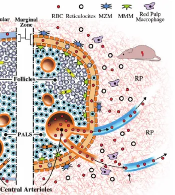
1.7.1 The splenic architecture ...37
1.7.2 Marginal Zone ...37
1.7.3 White pulp ...39
1.7.4 Red pulp ...41
1.7.5 Effects of aging on the spleen ...42
1.8 T CELL ACTIVATION ... 43
1.8.1 Impact of aging in T cell activation ...46
1.8.2 Macrophages and dendritic cells ...46
CHAPTER 2. IR-INDUCED SENESCENCE IN THE SPLENIC ENVIRONMENT INTERFERES WITH MURINE IMMUNE CELLS FUNCTIONS ... 48
2.1 ABSTRACT ... 49
2.2 KEY WORDS: ... 49
2.3 INTRODUCTION ... 50
2.4 RESULTS ... 52
2.4.1 Exposure to IR induces features of senescence in the spleen...52
2.4.2 Attrition of CD3+ and B220+ cell populations in the irradiated spleen ...53
2.4.3 Impaired proliferation of irradiated T cells is dependent on the splenic environment ...54
2.4.4 IR impairs macrophages and dendritic cells in the spleen ...56
2.5 DISCUSSION ... 57
2.6 MATERIALS AND METHODS ... 61
2.6.1 Animals and treatments ... 61
2.6.2 Bioluminescence ... 62
2.6.3 Gene expression ... 62
2.6.4 Flow cytometry analysis... 63
2.6.5 In vitro phagocytosis assay ... 64
2.6.6 Multiplex cytokine analysis ... 64
2.6.7 T-cells proliferation assays ... 64
IX
2.7 FIGURES... 67
2.8 REFERENCES... 81
CHAPTER 3. SUSTAINED P16INK4A EXPRESSION IS REQUIRED TO PREVENT IR-INDUCED TUMORIGENESIS IN MICE ... 85
3.1 ABSTRACT ... 85
3.2 KEYWORDS ... 86
3.3 INTRODUCTION ... 86
3.4 RESULTSANDDISCUSSION ... 88
3.4.1 Irradiated bone marrow stromal cells do not resume growth following p16INK4a deletion ...88
3.4.2 Increase BrdU incorporation in mice tissues following deletion of p16INK4a ...89
3.4.3 Sustained p16INK4a expression is necessary to limit cancer incidence ...91
3.5 ACKNOWLEDGMENTS ... 95
3.6 FIGURES... 96
3.7 REFERENCES... 112
CHAPTER 4. DISCUSSION, PERSPECTIVES AND CONCLUSIONS ... 115
4.1 General discussion ... 115
4.1.1 IR induces splenic aged-like alterations ... 116
4.1.2 IR induces a T cell proliferation impairment in function ... 117
4.1.3 T cell proliferation is partially limited by non-cell-autonomous mechanisms ... 118
4.1.4 Macrophages and DC cells ... 119
4.1.5 Splenic stroma cells ... 121
4.1.6 p16INK4a limits tissue proliferation and tumour progression ... 121
4.1.7 Is p16INK4a expression an universal marker of limited proliferation? ... 124
4.2 Perspectives ... 125
4.3 Conclusions ... 126
X
I.
List of tables
Table 1.1 Senescence mediator pathways ... 13
Table 1.2 Types of senescence ... 25
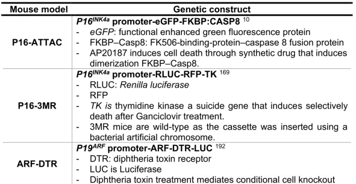
Table 1.3 Genetically modified mice to target senescent cells in vivo... 34
Table 1.4 Splenic cells ... 40
Table 1.5 Intrinsic age-related changes in the innate immune system ... 47
Supplementary Table 3.1 Cytokine levels in serum and spleen homogenates collected from mice irradiated or not 8 weeks prior to collection of tissues ... 110
XI
II. List of figures
Figure 1.1 The hallmarks of aging... 2
Figure 1.2 Molecular pathways of senescence ... 4
Figure 1.3 Senescent cells express specific characteristics ... 7
Figure 1.4 DDR-dependent and DDR-independent mechanisms that control the senescence-associated secretory phenotype (SASP). ... 11
Figure 1.5 DNA damage response (DDR) ... 15
Figure 1.6 p53 posttranslational regulation ... 17
Figure 1.7 The INK4B-ARF-INK4A locus ... 19
Figure 1.8 Transcriptional activators and repressors of p16INK4a promoter ... 20
Figure 1.9 Replicative senescence in vitro. ... 23
Figure 1.10 Multiple pathways are involved in oncogene-induced senescence. . 29
Figure 1.11 Murine splenic microarchitecture. ... 38
Figure 1.12 Priming of T cells activation ... 45
Figure 2.1 Exposure of mice to IR induces p16INK4a expression and a SASP in the spleen. ... 68
Figure 2.2 Attrition of T and B cell populations in the irradiated spleens ... 69
Figure 2.3 IR impairs T cells proliferation in vitro and in vivo ... 71
Figure 2.4 The irradiated splenic environment impairs T-cell proliferation ... 73
Figure 2.5 Impaired macrophage and DCs counts and function in the splenic environment. ... 76
Figure 3.1 Deletion of p16INK4a in irradiated bone marrow stromal cells allows for cell cycle progression but not cell growth. ... 96
Figure 3.2 Conditional deletion of IR-induced p16INK4a expression in mice. ... 98
Figure 3.3 Increase BrdU incorporation in irradiated mouse tissues following deletion of p16INK4a ... 99
Figure 3.4 Sustained p16INK4a expression is necessary to protect against cancer. ... 101
XII
Supplementary figure 2.1 Splenic cell counts following exposure to IR. ... 78 Supplementary Figure 2.2 Irradiation compromised the expression of
costimulatory molecules.. ... 79 Supplementary figure 2.3. Exposure to IR impacts splenic stromal cell
subpopulations. ... 80
Supplementary Figure 3.1 Deletion of p16INK4a in bone marrow stromal cells in vitro
... 103 Supplementary Figure 3.2 Efficient p16INK4a exon 1 deletion in various mice
tissues. ... 105 Supplementary Figure 3.3 High level BrdU incorporation in the spleen nonhematopoietic cells following p16INK4a inactivation in mice. ... 106
Supplementary Figure 3.4 Characterization of tumor types observed upon
XIII
III. List of abbreviations
ANOVA: Analysis of variance APC: Antigen-presenting cells ARF: Alternative Reading Frame ASF1a: Antisilencing function 1a ATM: Ataxia-telangectasia mutated ATR: ATM and rad3 related
BCL-2 : B-cell lymphoma 2 BER: Base excision repair
BM: Bone marrow
BMDM: Bone marrow‐derived macrophages BrdU: 5-bromo-2'-deoxyuridine
CDK: Cyclin-dependent kinase
CDKIs: Cyclin-dependent kinase inhibitors CDKN2a: Cyclin-dependent kinase inhibitor 2a C/EBPβ: CCAAT/enhancer-binding protein-β
CFSE: 6-carboxy-succinimidyl-fluorescein-ester dye CHK1: Checkpoint kinase-1
CHK2: Checkpoint kinase-2
COX: Cyclooxygenase
CTLA-4: Cytotoxic T lymphocyte-associated molecule-4
CTZ: Coelenterazine
DAPI: 4,6-diamidino-2-phenylindole DCs: Dendritic cells
DDR: DNA-damage response
DMP1: D binding myb-like protein 1
DNA-SCARSDNA: Segments with chromatin alterations reinforcing senescence DSB: Double strand breaks
DTR: Diphtheria toxin receptor
XIV Ets: E-26 transformation-specific EV: Extracellular vesicles
FBS: Fetal bovine serum FDCs: Follicular dendritic cells FOXO: The forkhead box O family FRCs: Fibroblastic reticular cells GATA4: GATA binding protein 4
GCV: Ganciclovir
Gy: Gray
H3K9Me: Lysine 9 methylated histone H3 TERT: Telomerase reverse transcriptase HIRA: Histone repressor A
HIV-1: Human immunodeficiency virus type 1 HMGA: High mobility group A
HP1: Heterochromatin protein 1 HR: Homologous recombination HRP: Horseradish peroxidase HSC: Hematopoietic stem cell
Id: DNA Binding 1
IF: Immunofluorescence
IGFBP-7: Insulin-like growth factor binding protein 7 INF-γ: Interferon gamma
IL: Interleukin
ILT2: Ig-like transcription 2 IP: Intraperitoneal injection IR: Ionizing radiation
ITSE: INK4a transcription silence element KC: keratinocyte chemoattractant
KIR: Killer immunoglobulin-like receptors KLRG: Killer cell lectin-like receptor subfamily G
XV
LAG-3: Lymphocyte activation gene 3 LPS: Lipopolysaccharides
LCMV-Arm: Lymphocytic choriomeningitis virus Amstrong strain MAdCAM-1: Addressin cell adhesion molecule 1
MCP-1: Chemoattractant protein-1
Mdm2: Mouse double minute 2 homolog MEFs: Mouse embryo fibroblasts
mH2A: Histone variant macro H2A
MiRNA: Micro RNA
MLR: Allogenic mixed lymphocyte reactions MMM: Marginal metallophilic-macrophages MMPs: Matrix Metalloproteinases
MMR: Mismatch repair
MRCs: Marginal reticular cells
mTOR : Mechanistic target of rapamycin
MVEs: Intracellular multi-vesicular endosomes
MZ: Marginal Zone
MZM: Marginal zone macrophages NER: Nucleotide excision repair NF1: Neurofibromatosis type I
NF-κB: Nuclear factor of the kappa light NHEJ: Nonhomologous end joining NK: Natural killers
NLRs: Nod-like receptors
8-oxo-dG: 8-oxo-2-deoxyguanosine
OIS: Oncogenic-induced senescence MAPK: Mitogen-activated protein kinase
PAMPs: Pathogen associated molecular patterns PALS: Periarteriolar lymphoid sheath
XVI PD: Population doublings PD1: Programmed death 1 PD-L1: Programmed death-ligand 1 PDNP: Podoplonin PE: Phycoerythrin PGE2: Prostaglandin E2
PI: Protease inhibitors
PI3K: Phosphatidylinositol kinase PML: Promyelocytic leukemia
PPARα: Peroxisome proliferator-activated receptor alpha PRR: Pathogen recognition receptors
PPRE: Peroxisome proliferator response element PRC: Polycomb transcription factor
Rb: Retinoblastoma
RE: p53-responsive element
RIPA: Radioimmunoprecipitation assay buffer RLRs: RIG-I like receptors
ROS: Reactive oxygen species
RPMI: Roswell Park Memorial Institute medium
RT: Room Temperature
SA-β-gal: Senescence-associated β-galactosidase SASP: Senescence associated secretory phenotype Sp1: Specificity protein 1
SSB: Single strand Breaks SSBR: Single-strand break repair
TAM: Tamoxifen
TGFβ: Transforming growth factor beta
Tim-3: T-cell immunoglobulin mucin domain-3 TIS: Senescence induced by genotoxic stressors TK: Thymidine kinase
XVII TLR: Toll like receptor
TNF-α: Tumor necrosis factor alpha TOR: Target of rapamycin
TRANCE: Tumour necrosis factor family member UTR: The 3`-untranslated region
UV: Ultraviolet
VEGF: Vascular endothelial growth factor
WHO: World health Organization
X-gal: 5-bromo-4-chloro-3-indolyl--D-galactopyranoside YB-1: Y box-binding protein 1
XVIII
To all my Family -A mi familia Greg and Emma Flora-
XIX
ACKNOWLEDGMENTS
I acknowledge that during these years I have met good people including my advisor, co-workers and friends that have helped me in so many ways. I want to express my profound gratitude to everyone that made this thesis a reality:
First and foremost, I would like to thank my PhD supervisor Dr Christian Beausejour for giving me an opportunity to work in his team, for his support and advice. I also express my profound gratitude for his always good character and positive attitude, this was an excellent place to learn and grow.
I thank to everyone in my lab, they have contributed in so many ways. I am profoundly thankful for Oanh Lee, for her advice in every single question that she answered. She had supported me and taught me so many techniques. I thank to my colleagues for their invaluable help and support: Basma, Cynthia, Gael, Mary-Lyn and Juliette. I especially emphasize the collaboration and support of the students who made an internship in my projects: Melissa Lelaider, Andrea Espinosa, Eliane Toleto-Cornu and Norbert Villeneuve. In addition, I am grateful for the old members that have come and gone: Cynthia, Mohamad, Vimal and Kerstin.
I would also like to thank my colleagues, friends and members of other labs. It was nice to collaborate with all of each of you.
XX
• Dr Decaluew and lab members: Chloe Berthe, Josse-Anne Joly, Sara Bourbonnais
• Dr Haddad and lab members: Dr Silvia Selleri, Hugo Roméro, Chloe Colas, William Lemieux, Aureliene Colamartino, Kathie Beland, Dr Simone Nicoletti.
• Dr Hickson and lab members: Sylvana Jananji, Zlatina Dragieva, Amel, Denise, Yvonne Ruella.
• Dr Duval and lab members: Pablo Cordero, Melanie Diaz, Dr Sabine Herblot and Assila Belounis.
I would also like to thank the animal facility for their amazing work and help: Denise, Dominique, Sonja, Rolando, Veronique, Edith, Marisol. In addition, I thank Ines for her advice in the Flow cytometry facility. I want to give special thanks to the following people who took their valuable time to read and actively review my thesis: Genelle Harrison, Yvonne Ruella, Gael Moquin-Baudry and Dr Christian Beausejour.
Last but not the least, I thank to my family for their unconditional love. To my friends in Montreal and Colombia; I thank them for their friendship, through these years they transformed winters in summers: Steven, Alexandru, Stefany, Jose, Steven, Nidia, Paula, Carlos, Rochy, David, Angie, and las princes.
1
Chapter 1: Introduction
1.1 Senescence as a hallmark of aging
In the recent decades, the proportion of the population over the age of 60 is higher than in any time in history and continues to grow and may outpace the younger population for many years to come. The societal effects of this demographic transformation are unknown but as the aged population worldwide increases, the incidence of age-related diseases will also rise. The World health Organization (WHO) highlights the importance to develop an effective health response to new disease patterns in aging populations to reduce the economic and social challenges 1.
Aging is defined as the time-dependent functional deterioration of physiological integrity leading to diminished function and augmented vulnerability to death. This phenomenon gathers important characteristic hallmarks that contribute to the aging process; these are genomic instability, telomere attrition, epigenetic alterations, loss of proteostasis, deregulated nutrient-sensing, mitochondrial dysfunction, cellular senescence, stem cell exhaustion, and altered intercellular communication (Figure 1.1) 2. Among these,
senescence is one of the most relevant factors affecting the aging population for many reasons. First, senescent cells accumulate in vivo during aging contributing to autonomous and non-autonomous effects that modulate the tissue microenvironment. Secondly, this accumulation limits the tissue regeneration capability, which influences the functional deterioration of organs. Despite a young chronological age of individuals, senescent cells can increase at a faster accumulation rate after exposure to stressors like
2
radiation, chemotherapy, high-fat nutrition; environmental toxicants such as arsenic or cigarette smoke provoking an accelerate-like age phenotype 3-9. Lastly, clearance of
senescent cells from tissues can lead to an extension of life span by rejuvenating organs and potentially improving functional as discussed in more details in future sections 10-13.
Although, senescence is considered a deleterious phenomenon in aging, in early stages of life plays an important role in embryogenesis, tissue regeneration, wound healing and cancer suppression.
Figure 1.1 The hallmarks of aging
During normal aging individuals harbor typical characteristics that cooperate to impair the function of tissues/organs. The word senescence can be traced to the Latin word senex, meaning old man. In cellular biology, senescence refers to a state of permanent cell growth arrest with characteristic features 8,9,14-17. Adapted from
3
1.2
Senescence phenotypeExperiments in cultured cells have identified a series of characteristics or markers in senescent cells that are summarized below. Moreover, not all senescent cells express all the markers, but when analyzed together they can define this state.
1.2.1 Cell cycle arrest
The cell cycle consists essentially of four basic phases: checkpoint Gap 1 (G1), DNA synthesis (S phase), checkpoint G2 and mitosis (M phase). In S phase, cells replicate the genomic DNA, which is distributed to two daughter cells in M phase. Most of the time the irreversible arrest of senescence occurs predominantly in G1/S checkpoint, triggered by telomeric-DNA damaged by eroded telomeres, or telomeric-independent DNA insult such as oncogene activation, loss of tumor suppressor genes and/or DNA-damage inductors
8,9,18-20. The molecular basis of this G1/S arrest results from the accumulation of
cyclin-dependent kinases (CDK)-inhibitors p16INK4a, p19ARF and p21Cip1 proteins. p16INK4a
protein arrests the cell cycle by limiting cyclin-dependent kinases 4 and 6 (CDK4/6) activity, impeding the retinoblastoma protein (Rb) phosphorylation. As consequence, Rb remains associated with the E2 factor (E2F) transcriptional factor localizing it to the cytoplasm, thus repressing transcriptional activity required to overpass G0/G1/S phase transition (Figure 1.2) 21. In addition, Rb activation in some cells organize the formation
of cytologically detectable regions of heterochromatin associated with senescence foci (SAHF). This facultative heterochromatin accumulates in many irreversible proliferation-arrested cells. In fact, SAFH is though to contribute to this permanent arrest of
4
proliferation by repressing-expression of proliferation-promoting genes necessary for the progression of the cell cycle 22-24.
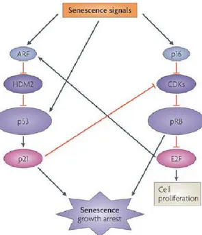
Figure 1.2 Molecular pathways of senescence
p16INK4a regulates cell cycle progression through hypophosphorylation of Rb.
p16INK4a binds to CDK4 and 6 to disable interaction of D-type cyclins. This
maintains Rb in a hypophosphorylated state with E2F repressionally bound and thus inhibiting cell cycle progression and arrest in G0/G1 phase. Adapted from Campisi & Di Fagagna, 20078.
In contrast, p19ARF inhibits the E3 ubiquitin ligase activity of mouse double minute 2
homolog (MDM2), leading to stabilization of p53 and inducing p53-dependent growth arrest or apoptosis depending on the biological context. In wild-type mouse embryo fibroblasts (MEFs), p19ARF and p16INK4a both accumulate significantly after passaging, but
5
than INK4a–Rb 25. P16INK4a expression is highly dynamic with temporal and spatial
compartmentalization with low level expression during embryogenesis, is undetectable in young healthy and proliferating cells, but its expression dramatically increases in chronological aging, wound healing and under cellular stress conditions 6,26-29. In
addition, cell arrest can occur in S and G2/M phases of the cell cycle depending on the cell type and the origin of stimulus 8,18-20. Notably, Irradiation (IR)-induced DNA damage
can also induce senescence during G2/M transition triggering p53/p21Cip1 response 30,31.
However, long-term G2 senescent cells can undergo G2slippage, bypass the M phase and directly entering G1 phase, consequently becoming senescent as tetraploid cells that fail to subsequently divide (4N DNA content) 31,32.
1.2.2 Altered morphology
The senescent cells are accompanied by changes in their morphology. Such changes include enlarged, irregular and flattened cytoplasm (in vitro), with increased vacuoles and lysosomal content associated to high expression of the lysosomal hydrolase β-galactosidase (encoded by GLB1 gene) 18,23,33.
1.2.3 Senescence-associated β-galactosidase activity
The β-galactosidase is a lysosomal hydrolase enzyme whose function is to cleave the β-glycosidic bond of a large variety of substrates such as gangliosides, glycoproteins and glycosaminoglycans. In 1995, Dimri and colleagues (1995) proposed using SA-β-gal to differentiate senescent fibroblasts and keratinocytes from quiescent and pre-senescent
6
cells. SA-β-gal remains one of the most extensively used biomarkers of cellular senescence in vitro and in vivo 34. In normal young cells, its activity can be determined
using chromogenic substrate the 5-bromo-4-chloro-3-indolyl β-D-galacto-pyranoside (X-Gal) at pH 4. In contrast, in senescent cells its activity is evaluated at pH of 5-6 (Figure 1.3). SA-β-gal presents limitations as parameter to define senescence both in vitro and in vivo, regardless of being considered a specific marker for senescence. For example, this enzymatic staining can be performed only on fresh frozen tissues; and in vitro SA-β-gal activity can be detected in cells in various nonsenescent states. Therefore, it is important to consider other senescent markers such as p16INK4a expression,
DNA-damage foci and the senescence-associated secretory phenotype (SASP), see below
6,10,23,34-41.
1.3 Epigenetic stress of senescent cells
1.3.1 SAHF a hallmark of senescence
Chromatin constitutes one of the most complex and dynamic macromolecular structures in the nucleus of a cell. The genomic DNA can be associated to a wide range of proteins and RNA molecules working in concert to mediate gene transcription or repression. The packaging of chromatin can be loosely packaged euchromatin and thus favoring access to the transcriptional machinery, or tightly packaged heterochromatin thereby limiting gene expression. Narita et al., (2013) observed that the nuclei of senescent cells contain 30–50 bright, punctate DNA-stained dense foci (Figure 1.3) that can be readily
7
distinguished from chromatin in normal cells 23,42-47. Cytologically, individual SAHF
appears as a compacted chromosome observed as punctuated DAPI- dense foci (Figure 1.3). In senescence, these heterochromatin structures are known as SAHF which correspond to highly compacted chromatin and they are not associated with cells undergoing quiescence. Importantly, these structures have also been shown to be different from constitutive heterochromatin because centromeres, telomeres, and other constitutive heterochromatin regions are not included in SAHF 23,43,48. Experimental
evidences in senescent cells have suggested that SAHF are responsible for sequestrating and silencing genes associated with the progression of the cell cycle; for example, E2F genes such as cyclin A related with proliferation 23,42-47. Thus, the formation of
heterochromatin in senescence is thought to contribute to permanent growth arrest 23,49 23,43,48.
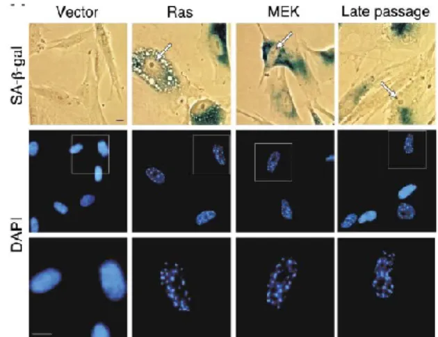
Figure 1.3 Senescent cells express specific characteristics
Cellular senescence induced by oncogene overexpression H-RasV12 (Ras) and
Mek1Q56P (Mek) or telomere attrition (late passage) possesses specific features,
8
(arrow) SA-β-gal expression and DAPI-dense foci termed senescence-associated heterochromatic foci (SAHF). Adapted from Narita et al., 2003 23.
SAHF also contribute to the tumor suppressive function of senescence by limiting the extent of DNA damaging signaling which may prevent senescent cells from undergoing apoptosis induced by high DNA damage signaling. Some senescent cells can also be identified by the formation of foci associated with a persistent activation of the general response to "DNA Damage Response (DDR)" 50. These DNA segments with
chromatin alterations reinforcing senescence (DNA-SCARS) contain proteins associated with DNA damage such as ATM, ATR, DNA-PK, phosphorylated histone H2AX (γ-H2AX) and p53 binding protein 1 (53BP1) 50,51. Another distinct type of senescence-specific
nuclear structures associated often at the periphery of PML nuclear bodies 51-53. PML
bodies contribute to senescence by recruiting pRB/E2F complexes and suppressing E2F target gene expression 53-55.
1.3.2 The nuclear Lamina and senescence
The integrity and shape of the nucleus is supported by an intricated mesh-like structure named the nuclear lamina. The nuclear lamina besides providing mechanical support, is involved directly or indirectly in functions such as chromatin structure, DNA replication, cell division and nuclear compartmentalization 56. Topologically, genes
generally expressed at low levels are organized into large areas known as lamina associated domains. These domains are depleted for active histone marks but include large domains of H3K9me3 and H3K27me3 enrichments and DNA hypomethylation 57.
9
Therefore, the nuclear lamina is considered to be a repressive chromatin environment associated to heterochromatin regions, including low gene density and repressed gene expression regions, with enrichment for repressed histone marks 58 57.
1.3.3 The Lamina A and B
The critical components of the nuclear lamina are Lamins A/C and B, alterations in their expression or functionality lead to disturbances in the integrity of the nuclear lamina and therefore are the caused of abnormal nuclear morphology, DNA damage, and chromosomal aberrations. In the context of accelerated aging or acute-induced senescence using multiple models of progerias the nuclear lamina instability is a key player in SAHF formation. For example, a single genetic alteration of Lamin A has a profound effect of on the mechanisms of aging and chromatin structure 59-62. Specifically,
progeria cells harbor mutations in Lamin A resulting in a reduced expression of Lamin B1. Similarly, replicative senescent fibroblasts, oncogene-induced senescence, and cells with high levels of chronic DNA damage decreased Lamin B1 expression 62-64. The loss of
Lamin B1 expression leads to the disassociation between the nuclear lamina and lamina associated domains during accelerated-aging (progeria) and chronic-induced senescence 61,65. In fact, it was recently shown that SAHF formation is aided by this loss
in anchoring of chromatin to the nuclear lamina 56-65. SAFH formation is suggested to be
the result of a structural rearrangement of the three dimensionally architecture change rather than a redistribution of epigenetic marks 61. Loss of Lamin B1 also appears to cause a redistribution of histone marks, including an enrichment in H3K27me3 and H3K4me marks within Lamin B1-associated domains and depletion in H3K27me3
10
marks in enhancers and genes 61,65. Experimental observations linked to the exclusive
acute-stress SAHF formation, with upregulation of heterochromatin markers such as HP1 and histone H2AX and absence of heterochromatin relaxation.
1.3.4 The senescence-associated secretory phenotype
The senescent cells bearing DNA-SCARS remain metabolically active, nonetheless upregulate a wide range of genes as part of the senescence-associated secretory phenotype (SASP) 15,66-68. The most remarkably increased genes encode a wide range
of secreted inflammatory proteins such as cytokines (e.g. interleukin (IL) IL-1α, IL-6, and IL-1β), chemokines (e.g. IL-8, MCP-1, KC), growth factors (e.g. bFGF, HGF/SF, IGFBPs), proteases (e.g. matrix metalloproteinase -MMP-) and other non-soluble extracellular matrix proteins (e.g. collagens, fibronectin, laminin); in addition to upregulation of genes, senescent cells secrete exosomes 9,67,69-71. The augmented secretion of SASP confers
multiple cell-autonomous and non-cell-autonomous activities. These biologically active factors provide both beneficial and/or deleterious responses in neighboring cells 9,15.
Understanding this apparent paradox may be particularly important depending on the senescence inducing stimuli and cellular context 69.
1.3.5 Regulation of SASP expression
The general understanding of how the expression of SASP is regulated remains poorly understood, and varies depending on the cell type and trigger of cellular senescence 72. SASP secretion is regulated through dependent and
DDR-11
independent mechanisms. In persistent DDR, signals emanating from DNA-SCARS activate nuclear factor of the kappa light (NF-κB) and CCAAT/enhancer-binding protein-β (C/EBPprotein-β) transcription factors of SASP genes including of IL-1α, IL-6 and IL-8 (Figure 1.4) 67,73. However, the DDR response is not considered to be the only regulator of SASP,
since the DDR is activated immediately after damage, whereas SASP, like other aspects of the senescent phenotype such as the activity of β-galactosidase, takes several days
72,67,73. So, SASP phenotype requires autocrine positive (continuing reading below).
Figure 1.4 DDR-dependent and DDR-independent mechanisms that control the senescence-associated secretory phenotype (SASP).
Different networks and their components interact to activate NF-κB and/or C/EBPβ transcriptional activators of SASP factors. Adapted from Lujambio (2016) 74.
12
1.3.6 Cell-autonomous reinforcement of senescence
SASP can reinforce the senescent phenotype in an autocrine manner through an amplification system. IL-1α is an essential positive regulator of IL-6/IL-8 expression. The IL-1α signalling network acts through p38MAPK and/or mammalian target of rapamycin (mTOR) pathways and therefore stimulates the expression and activity of NF-κB and C/EBPβ 75. In addition, recent evidence suggests that other SASP factors such as IL-6,
IL-8, CXCL1 (also named GROα, KC) and insulin-like growth factor binding protein 7 (IGFBP-7) act in an autocrine manner reinforcing senescence in a positive feedback loop
66,68.
1.3.7 Non-cell-autonomous activity of SASP
The SASP possesses the capacity to transmit signals to the normal neighboring cells. The paracrine-induced senescence is orchestrated by cytokines IL-1α, IL-1β, IL-6, VEGF and the transforming growth factor beta (TGFβ) signaling (Figure 1.4) 66,68,76.
Importantly, Ohanna et al., 2011 has shown that SASP factor MCP-1, (CCL2) expressed by senescent melanoma cells, induces DNA lesions in naive melanoma cells 77.
Paracrine effects of SASP in early stages of life are beneficial and play an important role in processes such as tissue regeneration, embryogenesis, tissue remodeling, limb regeneration (in salamanders) and wound healing and tumour suppression 78. However,
under the conditions of aging, SASP signaling is considered deleterious. An increased chronic inflammation favors the accumulation of senescent cells, which affects the repair,
13
turnover and regeneration of tissues leading to progression and age-related pathologies
79. In addition, the SASP factors secreted by senescent cells affect vital processes such
as growth, migration, tissue architecture, blood vessel formation and differentiation, processes that are highly controlled and whose deregulation can promote tumor growth
80.
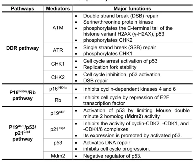
Table 1.1 Senescence mediator pathways
Pathways Mediators Major functions
DDR pathway
ATM
• Double strand break (DSB) repair • Serine/threonine protein kinase
• phosphorylates the C-terminal tail of the histone variant H2AX (γ-H2AX), p53 • phosphorylates CHK2
ATR • Single strand break (SSB) repair • phosphorylates CHK1
CHK1 • Cell cycle arrest activation of p53 • Replication fork stability
CHK2 • Cell cycle inhibition, p53 activation • DSB repair
P16INK4a/Rb
pathway
p16INK4a • Inhibits cyclin-dependent kinases 4 and 6
Rb • Inhibits cell cycle by repression of E2F transcription factor
P19ARF/p53/
p21Cip1
pathway
p19ARF • Activation of p53 by limiting Mouse double
minute 2 homolog (Mdm2) activity p21Cip1
• Inhibits the activity of cyclin-CDK2, -CDK1, and -CDK4/6 complexes
• Its expression is promoted by activated p53. p53 • Activates DNA repair
• inhibits cell cycle progression. Mdm2 • Negative regulator of p53.
14
1.4
Molecular pathways leading to senescenceThe cellular growth arrest in senescent cells is orchestrated and maintained by multiple tumour-suppressor genes from three important pathways: DDR, p19ARF/p53/p21Cip1 and p16INK4a/Rb (Figure 1.5) 9,14,81. These molecular cascades can
interact with each other, but they can also limit the progression of the cell cycle independently, responding to different stimuli and depending on the cell type.
1.4.1 DNA damage response: sending a SOS to repair, die or senesce
In specific situations, DNA-damaged or stressed cells are locked permanently in G1/S phase transition to limit the risks of becoming cancerous tumours 81,82. A common factor
in different senescence types is the presence of persistent DNA damage foci or DNA-SCARS; these lesions can impair genome replication and transcription and a wider-scale compromised genomic integrity and/or organismal viability 8.
15 Figure 1.5 DNA damage response (DDR)
DNA damage response requires diverse sensors, mediators and effectors to define the fate of cells with persistent DNA damage. Adapted from Campisi and d'Adda di Fagagna, 2007 8.
The wide diversity of DNA-lesion types requires specific repair mechanisms; mispaired DNA bases are replaced with correct bases by mismatch repair (MMR), and small chemical alterations of DNA bases are repaired by base excision repair (BER). More complex lesions, such as pyrimidine dimers and intra-strand crosslinks are corrected by nucleotide excision repair (NER). SSBs are repaired by single-strand break repair (SSBR), whereas DSBs are processed either by nonhomologous end joining (NHEJ) or homologous recombination (HR). The main DNA-damage sensor histone γ-H2AX is essential for the recruitment of 53BP1, BRCA1, MDC1, and the MRE11-RAD50-NBS1 complex (Figure 1.5) 82,83. Subsequently, those complex are targets for the kinases ATM
or ATR. In turn, both ATM and/or ATR transmit the DNA damage signal to their targets CHK2 and CHK1 respectively, which phosphorylate and activate several cell cycle inhibitor proteins, including the tumor suppressor gene p53 83. In turn, phosphorylated
p53 tetramers bind to specific DNA sequences, named the p53-responsive element (RE) and trigger the transcriptional activation of a multitude of target genes 84. Importantly,
16 1.4.2 p19ARF/p53 pathway
1.4.3 p53 activation
During cellular stress response, levels of p53 rise dramatically, which leads to different cell fates: apoptosis, autophagy, cell cycle arrest, DNA repair or senescence. The p53 protein is tightly regulated by an array of posttranslational modifications such as phosphorylation, ubiquitination, methylation, sumoylation, neddylation and acetylation; all targeting and modifying the C-terminal region of p53 85. In normal cells, p53 is a
short-lived protein that is constantly monoubiquitinated by an E3-ubiquitin ligase MDM2, promoting the export of nuclear p53 to cytoplasm for proteosomal degradation. Upon genotoxic stressors, rapid phosphorylation of p53 at Serine 15 (Ser-15) by ATM, and at Ser-20 by Chk2 results in dissociation of p53 from MDM2, leading to p53 stabilization, tetramerization and activation of its downstream processes. Likewise, phosphorylation of MDM2 at Ser-395 by ATM attenuates the capability of MDM2 in for subsequent p53 degradation, thereby enabling p53 accumulation 86,87.
17
Figure 1.6 p53 posttranslational regulation
Postransductional modifications of p53, methylation (Me), sumoylation (S), neddylation (N8), glycosylation (O-Glc), and ribosylation (ADP) Modified from Kruse and Gu, (2009) 86.
1.4.4 p53 and apoptosis
Acetylation of p53 on lysine 164 (K164) by CBP/p300 is required for the cell cycle arrest and apoptosis 88,89. Upon mono-ubiquitylation, p53 translocates from the nucleus to the
cytoplasm and then localizes in the mitochondria and induces apoptosis 90. At the outer
mitochondrial membrane p53 interacts with antiapoptotic members of the Bcl family such as Bcl-XL and Bcl-2 to promote the oligomerization of the proapoptotic factors Bak and Bax 91. In turn, Bak/Bax drives mitochondrial membrane permeabilization and release of
cytochrome c 92.
1.4.5 p53/p19ARF and senescence
The transcriptional activation of specific genes for cell cycle arrest and/or senescence programs mediated by p53 activation depends on the post-translations modifications and the combinations with other transcription factors. As a result, p53 increases the expression of cell cycle inhibitors and senescence mediators, p21Cip1 and of p19ARF.
p19ARF binds and retains MDM2 in the nucleolus; thus, when p19ARF is present p53
accumulates. In contrast, p53 activation leads to an autoregulatory negative feedback loop by promoting transcription of its own inhibitor Mdm2 93,94.
18 1.4.6 p16INK4a/Rb pathway regulation
1.4.7 The INK4a/ARF/INK4b locus
The INK4a/ARF/INK4b locus is located on chromosome 9 (p21) in humans, while the homolog in mice resides in chromosome 4, and encodes three important tumor suppressors genes: the cyclin-dependent kinase inhibitors (CDKIs) p15INK4b (INK4b), p16INK4a (INK4a) genes and p14ARF(ARF) 95. It is presumed
that p15INK4b and p16INK4a arose from an evolutionary duplication event as both genes
share high sequence homology and both proteins are almost indistinguishable in structure and biochemical properties; by acting as antagonist of the cyclin-dependent kinases (CDKs) that regulate progression through the G1 phase of the cell cycle. The p15INK4b gene has its own promoter and reading frame that is physically different from
p16INK4a and p14ARF. Interestingly, p16INK4a and p19ARF have different first exons 1α and
1β which bear no homology in their sequences and are transcribed from their own promoter 96. E1α and 1β are spliced to a common second and third exon; although p16INK4a and p14ARF share two exons, p14ARF mRNA is translated in an alternative reading
19 Figure 1.7 The INK4B-ARF-INK4A locus
The genomic structure of the INK4b-ARF-INK4a locus encodes three tumor suppressor genes p15INK4b, p19ARF (p14ARF in humans) and p16INK4a which are color
coded to represent the gene product that they are spliced into. Arrows represent the promotor of each gene. Adapted from Martin et al., 2014 98.
1.4.8 Transcriptional regulation of INK4a/ARF/INK4b locus
p16INK4a and p19ARF inhibit cell cycle and play critical and tightly controlled roles to
maintain tissue homeostasis and prevent cancer. In normal cells p16INK4a levels are
relatively low or undetectable, but its elevated expression in mice is enhanced during aging or in response to cellular stressors 6. Intriguingly, normal young mice exposed to IR
treatment experienced a premature senescence program, with a gradual increase of p16INK4a and p19ARF starting at 4 weeks post-IT treatment and reaching higher levels at
8-12 weeks although the reason for this delay is currently unknown 99,100. p16INK4a
transcriptional regulation is controlled at multiple levels of , and most of this regulation occurs through diverse regulatory elements and their combinatorial action 101-103.
1.4.8.1 TRANSCRIPTIONAL REGULATION OF P19ARF PROMOTER
p19ARF promoter is located in the INK4a/ARF/INK4b locus and contains a variety of
putative binding sites for transcription factors such as Sp1, cyclin D binding myb-like protein 1 (DMP1), Pokemon and the E2F family but its activation has remained poorly understood 104. The p19ARF protein exists at low or undetectable levels in most normal
cells and tissue types in embryogenesis 26,105. Like p16INK4a, the expression of p19ARF has
20
gene responds to low and chronic stress. p19ARF is induced in response to oncogenic
stress mainly by a sustained mitogenic signals from RasV12, H-ras, c-Myc, E2F-1, E1A,
v-Abl, T121 oncogenes 106,107. Overexpression of oncogenic RasV12 acts through the
Raf-MEK-ERK pathway and AP-1 signaling to activate Dmp1. In turn, DMP1 activates the p19ARF promoter only RasV12 activated the Dmp1 promoter 108. It is important to note
that many of these conclusions stem from highly tractable cell culture models, but the in vivo relevance is less clear in most cases.
1.4.8.2 REGULATORY DNA-MOTIFS FOR P16INK4A EXPRESSION
INK4a promoter contains its own activators sites to promote the subsequent recruitment of RNA polymerase and transcription of p16INK4a. Although the exact mechanism of
activation of INK4A is somewhat unclear, few models have been proposed (Figure 1.8) .
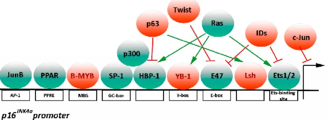
Figure 1.8 Transcriptional activators and repressors of p16INK4a promoter p16INK4a expression requires the recruitment of transcriptional factors (green) to
binding sites within INK4a promoter indicated by white rectangles. Transcriptional repressors (red) have an opposite function. Adapted from D’Arcangelo et al., 2017
21
Serving as an example, Ets-binding site binds E-26 transformation-specific 1 (Ets1) and E-26 transformation-specific 2 (Ets2) transcriptional factors which in turn can be activated by a variety of stress-associated signals, in particular the Ras/MAPk signaling pathway
22. In addition, Ets1, Ets2, E47 and/or c-Myc transcriptional factors can induce p16INK4a
expression by binding the E-box site located on the promoter. In replicative senescence, p16INK4a transcription is activated by increased expression of Ets1/2 and decreased
expression of DNA Binding 1 (Id) proteins22. Id1 is a helix loop helix transcription factor
that can negatively regulate p16INK4a expression by binding to phosphorylated Ets1/2
transcription factors. However, how Ets1/2 and Id1 protein regulation and expression change during aging is still elusive 110. Alternatively, AP1 binding site promotes p16INK4a
expression by binding JunB. Conversely, c-Jun acts as a JunB antagonist, downregulating p16INK4a expression. Another important regulatory element within
the p16INK4a promoter is the Sp1-binding site, a GC-rich region containing at least five
putative GC boxes that represent the putative binding target sites for Sp transcription factors. These are enhanced during cellular senescence mainly due to an increase in Sp1 binding affinity. Sp1 positively regulates p16INK4a transcription by directly binding
to DNA and recruiting P300 to the p16INK4a promoter 111,112.
1.5
Types of senescenceThe senescent phenotype depends on the stimulus that can differentiate two types of senescence. Currently, senescence is grouped in two categories, one of them is named replicative senescence and it is closely related to the finite number of cell divisions. This
22
is mainly regulated by telomere shortening which diminishes with each division leading an increase of the reactive oxygen species (ROS) levels and DDR 113,114. The other type
is premature senescence which is caused by a stressful exogenous stimulus such as DNA damage, oncogene activation, paracrine induced senescence 18,19.
1.5.1 Replicative senescence
1.5.2 Hayflick limit
The concept of cellular senescence was described for the first time in 1961 by Leonard Hayflick and Paul Moohead who observed a decline in the proliferative capability of normal embryonic human fibroblasts cultured in vitro after multiple passages. This finite number of cell cycle divisions or populations doubling (PD) corresponded approximately 50–60 rounds, and would be known as the Hayflick´s limit 115.
1.5.3 Telomere length defining the limit
Years later after Hayflick´s observations, Dr. Olovnikov hypothesized the molecular mechanism of Hayflick´s limit was the result of telomere attrition 116-118. In normal
eukaryotic cells, the telomeres protect the ends of chromosomes. These complex structures are composed of multiple proteins and range from 4000 to 15000 nucleotides in a tandem repeat sequence 5’-TTAGGG-3’ 119. To avoid telomere attrition, telomeres
23
complex RNA-dependent DNA polymerase known as telomerase reverse transcriptase (TERT) 120. In humans, hTERT is inactivated in most post-mitotic cells and its expression
is limited to embryonic stem cells, while in adults is confined to stem cells compartments
121. Consequently, in its absence differentiated cells experience a steady decay of
telomere length of 100-200pb per cell division, ticking as a cellular “molecular clock” (Figure 1.9) 114,121,122. The cellular micro-environment also plays an important role in
regulating the telomeric length and telomerase activity. Excessive oxidative stress causes a faster telomere attrition and antioxidants decelerate it 123,124. When telomeres reach
critical lengths, exposing the functional DNA ends, the DDR is engaged, followed by the activation of ataxia telangiectasia mutated (ATM) and ataxia telangiectasia and Rad3-related protein (ATR) kinases. These responses incur a growth arrest effected mainly by p53 ultimately leading to a cell-cycle arrest 125. Cells can bypass the cell cycle arrest by
oncogene activation and/or telomerase activation and can then undergo malignization 49.
24
Normal cells undergoing replicative senescence in vitro have low levels of telomerase or are telomerase negative and consequently experience telomere shortening with each cell division (black line) reaching a critical length of telomere triggering replicative senescence at the Hayflick´s limit. This proliferative checkpoint can be overcome by inactivation of Rb/p16INK4a or p53 pathways and ectopic
expression of hTERT leading to cell immortalization (dashed line). Pluripotent stem cells are telomerase positive (red line), so their telomeres length shorten in stem cells at rates slower than telomerase-negative somatic cells. Adapted from Cong et al., 2002 125.
1.5.4 Premature senescence
Regardless of the number of cell divisions, senescence can be induced prematurely by a plethora of non-telomeric stimuli including direct DNA-damaging stressors such as IR, chemotherapy drugs, reactive ROS, ultraviolet (UV), oncogene activation and high glucose concentrations 20,67,69,126-132. Moreover, senescent cells
bearing persistent DNA damage foci secrete pro-inflammatory cytokines that provoke non-cell autonomous induced premature senescence to neighboring normal cells
25 Table 1.2 Types of senescence
Types of senescence Inductor
Replicative senescence Replicative senescence Telomere erosion-dependent DNA damage
Premature senescence
ROS-induced senescence 8-oxo-2-deoxyguanosine, SSB and DSB.
DNA damage-induced senescence
Single- and double-strand DNA breakage
Oncogene induced
senescence (OIS) DNA replication stress Paracrine-induced
senescence
IL-1β, IL-1α, TGF- β ligands, VEGF, CCL2, CCL20
1.5.5 Oxidative stress induces senescence
During aging, cells undergo an intracellular accumulation of ROS 135,136. This
occurs due to a decrease in the activity of the enzymes responsible for eliminating or neutralizing these species. The increase in ROS is known to cause as much DNA damage as faster telomere shortening 137. Intracellular oxidative stress occurs when an increase
in the number of reactive molecules, causes nucleic acid, protein, and lipid damage 138.
ROS are highly reactive atoms or molecules with one or more unpaired electron(s) in their outermost shell and are formed when oxygen interacts with other specific molecules. Free radicals are unstable particles such as superoxide (O2•-), hydroxyl radical (OH•),
hydroperoxyl radical (HO2•), nitric oxide (NO•), nitrogen dioxide (NO2•), peroxyl (ROO•),
hydrogen peroxide (H2O2), ozone (O3), singlet oxygen (1O2), hypochlorous acid (HOCl),
nitrous acid (HNO2), peroxynitrite (ONOO–), dinitrogen trioxide (N2O3), and lipid peroxide
26
The pioneering work of Packer & Fuehr (1977) provided the first experimental evidence of the role of oxygen levels on in vitro senescence 140. They used the WI-38 human
fibroblast strain established by Hayflick and correlated the number of PDs with the amount of oxygen tension in the environment. Cells grown at 10% oxygen had a better capacity to proliferate as indicated by increased number of PDs while cells at 50% O2 possessed
a diminished life span and therefore, diminished PDs 140. Particularly in mice, fibroblasts
cultured in vitro will senesce after only 15-20 PDs despite having exceptionally long telomeres (>20 kb). This premature senescence makes it unlikely that telomere length plays a prominent role in normal mouse aging 141. This response is suggested to be due
to stressful culture conditions as a response of exposure to atmospheric oxygen concentrations of 20%, which is above biological levels of 3%. Mouse cells possess differences in oxygen sensitivity compared to human cells; murine fibroblasts have weaker protective mechanisms against ROS and growing at high levels of oxygen exhibit more damage than human cells 142. In recent studies, high oxygen levels in vitro culture
of MEFs have been demonstrated to accelerate the accumulation of DNA mutation, mostly by genomic rearrangements 143. On the other hand, telomere shortening in murine
cells is accelerated in response to ROS exposure and can be blocked by adding antioxidant in primary cell cultures lines.
According to the free radical theory of aging, oxidative DNA damage caused by ROS play a pivotal role in the aging process, these un-stable molecules make a significant contribution to genomic instability by causing single-strand base oxidative modification, the 8-oxo-2-deoxyguanosine (8-oxo-dG), which leads to single-strand nick formation.
27
These nicks or gaps are an important source of a subset of single-strand breaks (SSB) which in turn are at risk in becoming in double-strand DNA breaks (DSBs) if they persist until the next round of DNA replication, and thereby participates in downstream premature senescence signaling 144.
1.5.6 Ionizing Radiation
Direct damage to DNA, either by irradiation or by the use of damaging agents, can induce senescence. Normally the cellular response to damage includes the arrest of the cell cycle to carry out the repair. If the magnitude of the damage is very large, the response may include irreversible apoptosis or arrest of the cycle. Thus, the activated mechanism depends on the type of damage, the dose administered and the cell type treated 145,146.
Radiation is defined as the transmission of energy through waves or particles. IR corresponds X-rays and fast-moving subatomic particles, like alpha particles (α), beta particles (β) and neutrons (n). α particles are doubly charged and are relatively heavy with two protons and two neutrons and can be stopped by a sheet of paper. They lack energy to penetrate the outer dead layer of skin but are considered hazardous if ingested. β particles are much smaller with one negatively charged electron and can be stopped by a layer of clothing or millimetres of aluminum. Without any protection in the body, β particles can penetrate for a few centimetres and cause greater damage than α particles. The γ and X-rays are very short electromagnetic waves with high energy; both possess high penetrance ability in biological tissues and enough energy to ionize atoms and
28
disrupt molecular bonds in cells. In fact, exposure to radiation induces massive DNA damage, by depositing high energy on the DNA backbone resulting in SSB and/or DSB, and indirectly after electrons are removed from neutral water molecules to produce OH•.
These waves can be effectively blocked by several metres of concrete or few inches of lead. The intensity of X-rays is measured as the absorbed amount of energy per unit mass of tissue, defined as the dose of radiation and measured in gray (Gy), where 1Gy = 1 joule/kg of tissue.
1.5.7 Oncogene-Induced senescence
Sustained chronic proliferative capacity is the most fundamental hallmark for cancer development. Accordingly, oncogene-induced senescence (OIS) evolved as a powerful tumour suppressor mechanism to restrain aberrant cell proliferation of damaged or stressed cells bearing genetic alterations in proto-oncogenes and/or tumor suppressor genes 147-149. The first association between, constitutive activation of oncogenic Ras (H-RasV12) and OIS was described by Serrano et al., (1997) 18. They observed that
overexpression of H-RasV12 on normal human and murine fibroblasts induced a transient
arrest in growth accompanied with DNA damage and followed by activation of DDR and a strong growth arrest response by accumulation of p15INKb, p19ARF, p53 and p16INK4a
tumor suppressor genes 18,150. The induction of OIS is associated with senescent
hallmarks such as heterochromatin rearrangements, oncogene-induced DNA-damage foci, SASP and an increase in senescence-associated β-galactosidase (SA-β-Gal) activity 18,23,151,152. Normally, upon mitogenic signals the members of the Ras
proto-29
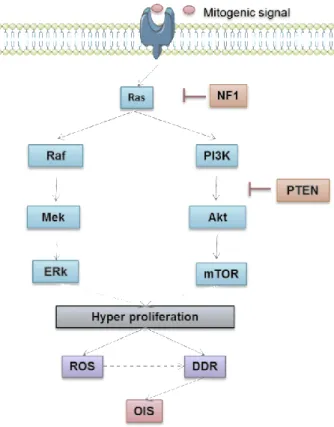
oncogene family (K-Ras, N-Ras and H-Ras) activate downstream mitogen-activated protein kinase (MAPK) and phosphatidylinositol kinase (PI3K) pathways which participate in the control of proliferation, differentiation, and survival of eukaryotic cells (Figure 1.10) . Genetic alterations within these growth-promoting genes transmit aberrant mitogenic signals; single mutations in Ras typically at codon 12, 13, or 61 can lead to hyperproliferative signaling. Overexpression of Ras effectors Raf, Mek1/2, Braf, PI3K, Akt provokes a burst in proliferation 18,147,153-156. Consequently, an aberrant DNA
hyper-replication promotes telomere-independent DNA damage through the accumulation of ROS and the collapse of DNA replication forks at genomic instability sites 147,152,157,158.
30
Ras activation promotes Raf/MEK/ERK and PI3K/Akt/mTOR pathways leading to cell growth. Ras activity can be repressed by NF1, and downstream PTEN constrain PI3K activity.
Phosphatase tensin homolog (PTEN) and neurofibromatosis type I (NF1) are tumor-suppressor genes thatnegatively control cell proliferation signaling (Figure 1.3). Their inactivation triggers sustained activation of PI3K cascade leading cells to senesce 159-161.
Promyelocytic leukemia (PML) protein inactivation also leads to senescence 132,162-165.
pVHL protein promotes the senescence program through stabilization and accumulation p27KIP, a cyclin-dependent kinase inhibitor which in turn leads to the Rb pathway 166,167.
1.6
The meaning of senesence in vivo1.6.1 The senescence is considered a double-edged sword
The paradigm of senescence as a "double-edged sword" is based on the observation that senescent cells have beneficial effects for tumour suppression, tissue homeostasis, limb and tissue regeneration, embryogenesis, and wound healing 6,26,28. However, senescent
cells can prevail for longer periods and accumulate during aging with deleterious role. Primarily, because of the decline in tissue regenerative ability from stress; and secondly because the production of chronic SASP factors by senescent cells are thought to drive undesired senescence in neighboring cells and favors age-related pathologies degenerative such as atherosclerosis, idiopathic pulmonary fibrosis, osteoarthritis, esteatosis, diabetes osteoporosis, neurodegeneration and atherosclerosis and cancer