Rhythmic Masticatory Muscle Activity during Sleep:
Etiology and Clinical Perspectives
par
Maria Clotilde Carra
Programme de Sciences Biomédicales Faculté de Médecine
Thèse présentée à la Faculté de Médecine en vue de l’obtention du grade de Doctorat
en Sciences Biomédicales option générale
Juin, 2012
Faculté des études supérieures et postdoctorales
Cette thèse intitulée:
Rhythmic Masticatory Muscle Activity during Sleep: Etiology and Clinical Perspectives
Présentée par: Maria Clotilde Carra
a été évaluée par un jury composé des personnes suivantes :
Dr Arlette Kolta, président-rapporteur Dr Gilles Lavigne, directeur de recherche
Dr Roger Godbout, membre du jury Dr Celyne Bastien, examinateur externe Dr Antonio Zadra, représentant du doyen de la FES
Résumé
L’activité rythmique des muscles masticateurs (ARMM) pendant le sommeil se retrouve chez environ 60% de la population générale adulte. L'étiologie de ce mouvement n'est pas encore complètement élucidée. Il est cependant démontré que l’augmentation de la fréquence des ARMM peut avoir des conséquences négatives sur le système masticatoire. Dans ce cas, l'ARMM est considérée en tant que manifestation d'un trouble moteur du sommeil connue sous le nom de bruxisme. Selon la Classification Internationale
des Troubles du Sommeil, le bruxisme est décrit comme le serrement et grincement des
dents pendant le sommeil. La survenue des épisodes d’ARMM est associée à une augmentation du tonus du système nerveux sympathique, du rythme cardiaque, de la pression artérielle et elle est souvent en association avec une amplitude respiratoire accrue. Tous ces événements peuvent être décrits dans le contexte d’un micro-éveil du sommeil.
Cette thèse comprend quatre articles de recherche visant à étudier i) l'étiologie de l’ARMM pendant le sommeil en relation aux micro-éveils, et à évaluer ii) les aspects cliniques du bruxisme du sommeil, du point de vue diagnostique et thérapeutique.
Pour approfondir l'étiologie de l’ARMM et son association avec la fluctuation des micro-éveils, nous avons analysé le patron cyclique alternant (ou cyclic alternating pattern (CAP) en anglais), qui est une méthode d’analyse qui permet d’évaluer l'instabilité du sommeil et de décrire la puissance des micro-éveils. Le CAP a été étudié chez des sujets bruxeurs et des sujets contrôles qui ont participé à deux protocoles expérimentaux, dans lesquels la structure et la stabilité du sommeil ont été modifiées par l'administration d'un médicament (la clonidine), ou avec l'application de stimulations sensorielles (de type vibratoire/auditif) pendant le sommeil. Dans ces deux conditions expérimentales caractérisées par une instabilité accrue du sommeil, nous étions en mesure de démontrer que les micro-éveils ne sont pas la cause ou le déclencheur de l’ARMM, mais ils représentent plutôt la «fenêtre permissive» qui facilite l'apparition de ces mouvements rythmiques au cours du sommeil.
Pour évaluer la pertinence clinique du bruxisme, la prévalence et les facteurs de risque, nous avons effectué une étude épidémiologique dans une population pédiatrique (7-17 ans) qui était vue en consultation en orthodontie. Nous avons constaté que le bruxisme est un trouble du sommeil très fréquent chez les enfants (avec une prévalence de 15%), et il est un facteur de risque pour l'usure des dents (risque relatif rapproché, RRR 8,8), la fatigue des muscles masticateurs (RRR 10,5), les maux de tête fréquents (RRR 4,3), la respiration bruyante pendant le sommeil (RRR 3,1), et divers symptômes liés au sommeil, tels que la somnolence diurne (RRR 7,4). Ces résultats nous ont amenés à développer une étude expérimentale pour évaluer l'efficacité d'un appareil d'avancement mandibulaire (AAM) chez un groupe d'adolescents qui présentaient à la fois du bruxisme, du ronflement et des maux de tête fréquents. L'hypothèse est que dans la pathogenèse de ces comorbidités, il y a un mécanisme commun, probablement lié à la respiration pendant le sommeil, et que l'utilisation d'un AAM peut donc agir sur plusieurs aspects liés.
À court terme, le traitement avec un AAM semble diminuer l'ARMM (jusqu'à 60% de diminution), et améliorer le ronflement et les maux de tête chez les adolescents. Cependant, le mécanisme d'action exact des AAM demeure incertain; leur efficacité peut être liée à l'amélioration de la respiration pendant le sommeil, mais aussi à l'influence que ces appareils pourraient avoir sur le système masticatoire. Les interactions entre le bruxisme du sommeil, la respiration et les maux de tête, ainsi que l'efficacité et la sécurité à long terme des AAM chez les adolescents, nécessitent des études plus approfondies.
Mots-clés: Bruxisme du sommeil, micro-éveil, adolescents, appareil d’avancement
Abstract
Approximately 60% of the general adult population experiences rhythmic masticatory muscle activity (RMMA) during sleep. The etiology of this movement is not yet understood. However, it has been demonstrated that an increased frequency of RMMA may have detrimental consequences on the stomatognathic system. In this case, RMMA is considered the manifestation of a sleep-related motor disorder known as sleep bruxism (SB). According to the definition of the International Classification of Sleep Disorders, SB is the activity of tooth grinding and clenching during sleep. The occurrence of SB-related activity, i.e., RMMA, is associated with rises of sympathetic tone, heart rate, blood pressure, and it is frequently concomitant with larger respiratory breaths. All these events can be described within a sleep arousal.
The present thesis includes four research articles aimed to study i) the etiology of RMMA during sleep in relation to sleep arousal; and ii) the clinical perspectives of SB assessment and management.
To further investigate the etiology of RMMA and its association with sleep arousal fluctuations we analyzed the cyclic alternating pattern (CAP), a scoring method to assess sleep instability and describe sleep arousal pressure. CAP was scored in SB subjects and controls that participated in two experimental protocols in which sleep architecture and stability were altered by either a medication (i.e., clonidine), or sensory stimulations (i.e., vibratory/auditory). Under these experimental conditions known to increase sleep instability, we were able to show that sleep arousal is not the trigger or cause of RMMA, rather the “permissive window” that facilitates the occurrence of RMMA during sleep.
To evaluate the clinical relevance of SB, we conducted a survey on a 7-17 year old orthodontic population to investigate the prevalence and risk factors associated with SB. It appeared that SB is a highly prevalent sleep disorders in children (15% of prevalence), and is a risk factor for tooth wear (odds ratio, OR 8.8), jaw muscle fatigue (OR 10.5), frequent headache (OR 4.3), loud breathing during sleep (OR 3.1), and several sleep complaints, such as daytime sleepiness (OR 7.4). These findings led us to design an experimental trial
using a mandibular advancement appliance (MAA) in adolescents in order to investigate the possible relationship between SB, snoring, and headache. We hypothesized that a common underlying mechanism related to breathing during sleep may be responsible for all concomitant conditions.
The short-term use of an MAA appeared to reduce SB (up to 60%), and improve snoring and headache complaints in adolescents. However, the precise mechanism of action of MAA remains under debate; its effectiveness can be either related to the improvement of breathing during sleep, or its influence on the masticatory system. The interactions between SB, breathing, and headache as well as the long-term effectiveness and safety of the MAA in adolescents need further investigations.
Riassunto
L’attività ritmica dei muscoli masticatori (ARMM) durante il sonno si osserva in circa il 60% della popolazione generale adulta. L'eziologia di questo movimento non è stata ancora del tutto compresa. Tuttavia, è dimostrato che un’aumentata frequenza di ARMM può avere conseguenze negative sul sistema stomatognatico. In questo caso, l’ARMM è considerato la manifestazione di un disturbo motorio del sonno noto come bruxismo. Secondo la Classificazione Internazionale dei Disturbi del Sonno, il bruxismo è l'attività di digrignamento e serramento dei denti durante il sonno. La comparsa di episodi di ARMM durante il sonno è associata a un aumento del tono del sistema nervoso simpatico, della frequenza cardiaca, della pressione arteriosa, ed è spesso in concomitanza con un aumentato volume inspiratorio. Le variazioni di questi parametri fisiologici sono compresi nel contesto di un arousal (micro risveglio) del sonno.
Questa tesi comprende quattro articoli di ricerca volti a studiare i) l'eziologia dell’ARMM durante il sonno in relazione all’arousal, ed a valutare ii) l’inquadramento clinico del bruxismo nel sonno.
Per approfondire l'eziologia dell’ARMM e l’associazione con l’arousal nel sonno, abbiamo analizzato il cyclic alternating pattern (CAP), che permette di valutare l'instabilità del sonno e descrivere la potenza degli arousals. Il CAP è stato esaminato in soggetti con bruxismo e soggetti controllo che hanno partecipato in due protocolli sperimentali, in cui la struttura e la stabilità del sonno sono stati modificati con la somministrazione di un farmaco (la clonidina), o con l’applicazione di stimolazioni sensoriali (di tipo vibratorio/uditivo) durante il sonno. In queste condizioni sperimentali caratterizzate da un’aumentata instabilità del sonno, siamo stati in grado di dimostrare che l’arousal non è la causa o il generatore dell’ARMM, ma piuttosto la "finestra permissiva" che facilita il verificarsi di questi movimenti ritmici durante il sonno.
Per valutare la rilevanza clinica del bruxismo, abbiamo condotto uno studio epidemiologico in una popolazione pediatrica afferente alla clinica di ortodonzia per studiare la prevalenza e i fattori di rischio associati al bruxismo. Questa ricerca ha
evidenziato che il bruxismo è un comune disturbo del sonno nei bambini (con una prevalenza del 15%), ed è un fattore di rischio per usura dentale (odds ratio, OR 8.8), fatica dei muscoli masticatori (OR 10.5), mal di testa frequenti (OR 4.3), respirazione rumorosa durante il sonno (OR 3.1), e diversi sintomi legati al sonno, quali la sonnolenza diurna (OR 7.4). Questi risultati ci hanno portato a progettare uno studio sperimentale per valutare l’efficacia di un apparecchio di avanzamento mandibolare (AAM) in un gruppo di adolescenti che presentavano al contempo bruxismo, russamento e frequenti cefalee. L’ipotesi è che nella patogenesi di tali comorbidità, vi sia un meccanismo comune, probabilmente legato alla respirazione durante il sonno, e che l’utilizzo di un AAM possa quindi avere un beneficio multiplo.
Il trattamento a breve termine con un AAM sembra diminuire l’ARMM (fino al 60%) e migliorare il russamento e i mal di testa negli adolescenti. Tuttavia, l'esatto meccanismo di azione degli AAM rimane incerto; la loro efficacia può essere correlata sia al miglioramento della respirazione durante il sonno, ma anche all’influenza che questi apparecchi svolgono sul sistema masticatorio. Le interazioni tra il bruxismo nel sonno, la respirazione, e le cefalee, così come l'efficacia e la sicurezza a lungo termine degli AAM negli adolescenti, necessitano di ulteriori studi clinici.
Keywords: Bruxismo nel sonno, arousal, età pediatrica, apparecchi di avanzamento
Abbreviations
ADHD Attention Deficit Hyperactivity Disorder CAP Cyclic Alternating Pattern
ECG Electrocardiogram
EEG Electroencephalogram
EMG Electromyogram
EOG Electrooculogram
HA Headache
MAA Mandibular Advancement Appliance
NREM Non-Rapid Eye Movement
PSG Polysomnigraphy
REM Rapid Eye Movement
RLS Restless Leg Syndrome
RMMA Rhythmic Masticatory Muscle Activity
SB Sleep Bruxism
SDB Sleep-Disordered Breathing SEM Standard Error of the Mean
SWA Slow Wave Activity
SWS Slow Wave Sleep
TC Tooth Clenching
TMD TemporoMandibular Disorders
TMJ TemporoMandibular Joint
Table of Contents
Introduction...1
Chapter 1: Literature Review...3
1.1 Historical Aspects of Bruxism...4
1.2 Definition and Classification of Sleep Bruxism ...4
1.3 Assessment and Diagnosis of Sleep Bruxism...6
1.3.1 Clinical Diagnosis of Sleep Bruxism...9
1.3.2 Ambulatory Assessment of Sleep Bruxism ...11
1.3.3 Polysomnographic Diagnosis of Sleep Bruxism ...12
1.4 Epidemiology of Sleep Bruxism...13
1.5 The Masticatory System during Wake and Sleep...14
1.6 Etiology and Pathogenesis of Sleep Bruxism ...17
1.6.2 Sleep Arousal...17
1.6.3 Autonomic Sympathetic-Cardiac Activity...23
1.6.4 Neurochemicals ...27
1.6.5 Genetic Factors ...27
1.6.6 Psychosocial Factors: Stress, Anxiety, and Behavior...28
1.6.7 Exogenous Factors and Comorbidities ...29
1.7 Sleep Bruxism and Sleep Quality ...33
1.8 Management of Sleep Bruxism...35
1.8.1 Behavioral Strategies ...36
1.8.2 Oral Appliances ...36
1.8.3 Pharmacotherapy ...39
Chapter 2: Thesis Objectives and Hypotheses...42
2.1 Objectives ...43
2.2 Hypotheses...44
2.2.2 Second Hypothesis...45
2.3 Materials and Methods...46
Chapter 3: Research Articles ...47
3.1 First article: “Clonidine Has a Paradoxical Effect on Cyclic Arousal and Sleep Bruxism during NREM Sleep” ...48
3.2 Second Article: “Sleep Bruxism and Sleep Arousal: an Experimental Challenge to Assess the Role of Cyclic Alternating Pattern”. ...67
3.3 Third Article: “Prevalence and Risk Factors of Sleep Bruxism and Wake-Time Tooth Clenching in a 7-17 Year Old Population”...86
3.4 Fourth Article: “Sleep Bruxism, Snoring, and Headache in Adolescents: an Experimental Trial with a Mandibular Advancement Appliance” ...113
Chapter 4: General Discussion ...136
4.1 CAP Phase A as the “Permissive Window” for Rhythmic Masticatory Muscle Activity during Sleep ...137
4.1.1 Evidence from the Experimental Trial with Clonidine...138
4.1.2 Evidence from the Experimental Trial applying Sensory Stimulations during Sleep...141
4.2 Sleep Bruxism, Snoring, and Headache: a Triad that Needs Further Investigations 143 4.2.1 Evidence from the Epidemiological Survey on a Pediatric Population...144
4.2.2 New Insights on the Role of Mandibular Advancement Appliances...146
4.3 Scientific and Clinical Relevance of the Findings...152
4.4 Study Limitations and Future Directions...155
4.4.1 What I Would Do Differently...156
4.4.2 What Needs to Be Done...157
Conclusion ...160
List of Tables
Table 1.1 Methods for assessing sleep bruxism. ...8
Table 1.2 American Academy of Sleep Medicine (AASM) clinical diagnostic criteria for sleep bruxism. ...10
Table 1.3 Polysomnographic research diagnostic criteria for sleep bruxism ...12
Table 1.4 Etiology and pathophysiology of sleep bruxism. ...18
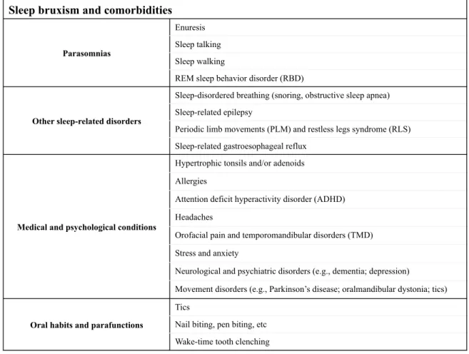
Table 1.5 Sleep bruxism and comorbidities...31
Table 1.6 Management of sleep bruxism...37
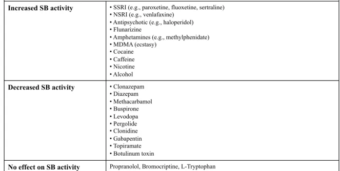
Table 1.7 Effect of medications and chemical substances on sleep bruxism and SB-like activities. ...41
Table 3.1.1 CAP variables during placebo and clonidine nights...61
Table 3.2.1 Demographic, RMMA/SB, sleep, and CAP variables during baseline and experimental nights in 8 controls and 8 SB subjects. ...82
Table 3.3.1 Prevalence of variables in the study population (n=604). ...101
Table 3.3.2 Craniofacial morphology and dental characteristics for control (CTL), sleep bruxism (SB) and wake-time tooth clenching (TC) subjects...102
Table 3.3.3 Temporomandibular (TMD) signs and symptoms, oral habits and parafunctions for control (CTL), sleep bruxism (SB) and wake-time tooth clenching (TC) subjects. ...102
Table 3.3.4 Sleep complaints for control (CTL), sleep bruxism (SB) and wake-time tooth clenching (TC) subjects. ...103
Table 3.3.5 Behavioral complaints for control (CTL), sleep bruxism (SB) and wake-time tooth clenching (TC) subjects. ...103
Table 3.3.6 Dental, temporomandibular disorders (TMD), sleep and behavioral complaints. ...104
Table 3.4.1 Demographic, dental and oropharyngeal data for the study sample (n=10) assessed during the clinical examination. ...129
Table 3.4.2 Polysomnographic variables for the baseline night and the three nights with the mandibular advancement appliance...130 Table 3.4.3 Self-report results from the questionnaire assessing the mandibular advancement appliance (MAA). ...132
List of Figures
Figure 1.1 Hypnogram and polysomnographic tracing showing an episode of rhythmic masticatory muscle activity (RMMA) during sleep. ...7 Figure 1.2 Schematic representation of cyclic alternating pattern (CAP). ...22 Figure 1.3 Genesis of an RMMA episode (schematic representation of the cascade of physiologic events that precedes RMMA onset) ...26 Figure 1.4 Comorbid sleep bruxism, headache, and sleep-disordered breathing: putative mechanisms...33 Figure 3.1.1 CAP and RMMA distribution over NREM/REM sleep cycles...59 Figure 3.1.2 Cross-correlation between RMMA/SB episodes and CAP phases. ...60 Figure 3.2.1 Comparison between control and SB subjects for RMMA/SB index, sleep arousal index and number of CAP phase A3 in baseline and experimental (VT/AD) nights...79 Figure 3.2.2 RMMA/SB activity (episodes/h) and CAP phases A1, A2, A3 (number/h)....80 Figure 3.2.3 Power spectral analysis of EEG delta (0.5–4.0 Hz), theta (4.0–8.0 Hz) and alpha (8.0–13.0 Hz) activities. ...81 Figure 3.4.1 The mandibular advancement device. ...128
Ai miei cari genitori, che non hanno mai smesso di contare i giorni che mancavano al traguardo.
Acknowledgements
My PhD has been a fantastic journey in a very beautiful country, which enlarged my horizon and opened my mind. For this inestimable experience I need to say a Grand Merci to many people…
Firstly, I would like to thank my director, Prof Gilles Lavigne, who has been for me a supervisor, a teacher, and a mentor. He taught me to be a researcher with data and a doctor with patients, to be critical with results and daring with ideas. I also thank him for his great understanding and sustenance all along my journey. He welcomed me in such a way that I never felt homesick in Montreal, and he let me go home anytime I needed to taste some Parmigiano … I am very fortunate to have worked with him and to have been part of his exceptional team.
I would like to say a special thank you to Prof Nelly Huynh, who was like a co-director in all my works. She supervised me, she advised me, and mostly she helped me every day until the end, sharing the good and the difficult moments. My project would have not been possible without her. But I would also like to thank my friend, Nellina, who accompanied me throughout my PhD with her capacity, diplomacy, and elegance.
I would like to thank Pierre Rompré for teaching me statistics and guiding me in all the analyses with rigor and enthusiasm, even when we had to deal with “fruit salads”. His contribution has been cardinal in all my works.
I would like to thank my great friend Angela Nashed, for her help and solidarity during all my research projects. Thank you for teaching me English, for making me run the 10 km marathon, and for always listening to me and giving me a smile!
I would like to thank Carmen Remo for her constant help and support. But mostly for her precious friendship that, together with Raymond, always reminded me that “le meilleur est toujours à venir”.
I would like to thank Christiane Manzini for her advices and kindness. In these years she has been such a positive example of determination and strength, which I will always remember and admire.
I would like to send my gratitude to Drs Claude Remise, Hicham El-Khatib, Athena Papadakis, and Paul Morton at the Clinique d’Orthodontie de l’Université de Montréal, and to the entire research lab at the Université of Montréal and at the Hôpital Sacré-Coeur de Montréal. In particular, I would like to thank Regis Schawb, Sophie Pelletier, Hajar El-Alaoui, Isabelle Roy, Samar Khoury and Hélène Labrecque for their precious help.
I would like to thank Prof Florin Amzica and Dr Raffaele Ferri, for their contributing mentorship in research.
I would like to thank Prof Guido Macaluso, who saw in me the potential researcher and showed me the way to Montreal.
I would like to thank my friend Chiara Ferrari, who was always there when I needed to hear some Italian words from overseas, and my friend and colleague, Massimo Manchisi, who never forgets me even from far away!
I would like to thank my sister and best friend, Ginny. I wish she were here all the time. My Mum and Dad, who supported and uplifted me every single day of this long journey, with enthusiasm, comprehension, and love. This work would have never been accomplished without them.
I would like to thank all the people that during these years share with me a bit of their life, their feelings, their thoughts. All of them are part of invaluable memories that I will carry with me forever.
Few episodes of rhythmic masticatory muscle activity during sleep occur in approximately 60% of the general adult population as a physiologic jaw movement probably related to swallowing and breathing. However, this motor behavior may fall into a pathological range if occurring with increased frequency during sleep and if associated with clinical signs and symptoms. In this case, we talk about sleep bruxism, a sleep-related movement disorder included in the International Classification of Sleep Disorders as the oral parafunction of grinding and clenching of the teeth during sleep.
Although the precise etiology of sleep bruxism remains unclear, its pathophysiology is researched in the complex mechanisms that regulate sleep. Sleep is a highly organized brain state of quiescence that entails several important functions, such as physical and psychological recovery, biochemical refreshment, memory consolidation and emotional regulation. Within sleep, physiological, endocrine and neurological functions follow a cyclic fluctuation controlled by the homeostatic and ultradian drives. It seems probable that also phasic events during sleep, such as sleep arousals and sleep bruxism, obey to this fluctuating pattern of occurrence.
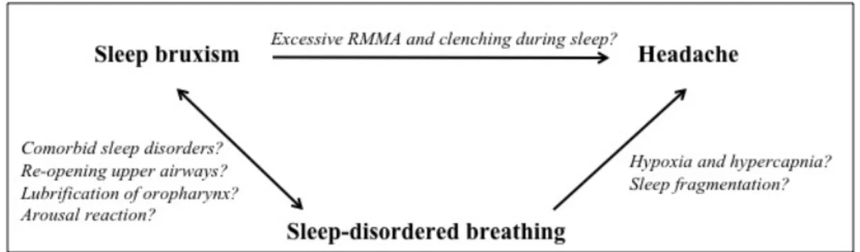
From a clinical perspective, sleep bruxism has been frequently described in association with other sleep disorders (e.g., obstructive sleep apnea), pain complaints (e.g., headache), and, especially in children, with behavioral problems (e.g., inattention and hyperactivity). Thus, tooth-grinding should be considered more than an oral parafunction causing tooth wear, rather it should be accounted in a wider clinical assessment of the patient’s health.
The present thesis aims to better understand the pathogenesis and regulation of rhythmic masticatory muscle activity during sleep to untimely provide support for an evidence-based management of sleep bruxism.
1.1 Historical Aspects of Bruxism
The word “bruxism” originates from the Greek word βρυγµός (brygmós), meaning gnashing of the teeth. The first description of the phenomenon in the scientific literature is dated 1907, when the French term bruxomanie was used to describe an involuntary and ”nervous” grinding of the teeth, as observed in patients who were afflicted with lesions in the central nervous system like meningitis, dementia, and epilepsy (1). Later in 1931, Frohman, a physician, was one of the earliest to use the word bruxism, defined as a problem of a dental nature resulting from non-physiological movements of the mandible related to psychological factors (2). From then on, multiple definitions and several terms have been referred to bruxism: “occlusal habit neurosis”, “neuralgia traumatic”, “teeth gnashing-grinding”, and “parafunction” (3, 4). Few authors also attempted to distinguish between different forms of bruxism. Miller alluded to bruxism to indicate the teeth grinding during sleep, whereas bruxomania was used to denote the habit of grinding during daytime (5). Ramfjord and Ash described clenching as a “centric bruxism”, while grinding as “eccentric bruxism” (6). From the perspective of different medical disciplines, bruxism ranged from being considered a neurological tic or automatism, to a parasomnia or a sleep-related movement disorder (7). The many descriptions and classifications applied to this disorder merely reflect the variety of etiologic factors that over the years have been deemed to cause bruxism.
1.2 Definition and Classification of Sleep Bruxism
According to the Glossary of Prosthodontic Terms, bruxism is considered an oral parafunction consisting of involuntary rhythmic or spasmodic nonfunctional gnashing, grinding, or clenching of the teeth (8). Although this definition describes the main movement-related characteristics of the disorder, it lacks a substantial and important
distinction between the wake and sleep states in which this oral parafunction may occur. There is clinical and research evidence to consider the wake-time habit of clenching, grinding, or gnashing the teeth a distinct nosologic entity, probably with different etiology and pathophysiology, that should be distinguished from bruxism during sleep (7).
The American Academy of Orofacial Pain, indeed, defines bruxism as the diurnal or nocturnal parafunctional activity of clenching, bracing, gnashing, and grinding of the teeth (9). However, the use of the words “diurnal” and “nocturnal” is obsolete; the more precise “wake-time” and “sleep-related” terms should be preferred since they respect the fact that being awake or asleep does not always coincide with daytime and nighttime, respectively.
According to the International Classification of Sleep Disorders, second edition (ICSD-II), published by the American Academy of Sleep Medicine in 2005 (10), sleep bruxism (SB) is classified as a sleep-related movement disorder. The characteristic electromyography (EMG) pattern of SB is found in repetitive and recurrent episodes of rhythmic masticatory muscle activity (RMMA) of the masseter and temporalis muscles that are usually associated with sleep arousals (7, 10). The RMMA shows a frequency of 1 Hz and typically occurs cyclically during sleep (Figure 1.1). RMMA episodes are observed in 60% of the general adult population as physiological activity of the jaw muscles during sleep (11, 12). Many other forms of masticatory and facial muscle activity are also observed during sleep, such as swallowing, coughing, sleep talking, smiling, lip sucking, jaw movements, and myoclonus (7, 13). These orofacial activities account for approximately 85% of EMG events scored on the masseter and temporalis muscles in control subjects and 30% in SB subjects (14-16). In fact, RMMA frequency is three times higher in SB subjects than in controls, and is typically associated with tooth grinding sounds (in 45% of cases), as reported by the patient, bed partner, parents, or siblings (7).
SB may be an extreme manifestation of a physiological orofacial motor behavior during sleep (RMMA and chewing-like activity) whereby certain factors increase its occurrence until it falls into the pathological range of jaw-muscle activity. Therefore, SB
refers to the sleep motor disorder, whereas RMMA is the characteristic EMG pattern that is scored during sleep to make a polysomnographic diagnosis of SB.
1.3 Assessment and Diagnosis of Sleep Bruxism
The assessment and diagnosis of SB are often challenging. Generally, the assessment is based on reports of tooth-grinding sounds during sleep and the presence of clinical signs and symptoms (10). However, only an electromyographic (EMG) recording of the masticatory muscles can confirm the SB diagnosis. A number of portable diagnostic tools have been developed to record masseter and/or temporalis EMG activity during sleep in order to avoid using the more sophisticated but highly cost- and time-consuming polysomnography (PSG). However, the reliability of most portable devices has not yet been validated, and their use may be considered only as a support in the clinical assessment of SB. In fact, the SB diagnosis is usually clinical, although the gold standard remains a full-night PSG with audio-video recording (Table 1.1). The future direction for SB assessment would be to develop a handy tool that can directly, reliably, and rapidly measure ongoing bruxism activity, and that can be used in both clinical (for diagnosis, treatment outcome evaluation, and follow-up) and research settings.
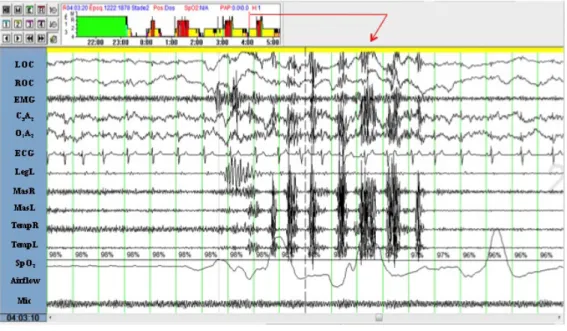
Figure 1.1 Hypnogram and polysomnographic tracing showing an episode of rhythmic masticatory muscle activity (RMMA) during sleep.
The full night hypnogram (graph in the upper left represents sleep stage distribution in non-REM sleep 1, 2, 3, 4 and non-REM sleep) and a 20-sec polysomnographic page with a clear example of RMMA during sleep are shown. The subject is in non-REM sleep stage 2. RMMA is defined when at least 3 consecutive EMG bursts (frequency 1 Hz) lasting ≥ 0.25 sec are scored on the masseter and temporalis channels. Corresponding with the RMMA episode, note the increased frequency in cortical activity (EEG central (C3A2) and occipital
(O1A2) derivations), increased heart rate (on the ECG channel), and increased amplitude of
respiratory airflow (naso-cannula). Immediately before the RMMA onset, an increase in the EMG activity of the suprahyoid muscle (EMG channel) and a leg movement (LegL channel) are observed (From ambulatory PSG recording Siesta, Compumedics).
LOC: left electrooculogram; ROC: right electrooculogram; EMG: electromyographic
activity of the suprahyoid muscle; C3A2: the central derivation of the electroencephalogram
(EEG); O1A2: the occipital derivation of the EEG; ECG: electrocardiogram; LegL: EMG of
the left tibialis muscle; MasR and MasL: EMG of the right and left masseter muscles;
TempR and TempL: EMG of the right and left temporalis muscles; SpO2: oxygen saturation
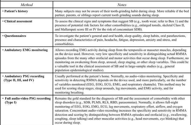
Table 1.1 Methods for assessing sleep bruxism.
SB: sleep bruxism; SDB: sleep-disordered breathing; EMG: electromyogram; RMMA:
rhythmic masticatory muscle activity; EEG: electroencephalogram; EOG:
elecrooculogram; ECG: electrocardiogram; PLMS: periodic limb movement during sleep; RLS: restless leg syndrome; RBD: REM sleep behavior disorder
(Carra MC. based on (7, 17))
Methods for assessing sleep bruxism (order of increasing reliability)
Method Notes
•! Patient’s history Many subjects may not be aware of their tooth-grinding habit during sleep. More reliable if the bed partner, parents, or siblings report current tooth grinding sounds during sleep.
•! Clinical assessment To assess the clinical signs and symptoms that suggest SB (e.g., tooth wear; refer to Box 1) and the presence of potential risk factors for other comorbidities (e.g., enlarged tonsils, skeletal Class II, and Mallampati score III or IV for the risk of concomitant SDB).
•! Questionnaires To investigate the patient’s general and oral health, sleep quality, sleep habits, oral parafunctions, presence and characteristics of pain, headache, fatigue, depression, anxiety and stress, and comorbidities.
•! Ambulatory EMG monitoring Allows recording EMG activity during sleep from the temporalis or masseter muscles, depending on the device used. However, very low specificity and sensitivity in distinguishing actual RMMA episodes from the many other orofacial and motor activities that occur dung sleep. Furthermore, no monitoring on awakening from sleep, arousal, sleep staging, or other sleep variables. This could be a valuable tool in the clinical assessment of SB and in large-sample studies (e.g., general population epidemiological studies).
•! Ambulatory PSG recording (Type II, III, and IV)
Usually performed at the patient’s home. Normally, no audio-video monitoring. Specificity and sensitivity in detecting RMMA depends on the device used, and more particularly, on the number of variables monitored (EEG, EOG, ECG, EMG, and respiratory channels). This method may be used for scoring sleep stages, sleep arousals, leg movements, and EMG activity, and for monitoring breathing.
•! Full audio-video PSG recording (Type I)
Remains the gold standard for the diagnosis of SB and the assessment of comorbidity with other sleep disorders (e.g., SDB, PLMS, RLS, RBD, parasomnias). Normally, it allows full-night monitoring of EEG, EOG, EMG, ECG, leg movements, respiratory effort, airflow, and oxygen saturation. Concomitant audio-video recording increases the specificity and sensitivity in RMMA detection and scoring by distinguishing between RMMA episodes and orofacial (e.g., swallowing, coughing, sleep talking) and other muscular activities (e.g., head movements, eye blinking) that occur during sleep.
1.3.1 Clinical Diagnosis of Sleep Bruxism
The clinical diagnosis of SB should be based on the international diagnostic criteria proposed by the American Academy of Sleep Medicine (Table 1.2) (10, 18). Grinding sounds due to tooth contacts are the pathognomonic sign of SB, and they are usually reported by the patient, bed partner, siblings, or parents. However, not all RMMA episodes are accompanied by tooth grinding, and many patients or family members may not be aware of this.
A clinical examination of the oral cavity allows identifying signs and symptoms that are markers of tooth-grinding activity and a clenching habit. These signs and symptoms include hypertrophy of the masseter and temporalis muscles, tongue indentation, tooth wear, jaw muscle tenderness or pain on digital palpation, and reports of morning headache (7, 19). However, none of these signs and symptoms constitutes direct proof of current SB activity. For example, although tooth wear is widely reported in the literature as the classic dental sign of bruxism (both awake and during sleep), it may be related to many other factors that can induce attrition and erosion on dental surfaces (e.g., age, occlusal conditions, enamel characteristics, diet, carbonated drinks, medications, gastroesophageal reflux, and alimentary disorders)(7, 19-23). Moreover, it was recently demonstrated that tooth wear cannot be used as an absolute criterion to assess SB severity: no difference in tooth wear grade was found between low and high frequency of muscle contractions in young adults with SB (21).
During the clinical examination, dental clinicians can also identify early risk factors for SB and other sleep or medical disorders (e.g., sleep-disordered breathing), and promote further investigations when necessary. In particular, the risk of having or developing sleep-disordered breathing (SDB) increases with retrognathia, micrognathia, macroglossia, adenotonsillar hypertrophy, and a Mallampati score of III and IV (24). The Mallampati score qualifies oropharyngeal obstruction, with I standing for no obstruction (tonsils, pillars, and soft palate are clearly visible) and IV for high obstruction (where only the hard
palate is visible)(25). In addition, clinicians can directly observe breathing habits (mouth breathing vs. nasal breathing), behavioral attitudes (agitation, anxiety), and a tendency to fall asleep. Although it remains under investigation, some of these factors have been associated with an increased risk for both SB and SDB.
Appropriate questionnaires can also be used to investigate general health, quality of life, pain, headache, sleep quality, and sleepiness. Some questionnaires have been validated for both clinical and research purposes (e.g., the Pittsburg Sleep Quality Index and the Epworth Sleepiness Scale). Questionnaire assessments may give the clinician an indication of the risk of comorbidity between SB and other, more severe sleep disorders, such as SDB or restless leg syndrome (RLS) (Table 1.1).
Table 1.2 American Academy of Sleep Medicine (AASM) clinical diagnostic criteria for sleep bruxism.
*None of these signs and symptoms constitutes direct proof of current SB activity. Full-night PSG with audio-video recording remains the gold standard for SB diagnosis.
(Carra M.C. based on (7, 10))
AASM clinical diagnostic criteria for sleep bruxism
1)! Patient history:Recent patient’s and/or parent’s and/or sibling’s report of tooth grinding sounds occurring during sleep for at least 3 to 5 nights per week in the last 6 months
2)! Clinical evaluation:* - Abnormal tooth wear
- Hypertrophy of the masseter muscles on voluntary forceful clenching
- Discomfort, fatigue, or pain in the jaw muscles (and transient morning jaw muscle pain and headache)
3)! Jaw muscle activity cannot be better explained by another current sleep disorder, medical or neurologic disorder, medication use, or substance use disorder.
1.3.2 Ambulatory Assessment of Sleep Bruxism
A number of portable EMG monitoring systems have been developed to assess SB activity. They differ in degree of complexity, ranging from miniature self-contained EMG detectors to ambulatory PSG systems (levels II, III, and IV)(17), which allow monitoring only a limited number of channels (Table 1.1). These devices enable multiple-night recordings in the patient’s home at minimal expense, and could be useful research tools in large sample studies. However, the lack of standardized scoring criteria and evidence-based validity limit their application to both clinical and research settings.
Because automatic EMG detectors and analyzers usually use a unique algorithm for RMMA activity scoring, their validity remains to be demonstrated. Conversely, ambulatory PSG recordings provide very good quality EMG signals, and depending on their complexity, they can usually assess other sleep parameters, such as sleep EEG (essential for sleep staging) or respiratory variables. In addition, on the masseter and/or temporalis EMG channels, RMMA episodes can be distinguished as phasic, tonic, or mixed. Furthermore, episode and burst frequency and muscular strength can be calculated (Table 1.3)(7). However, ambulatory PSG is usually performed in the patient’s home without audio-video monitoring. This may lead to overestimation of RMMA episodes due to confounding and non-SB-specific motor activities during sleep. We are currently validating RMMA scoring criteria on ambulatory PSG recordings, and have observed a modest concordance rate between RMMA scored with and without video on the same night (Carra et al.,
unpublished data). Although preliminary, this finding suggests that, in the absence of
audio-video recording, more rigorous criteria should be applied to the clinical assessment and EMG scoring of SB-related activity.
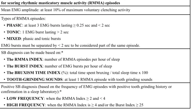
Table 1.3 Polysomnographic research diagnostic criteria for sleep bruxism
*Best level of reliability when performing audio-video PSG recordings and the presence of
at least 2 RMMA episodes associated with tooth-grinding sounds.
(Carra MC. based on (7, 10, 15, 26-29)).
1.3.3 Polysomnographic Diagnosis of Sleep Bruxism
PSG for SB is mainly used for research purposes (Table 1.1). The research diagnostic criteria have been developed on the basis of PSG with audio-video recordings performed in a hospital setting with a sleep technician attending full-night monitoring (15, 18). This PSG (referred to as level I)(17) allows assessing several sleep physiological parameters (e.g., electroencephalogram, electrooculogram, electromyogram, electrocardiogram, airflow, respiratory effort, oxygen saturation), while audio-video recording enables documenting tooth grinding sounds and distinguishing between RMMA
Polysomnographic research diagnostic criteria for sleep bruxism for scoring rhythmic masticatory muscle activity (RMMA) episodes
Mean EMG amplitude: at least 10% of maximum voluntary clenching activity Types of RMMA episodes:
•! PHASIC: at least 3 EMG bursts lasting ! 0.25 sec and < 2 sec •! TONIC: 1 EMG burst lasting > 2 sec
•! MIXED: phasic and tonic bursts
EMG bursts must be separated by < 2 sec to be considered part of the same episode. SB diagnosis can be made based on:*
•! The RMMA INDEX: number of RMMA episodes per hour of sleep •! The BURST INDEX: number of EMG bursts per hour of sleep
•! The BRUXISM TIME INDEX (%): total time spent bruxing / total sleep time x 100 •! TOOTH-GRINDING SOUNDS: at least 1 RMMA episode with tooth grinding sounds
Positive SB diagnosis (based on the frequency of EMG episodes with positive tooth grinding history or confirmation in a sleep laboratory):*
•! LOW FREQUENCY: when the RMMA Index ! 2 and < 4
•! HIGH FREQUENCY: when the RMMA Index is ! 4 and/or the Burst Index ! 25
and orofacial (e.g., swallowing) and other muscular activity (e.g., head movements) during sleep. The validated criteria for a sleep laboratory diagnosis of SB showed 72% sensitivity and 94% specificity (15). Based on RMMA index (number of episodes/h of sleep), the diagnosis of SB is made when RMMA index ≥ 2 (low-frequency SB – mild bruxism). A RMMA index greater than 4 is considered moderate to severe bruxism (high-frequency SB) (Table 1.3)(7, 15, 30).
PSG recordings are not usually indicated for subjects who report SB only. However, the clinician should refer the patient to a sleep physician for further investigation and diagnosis if other sleep disorders are suspected (e.g., sleep apnea, sleep-related epilepsy, REM sleep behavior disorder, periodic limb movement, or other neurological disorder).
1.4 Epidemiology of Sleep Bruxism
In large population-based studies, it is difficult to assess SB by objective measures such as PSG recordings. The epidemiology of SB is therefore largely determined by questionnaires, self-reports, and/or clinical findings (e.g., tooth wear).
SB is reported by 8% of the general adult population (31, 32). It typically peaks during childhood (with prevalence approaching 40% below the age of 11 years)(33-38), and tends to decrease after adulthood. During elderly (>60 years of age) SB is reported with a low prevalence (3%)(31, 32). However, this may be explained by the presence of edentulism, denture wearing, and changes in sleeping behaviors (i.e., in isolation) that are often observed in elderly populations. Overall, no gender difference has been observed (31, 38, 39).
Many confounding factors should be considered when interpreting the epidemiology of SB. The wide prevalence range (from 8% to 40%) reported in the
literature is most probably because many studies failed to distinguish between wake-time and sleep-related bruxism or to assess the presence of medical comorbidities that may influence its occurrence. Indeed, SB is frequently concomitant (approximately one third of the subjects) with wake-time bruxism, which is characterized mainly by a tooth clenching habit (40). Wake-time bruxism tends to increase with age, with an estimated prevalence of 13% in children (38) and over 30% in adults (41-43).
Notwithstanding the limitations related to its objective assessment, SB is a common sleep disorder and its actual prevalence is probably much higher since individuals are often un-aware of sleep-related motor behaviors, especially if subjects sleep alone.
1.5 The Masticatory System during Wake and Sleep
Rhythmic masticatory movements, like chewing, are controlled by the central nervous system. Specifically, it is the trigeminal system that is responsible for the activation and regulation of the masticatory muscles (44). Indeed, the muscles of the jaw are innervated by trigeminal motoneurons located in the trigeminal motor nucleus, in the rostral part of the pons. Trigeminal motoneurons receive both excitatory (i.e., glutamatergic) and inhibitory (i.e., GABA-ergic and glycinergic) synapses. The excitatory and inhibitory inputs onto trigeminal motoneurons are coming from premotoneurons surrounding the trigeminal motor nuscleus. During mastication, trigeminal motoneurons fire to produce very regular and repetitive jaw movements on the three planes of the space. These jaw movements are determined by the activation (i.e., contraction) of the jaw-opening muscles (e.g., digastric muscles) during mouth jaw-opening, and of the jaw-closing muscles (e.g., masseter and temporalis muscles) during mouth closing. The rhythmical and alternated activation of the jaw muscles is produced by the brainstem nuclei that compose the central pattern generator (CPG) (45). The CPG is responsible of initiating and
maintaining the motor activity through pattern generation and rhythm generation. These activities in the CPG can be triggered both from the cortex and from the sensory signals from the periphery. In facts, although chewing can continue without feedback, the movement is fine-tuned by sensory signals acting through the action of reflexes (e.g., the stretch reflexes; the periodontal reflexes) (46). This mechanism guarantees that muscle activity, muscle force, and jaw position are adjusted to deal with the changing conditions that occur when different foods are being chewed. Therefore, mastication is neither a purely voluntary movement nor a purely reflex (44, 46). A complex combination of both drives the rhythmicity and coordination of the masticatory muscles, whose movement is centrally programmed (47).
Chewing-like rhythmic jaw movements during wakefulness can also be cortically evoked by electrical stimulation, resulting in a pattern of jaw-opening and jaw-closing muscle activity that resembles natural food chewing (48). Conversely, rhythmic masticatory movements during sleep (e.g., RMMA) occur spontaneously without any food triturating purpose, and with a co-contraction of both the jaw-closing and jaw-opening muscles. Moreover, RMMA occurs without apparent cortical involvement—unlike chewing, which is initiated at the cortical level— and it is strongly influenced by autonomic nervous system activity and arousals during sleep (20, 49, 50). Nevertheless, many studies suggest that SB is centrally regulated—probably in the brainstem—and its genesis is more likely multifactorial (7, 51-53).
The brainstem neuronal networks which control the genesis of rhythmic masticatory movements during wakefulness has been studied in animal models, which allowed describing the cortico-bulbar pathway that drives mastication (45, 47). In particular, it seems that cortico-bulbar inputs to controlateral brainstem structures first activate a relay in the medial pontomedullary reticular formation that eventually reaches the trigeminal motor nuclei and activates jaw-opening or jaw-closing muscles to produce jaw movements (47, 54). During sleep, these cortico-bulbar influences are not dominant, rather they seem to be partially de-activated to preserve sleep continuity (55, 56). Interestingly, several of the
brainstem reticular nuclei that are involved in the masticatory regulation are also involved in sleep genesis and maintenance, as well as in respiratory control (51).
The ascending arousal system originating in the brainstem is crucial in maintaining wakefulness with well-defined cell groups involved in two major pathways (57). The major inputs come from the cholinergic pedunculopontine and laterodorsal tegmental nuclei. These nuclei activate thalamic relay neurons and the thalamic reticular nucleus that are crucial for transmission of information to the cerebral cortex. The other pathway originates in centers in the upper brainstem and caudal hypothalamus, including the locus coeruleus, dorsal and median raphe nuclei, ventral periaqueductal gray matter, and the tuberomammillary nucleus. This ascending arousal pathway bypasses the thalamus, activating the lateral hypothalamus and basal forebrain and then the cerebral cortex. The activity of both the wake-promoting pathways is inhibited by a system of gamma-aminobutyric acid (GABA)-containing neurons, in which the lateral hypothalamic ventrolateral preoptic nucleus (VLPO) appears to play a key role (58). Wake- and sleep-promoting systems have mutually inhibitory influences on each other. Indeed, during wakefulness the activity of the VLPO is strongly inhibited by the noradrenergic locus coeruleus and other wake-promoting centers. As the activity of these centers decreases at sleep onset, the VLPO becomes active, in turn reciprocally inhibiting wake-promoting neuron activity. This mutually inhibitory self-reinforcing loop has the characteristic of a “flip-flop switch” that acts to produce stable states of wakefulness or sleep with sharp transitions between them (57). Glycinergic and GABAergic inhibitory neurons active during sleep are known to directly project to the respiratory centers, such as the hypoglossal motor nucleus (59), and to influence the activity of the masticatory CPG nuclei. In fact, during sleep there is a general reduction in muscle tone in comparison with wakefulness. This could result from a reduction in the cortico-bulbar drive, and/or tonic hyperpolarization of masticatory muscle motoneurons, making them less excitable, and/or a reduction in the arousal driven monoaminergic tone (51). Similar to the decrease in postural muscle tone, sleep causes the suppression of masticatory muscles activity, which typically
occur immediately at sleep onset indicating a primary suppressant effect of sleep neural mechanisms. This inhibition (i.e., decrease in trigeminal motoneurons excitability) is higher during REM sleep, a sleep stage characterized by powerful generalized muscle “atonia” (or better hypotonia) of the limb and jaw muscles. However, even during REM sleep transient and sudden periods of increased trigeminal motoneurons excitability are observed, and RMMA activity can be manifest (50, 51).
1.6 Etiology and Pathogenesis of Sleep Bruxism
The exact etiology and pathophysiology of SB are still unknown (60). The putative etiologic mechanisms for the genesis of RMMA during sleep include sleep arousal, autonomic sympathetic cardiac activation, genetic predisposition, neurochemicals, psychosocial components, exogenous factors, and comorbidities (Table 1. 4).
1.6.2 Sleep Arousal
As stated in the ICSD-II definition, the majority of RMMA episodes occur in association with sleep arousals (10, 18, 61). This association was first observed by Reding et al. (62) in 1968 and by Satoh and Harada (63) in 1971, who described tooth-grinding activity as an “arousal reaction”. Since then, many studies have used polysomnography and electrophysiology to investigate the complex relationship between SB and sleep arousal (60, 61, 64-67).
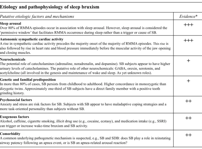
Table 1.4 Etiology and pathophysiology of sleep bruxism.
*Strength of available scientific evidence.
+ weak evidence; ++ moderate evidence; +++ strong evidence
(Carra MC. based on (7, 20, 51, 60, 61, 64, 68-74)).
1.6.2.1 The sleep arousal system
Sleep arousal is defined as a brief awakening from sleep (for at least 3 seconds) characterized by an abrupt shift toward higher EEG frequencies (including alpha, theta and/or frequencies greater than 16 Hz, but not spindles), generally associated with cortical, autonomic and behavioral activations in the absence of the return of consciousness (18). Arousals normally reoccur from 6 to 14 times per hour of sleep as the response of the
Etiology and pathophysiology of sleep bruxism
Putative etiologic factors and mechanisms Evidence*
Sleep arousal
Over 80% of RMMA episodes occur in association with sleep arousal. However, sleep arousal is considered the ‘permissive window’ that facilitates RMMA occurrence during sleep rather than a trigger or cause of SB.
+++
Autonomic sympathetic cardiac activity
A rise in sympathetic cardiac activity precedes the majority onset of the majority of RMMA episodes. This rise is also followed by rise in heart rate and blood pressure immediately before the muscular activity of the jaw opening and closing muscles.
+++
Neurochemicals
The potential role of catecholamines (adrenaline, noradrenalin, and dopamine). SB subjects appear to have higher urinary levels of catecholamines. The putative role of other neurochemicals: GABA, orexin, serotonin, and acetylcholine (all involved in the genesis and maintenance of wake and sleep. As yet unknown roles).
+
Genetic and familial predisposition
In more than 80% of cases, SB persists from childhood to adulthood. Higher concordance in monozygotic than dizygotic twins. Approximately one-third of SB subjects have a direct family member with a positive tooth grinding history.
+
Psychosocial factors
Anxiety and stress are risk factors for SB. Subjects with SB appear to have maladaptive coping strategies and a more task-oriented personality than subjects without SB.
++
Exogenous factors
Alcohol, caffeine, cigarette smoking, illicit drug use (e.g., cocaine, ecstasy), and medication intake (e.g., SSRI) can trigger or increase wake-time bruxism and SB activity.
++
Comorbidity
A common underlying pathogenetic mechanism is suspected, e.g., SB and SDB: does SB play a role in reinstating airway patency following an apnea event, or is SB an apnea-related arousal reaction?
sleeping brain to external (environmental) and internal (physiological or pathological) stimuli. In the presence of certain sleep disorders, breathing anomalies or chronic pain, arousals are more frequent.
Although sleep is defined as a reversible behavioral state of reduced responsiveness to environmental stimuli, the sleeping brain is still active and able to control the autonomic, metabolic and hormonal changes that take place within the body. Some cerebral areas remain gradually and partially activated and they are able to scan and weight information filtered through the thalamocortical pathways, and simultaneously trigger behavioral responses to given external stimuli. Such activation is stimulated by the arousal system (57, 75). The arousal system comprises cholinergic, monaminergic, histaminergic, and orexinergic neurons located in the brainstem, which fire at different rate during wakefulness, NREM and REM sleep (57, 76). Their activity finely regulates the sleep-wake transition and the sleep cycle (57). Sleep arousals can be seen as the body’s attempt to prepare the sleeping individual (who is in a low vigilance state) to react to a potential risk, i.e., a « fight or flight » state. However a high arousal index (number of arousals per hour of sleep) is also a sign of sleep fragmentation, a detrimental alteration of sleep architecture, and thus a poor quality of sleep (75).
The arousal threshold (the probability to elicit an arousal from sleep) changes throughout the night. In the wake-to-sleep transition, usually sleep stage N1, the arousal threshold is very low and thus sleep can be easily discontinued by environmental stimuli. In stage N2 and N3 an incrementally more intense stimulus is required to produce arousal. Conversely, the arousal threshold during REM sleep is quite variable throughout the night (77, 78). Beside the behavioral state, the arousal response depends also on the nature of sensory stimulations (e.g., intensity, modality) (79).
RMMA episodes are observed predominantly during sleep stages N1 and N2 (light
sleep), whereas only the 10% of episodes occur during REM sleep (7, 61). Between 70%
autonomic-cardiac activations (See Section 1.5.2). Although the number and index of arousal do not generally differ between young otherwise healthy SB subjects and controls, experimental evidence suggested that SB subjects may have a higher responsiveness to sleep arousal (65). Whether this condition may influence the onset and recurrence of RMMA is unclear.
1.6.2.2 The cyclic alternating pattern
The cyclic alternating pattern (CAP) is a marker of cerebral activity occurring under conditions of reduced vigilance (e.g., sleep). CAP is considered the expression of a basic arousal modulator, which represents states of sleep instability but also belongs to physiological sleep (80-82). Terzano and colleagues have extensively studied the features of CAP for 30 years, and they have demonstrated that most of the arousal-related phasic events, which can appear either spontaneously or after external perturbation, follow this cyclic and rhythmic time organization during NREM sleep (83, 84). CAP is described as the structural framework that ties together both sleep-preserving features (low EEG frequency, high amplitude bursts) and sleep-disrupting events within 20-40 seconds periodicity.
During NREM sleep, CAP is composed by the alternation of two EEG patterns (phase A and phase B) each lasting 2 to 60 seconds. Phase A is considered the active phase associated with heightened arousal levels, while phase B corresponds to the periodic replacement of the background EEG activities peculiar to the specific NREM sleep stage. On the basis of the reciprocal correlations between EEG and polygraphic parameters, it is possible to distinguish three types of CAP phase A (of increasing arousal pressure)(85):
- Phase A1: comprising exclusively synchronized EEG patterns that generally show only slight simultaneous variations in muscle tone and autonomic functions. It represents the weakest arousal power;
- Phase A2: composed of a mixture of slow and fast rhythms. It represents a transition phase and fits an intermediate arousal power level;
- Phase A3: characterized predominantly by EEG desynchronized patterns. It corresponds to the most powerful arousal pressure, as it is usually accompanied by relevant changes in muscle tone, heart rate and respiratory activity.
When the interval between 2 consecutive phase A and phase B exceeds 60 seconds, the CAP sequence ends and sleep enters the non-CAP (NCAP) mode characterized by stable EEG rhythms with very few and randomly distributed arousal-related phasic events (Figure 1.2). On the basis of the reciprocal occurrence and respective meaning of both CAP (marker of unstable sleep) and NCAP (stable and consolidated sleep), it is possible to define the sleep variable CAP rate (the ratio of total CAP time over the total NREM sleep time) as the measure of arousal instability during sleep (83). CAP rate is enhanced when sleep is disturbed by internal or external factors and its variations correlate with the subjective appreciation of sleep quality, with higher CAP rate associated with poorer sleep quality (82). CAP rate in normal sleepers shows a low intra-individual variability from night-to-night, while remarkable age-related differences have been reported. In particular, CAP rate was found to be progressively increasing from pre-school age to adolescence, and then U-shaped with a minimum in young adults and increasing again in late adulthood and elderly (86-88).
The distribution of CAP has been proven to be different across the sleep cycle. CAP sequences normally predominate in close temporal connection with major dynamic events, such as falling asleep, sleep stage shift, NREM/REM sleep transitions, nocturnal awakenings and body movements. It has been hypothesized that the abundance of A1 subtypes in the descending phase of the sleep cycle is the EEG expression of the cerebral mechanisms involved in the build-up and maintenance of deep NREM sleep, whereas subtypes A2 and A3 are predominant in the period preceding the onset of REM sleep (ascending phase) and may express the subject’s arousability (89). This hypothesis is also supported by the topographic distribution of CAP A phases. Indeed, the A1 activity is
thought to be generated primarily by the frontal lobes where also slow waves are predominantly detected, whereas CAP A2 and A3 activity is thought to be generated by the posterior brain regions (90).
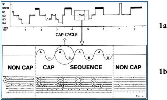
Figure 1.2 Schematic representation of cyclic alternating pattern (CAP).
1a. Histogram of physiological sleep in which CAP sequence (comb-like oscillation) and
NCAP sequence (horizontal stretches) are outlined. 1b. A specimen of sleep stage 2 is expanded to highlight the sequence of 4 CAP cycles (phase A + phase B) framed between portion of NCAP. (From Parrino et al. 1996, J Clin Neurophysiol)(91).
W, wakefulness; SREM, REM sleep; S1, S2, S3, S4, NREM sleep stages.
1a
.
CAP and arousals are considered active adaptive responses of the sleep regulatory mechanisms, which tend to remove the stimulus-disturbance effect and re-establish an internal equilibrium. The total amount of CAP can be seen as the effort to maintain sleep at the microstructural level. While a limited amount of CAP is considered physiological, larger quantities reflect the brain difficulties to consolidate and preserve sleep and they may be associated with detrimental effects. The arousal system plays a cardinal neurophysiologic role in protecting and tailoring sleep duration and depth. CAP oscillation participates in the dynamic organization of sleep. Physiologic, paraphysiologic and pathologic motor activities during NREM sleep are associated with a stereotyped arousal pattern characterized by an initial increase in EEG delta power and heart rate, followed by a progressive activation of faster EEG frequencies (91-93).
In this perspective, there is evidence that supports the hypothesis that the frequency of RMMA episodes is modulated by the cyclic occurrence of sleep arousals, i.e. CAP (60, 61, 64). As previously mentioned, RMMA episodes are more frequently observed in NREM sleep stages 1 and 2 (light sleep), in sleep stage shifts, and especially in the transition period from non-REM to REM sleep (64). Over 80% of RMMA episodes are time-correlated with CAP phase A, and they recur in rhythmic clusters, with a periodicity of 20 to 30 seconds, which is similar to the physiological arousal rhythm of CAP.
Notwithstanding this association between sleep arousal and the occurrence of SB, the role of CAP and sleep instability in the pathogenesis of RMMA remains to be elucidated.
1.6.3 Autonomic Sympathetic-Cardiac Activity
Recent evidence on SB pathophysiology highlights the role of the autonomic nervous system (60, 64, 94). It has been well demonstrated that RMMA onset is associated
with a sequence of physiological events that occur within a sleep arousal. Briefly, the genesis of most RMMA episodes is preceded by the following cascade of events (7): - A rise in the autonomic sympathetic-cardiac activity with a concomitant withdrawal of
parasympathetic influences (from 8 to 4 minutes before RMMA onset)(64)
- The appearance of rapid-frequency EEG cortical activity (sleep arousal; approximately 4 seconds before RMMA onset)(60)
- A rise in heart rate of about 25% (beginning 1 second before RMMA onset), concomitant with
- An increase in jaw opener muscle tone (the suprahyoid muscle, probably responsible for mandible protrusion and airway opening), concomitant with
- An increase in the amplitude of the respiratory effort (nasal airflow)(69), preceding or concomitant with
- A rise in diastolic and systolic blood pressure (95)
- And finally, an observable EMG incident in the jaw-closing muscles (masseter and temporalis), scored as RMMA with or without tooth-grinding sounds (7). Almost 60% of RMMA episodes are followed in the 5 to 15 seconds after onset by swallowing (96) (Figure 1.3).
The activity of autonomic nervous system is physiologically modulated during sleep (97, 98). NREM sleep is characterized by a period of relative autonomic stability, with a vagal nerve dominance and sinusoidal modulation of heart rate variation due to a coupling with respiratory activity. Hypotension, bradycardia, and reduction of cardiac input are also progressively observed with deepening stages of NREM sleep. Conversely, during REM sleep the brain’s increased excitability can result in major surges in cardiac sympathetic nerve activity, striking fluctuations of blood pressure and heart rate, and marked episodes of tachycardia and bradycardia (97). Finally, as describe above, significant autonomic changes (approximately 90% increase in sympathetic activity and 35% decrease in
parasympathetic activity)(99) accompany electrocortical arousals from sleep as well as periodic movements during sleep (64, 95, 98).
The autonomic nervous system activity can be non-invasively assessed by analyzing the heart rate variability during both wakefulness and sleep (100). Power spectral analysis of heart rate can then be evaluated as low frequency (LF: 0.04-0.15 Hz) and high frequency (HF: 0.15-0.5 Hz) ranges of modulation, reflecting the sympathetic+parasympathetic, and parasympathetic activities respectively. By calculating the LF/HF ratio, an index of sympathovagal balance can be obtained (100, 101). It has been reported that SB subjects have higher LF power and higher LF/HF ratio during wake compared to healthy controls, supporting that heart rate variability in SB subjects might be altered toward an increase in sympathetic activity also during wakefulness (94). This hypothesis was already suggested by the observation of phenomena like peripheral vasoconstriction, tachycardia and skin potential changes during tooth-grinding events, which might be a consequence of increased sympathetic activity (67). However, the power spectral analysis of heart rate variability during sleep showed no difference between SB subjects and controls for LF and HF powers, neither for the sympathovagal balance, even though a rise in sympathetic cardiac activity is registered in the minutes preceding RMMA episodes (64).
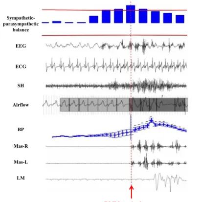
Figure 1.3 Genesis of an RMMA episode (schematic representation of the cascade of physiologic events that precedes RMMA onset)
EEG: electroencephalogram; ECG: electrocardiogram; SH: EMG of the suprahyoid muscle; BP: blood pressure; Mas-R and Mas-L: EMG of the right and left masseter muscles; LM: laryngeal movements.
(Lavigne G., Huynh N. based on (60, 64-66, 69)).
EEG ECG SH Airflow BP Mas-R Mas-L LM
RMMA episode onset
Sympathetic-parasympathetic balance
Correlation between RMMA onset and a rise in sympathetic activity
Sleep arousal Tachycardia
Increased jaw opening muscle activity
Large respiratory breaths
A rise in systolic and diastolic blood pressure
Rhythmic masticatory muscle activity (RMMA)