PRÉCISIONS SUR L'ANATOMIE DE L'OSTÉOLÉPIFORME EUSTHENOPTERON FOORDI DU DÉVONIEN SUPÉRIEUR
DE MIGUASHA, QUÉBEC
MÉMOIRE
PRÉSENTÉ À
L'UNIVERSITÉ DU QUÉBEC À RIMOUSKI
Comme exigence partielle du programme de Maîtrise en Gestion de la Faune et de ses Habitats
PAR
JOËL LEBLANC
Avertissement
La diffusion de ce mémoire ou de cette thèse se fait dans le respect des droits de son auteur, qui a signé le formulaire « Autorisation de reproduire et de diffuser un rapport, un mémoire ou une thèse ». En signant ce formulaire, l’auteur concède à l’Université du Québec à Rimouski une licence non exclusive d’utilisation et de publication de la totalité ou d’une partie importante de son travail de recherche pour des fins pédagogiques et non commerciales. Plus précisément, l’auteur autorise l’Université du Québec à Rimouski à reproduire, diffuser, prêter, distribuer ou vendre des copies de son travail de recherche à des fins non commerciales sur quelque support que ce soit, y compris l’Internet. Cette licence et cette autorisation n’entraînent pas une renonciation de la part de l’auteur à ses droits moraux ni à ses droits de propriété intellectuelle. Sauf entente contraire, l’auteur conserve la liberté de diffuser et de commercialiser ou non ce travail dont il possède un exemplaire.
TABLE DES MATIÈRES
TABLE DES MATIÈRES ... .ii
LISTE DES TABLEAUX ... v
LISTE DES FIGURES ... vi
INTRODUCTION GÉNÉRALE ... xi
RÉFÉRENCES ... xiv
REMERCIEMENTS ... xvi
CHAPITRE 1 NEW DATA AND NEW METHODS ON AN OLD PROBLEM: THE CHOANA REVISITED ... 1
ABSTRACT ... 2
RÉsuMÉ ... 3
INTRODUCTION ... 5
MATE RIAL AND METHODS ... 8
RESULTS ... 9 Phylogenetic analyses ......................................... 13 DISCUSSION ... 16 ACKNOWLEDGEMENTS ... 18 REFERENCES ... 19 Tables ... 23 Figures ... 25
CHAPITRE 2
DEVELOPMENTAL MODULARITY AND SALTA TORY ONTOGENY IN THE
LATE DEVONIAN OSTEOLEPIFORM EUSTHENOPTERON FOORDI .... 32
ABSTRACT ... 33
RÉSUMÉ ... 34
INTRODUCTION ... 36
MATERIAL AND METHODS ... 39
Specimens ....................................................... 3 9 Sequence of ossification ......................... 39 Measurements ............... 41 Bane terminology .......... 41 RESULTS ... 42 Skull ............................ 43
Sequences of ossification of the fins ........................... .44
Pectoral fins ..................... 45
Pelvic fins ............................ 45
First dorsal fin ................ 45
Second dorsal fin ....... 46
Analfin ......................... 46
Vertebral column and caudal fin ............ .47
Disparity and reliability of the sequence ............. .49
Basal seules ................... 53 Lepidotriehia ...... 54 Ontogenelie palhway ....................................... 55 DISCUSSION ... 57 CONCLUSION ... 61 . ACKNOWLEDGMENTS ... 62 REFERENCES ... 63 Tables ... 68 Figures ... 70 CONCLUSION GÉNÉRALE ... 85
LISTE DES TABLEAUX
Chapitre 1
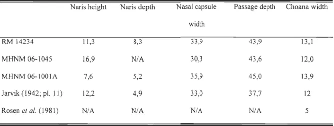
Table 1. Relative dimensions of nasal capsule related structures among three specimens of Eusthenopteron foordi. AU measurements are given in percentage; naris height: proportion
of snout height (lateral view); naris depth: proportion of snout width (scan view); nasal
capsule width: proportion of snout width; passage depth: proportion of snout height; choana
width: proportion of snout width ... 19
Table 2. Results ofphylogenetic analyses using PAUP*4.0bl0 on four previously
published matrices ... 20
Chapitre 2
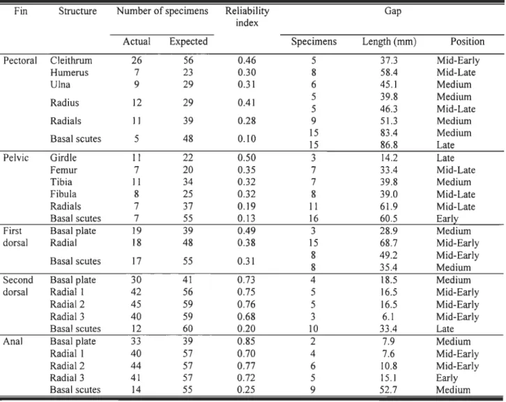
Table 1. Major axis and 95% confidence interval for the slope for six fin position in
relation to the standard length of Eusthenopteronfoordi (x = 10glO converted data, y = 10glO
converted standard length). These equations correspond to the scatter plots of figure 3 ... 56 Table 2. Ossification sequence reliability for each appendicular skeleton structure for 56 Eusthenopteronfoordi. See explanations in the text for the definition and interpretation of
LISTE DES FIGURES
Chapitre 1
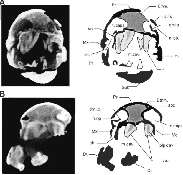
Figure 1. Cross section in the nasal capsule region of Eusthenopteronfoordi. A. Specimen MHNM 06-1001A (scan 8/36); B. Specimen MHNM 06-1045 (scan 8/16); C. Specimen RM 14234 (scan 102/110). Left column: Computed Tomography. Right column:
Interpretation of the CT scan; black = bone, dark grey = ethmosphenoid, light grey
=
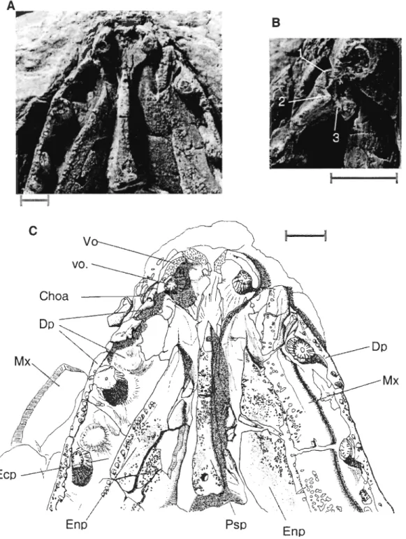
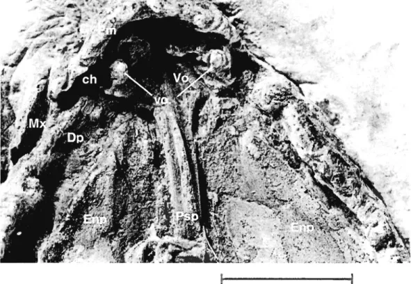
teeth. Abbreviations, a.Te.: anterior tectal, ch.: choana, drnl.p.: dermintermedial process, DpI.: dermopalatine, Dt.: dentary, Ecpt.: ectopterygoid, Enpt.: entopterygoid, Ethrn.:ethmosphenoid, f.: fang, GuI.: gular plate, ioc: infraorbital canal, I.Te: lateral tectal, rn.cav.: mouth cavity, Mx.: maxillary, n.caps.: nasal capsule, n.op.: nasal opening, plp.cav.: pulp cavity, Prnx.: premaxillary, Pr.: postrostral, Psph.: parasphenoid, soc: supraorbital canal, Vo.: vomer, vo.f.: vomerine fang ... 25 Figure 2. Eusthenopteron foordi, specimen MHNM 06-719. A. Ventral view of the palate; B. Close-up of the choana area; C. Camera-Iucida drawing of the specimen. Scale bars: 1 cm. 1,2 and 3 identify facets on the head of the dermopalatine. DpI.:
dermopalatine, Ecpt.: ectopterygoid, Enpt.: entopterygoid, Mx.: maxillary, Psph.:
parasphenoid, Vo.: vomer, vo.f.: vomerine fang ... 27 Figure 3. Eusthenopteronfoordi, specimen MHNM 06-220. Ventral view of the palate. Scale bar ; 1 cm. ch.: choana, DpI.: dermopalatine, Enpt.: entopterygoid, Mx.: maxillary,
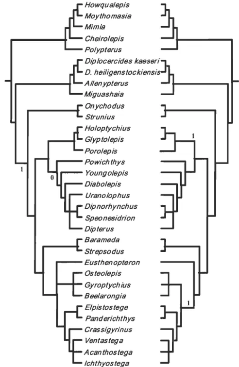
Figure 4. Strict consensus trees obtained from previously published matrices. Trees
obtained from: A. Cloutier & Ahlberg (1996); B. Zhu & Schultze (1997); C. Zhu & Schultze (2001); D. Johanson & Ahlberg (2001) matrices. 1 identifies appearance of the
choana char acter, 0 identifies reversaI of this character. Trees from original matrices are numbered on the right side of the branches, whereas trees from modified matrices (choana considered absent in Eusthenopteron) are numbered on their left side. C and D only have
one tree because the topology remained the same after the matrix modification. Trees B and D show double appearance and/or reversaI because it was not possible to know if the authors used "ACCTRAN" or "DELTRAN" options ... 29
Chapitre 2
Figure 1. Scatter diagram of standard length (mm) plotted against distance (mm) between
insertion points of fins in Eusthenopteronfoordi. The lower regression line (0)
corresponds to the distance between insertion points of the first and second dorsal fins (D 1-D2); the upper regression line (0) corresponds to the distance between the insertion point of
the second dorsal fin and the insertion point of the epichordallobe of the caudal fin (D2-C).
Data were obtained from Thomson and Hahn (1968), Schultze (1984), and original
measurements ... 70
Figure 2. Sequence of ossification of Eusthenopteron foordi for the pectoral, pelvic, dorsal
and anal fins. Observed skeletal elements (dark rectangles) are recorded for 65 specimens. For each specimen, the standard length (S.L.) is given; a star indicates an estimated length
parts owing to partial preservation of fossils. A and B designate part and counterpart of a specimen. Bas.: basal plate; Clei.: c1eithrum; Fern.: femur; Fib.: fibula; Hum.: humerus; RI, R2 and R3: first, second and third radial in an anterior to posterior position;
RadlRads.: radiaVradials; Seut.: basal scutes ... 71
Figure 3. Scatter diagrams of body measurements (mm) versus standard length (mm) in
Eusthenopteronfoordi. A, Distance from posterior margin of the operculum to the insertion of the first dorsal fin; B, distance from posterior margin of the operculum to the insertion of
the second dorsal fin; C, distance from posterior margin of the operculum to the insertion of the pelvic fins; D, distance from posterior margin of the operculum to the insertion of the
anal fin; E, distance from posterior margin of the operculum to the insertion of the
epichordallobe of the caudal fin; F, distance from posterior margin of the operculum to the
insertion of the hypochordallobe of the caudal fin. Regression lines are illustrated ... 73
Figure 4. Small specimen of Eusthenopteronfoordi (UL 265). Enlarged view of the
pectoral girdle region, showing the first ossified branchial elements. b.se.: basal scute; Cbr.: ceratobranchials; Dt.: dentary; I.Ano.: anoc1eithrum; I.CI.: left c1eithrum; I.Cla.: left
c1avic1e; 1.l.Gu.: left lateral gular plate; Op.: opercular; pee.f.: pectoral fin; r.CI.: right c1eithrum; r.Cla.: right c1avic1e; r.l.Gu.: right lateral gular plate; sbm.: submandibular; Sc.: scales. Scale bar equal 5 mm ... 75 Figure 5. Schematic representation of the ossification of each fin in Eusthenopteron
foordi. Stages are progressive from left to right. Solid lines correspond to fin rays, dark
grey elements are endochondral bony structures and light grey elements are basal scutes. Arrows indicate direction of ossification ... 76
Figure 6. Caudal region of Eusthenopteronfoordi, specimen MHNM 06-59 (S.L. ca. III
mm). Rod-shaped basal elements in both the second dorsal fin and the anal fin in early stage of development. A. rad.: anal fin radiaIs; b.pl.: basal plate; D2 rad.: second dorsal
[m radiaIs; H.sp.: haemal spines; N.sp.: neural spines; V.c.rad.: ventral caudal radiaIs. Scale bar equals 10 mm ... 78 Figure 7. Sequence of ossification of Eusthenopteronfoordi for the vertebral column and
the caudal fin. Skeletal elements present in each of the 65 observed specimens, identified by their standard length (same order as in figure 2). The light grey shadowed are as are
missing parts of the fossils. Legend: _Neural spines; ~ Haemal spines;
!IIIllD
Intercentra; ~ Pleurocentra;c=:J
Caudal ventral radiaIs ... 79Figure 8. Specimen of Eusthenopteronfoordi (UL 95, S.L.: 59.9 mm). A, Complete specimen. B, Details of the anterior ventral radiaIs of the caudal fin. Three of them show evidence of collapsing (longitudinal grooves) owing to taphonomic process. Scale bars
equal 1 mm ... 81
Figure 9. Three ofthe smallest specimens of Eusthenopteronfoordi, showing the early ossification of the basal scutes. A, AMNH 7687A; B, AMNH 7650; C, UL 121A; D,
enlargement of the anal fin region of specimen UL 121 A. Scale bars equal 5 mm ... 82
Figure 10. Anal fin of Eusthenopteronfoordi showing the segmentation and branching of the lepidotrichia. Cireles indicate branching. A, FMNH UC 2127 (S.L.: 102.3 mm); B,
FMNH UF 45 (S.L.: ca. 252 mm). b.PI.: basal plate; ra.: radial. Scale bars equal 5 mm ... 83
Figure 11. Ontogenetic pathway of Eusthenopteronfoordi. The cwnulative nwnber of
ossified elements for 65 specimens is traced against the specimen standard length. Values 1 to 6 are rate of development expressed in terms of the mean nwnber of new elements for each millimetre of growth (elements/mm) for each step and threshold ... 84
INTRODUCTION GÉNÉRALE
La paléontologie moderne ne se contente plus de livrer de simples descriptions anatomiques des organismes fossilisés qu'elle étudie. Elle arrive aujourd'hui, par des méthodes de pointe, à découvrir la façon dont vivaient ces êtres disparus, les distances phylogénétiques qui les séparent, la nature de l'habitat dans lequel ils évoluaient. Certaines espèces sont plus fréquemment trouvées (e.g., plusieurs espèces de trilobites), d'autres se voient accorder une grande importance dans l'histoire de la vie (e.g., l'Archaeopteryx
lithographica), d'autres encore acquièrent une renommée souvent due à leurs
impressionnantes caractéristiques physiques (e.g., le Tyranosaurus rex).
Le Québec aussi a son fossile « populaire ». Trouvé dans les falaises de Miguasha en Gaspésie, l' Eusthenopteron foordi Whiteaves 1881 est un poisson sarcoptérygien qui vivait il y a environ 370 millions d'années (Frasnien, Dévonien supérieur). Connu depuis plus de 125 ans, le nombre de spécimens récoltés dépasse aujourd'hui 2 200 (Parent et Cloutier, 1996) et les publications sur son sujet se chiffrent à près de 200 (sans compter toutes celles qui ne font que mentionner l'espèce; Cloutier, 1996). Il est devenu,
notamment grâce aux travaux de Jarvik (1942, 1980), l'un des vertébrés fossiles les mieux connus et décrits et il fut élevé assez tôt au rang de taxon clé dans la transition menant des « poissons » aux tétrapodes (Cope, 1892), principalement à cause des particularités anatomiques de ses nageoires paires qui rappellent les membres des tétrapodes.
Dès lors, à cause de son statut « hybride » entre aquatique et terrestre, une relative importance fut accordée à l'étude du museau de l' Eusthenopteron foordi et de ses conduits
nasaux (Jarvik, 1942; Panchen, 1967) afin de déterminer à quel point il se rapprochait phylogénétiquement des tétrapodes. Ici, la structure qui a le plus attiré l'attention est la choane, i.e. l'orifice dans le palais de la majorité des rhipidistiens qui relie la cavité buccale
à la cavité nasale et permet la respiration aérienne lorsque la bouche est fermée. Des
choanes étaient considérées présentes chez E. foordi jusqu'en 1981, date à laquelle Rosen
et al. (1981) ont publié une nouvelle description de l'animal dans laquelle les orifices palataux ne sont pas considérés comme des choanes fonctionnelles. Les années suivantes
ont vu la publication de nombreuses critiques, aussi bien de ceux qui considèrent la choane
présente chez E. foordi (Jarvik, 1981; Holmes, 1985; Panchen et Smithson, 1987; Schultze, 1987, 1991), que de ceux qui n'y croient pas (Chang, 1991; Forey et al., 1991). Mais aucun
de ces articles n'est le résultat de travaux d'observation sur de nouveaux spécimens, tous
les auteurs se contentant de citer des travaux antérieurs. Le premier chapitre du présent mémoire est le fruit de l'étude de nouveaux spécimens d' Eusthenopteron foordi par des
méthodes inédites et propose de mettre un terme à ce débat vieux de 24 ans.
Mais les fossiles ne permettent pas seulement des études anatomiques. Des réalités
plus dynamiques, comme le développement et la croissance, peuvent aussi être
investiguées. Depuis une trentaine d'années, l'étude du développement des vertébrés dans
une perspective phylogénique a pris une grande ampleur dans les laboratoires d'évolution
(Gilbert et al., 1996; Holland, 1999; Goodman et Coughlin, 2000; Arthur, 2002). Les différentes espèces ne sont plus comparées entre elles seulement selon leurs caractéristiques
morphologiques et anatomiques, mais aussi selon les ressemblances et différences entre
ontogénétiques d'un animal donné permet (Mabee et Trendler, 1996; Smith, 2001; Jeffery et al., 2002 a,b; Grunbaüm, 2004) (a) d'estimer sa proximité phylogénétique relative avec les autres espèces dont on connaît aussi l'ontogénie, (b) de voir les nouveautés acquises dans le développement des espèces durant l'évolution, et (c) d'utiliser des séquences ontogénétiques détaillées comme caractères pouvant être codés dans l'élaboration d'arbres phylogénétiques. Cette branche relativement récente des sciences de la vie, appelée biologie évolutive du développement, ou évo-dévo, permet entre autres d'affiner les liens phylogénétiques entre espèces lors de reconstructions cladistiques.
L'établissement des patrons de développement, appliqué surtout aux espèces vivantes (Mabee, 1993, 2000; Cubbage et Mabee, 1996; Mabee et Trendler, 1996; GrunbaÜID et al., 2003), est parfois tenté chez des espèces fossiles (Cote et al., 2002; Chipman et Tchemov, 2002). Il faut pour cela avoir accès à suffisamment d'individus d'âges différents conservés dans un excellent état afin de voir les structures présentes à tous, ou du moins à plusieurs stades de croissance. Les spécimens d'Eusthenopteronfoordi
trouvés dans la Formation d'Escuminac (Miguasha) étant relativement nombreux et de toutes tailles, ils permettent l'étude de la croissance chez ce poisson. Les résultats des observations sur un échantillon de 65 spécimens, de tailles comprises entre 27 et 270 mm, sont présentés au deuxième chapitre de ce mémoire. Il a été possible de reconstruire une trajectoire ontogénétique pour l'espèce et celle-ci pourra servir de comparatif pour les éventuelles études du même type chez d'autres espèces.
Ensemble, ces deux chapitres constituent un complément d'information sur cette espèce remarquable qui laisse apparaître de nouvelles possibilités d'investigation à chaque
nouvelle étude. Le premier prétend régler une controverse, et le suivant apporte de
nouvelles données dans un domaine en plein essor, la biologie évolutive du développement.
RÉFÉRENCES
Arthur, W. 2002. The emerging conceptual framework of evolutionary developmental biology. Nature 415:757-764.
Cope, E. D. 1892. On the phylogeny ofthe Vertebrata. Proceedings of the American Philosophical Society 30:278-281.
Cubbage, C. C. and P. M. Mabee. 1996. Development of the cranium and paired fins in the zebrafish Danio rerio (Ostariophysi, Cyprinidae). Journal of Morphology
229:121-160.
Forey, P. L., B. G. Gardiner and C. Patterson 1991. The lungfish, the coelacanth, and the cow revisited. pp. 145-172 in H.-P. Schultze and L.Trueb (eds), Origins of the major groups of tetrapods: controversies and consensus. Cornell University Press,
Ithaca.
Gilbert, S. F., J. M. Opitz and R. A. Raff 1996. Resynthesizing evolutionary and developmental biology. Developmental Biology 173 :357-372.
Goodman, C. S. and B. C. Coughlin. 2000. The evolution of evo-devo biology. Proceedings of the National Academy of Sciences 97(9):4424-4425.
Grünbaum, T. 2004. Étude des séquences de chondrification et d'ossification durant le développement du système caudal chez cinq espèces de poissons cypriniformes :
interaction entre l'ontogénie et la phylogénie. Mémoire de maîtrise. Université du Québec, Rimouski.
Holland, P. W. H. 1999. The future of evolutionary developmental biology. Nature 402:C41-C44.
Jeffery, J. E., O. R. P. Bininda-Emonds, M. 1. Coates and M. K. Richardson. 2002a.
Analyzing evolutionary patterns in amniote embryonic development. Evolution and Development 4:292-302.
Jeffery, J. E., M. K. Richardson, M. 1. Coates and
o.
R. P. Bininda-Emonds. 2002b. Analyzing developmental sequences within a phylogenetic framework. Systematic Biology 51:478-491.Mabee, P. M. 1993. Phylogenetic interpretation of ontogenetic change: sorting out the actual and artefactual in an empirical case study of centrarchid fishes. Zoological Journal of the Linnean Society 107:175-291.
Mabee, P. M. 2000. Developmental data and phylogenetic systematics: evolution of the
vertebrate limb. American Zoology 40:789-800.
Smith, K. K. 2001. Heterochrony revisited: the evolution of developmental sequences. Biological Journal of the Linnean Society 73: 169-186.
REMERCIEMENTS
Ça porte bien son nom, une maîtrise... On y apprend la maîtrise de soi et de ses envies de faire autre chose ...
Ces trois années passées à Rimouski m'ont bien servies et m'ont permis de me connaître, de savoir ce que j'aimais et ce que j'aimais moins. L'essentiel de mes remerciements ira à Richard Cloutier, mon directeur de recherche. Il a eu la difficile tâche de piloter un étudiant pour qui les études de maîtrise étaient vues comme un projet parmi de nombreux autres. Malgré mon inconstance, malgré mes pannes de motivations, malgré nos déceptions réciproques, il a gardé le cap, a continué à me pousser sans cesse, en dépit des orages
auxquels il avait lui-même à faire face. Le présent mémoire est le fruit de cette persévérance: sans l'énergie de Richard, le projet se serait probablement éteint assez rapidement. Le résultat final est à l'honneur de cet homme de grandes qualités.
Les quelques centaines d'heures passées dans les sous-sols de musées à dessiner des fossiles ou à jongler avec les bases de données pour en obtenir des résultats cohérents en valaient bien la peine finalement. Avec le recul, je suis fier de présenter un rapport étoffé et rigoureux, résultat d'un important travail d'observation et d'acquisition de données. C'est souvent en se retournant qu'on peut contempler l'ampleur du chemin parcouru. Et celui que je peux voir a été tortueux, mais gratifiant.
Je m'en voudrais de passer sous silence l'apport de Claudette et Gaston, mes parents, l'homme et la femme de ma vie, qui oeuvrent sans relâche en coulisse pour me supporter et m'aider dans tous les projets que j'entreprends, aussi farfelus soient-ils. Merci pour cet amour inconditionnel.
CHAPITRE 1
New
data and new methods on an old problem: the choana
revisited.
Joël L
e
blanc and Richard Cloutier
Laboratoire de Biologie évolutive, Université du Québec à Rimouski, 300 allée des Ursulines, Rimouski, Québec, G5L 3Al, Canada
richard _ cloutier@uqar.qc.ca
ABSTRACT
Interrelationships among sarcopterygians and among osteolepiforms more
specifically have been the focus of numerous analyses and debates during the past 20 years. Differences among phylogenetic hypotheses resulted from (1) different methodological approaches, (2) the selection of taxa, and (3) the selection and homology of characters. In numerous sarcopterygian phylogenies, the Late Devonian tristichopterid Eusthenopteron
foordi (Escuminac Formation, Miguasha, eastem Canada) was selected as the sole
representative of osteolepiforms. The presence of a choana - a character considered by different authors as a "key" character in relation to the origin of tetrapods - in E. foordi has
been questioned frequently. Various critics and scenarios have been suggested to negate the presence of a choana in this species; the palatal opening has been interpreted as (l) part of a mobile joint between the snout and the cheeklpalate unit, (2) a duct for nerves and blood vessels, (3) a pit to accommodate a coronoid fang during mouth occlusion, and (4) an artefact owing to mechanical or acid preparation of the specimens. New material of E.
foordi has been prepared in order to display the palatal anatomy. In addition, non-invasive
CT -scans have been performed on three-dimensional unprepared specimens. The presence of a choana in E. foordi is confirmed. However, the size of the palatal opening and the relative position of coronoid fangs differ from previous interpretations. Independently from the presence of a choana, the phylogenetic coding of E. foordi (with or without a
choana) and the deletion or addition of the choanal character have little impact on the length of the trees, number of equally most parsimonious trees, and the resolution of sarcopterygian and osteolepiform interrelationships.
RÉSUMÉ
Les relations phylogénétiques entre les sarcoptérygiens, et plus spécifiquement entre ostéolépiformes, ont été l'objet de nombreuses analyses et débats au cours des 20 dernières années. Les différences entre hypothèses phylogénétiques sont dues à (1) des approches méthodologiques différentes, (2) le choix des taxons et (3) le choix et l'homologie des caractères. Dans plusieurs phylogénies des sarcoptérygiens, le tristichoptéride
Eusthenopteronfoordi du Dévonien supérieur (Formation d'Escuminac, Miguasha, est du
Canada), a été utilisé comme unique représentant des ostéolépiformes. La présence d'une choane - un caractère considéré par différents auteurs comme un caractère « clé » en ce qui concerne l'origine des tétrapodes - chez E. foordi a été souvent mise en doute. Des
critiques et scénarios variés ont été avancés pour réfuter la présence d'une choane chez cette espèce, l'ouverture palatale étant alors interprétée comme (1) une partie d'une articulation mobile entre le museau et l'ensemble palais-joue, (2) un conduit laissant passage à des nerfs et des vaisseaux sanguins, (3) une fossette pouvant recevoir un croc coronoïdien lors de l'occlusion buccale et (4) un artefact dû à la préparation des spécimens, à l'acide ou par des procédés mécaniques. De nouveaux spécimens d' E. foordi ont été préparés afin de révéler l'anatomie du palais. De plus, des tomographies assistées par ordinateur ont été réalisées sur des spécimens tridimensionnels non préparés. La présence d'une choane chez E. foordi est maintenant confirmée. Toutefois, les dimensions de l'ouverture palatale et la position relative des crocs coronoïdiens diffèrent des
interprétations antérieures. Indépendamment de la présence d'une choane, le codage (avec ou sans choane) et le retrait ou l'ajout du caractère choane dans les analyses
phylogénétiques n'a que peu d'influence sur le nombre d'arbres également parcimonieux, sur leur longueur et sur la résolution des relations entre sarcoptérygiens et entre
INTRODUCTION
F or the past 20 years, numerous controversies have challenged sarcopterygian
systematics. Reasons for these inconsistencies are related to the material itself (e.g., rarity of specimens, incompleteness of fossils, poor fossil preservation) or to methodological
considerations (e.g., different approaches, selection of taxa, selection and homology of
characters). Until recently, hypotheses of sarcopterygian interrelationships greatly vary among authors (see Schultze 1991). The proposed tetrapod sister-group, for instance, was successively interpreted as the dipnoans (Rosen et al. 1981), osteolepiforms (Schultze 1987, Panchen and Smithson 1987) and elpistostegalians (Cloutier and Ahlberg 1996, Zhu and Schultze 2001).
Most recent phylogenetic analyses of sarcopterygians utilize large and diversified data matrices including up to 216 characters (Zhu and Schultze 2001). Among these characters, the presence of a choana has generated special interest because of its functional interpretation related to the terrestrialization of vertebrates (J arvik 1942, Panchen 1967,
Jurgens 1971). According to Schultze (1987), the choana Can be defined in two
complementary ways: (1) an opening in the palate surrounded primitively by four dermal
bones -the premaxiIla, maxilla, vomer and palatine - (morphological definition), and (2) a
palatal opening for a pathway beginning with one opening outside the mouth, the external naris (in tetrapods, the pathway for air, with the mouth closed; functional definition).
Characters related to the choana have been used to diagnose different taxa by different authors. Subtle differences concern the character definition itself as weIl as the taxonomic definition of the clades. Rosen et al. (1981, char. 42) considered the presence of
a choana as a synapomorphy of Dipnoi plus Tetrapoda, a clade that they referred to as the Choanata. In opposition, Schultze (1987, char. 42) considered the presence of the choana as a synapomorphy shared by Osteolepiforrnes, Elpistostegalia and Tetrapoda, a clade he also called the Choanata, but which excludes the Dipnoi. Vorobyeva and Schultze (1991, char. 24) used the presence of a "choana between premaxilla, maxilla, vomer and
derrnopalatine" as a synapomorphy of the Choanata (i.e., [Osteolepiforrnes
+
Rhizodontida + [Elpistostegalia + Tetrapoda]]). Cloutier and Ahlberg (1996, char. 48) considered the presence of a "palatal opening (choana) surrounded by premaxilla, maxilla, derrnopalatine, and vomer" as an homoplasy occurring (1) in Porolepiforrnes and (2) in Rhipidistia. The usage of Rhipidistia by Cloutier and Ahlberg (1996) inc1udes the Dipnoiformes; however, this clade is characterized by a reversaI for the choana character. In Zhu and Schultze's (2001; char. 113) analysis, the presence of a "palatal opening surrounded by premaxilla, maxilla, derrnopalatine and vomer" is homoplastic occuring in (1) the clade [Holoptychius+
Glyptolepis] and (2) the Osteolepidida (i.e., [Osteolepiformes + [Eusthenopteron+
[Elpistostegalia + Tetrapoda] ]
D
.
In contrast to Cloutier and Ahlberg (1996) and Zhu and Schultze (2001), Clement (2001) mentioned that a choana is absent from porolepiforrns, a view originally expressed by Jarvik (1972). Zhu and Ahlberg (2004) recently resolved a debate concerning the origin of the choana and proposed a reconstruction of Kenichthyscampbelli in which a transitional state between the posterior naris of bony fishes and the
palatal choana of tetrapodomorphs is visible, breaking down the suture between the premaxilla and maxilla.
As it can be seen, no general consensus emerges from past analyses. Independently from the phylogenetic hypotheses, the occurrence of a choana in Eusthenopteron foordi is still debated. Bryant (1919), Stensio (1921,1922), Watson (1926), Holmgren and Stensio (1936) and Jarvik (1937, 1942,1980) described a choana in Efoordi ("internaI nares" of Bryant, "fenestra exochoanalis" of Jarvik). But in 1981, Rosen et al. published a new description of E foordi based on four specimens from The Natural History Museum, in London (UK), one of them having been prepared with acetic acid (BMNH P. 60310). Their reconstruction differs in many points from that of Jarvik (1942), particularly by a much smaller size of the palatal opening. They argued that the foramen described as a choana by Jarvik (1942) was not such a structure according to the functional definition and they
suggested three hypotheses for its use: (1) a part of a mobile joint between the snout and the cheek/palate unit, (2) a duct for nerves and blood vessels, and (3) a pit to accommodate a coronoid fang during mouth occlusion. Furthermore, Chang (1991, p.ll) reviewed Jarvik's interpretation of the choana in two specimens (SMNH P.341 and P.222) and suggested that "the only "rhipidistian" that might have possessed a passage between the nasal and mouth cavities is Eusthenopteron foordi, but as Rosen et al. (1981) pointed out, its presence is questionable." In fact, she favoured Rosen et al. 's interpretation of a mobile articulation between the palatoquadrate-cheek unit and the snout.
Since the publication of Rosen et al. (1981), numerous critics have been published to demonstrate the presence of a choana in osteolepiforms and its absence in dipnoans
(Schultze 1981, 1987, 1991, Jarvik 1981, Campbell and Barwick 1984, Holmes 1985, Panchen and Smithson 1987), but new material of E foordi has never been prepared and
studied. The number of specimens of E. foordi now exceeds 2100 specimens (Parent and Cloutier 1996, Cloutier et al. 1997) and new specimens are discovered every year by the personnel of the Parc National de Miguasha. Evidently, a small number of specimens display choanal characteristics. Previous methods for examination of fossils, such as seriaI grounding for Jarvik (1942) or acid preparation for Rosen et al. (1981), have been criticized for their potential destructive effects and the possibility of "generating" artiticial choanas. Here we report the tirst results of examination of new specimens since 1981, using CT scan on four three-dimensional skulls, a non-invasive method to study internaI morphology.
MATERIAL AND METHODS
Three specimens of Eusthenopteronfoordi (MHNM 06-1045, 06-1001A, RM
14234) and one specimen of the elpistostegalian Elpistostege watsoni (MHNM 06-538) were scanned with a medical CT scan. Two specimens of E. foordi (MHNM 06-719, 06-220) offering direct ventral view of the palate were also used. MHNM specimens were scanned with a Picker Scanner with the following parameters: 130 kV, 125 mA, slice thickness 2.0 mm and interslice spacing 2.0 mm. Specimen RM 14234 was scanned with a Somatom Volume Access with the following parameters: 140 kV, 140 mA, slice thickness 0.3 mm and interslice spacing 0.5 mm. The images were processed with Osiris 4.15 (Medical Imaging Software). Corrected measures were taken on cross sections after inclination of images with Corel PHOTO-PAINT 8.369 software to compensate for specimen deformation.
In the text, head length is as defined by Schultze (1984, measurement no. 5) and the snout height is the vertical measure between the ventral margin of the upper jaw
(premaxilla or maxilla, depending on which level) and the top of the skull at the same level. Phylogenetic analyses were performed using PAUP* 4.0b10 (SWOFFORD 2002). Four different matrices were used (Cloutier and Ahlberg 1996, Zhu and Schultze 1997,
Johanson and Ahlberg 2001, Zhu and Schultze 2001).
Institution abbreviations: BMNH, The Natural History Museum, London, UK; MHNM, Musée d'Histoire Naturelle de Miguasha, Québec, Canada; RM, Redpath Museum, McGill University, Montréal, Québec, Canada.
RESULTS
The skull of specimen RM 14234 (length ofhead: 13,5 cm) is only slightly
deformed. The specimen is compressed lateraIly and the left side is slightly higher than the right side, but this was corrected by computerized images processing.
Only the left nasal area of specimen MHNM 06-1045 (length of head: 14,5 cm) is weIl preserved. The right nasal capsule is incomplete because the right mandible and maxilla are crushed against the right side elements. The overaIl shape of the snout itself is perfectly preserved, without apparent deformation. The resolution of the cross sections (2 mm thickness) and the large interval between each ofthem (2 mm) render the interpretation less precise.
Specimen MHNM 06-1001 A is one of the largest known three-dimensional skulls of E. foordi (length ofhead: 28,0 cm). Numerous skull roofing and cheek bones are missing; nevertheless, the skull is in an excellent three-dimensional shape. As for MHNM 06-1045, cross sections were done at every 2 mm and each slice is 2 mm thick. Nasal regions are visible on both sides of the head.
The resolution of CT cross sections (fig. 1) on aIl three specimens allow us to describe the general morphology of the nasal capsule without preparing specimens mechanically or with acid. On both sides of the head, the anterior external naris is
surrounded dorsally by the anterior tectal and ventrally by the lateral tectal. The maximum relative height of the external naris varies among individuals; 7,6% (MHNM 06-1001A), 11,3% (RM 14234) and 16,9% (MHNM 06-1045) of the snout height. It is nearly
symmetrical on specimens where both openings are visible.
The naris outline itself is pear-shaped, with the narrow part pointing toward the orbit. The external naris on MHNM 06-1045 reveals that, as in Bjerring's (1989: Fig. 2E, p.77) illustrations, the inner surface of the canal (upper surface of the dermintermedial process) is smooth (in opposition with the condition in Gogonasus andrewsae, where the nasal opening is ornamented with denticles, Long et al. 1997). The canalleading from the external nostril to the nasal cavity is only visible and measurable on specimen RM 14234. It has a maximum depth of 1,8 mm and the snout width at this level is 21,7 mm. From the naris, the canal is oriented medially and perpendicular to mid-body plan. The canal corresponds solely to the thickness of the anterior tectal dorsally. The canal enters the capsule antero-Iaterally in the upper part of the capsule.
The nasal capsule lies adjacent to the ethmoidal wall. The paired capsules occupy approximately one third of the snout width and are separated widely by the ethmosphenoid. Inside the capsule, the dermintermedial process extends anteroposteriorly along the lateral wall of the nasal capsule; it divides incompletely the nasal capsule into dorsal and ventral chambers. The process never extends more than half of the capsule width.
Most of the nasal capsule floor is open, leading to the mouth cavity. From the opened floor of the nasal cavity to the palate, the radiographie sections reveal a passage formed by the ventral tooth-bearing laminas of the premaxilla and by the vomer; this passage corresponds to the choana. More posteriorly, this choana is delimiteà by the maxilla and the dermopalatine. The depth of the choana is 7,5 mm for a 17, 1-mm high snout in RM 14234 (relative depth of 43,9%),9,8 mm for a 22,5 mm-high snout in MHNM 06-1045 (43,6%) and 12,6 mm for a 27,7 mm-high snout in MHNM 06-1001A (45,0%). The elongated choana extends posteriorly from the level of the posterior margin of the external nasal opening to the level of the first dermopalatine teeth. In palatal view, the choana itselfis 2,7 mm wide at a level where the palate is 20,6 mm wide in RM 14234
(relative width of 13,1 %),3,6 mm in a 29,7 mm-wide snout in MHNM 06-1045 (12,0%) and 6,0 mm in a 43,3 mm-wide snout in MHNM 06-1 001A (13,9%). Relative dimensions of the structures associated with the nasal capsule are summarized in Table 1.
On the left side of specimen RM 14234, the dermopalatine is displaced, pointing upward in the nasal capsule and its extremity almost touches the external naris.
Specimen MHNM 06-1001A is preserved with the mandible in natural occluding position. Radiographic sections reveal that the tip of the anteriormost coronoid fang
occludes directly on the flat surface of the dermopalatine, and does not tit into a pit as suggested by the third hypothesis of Rosen et al. (1981).
Bones surrounding the choana are visible in specimens MHNM 06-719 (Fig. 2) and 06-220 (Fig. 3). On both specimens, only structures ofthe right side ofthe palate are clearly visible because of the displacement of left maxilla. The choana is surrounded by four bones: anterolaterally by the posterior part of the premaxilla, posteroiaterally by the anterior part of the maxilla, anteriorly and medially by the vomer and posteriorly by the anterior part of the dermopalatine.
The anterior process of the dermopaÏatine of specimen MHNM 06-719 (Fig. 2B) bears three articular facets, aIl pointing more or less directly toward the lateroposterior margin of the vomer. This situation is also represented in Rosen et al. (1981)
reconstruction, but the facets of specimen MHNM 06-719 were fairly different both in position and in orientation from those of Rosen et al. The large st facet (1, Fig. 2B) is pointing forward and its tip disappears dorsally to the vomer in palatal view. The two other facets lie ventral and dorsal to the tirst one. The most ventral one (2, Fig. 2B) is facing dorsally but is not articulated with the vomer. The most dorsal facet (3, Fig. 2B) is facing ventrally but is posteriorly situated too far to articulate with the vomer's margin. This last facet would actually touch and skirt the circular base of the tirst dermopalatine fang ifit was still in place.
Phylogenetic analyses
In order to investigate the importance of the choana character in terms of
phylogenetic resolutions, previously published data matrices were reanalysed. Four data matrices have been studied: Cloutier and Ahlberg (1996: 140 characters and 33 taxa), Zhu and Schultze (1997: 140 characters and 33 taxa; 2001: 216 characters and 33 taxa) and Johanson and Ahlberg (2001: 99 characters and 30 taxa). The data matrices used by
Cloutier and Ahlberg (1996) and Zhu and Schultze (1997) were created to study
sarcopterygian interrelationships, whereas the Zhu and Schultze (2001) matrix was
designed to address basal osteichthyan interrelationships. Johanson and Ahlberg (2001) constructed their matrix to investigate osteolepiform interrelationships. Eusthenopteron foordi was inc1uded in these four data matrices, in each ofthese analyses, a choana was also
considered to be present. For each one of the data matrices, three analyses were performed reconsidering the co ding of E. foordi : Cl) one analysis in which a choana was considered absent (plesiomorphic) in E. foordi, (2) one analysis corresponding to our observation in
which a choana is present in E. foordi (apomorphic) and (3) one analysis in which the
choana character is deleted. The analyses were performed with PAUP 4.0blO* (Swofford
2002), using the heuristic search, aIl characters being entered unordered and unweighted.
Tree length and number of equaIly parsimonious trees might differ from the original
publications because sorne authors choose to order sorne characters (Johanson and Ahlberg
200 1, Zhu and Schultze 2001) or used different versions of P AUP. Tree statistics are
The data matrix previously published by Cloutier and Ahlberg (1996) gave 54 equally parsimonious trees at 278 steps (277 steps in the original paper). The deletion of the choana character (char. 48) gave 54 trees at 276 steps and the strict consensus tree (Fig
4A) remains unchanged. This difference of two steps is explained by the fact that, in the
original tree, the choana occurs once, at the node [Onychodontida
+
Rhipidistia], but is reversed in Dipnoiformes. In the new analysis, these two steps disappear. Considering a choana absent in E. foordi, 18 trees were found at 278 steps. In the strict consensus tree, the original Tetrapodomorpha trichotomy is resolved; E. foordi is considered thesister-group of the remaining osteolepiforms (Fig. 4A). The choana character appears
independently at the base of the Osteolepidida excluding E. foordi and at the base of the Porolepiformes. The choana character is thus responsible for two steps in this new
cladogram, explaining the fact that the number of steps is similar to the original number of trees (278). The remaining part of the topology remains unchanged.
Analyses performed on Zhu and Schultze's (1997) data matrix give results similar
to those obtained after manipulations on Cloutier and Ahlberg' s (1996) matrix; most of the data matrix is identical in both papers. A total of 54 trees at 274 steps were obtained when removing the choana character (char. 48), whereas the original analysis provided 54 trees at
276 steps. The difference oftwo steps corresponds to the appearance of the choana at the
node including Youngolepis plus more derived taxa (ACCTRAN) or the porolepiforms clade plus more derived taxa (DELTRAN), and its reversaI at the base of [[Diplocercides
kaeseri + D. heiligenstockiensis] + Allenypterus] (DEL TRAN) or [actinistians +
know if the authors used ACCTRAN or DELTRAN option in their original analysis. We obtain 18 trees at 276 steps when considering the choana absent for E. foordi. The
Tetrapodomorpha trichotomy is resolved here the same way it was in Cloutier and Ahlberg (1996) and the main topology remains unaltered (fig. 4B). We still have two steps for the choana character, because it appears independently in [Holoptychius
+
Porolepis] or in porolepiforms and osteolepiforms (excluding E. foordi).With Zhu and Schultze's (2001) matrix, the analysis without changing the data matrix gave 60 trees at 461 steps [45 trees at 464 steps in the original paper - the presence of 15 additional three step shorter trees is because aIl characters were analysed unordered in contrast with Zhu and Schultze (2001)]. Removing the choana character (char. 113)
reduces the number ofsteps to 459, whereas the number oftrees remains the same. These two steps correspond to the presence of a choana in crossopterygians (sensu Zhu and Schultze) above Powichthys and a reversaI in actinistians or [actinistians + onychodonts]. A total of 60 trees at 462 steps were found when E. foordi was considered as lacking a choana. The choana character is responsible for three steps in this new cladogram: the two former ones and another reversaI for E. foordi. In both cases, the topology remains
unchanged (FigAC).
J ohanson and Ahlberg' s (200 l) matrix gives six equally parsimonious trees at 193 steps (six trees at 197 steps in the original paper). With the choana character (char. 28) omitted, we obtain six trees at 192 steps with no effect on the topology. The difference of one step corresponds to the appearance of a choana at the no de Tetrapodomorpha including
choana character coded
a
for E. foordi, we obtain six trees at 194 steps with no effect on the topology (Fig. 4D). The two steps correspond to the appearance of the choana still at the base of Tetrapodomorpha (with or without Kenichthys) at the same location, but a reversaI is added for Eusthenopteron. In this matrix, Johanson and Ahlberg (200 1) considered the choana as absent in Glyptolepis, in contrast to Cloutier and Ahlberg (1996) and Zhu and Schultze (1997, 2 00 1 ).DISCUSSION
Rosen et al. (1981) argued that the structure long considered as a choana in Eusthenopteron foordi by numerous authors was not compatible with the functional defmition of a choana, arguing that the canalleading from the mouth cavity to the nasal capsule was too small and obstructed. They suggested three hypotheses for the
interpretation of the palatal opening: (1) part of a mobile joint between the snout and the cheek/palate unit, (2) a duct for nerves and associated blood vessels, or (3) a pit to
accommodate a coronoid fang during mouth occlusion. Chang (1991) favoured Rosen et al.' s mobile articulation hypothesis, also suggesting that palatal bones positions and dimensions were greatly variable among specimens of E. foordi. However, this suggested variation has never been described.
Radiographie sections on the three specimens of Eusthenopteron foordi clearly reveal a non-obstructed duct leading from the nasal capsule to the mouth cavity on both sides of the head. The relative maximum width of the choana for each specimen was
measured directly on the radiographic section on which the choana was the widest. The relative maximum size ofthe choana varies from 12,0% to 13,9% of the snout width.
The observed relative width of the choana agrees with J arvik' s (1942)
reconstruction (12 to 15%), and consequently differs from Rosen et al. (1981) one (5%). The position of the coronoid fangs during occlusion reveals that none of them was in the choana when the mouth was closed, thus refuting the third alternative hypothesis of Rosen et al. (1981) proposed to explain the presence ofthat orifice in the palate.
Except for the relative proportions of the external nostril, the anatomy of the nasal region also shows relatively constant proportions among the three specimens (Table 1). This consistency among the measurements challenges Chang' s (1991) proposition that the anatomy could be greatly variable among specimens.
Our description of the main palatal bones corresponds to the classic reconstructions of Eusthenopteronfoordi (Bryant 1919, Jarvik 1937, 1942). However, the anterior end of the dermopalatine bears three articular facets, two of them being too far to the
latero-posterior margin of the vomer to articulate with it, as described by Rosen et al. (1981). Our observed specimens do not seem to have any displacement of the palatal bones, revealing a wide open choana like in Jarvik's reconstructions and a dermopalatine wearing three articular facets at its head, like in Rosen et al.'s reconstruction.
The present results, those of Clement (2001) (confirrning the absence of choana in porolepiforms), and those of Zhu and Ahlberg (2004) (proposing a solution to the debate on the origin ofthe choana and stating that the dipnoan "choana" is not homologous to that of
tetrapodomorphs), will clarify the long-time uncertainty about the presence/absence of choanal aperture in sorne clades.
Despite this, our phylogenetic analyses of previously published matrices (Cloutier
and Ahlberg 1996, Zhu and Schultze 1997, Johanson and Ahlberg 2001, Zhu and Schultze
2001, table 2) shows that the choana character does not have the phylogenetic importance that is often given to it. Deletion of the character and differential determination of that character state for Eusthenopteron foordi (absent or present) have little influence if any on
the final topology of sarcopterygian relationships. In comparison with pre-Hennigian
hypotheses to identify tetrapod ancestry, modern analyses take into account an important number of characters [from 99 (Johanson and Ahlberg 2001) to 216 characters (Zhu and
Schultze 2001)]. When aIl ofthem are equally weighted, most ofthem don't have enough
importance to significantly change cladograms configuration aIl al one (principle of total evidence). Despite its probable evolutionary importance, the choana is no more than a character among others in phylogenetic studies.
ACKNOWLEDGEMENTS
We wish to thank André Chagnon and Bernard Long from the Institut National de la
Recherche Scientifique (Québec City, Canada) and Michel Bovo from Centre Hospitalier
Régional Restigouche (CampbeIlton, New Brunswick, Canada) for CT Scan expertise.
Robert L. Carroll (Mc Gill University) and Marius Arsenault (Musée d'Histoire Naturelle
de Miguasha) loaned the specimens. Robert Chabot shot, developed and printed the
(238612) and by the Fondation Gérard D. Lévesque. Jean Ferron and Hans Larsson commented earlier versions of the manuscript.
REFERENCES
Bjerring, H. C. 1989. Apertures of craniate olfactory organs. Acta Zoologica 70:71-85. Bryant, W. L. 1919. On the structure of Eusthenopteron. Bulletin of the Buffalo Society of
Natural Sciences 13:1-59.
Campbell, K. S. W. and R. E. Barwick 1984. The choana, maxillae, premaxillae and
anterior palatal bones of early dipnoans. Proceedings of the Linnean Society of New South Wales 107:147-170.
Chang, M.-M. 1991. "Rhipidistians", dipnoans and tetrapods. pp.3-28 in H.-P. Schultze and L. Trueb, (eds), Origins of the higher groups oftetrapods, controversy and consensus, Cornell University Press, New York.
Clement, G. 2001. Evidence for lack of choanae in the Porolepiformes. Journal of Vertebrate Paleontology 21(4):795-802.
Cloutier, R, and P. E. Ahlberg. 1996. Morphology, characters and interrelationships of basal sarcopterygians. pp. 445-479 in M. L. 1. Stiassny, L. R Parenti and G. D.
Johnson (eds), Interrelationships of Fishes. Academic Press, New York.
Cloutier, R, S. Loboziak, A.-M. Candilier and A. Blieck. 1997. Biostratigraphy of the
Upper Devonian Escurninac Formation, Eastern Quebec, Canada: a comparative study based on miospores and fishes. Review of Palaeobotany and Palynology 93:191-215.
Holmes, E. B. 1985. Are lungfishes the sister-group oftetrapods? Biological Journal of the
Linnean Society 25:379-397.
Holmgren, N. and E. A. Stensio 1936. Kranium und Visceralskelett der Akranier,
Cyclostomen und Fische. pp. 223-500 in L. Bolk, E. Goppert, E. Kallius and W.
Lubosch (eds), Handbuch der vergleichenden Anatomie der Wirbeltiere. VI.
Skelettsystem, Urban and Schwarzenberg, Berlin, Wien.
Jarvik, E. 1937. On the species of Eusthenopteron found in Russia and the Baltic States. Bulletin of the Geological Institution of the University of Uppsala 27:63-127. Jarvik, E. 1942. On the structure of the snout of crossopterygians and lower gnathostomes
in general. Zoologiska Bidrag Uppsala 21 :235-675.
Jarvik, E. 1972. Middle and Upper Devonian Porolepiformes from East Greenland with
special reference to Glyptolepis groendlandica n. sp. and a discussion on the structure of the head in the Porolepiformes. Meddelelser om Gmn1and 187: 1-295. Jarvik, E. 1980. Basic structure and evolution of vertebrates. Vol. 1. Academic Press, New
York.
Jarvik, E. 1981. Lungfishes, tetrapods, paleontology, and plesiomorphy. D. E. Rosen, P. L.
Forey, B. G. Gardiner and C. Patterson 1981. Systematic Zoology 30:378-384.
Johanson, Z. and P. E. Ahlberg 2001. Devonian rhizodontids and tristichopterids
(Sarcopterygii; Tetrapodomorpha) from East Gondwana. Transactions of the Royal
Society of Edinburgh: Earth Sciences 92:43-74.
Jurgens, J. D. 1971. The morphology of the nasal region of Amphibia and its bearing on
Long, 1. A., R. E. Barwick and K. S. W. Campbell. 1997. Osteology and functional morphology of the osteolepiform fish Gogonasus andrewsae from the Upper Devonian Gogo formation, Western Australia. Records of the Western Australian Museum Supplements 53:1-89.
Panchen, A. L. 1967. The nostril of choanate fishes and early tetrapods. Biological Review 42:374-420.
Panchen, A. L. and T. R. Smithson 1987. Character diagnosis, fossils and the origins of tetrapods. Biological Review 62:341-438.
Parent, N. and R. Cloutier. 1996. Distribution and preservation offossils in the Escuminac
Formation. pp. 54-78 in H. -P. Schultze and R. Cloutier (eds), Devonian Fishes and
Plants of Miguasha, Quebec, Canada. Verlag Dr. Friedrich Pfeil, München. Rosen, D. E., P. L. Forey, B. G. Gardiner and C. Patterson 1981. Lungfishes, tetrapods,
paleontology and plesiomorphy. Bulletin of the American Museum ofNatural History 167:159-276.
Schultze, H.-P. 1981. Hennig und der Ursprung der Tetrapoda. PaHiontologie Zeitung
55:71-86.
Schultze, H.-P. 1984. Juvenile specimens of Eusthenopteronfoordi Whiteaves, 1881
(osteolepiform rhipidistian, Pisees) from the Late Devonian of Miguasha, Quebec,
Canada. Journal ofVertebrate Paleontology 4:1-16.
Schultze, H.-P. 1987. Dipnoans as sarcopterygians. Journal of Morphology Supplements
Schultze, H. -P. 1991. A comparison of controversial hypothesis on the origin of tetrapods.
pp. 29-67 in H.-P. Schultze and L. Trueb (eds), Origins of the higher groups of
tetrapods, controversy and consensus, Comell University Press, New York.
Stensio, E. 1921. Triassic fishes from Spitzbergen. Part 1. A. Holzhausen, Vienna, 307p.
StensiO, E. 1922. Notes on certain crossopterygians. Proceedings of the Zoological Society
of London 1922:1241-1271.
Swofford, D. L. 2002. PAUP*. Phylogenetic analyses using parsimony (*and Other
Methods). Version 4. Sinauer Associates, Sunderland, Massachusetts.
Vorobyeva, E. 1. and H. -P. Schultze. 1991. Description and systematics of panderichtyid
fishes with comments on their relationship to tetrapods. pp. 68-109 in H. -P.
Schultze and L. Trueb (eds), Origins of the Higher Groups of Tetrapods:
Controversies and Consensus, Cornell University Press, New York.
Watson, D. M. S. 1926. The evolution and origin of the Amphibia. Philosophical
Transactions of the Royal Society of London (B) 214:189-257.
Zhu, M. and H.-P. Schultze. 1997. The oldest sarcopterygian fish. Lethaia 30:293-304.
Zhu, M. and H.-P. Schultze. 2001. Interrelationships of basal osteichthyans. pp. 289-314 in
P. E. Ahlberg (ed.), Major events in early vertebrate evolution. Paleontology,
phylogeny, genetics and development. Systematics Association Special Volume
Series 61. Taylor and Francis, London and New York.
Zhu, M. and P. E. Ahlberg. 2004. The origin of the internaI nostril oftetrapods. Nature
Table 1 Relative dimensions of nasal capsule related structures among three specimens of
Eusthenopteron foordi. AlI measurements are given in percentage; naris height: proportion
of snout height (lateral view); naris depth: proportion of snout width (scan view); nasal capsule width: proportion of snout width; passage depth: proportion of snout height; choana width: proportion of snout width.
Naris height Naris depth Nasal capsule Passage depth Choana width
width RM 14234 Il,3 8,3 33,9 43,9 13,1 MHNM 06-1045 16,9 NIA 30,3 43,6 12,0 MHNM 06-1001A 7,6 5,2 35,9 45,0 13,9 Jarvik (1942; pl. Il) 12,2 4,9 33,0 37,7 12 Rosen et al. (1981) NIA NIA NIA NIA 5
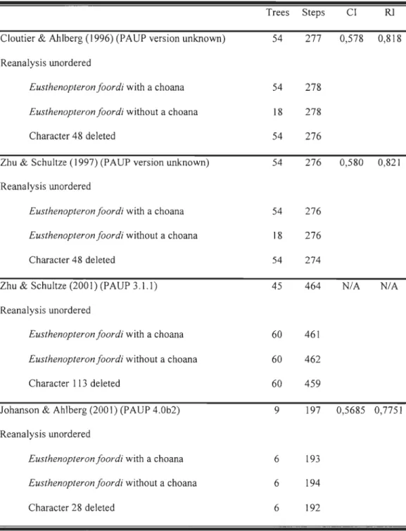
Table 2 Results ofphylogenetic analyses using PAUP*4.0blO on four previously
published matrices.
Trees Steps CI RJ
Cloutier & Ahlberg (1996) (PAUP version unknown) 54 277 0,578 0,818
Reanalysis unordered
Eusthenopteron foordi with a choana 54 278
Eusthenopteron foordi without a choana 18 278
Character 48 deleted 54 276
Zhu & Schultze (1997) (PAUP version unknown) 54 276 0,580 0,821
Reanalysis unordered
Eusthenopteron foordi with a choana 54 276
Eusthenopteron foordi without a choana 18 276
Character 48 deleted 54 274
Zhu & Schultze (2001) (PAUP 3.l.l) 45 464 NIA NIA Reanalysis unordered
Eusthenopteron foordi with a choana 60 461
Eusthenopteron foordi without a choana 60 462
Character 113 deleted 60 459
Johanson & Ahlberg (2001) (pAUP 4.0b2) 9 197 0,5685 0,7751
Reanalysis unordered
Eusthenopteron foordi with a choana 6 193
Eusthenopteron foordi without a choana 6 194
B
~--soc n.caps. Mx. - -. Vo. ch. DtFigure 1 Cross section in the nasal capsule region of Eusthenopteronfoordi. A. Specimen MHNM 06-1001A (scan 8/36); B. Specimen MHNM 06-1045 (scan 8/16); C. Specimen RM 14234 (scan 102/110). Left column: Computed Tomography. Right column: Interpretation ofthe CT scan; black = bone, dark
grey = ethmosphenoid, light grey = teeth. Abbreviations, a.Te.: anterior tectal, ch.: choana, dml.p.: dermintermedial process, DpI.: dermopalatine, Dt.: dentary, Ecpt.: ectopterygoid, Enpt.: entopterygoid, Ethm.: ethmosphenoid,
f.: fang, GuI.: gular plate, ioc: infraorbital canal, I.Te: lateral tectal, m.cav.:
mouth cavity, Mx.: maxillary, n.caps.: nasal capsule, n.op.: nasal opening,
plp.cav.: pulp cavity, Pmx.: premaxillary, Pr.: postrostral, Psph.:
Pr
B
c
vo. Choa Dp Mx Mx EnpPSP
EnpFigure 2 Eusthenopteronfoordi, specimen MHNM 06-719. A.Ventral view of the palate; B. Close-up of the choana area; C. Camera-lucida drawing of the specimen.
Scale bars: 1 cm. 1,2 and 3 identify facets on the head of the dermopalatine. DpI.:
dermopalatine, Ecpt.: ectopterygoid, Enpt.: entopterygoid, Mx.: maxillary, Psph.:
Figure 3 Eusthenopteronfoordi, specimen MHNM 06-220. Ventral view of the
palate. Scale bar ; 1 cm. ch.: choana, DpI.: dermopalatine, Enpt.: entopterygoid, Mx.: maxillary, Pmx.: premaxillary, Psph.: parasphenoid, Vo.: vomer, vo.f.:
Mimia Cheirolepis Polypterus Diplocercides kaeseri D. heiligenstockiensi AI/enypterus Miguashaia Onychodus Strunius Holoptychius Glyptolepis Porolepis Powichthys 1 Youngolepis Diabolepis Uranolophus Dipnorhynchus Speo nesidrion Dipterus Barameda Strepsodus Eusthenopteron Osteolepis Gyroptychius Beelarongia Elpistostege Panderichthys Crassigyrinus Ventastega A can thos tega Ichthyostega
Figure 4 Strict consensus trees obtained from previously published matrices. Trees
obtained from: A. Cloutier & Ahlberg (1996); B. Zhu & Schultze (1997); C. Zhu &
Schultze (2001); D. Johanson & Ahlberg (2001) matrices. 1 identifies appearance of the choana character, 0 identifies reversaI of this character. Trees from original matrices are numbered on the right side of the branches, whereas trees from modified matrices
(choana considered absent in Eusthenopteron) are numbered on their left side. C and D
only have one tree because the topology remained the same after the matrix
modification. Trees B and D show double appearance and/or reversaI because it was not possible to know if the authors used "ACC1RAN" or "DELTRAN" options.
B
1 Howqualepis Moythomasia Mimia Cheirolepis Polypterus Uranolophus Dipnorhynchus Speonesidrion Dipterus Diabolepis Youngolepis ... - - - Powichthys 1 Holoptychius Glyptolepis Porolepis Diplocercides kaeseri D. heiligenstockiensis A/lenypterus Miguashaia Onychodus Strunius Strepsodus Barameda , . . - - Eusthenopteron Osteolepis ... - Gyroptychius Beelarongia Elpistostege Panderichthys Cras sigyrinus Ichthyostega A can thos tega VentastegaDiabolepis Mimia Dipterus Moythomasia Kenichthys Cheirolepis Barameda Polypterus Go 010 ogongia Dialipina
-
.
pGogonasus Speonesydrion Dipnorhynchus Osteolepis Uranolophus Ectosteorachis Diabolepis Megalichthys Youngolepis Cladarosymblema Powichthys Medoevia Holoptychius Bee/arongia Glyptolepis Koharalepis Porolepis Canowindra Onychodus Gyroptychius Strunius Panderichthys Diplocercides Ichthyostega Miguashaia Barameda A can thos tegaStrepsodus 0 Eusthenopteron Kenichthys Tristichopterus Bee/arongia Jarvikina Gyroptychius Notorhizodon Osteolepis Platycephalichthys Eusthenopteron
Cabon nichth ys Panderichthys
Mandageria Elpistostege
Eusthenodon Crassigyrinus
Ichthyostega w
CHAPITRE 2
De
v
elopmental Modularity and Saltato
ry
Ontogen
y
in the Late
De
v
onian Osteolepiform Eusthenopteron foordi.
Joël Leblanc and Richard Cloutier
Laboratoire de Biologie évolutive, Université du Québec à Rimouski, 300 allée des Ursulines, Rimouski, Québec, G5L 3AI, Canada
richard _ cloutier@uqar.qc.ca
ABSTRACT
Sixty-five specimens of the Late Devonian osteolepiform Eusthenopteronfoordi
were studied in order to reconstruct its sequence of ossification. The specimens ranging
from 27.0 to 270.6 mm in standard length represent a size series that was treated like a growth series thus revealing the growth pattern of the species. The presence and absence of aIl postcranial bony elements, with special emphasis on fin-systems, were recorded. The pectoral girdles (cleithra, clavicles and anocleithra) and the basal scutes at the base of unpaired fins are among the earliest postcranial structures to ossify. In the fins, the distal elements are the first to ossify, foIlowed by proximal elements. The caudal part of
vertebral column, as weIl as the distal bones of the second dorsal and anal fins, are the first endochondral bones to ossify. After a relative stagnation of the development, during which the other fins partly develop, the anterior part of the vertebral column suddenly settles in a short time. The general pattern of ossification shows a saltatory ontogeny: numerous bones appear in short time laps, and these events are separated by periods of slower development. A first threshold of accelerated development occurs at a standard length of ca. 42 mm
(ossification of the posterior propulsive system), and a second one occurs at ca. 160 mm
(ossification of the anterior part of vertebral column). Isometric growth for postcranial skeleton is confirmed in E. foordi. Study of the growth series also reveals a developmental modularity: the second dorsal and anal fins, appearing and growing in synchronicity,
together form a module. This is comparable with the medial fins developmental modularity of aU living actinopterygian and this is the first description of such a development for a fossil species.
RÉSUMÉ
Soixante-cinq spécimens de l' ostéolépiforme Eusthenopteron foordi du Dévonien
tardif ont été étudiés afin de reconstituer sa séquence d'ossification. L'échantillon,
constitué de spécimens dont les longueurs standard allaient de 27,0 à 270,6 mm, représente
une «série de taille» qui fut traitée comme une série de croissance révélant ainsi le patron de croissance de l'espèce. Les observations furent faites sur la base de la présence ou
l'absence de tous les éléments osseux du squelette post-crânien, avec une attention
particulière portée aux systèmes natatoires. Les ceintures pectorales (cleithra, clavicules,
anocleithra) et les scutelles à la base de toutes les nageoires impaires sont parmi les
premières structures postcrâniennes à s'ossifier. Dans les nageoires, les éléments distaux
s'ossifient d'abord, suivi par les éléments proximaux. La partie postérieure de la colonne
vertébrale, située dans la nageoire caudale, tout comme les os distaux de la seconde
nageoire dorsale et de la nageoire anale, sont les premiers os endochondraux à s'ossifier. Après une stagnation relative du développement, pendant laquelle les autres nageoires se
développent partiellement, la partie antérieure de la colonne vertébrale prend soudainement
place en peu de temps. Finalement, la majeure partie du reste de la croissance est marquée
par l'ajout constant et continuel d'éléments, principalement dans la partie médiane de la
colonne vertébrale. Le patron général d'ossification révèle une ontogénie saltatoire :
plusieurs os apparaissent en peu de temps, et ces événements sont séparés par des périodes
de développement plus lent. Une première poussée de développement accéléré se produit à
une longueur standard d'environ 42 mm (ossification du système propulsif postérieur), et
vertébrale). Une croissance isométrique du squelette postcrânien est confirmée chez E.
foordi. L'étude de cette série de croissance révèle aussi une modularité developpementale:
la deuxièeme nageaoire dorsale et la nageoire anale, apparaissant et se développant en
synchronisme, forme un module. Celui-ci est comparable à la modularité
développementale retrouvée dans les nageoires médianes de tous les acitnoptérygiens
vivants et il s'agit de la première description d'un tel développement chez une espèce fossile.
INTRODUCTION
Skeletal ontogeny can be described in terms of morphometric changes (i.e., size and shape) as well as anatomical (e.g., ossification pattern) and histological changes. In either case, one has to observe growth series in living animais (e.g., Arratia et al., 2001;
Grünbaum et al., 2003) or size series in extinct animaIs (Schultze, 1984; Carroll et al., 1999; Chipman and Tchernov, 2002; Cote et al., 2002). With respect to fossils, as many different sized specimens as possible of the same species are needed, allowing to compare the anatomy of the smallest individuals up to those of the large st individuals. Furthermore, the estimate of intraspecific variation would be more accurate if based on larger sample size. Size series of extinct species are rare and younger individuals are especially
uncommon. A size series is not exactly equivalent to a growth series; it is actually a static sample of a species or a population (Schultze, 1984). But, as explained by Schultze (1984), it is reliably representative of a growth series. Such inferences of growth series from size series have been carried out for a few fossil fishes: the placoderm Bothriolepis canadensis (Werdelin and Long, 1986) and B. nitida (Weems, 2004), the actinopterygian Elonichthys
(Schultze and Bardack, 1987), the dipnoan Scaumenacia curta (Cloutier, 1997), and the osteolepiform Eusthenopteron foordi (Thomson and Hahn, 1968; SchuItze, 1984).
Eusthenopteronfoordi Whiteaves 1881, from the Upper Devonian Escuminac Formation of Miguasha, Québec, Canada, is one ofthese rare fossil species to be a candidate for such an ontogenetic study. Phylogeneticaly, Eusthenopteron is informative because it belongs to the Tetrapodomorpha, the clade inclusive of elpistostegalians and tetrapods (Cloutier and Ahlberg, 1996; Zhu and Schultze, 2001). For the past 120 years,