Role of cardiovascular imaging in cancer
patients receiving cardiotoxic therapies:
a position statement on behalf of the Heart
Failure Association (HFA), the European
Association of Cardiovascular Imaging
(EACVI) and the Cardio-Oncology Council of
the European Society of Cardiology (ESC)
Jelena ˇ
Celutkien ˙e
1,2*
†, Radek Pudil
3, Teresa López-Fernández
4, Julia Grapsa
5,
Petros Nihoyannopoulos
6,7, Jutta Bergler-Klein
8, Alain Cohen-Solal
9,
Dimitrios Farmakis
10,11, Carlo Gabriele Tocchetti
12, Stephan von Haehling
13,
Vassilis Barberis
14, Frank A. Flachskampf
15, Indr ˙e ˇ
Ceponien ˙e
16,
Eva Haegler-Laube
17, Thomas Suter
17, Tomas Lapinskas
16, Sanjay Prasad
18,19,
Rudolf A. de Boer
20, Kshama Wechalekar
21, Markus S. Anker
22,
Zaza Iakobishvili
23,24, Chiara Bucciarelli-Ducci
25, Jeanette Schulz-Menger
26,27,
Bernard Cosyns
28, Oliver Gaemperli
29, Yury Belenkov
30, Jean-Sébastien Hulot
31,
Maurizio Galderisi
32†, Patrizio Lancellotti
33, Jeroen Bax
34, Thomas H. Marwick
35,
Ovidiu Chioncel
36,37, Tiny Jaarsma
38,39, Wilfried Mullens
40, Massimo Piepoli
41,42,
Thomas Thum
43, Stephane Heymans
44,45,46, Christian Mueller
47, Brenda Moura
48,
Frank Ruschitzka
49, Jose Luis Zamorano
50,51,52, Giuseppe Rosano
53,54,
Andrew J.S. Coats
55, Riccardo Asteggiano
56, Petar Seferovic
57, Thor Edvardsen
58,59,
and Alexander R. Lyon
19,60†1Clinic of Cardiac and Vascular Diseases, Institute of Clinical Medicine, Faculty of Medicine, Vilnius University, Vilnius, Lithuania;2State Research Institute Centre For Innovative
Medicine, Vilnius, Lithuania;3First Department of Medicine - Cardioangiology, Charles University Prague, Medical Faculty and University Hospital Hradec Králové, Hradec
Kralove, Czech Republic;4Cardiology Department, La Paz University Hospital, IdiPAz, Ciber CV, Madrid, Spain;5Department of Cardiology, St Bartholomew Hospital, Barts
Health Trust, London, UK;6Unit of Inherited Cardiovascular Diseases/Heart Center of the Young and Athletes, First Department of Cardiology, Hippokrateion General Hospital,
National and Kapodistrian University of Athens, Athens, Greece;7National Heart and Lung Institute Imperial College London, Hammersmith Hospital, London, UK;8Department
of Cardiology, Medical University of Vienna, Vienna, Austria;9UMR-S 942, Cardiology Department, Hôpital Lariboisière, AP-HP, Université de Paris, Paris, France;10University of
Cyprus Medical School, Nicosia, Cyprus;11Cardio-Oncology Clinic, Heart Failure Unit, Department of Cardiology, Athens University Hospital Attikon, National and Kapodistrian
University of Athens, Athens, Greece;12Department of Translational Medical Sciences, and Interdepartmental Center for Clinical and Translational Research (CIRCET), Federico
II University, Naples, Italy;13Department of Cardiology and Pneumology, University of Göttingen Medical Centre, Göttingen, Germany;14American Medical Center, American
Heart Institute, Nicosia, Cyprus;15Department of Medical Sciences, Uppsala University, Clinical Physiology and Cardiology, Akademiska Hospital, Uppsala, Sweden;16Department
of Cardiology, Medical Academy, Lithuanian University of Health Sciences, Kaunas, Lithuania;17Department of Cardiology, Inselspital, University of Bern, Bern, Switzerland;
*Corresponding author. Clinic of Cardiac and Vascular Diseases, Institute of Clinical Medicine, Faculty of Medicine, Vilnius University, Santariški ¸u 2, LT-08661, Vilnius, Lithuania. Tel: +370 616 00180, Fax: +370 5 2501742, Email: jelena.celutkiene@santa.lt
18Department of Cardiac Magnetic Resonance, Royal Brompton Hospital, London, UK;19National Heart and Lung Institute, Imperial College, London, UK;20Department of
Cardiology, University Medical Center Groningen, University of Groningen, Groningen, The Netherlands;21Department of Nuclear Medicine, Royal Brompton Hospital, London,
UK;22Division of Cardiology and Metabolism, Department of Cardiology, Charité; and Berlin Institute of Health Center for Regenerative Therapies (BCRT); and DZHK (German
Centre for Cardiovascular Research), partner site Berlin; and Department of Cardiology, Charité Campus Benjamin Franklin, Berlin, Germany;23Sackler Faculty of Medicine, Tel
Aviv University, Tel Aviv, Israel;24Tel Aviv-Jaffa District, Clalit Health Services, Tel Aviv, Israel;25Bristol Heart Institute, Bristol NIHR Biomedical Research Centre and Clinical
Research and Imaging Centre (CRIC) Bristol, University Hospitals Bristol NHS Trust and University of Bristol, Bristol, UK;26Working Group on Cardiovascular Magnetic
Resonance, Experimental and Clinical Research Center a joint cooperation between the Charité - Universitätsmedizin Berlin, Department of Internal Medicine and Cardiology and the Max-Delbrueck Center for Molecular Medicine, and HELIOS Klinikum Berlin Buch, Department of Cardiology and Nephrology, Berlin, Germany;27DZHK (German
Centre for Cardiovascular Research), partner site Berlin, Berlin, Germany;28Department of Cardiology, CHVZ (Centrum voor Hart en Vaatziekten), ICMI (In Vivo Cellular and
Molecular Imaging) Laboratory, Universitair Ziekenhuis Brussel, Brussels, Belgium;29Heart Clinic Hirslanden, Hirslanden Hospital, Zurich, Switzerland;30I.M. Sechenov’s First
Moscow State Medical University of Ministry of Health (Sechenov University), Moscow, Russia;31Université de Paris, CIC1418, Paris Cardiovascular Research Center, INSERM,
Paris, France;32Department of Advanced Biomedical Sciences, Federico II University Hospital, Naples, Italy;33University of Liège Hospital, GIGA Cardiovascular Sciences,
Department of Cardiology, CHU SartTilman, Liège, Belgium;34Department of Cardiology, Leiden University Medical Centre, Leiden, The Netherlands;35Baker Heart and
Diabetes Institute, Melbourne, Australia;36Emergency Institute for Cardiovascular Diseases C.C. Iliescu, Bucuresti, Romania;37University of Medicine Carol Davila, Bucuresti,
Romania;38Department of Health, Medicine and Caring Sciences, Linköping University, Linköping, Sweden;39Julius Center for Health Sciences and Primary Care, University
Medical Center Utrecht and Utrecht University, Utrecht, The Netherlands;40Ziekenhuis Oost Limburg, University Hasselt, Genk, Belgium;41Heart Failure Unit, Cardiology,
Guglielmo da Saliceto Hospital, Piacenza, Italy;42University of Parma, Parma, Italy;43Hannover Medical School, Institute of Molecular and Translational Therapeutic Strategies
(IMTTS), Hannover, Germany;44Department of Cardiology, CARIM School for Cardiovascular Diseases Faculty of Health, Medicine and Life Sciences, Maastricht University,
Maastricht, The Netherlands;45William Harvey Research Institute, Barts Heart Centre, Queen Mary University of London, Charterhouse Square, London, UK;46Department of
Cardiovascular Sciences, Centre for Molecular and Vascular Biology, KU Leuven, Leuven, Belgium;47Department of Cardiology and Cardiovascular Research Institute Basel
(CRIB), University Hospital Basel, University of Basel, Basel, Switzerland;48Cardiology Department, Military Hospital, and CINTESIS, CardioCare, Faculty of Medicine, Porto
University, Porto, Portugal;49University Heart Center, Department of Cardiology, University Hospital Zurich, Zurich, Switzerland;50Cardiology Department, University Hospital
Ramón y Cajal, Madrid, Spain;51University Alcala, Madrid, Spain;52CIBERCV, Instituto de Salud Carlos III (ISCIII), Madrid, Spain;53Department of Medical Sciences, IRCCS San
Raffaele Pisana, Rome, Italy;54Cardiology Clinical Academic Group, St George’s Hospitals NHS Trust University of London, London, UK;55IRCCS San Raffaele Pisana, Rome, Italy; 56Cardiologist in Practice, Turin, Italy;57University of Belgrade Faculty of Medicine and Serbian Academy of Sciences and Arts, Belgrade, Serbia;58Department of Cardiology, Oslo
University Hospital, Rikshospitalet, Oslo, Norway;59Faculty of Medicine, University of Oslo, Oslo, Norway; and60Cardio-Oncology Service, Royal Brompton Hospital, London,
UK
Received 22 February 2020; revised 8 June 2020; accepted 1 July 2020 ; online publish-ahead-of-print 21 August 2020
Cardiovascular (CV) imaging is an important tool in baseline risk assessment and detection of CV disease in oncology patients receiving cardiotoxic cancer therapies. This position statement examines the role of echocardiography, cardiac magnetic resonance, nuclear cardiac imaging and computed tomography in the management of cancer patients. The Imaging and Cardio-Oncology Study Groups of the Heart Failure Association (HFA) of the European Society of Cardiology (ESC) in collaboration with the European Association of Cardiovascular Imaging (EACVI) and the Cardio-Oncology Council of the ESC have evaluated the current evidence for the value of modern CV imaging in the cardio-oncology field. The most relevant echocardiographic parameters, including global longitudinal strain and three-dimensional ejection fraction, are proposed. The protocol for baseline pre-treatment evaluation and specific surveillance algorithms or pathways for anthracycline chemotherapy, HER2-targeted therapies such as trastuzumab, vascular endothelial growth factor tyrosine kinase inhibitors, BCr-Abl tyrosine kinase inhibitors, proteasome inhibitors and immune checkpoint inhibitors are presented. The indications for CV imaging after completion of oncology treatment are considered. The typical consequences of radiation therapy and the possibility of their identification in the long term are also summarized. Special populations are discussed including female survivors planning pregnancy, patients with carcinoid disease, patients with cardiac tumours and patients with right heart failure. Future directions and ongoing CV imaging research in cardio-oncology are discussed.
...
Keywords Imaging • Cardio-oncology • Cardiotoxicity • Heart failure • Echocardiography • Cardiac
magnetic resonance • Computed tomography • Global longitudinal strain • Nuclear imaging
Introduction
Cardiovascular disease (CVD) and cardiovascular (CV) complica-tions in cancer patients present a growing medical problem, causing substantial morbidity and premature mortality in this population. An increasing prevalence of pre-existing CVD and the CV toxi-city of both established and emerging cancer treatments includ-ing anthracycline (AC) chemotherapy, targeted therapies such as trastuzumab, proteasome inhibitors (PIs), immune checkpoint inhibitors (ICI) and vascular endothelial growth factor inhibitors (VEGFi), along with biological treatments and radiation therapy collectively contribute to this new epidemic. There is an urgent clinical need to modernize and validate monitoring algorithms for ...
the early detection of CVD in cancer patients receiving potentially cardiotoxic treatments, and to intervene prior to the develop-ment of manifest CVD. Considerations are also needed as to which cancer survivors require screening after completion of oncology treatment.
Contemporary cardiac imaging is a valuable instrument to help in multiple ways—for baseline risk stratification, timely diagnosis of early CVD and of cardiac dysfunction, both during and fol-lowing treatment, for the identification of cancer patients who may benefit from cardioprotective treatments whilst continuing oncology treatment, and prognostication to select cancer patients who may require long-term CVD follow-up. The Imaging and Cardio-Oncology Study Groups of the Heart Failure Association
(HFA) of the European Society of Cardiology (ESC) in collaboration with the European Association of Cardiovascular Imaging (EACVI) and the Cardio-Oncology Council of the ESC have evaluated the current evidence for the role of CV imaging including echocardio-graphy, cardiac magnetic resonance (CMR), computed tomography (CT) and nuclear testing before, during and after cancer therapy. This position statement summarizes their consensus regarding the application of modern cardiovascular imaging in cancer patients. It focuses on the detection and assessment of myocardial dys-function and heart failure (HF), the optimal timing for monitoring in various cardiotoxic cancer treatments, special populations and future developments in this field. The authors aim to provide car-diologists, oncologists, haematologists and general medical physi-cians with a framework for using cardiac imaging for the timely diagnosis of CV involvement and for prevention of CVD in can-cer patients and survivors. This position statement examines CV imaging and has been developed in parallel to a position statement addressing the role of cardiac biomarkers in cancer patients and detailed cancer-treatment specific baseline risk assessment.1 CV imaging and cardiac biomarkers in baseline risk assessment and in surveillance receiving cardiotoxic cancer therapies are synergis-tic and complementary approaches, and it is important that they should be considered together. A future HFA position statement will review the evidence and provide details of the specific treat-ment interventions recommended for the different cardiotoxicities detected using CV imaging for a range of cancer therapies. These are complex and beyond the scope of this article.
We emphasize that the suggested surveillance pathways and frequencies of use are based on expert opinion and experience, since validation studies are lacking in this area, especially with regard to cost-effectiveness and effect on long-term outcomes.
General principles
Cardiac imaging in general, and echocardiography in particular, play a central role in the expanding field of cardio-oncology. Given that the current definitions of cardiotoxicity in many guidelines and oncology trials are based on a reduction of left ventricular ejection fraction (LVEF),2,3many oncologists restrict cardiotoxicity evalua-tion to measurement of this single parameter only. However, it is well known in contemporary cardiology that a normal LVEF does not exclude significant myocardial dysfunction.4,5In addition, there are important limitations of serial measurement of LVEF such as physiological temporal and operator variability, and haemodynamic load-dependence. Concurrent measurement of blood pressure may help to avoid misinterpretations in cases of blood pressure and blood volume changes due to fluid excess during intravenous chemotherapy or fluid loss due to adverse reactions.6 Temporal variability of LVEF measured by two-dimensional (2D) echocardio-graphy using biplane Simpson’s method has been reported to be approximately 10%, with the same level of 10% seen for inter- and intra-observer variability.7 Thus, the LVEF should be reassessed to confirm the development of subclinical left ventricular (LV) dysfunction.2
Three-dimensional (3D) echocardiography should be utilized for the assessment of LVEF and cardiac volumes when available and ...
...
...
with appropriate expertise and experience due to its lower inter-, intra-observer and test–retest variability.7 Adequate inter-reader agreement in an echocardiography laboratory may be achieved by standardizing the analytical approach through dedicated qual-ity audit sessions.83D echocardiography is likely to become more widely accepted in routine practice due to improved image acqui-sition and the implementation of semi- or fully automated analysis algorithms.4 The feasibility of 3D LVEF in breast cancer patients with adequate echocardiographic images was 88% at baseline and 66% after AC therapy, reduced during follow-up due to concomi-tant radiotherapy (RT), left mastectomy, left breast prosthesis and other patient factors.9
When transthoracic echocardiographic image quality is inade-quate for the application of Simpson’s method, which is more com-mon in cancer patients who have previously undergone left breast or left chest surgery and/or RT, and sometimes in very cachec-tic patients, adding contrast media or using alternative imaging modalities such as CMR can be considered for serial monitoring of LV size and function. The latter technique, although less feasible and more expensive, has improved accuracy and reproducibility with the coefficient of variation for CMR LVEF being reported at approximately 4%.10 The historical method of planar imag-ing, multigated acquisition (MUGA) scan, used for serial assess-ment in earlier clinical trials, is not recommended as a first-line cardiac imaging modality, due to exposure to ionizing radiation and advances in ultrasound and CMR modalities.11 Single photon emission computed tomography (SPECT) MUGA acquired with high-sensitivity cadmium zinc telluride cameras can be done with lower radiation dose, faster image acquisition time and improved reproducibility.11
Global longitudinal strain (GLS) has emerged as a new marker of subclinical ventricular dysfunction demonstrating stronger asso-ciation with prognosis than LVEF in non-oncology heart disease populations.12,13 This reflects the fact that LV longitudinal tion may be reduced first and this component of ventricular func-tion has a limited influence upon LVEF.14 Several researchers have reported a higher sensitivity and either a non-inferior or supe-rior test–retest reliability of GLS compared to LVEF.5,14,15A num-ber of observational studies show potential for reduction in GLS to accurately predict a future decrease in LVEF and significant cardiotoxicity.14,16,17
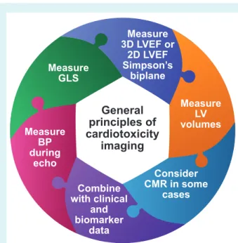
A recent study in 116 patients with human epidermal growth factor receptor 2 (HER2)-positive breast cancer supported the serial surveillance using GLS to guide cardioprotection and main-tain patients on uninterrupted trastuzumab therapy.18The ongoing SUCCOUR study is prospectively assessing the value of initiating cardioprotective medication triggered by the reduction of GLS vs. waiting for a decline in 3D LVEF.19 GLS should be based on three apical (long-axis) views and not replaced by single-view longitu-dinal strain due to substantial disagreement in the diagnosis of cardiotoxicity.20 Although less feasible and reproducible, 2D- or 3D-derived global circumferential strain may also serve as addi-tional markers of myocardial dysfunction but require more studies for validation.21,22 GLS surveillance may become a more sensitive strategy for early detection of cardiotoxicity and guide timing of cardioprotective treatment (Figure 1).
Figure 1 General principles of imaging for cardiotoxicity. 2D, two-dimensional; 3D, three-dimensional; BP, blood pressure; CMR, cardiac magnetic resonance; GLS, global longitudinal strain; LV, left ventricular; LVEF, left ventricular ejection fraction.
Several cardiotoxic cancer treatments including AC and trastuzumab have been shown to cause a persistent reduc-tion in LVEF and GLS.6 Other cancer drugs may cause different forms of myocardial toxicity where LVEF reduction is not the primary manifestation. For example, ICIs cause myocarditis, which can lead to severe HF, cardiogenic shock and death, but in 38% of cases may also occur even without a fall in LVEF.23,24 Thus, decision-making concerning the continuation or inter-ruption of such potentially life-saving therapy should no longer rely solely on the single, surrogate echocardiographic parameter (LVEF) which mainly reflects changes in LV volumes, rather than function.
Several small studies have analysed the serial measurement of LV diastolic function using tissue and transmitral Doppler (E/e′) in various cancer populations.25,26Most have not found improved sensitivity compared with measurements of LV systolic function for detection of cardiotoxicity. A sequential relation between diastolic and systolic impairment has not been proven, either in experimental, or in clinical settings. Initial investigations of left atrial size and function have shown that early atrial dilatation and a reduction in conduit and reservoir strain may be potential markers of cardiotoxicity.27,28
Current recommendations of screening for cardiotoxicity using serial LVEF measurement remain sub-optimally implemented in the majority of patients with breast cancer.29,30 In one study baseline evaluation was performed in only 74% of patients receiving HER2-targeted therapy, and only 46% were assessed repeatedly during treatment.31 Quality of care may be improved by establishing dedicated cardio-oncology services delivering ...
...
...
Figure 2 Cardio-oncology interactions. HF, heart failure; LV, left ventricular.
structured pathways for baseline risk stratification and surveillance (Figure 2).32,33
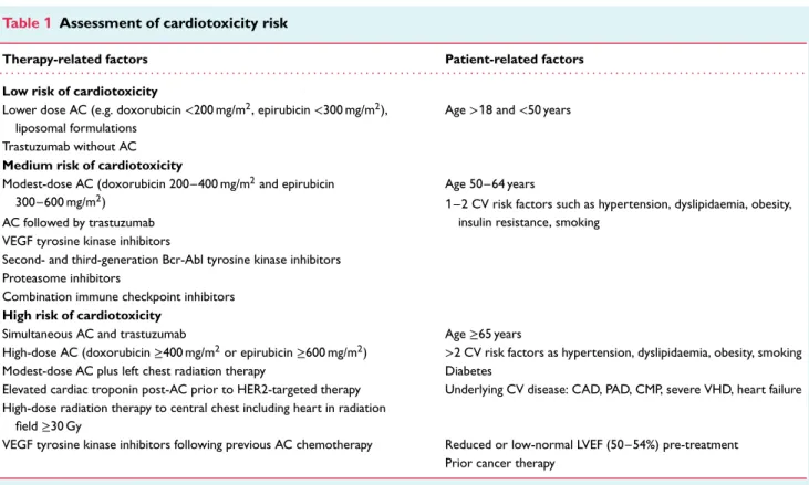
Assessment of cardiotoxicity risk
Systematic cardiac surveillance with more sensitive technologies and a higher frequency of measurements will lead to a greater inci-dence of detected cardiotoxicity.31In order to maintain a balance between the rational use of resources and maximal patient safety, we recommend a personalized approach taking into account the patients’ baseline risk of cardiotoxicity (Table 1). Cancer patients scheduled to receive potentially cardiotoxic cancer therapies are evaluated pre-treatment for cardiotoxicity risk and stratified into three categories (low, medium and high) according to the baseline CV profile and risk factors, pre-existing CVD, type and dose of cancer therapy.
New information on the risk of myocardial dysfunction was obtained analysing follow-up data in adult survivors of childhood cancer.34 Even in this relatively young population, the effect size of traditional risk factors for HF, including hypertension, insulin resistance, obesity, was comparable or even higher than effect size of cancer treatment-related risk factors, such as an AC dose, RT, or current age. Traditional risk factors, including age, coro-nary artery disease, diabetes, hypertension, atrial fibrillation, renal
Table 1 Assessment of cardiotoxicity risk
Therapy-related factors Patient-related factors
. . . .
Low risk of cardiotoxicity
Lower dose AC (e.g. doxorubicin<200 mg/m2, epirubicin<300 mg/m2),
liposomal formulations
Age>18 and <50 years Trastuzumab without AC
Medium risk of cardiotoxicity
Modest-dose AC (doxorubicin 200–400 mg/m2and epirubicin
300–600 mg/m2)
Age 50–64 years
1–2 CV risk factors such as hypertension, dyslipidaemia, obesity, insulin resistance, smoking
AC followed by trastuzumab VEGF tyrosine kinase inhibitors
Second- and third-generation Bcr-Abl tyrosine kinase inhibitors Proteasome inhibitors
Combination immune checkpoint inhibitors High risk of cardiotoxicity
Simultaneous AC and trastuzumab Age≥65 years
High-dose AC (doxorubicin≥400 mg/m2or epirubicin≥600 mg/m2) >2 CV risk factors as hypertension, dyslipidaemia, obesity, smoking
Modest-dose AC plus left chest radiation therapy Diabetes
Elevated cardiac troponin post-AC prior to HER2-targeted therapy Underlying CV disease: CAD, PAD, CMP, severe VHD, heart failure High-dose radiation therapy to central chest including heart in radiation
field≥30 Gy
VEGF tyrosine kinase inhibitors following previous AC chemotherapy Reduced or low-normal LVEF (50–54%) pre-treatment Prior cancer therapy
Abr, active Bcr-related; AC, anthracycline; Bcr, breakpoint cluster region; CAD, coronary artery disease; CMP, cardiomyopathy; CV, cardiovascular; HER2, human epidermal growth factor receptor 2; LVEF, left ventricular ejection fraction; PAD, peripheral artery disease; VEGF, vascular endothelial growth factor; VHD, valvular heart disease.
failure, have also been predominant predictors of prevalent HF or cardiomyopathy in older women (mean age 74 years) after adjuvant trastuzumab therapy.35 If LVEF falls to a marginally normal range (50–54%) before treatment, the incidence of HF rises remarkably in cancer patients receiving AC and trastuzumab.36,37New targeted therapies including VEGF tyrosine kinase inhibitors (VEGF-TKIs), second- and third-generation Bcr-Abl TKIs for chronic myeloid leukaemia, and PIs for multiple myeloma (MM), are associated with an increased risk of HF and other CV toxicities.
Definitions of cardiotoxicity
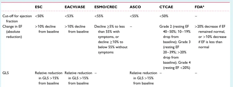
The cancer therapy-related cardiac dysfunction (CTRCD) defini-tion, which is adopted in the 2016 ESC Cardio-Oncology position statement, is defined as any reduction of LVEF to below 50% or a>10% reduction from baseline falling below the lower limit of normal.2,3 Current echocardiography recommendations set low normal value of 2D LVEF as 54% for women and 52% for men38 and hence in the previous EACVI position statement a reduction of LVEF below 53% was classified as abnormal.2
Changes in the myocardial deformation parameter GLS may also be considered an early sign of CTRCD.39–42 When detected it correlates with focal and diffuse fibrosis.43During follow-up LV GLS falling below (−)18% into the abnormal range (0% to −17.9%) or a>15% relative decrease of this marker and to below the lower limit of normal may be considered abnormal.2,3,12 ...
There is a variation in the definition of CTRCD across guidelines, position statements and oncology trials (Table 2); numerous mech-anisms of cardiotoxicity inherent to different cancer drug classes add to the complexity of this condition. Latest accumulating data on the specific incidence and reversibility of cardiotoxicity have forced the authors to abandon the outdated concept of type I and type II cardiotoxicity.44The recently proposed Royal Brompton Hospi-tal classification of myocardial toxicity incorporated alterations of biomarkers and/or GLS as evidence of early biochemical, functional or early mixed cardiotoxicity where oncology treatment should continue but consideration to start cardioprotective medication or implement closer monitoring is advised.32
Cardiovascular imaging
at baseline pre-treatment
It is essential to evaluate cardiac function with echocardiogra-phy before starting potentially cardiotoxic therapy in every can-cer patient as a baseline for monitoring and for risk stratifica-tion (online supplementary Figure S1, online supplementary Video
S1). The most relevant parameters for initial and subsequent
echocardiographic assessment are presented in Table 3. CMR is recommended in cases with poor quality echocardiographic images, in patients with complex pre-existing heart diseases (for example hypertrophic or dilated cardiomyopathy). In patients with suspected angina, stress echocardiography, vasodilator stress CMR or SPECT are recommended to diagnose the presence and extent
Table 2 The difference in published definitions of cardiotoxicity
ESC EACVI/ASE ESMO/CREC ASCO CTCAE FDAa
. . . .
Cut-off for ejection fraction <50% <53% <55% <55% <50% – Change in EF (absolute reduction) >10% decline from baseline >10% decline from baseline Decline≥5% to less than 55% with symptoms, or decline≥10% to below 55% without symptoms – Grade 2 (resting EF 40–50%; 10–19% drop from baseline); Grade 3 (resting EF 20–39%;>20% drop from baseline); Grade 4 (resting EF<20%) >20% decrease if EF remained normal, or>10% decrease if EF is less than normal GLS Relative reduction in GLS>15% from baseline Relative reduction in GLS>15% from baseline – Relative reduction in GLS>15% from baseline – –
ASCO, American Society of Clinical Oncology; ASE, American Society of Echocardiography; CREC, Cardiac Review and Evaluation Committee; CTCAE, Common Terminology Criteria for Adverse Events (US Departments of Health and Human Services); EACVI, European Association of Cardiovascular Imaging; EF, ejection fraction; ESC, European Society of Cardiology; ESMO, European Society of Medical Oncology; FDA, US Food and Drug Administration; GLS, global longitudinal strain.
aFor anthracyclines.
Table 3 Parameters relevant for cardio-oncology surveillance: echocardiography protocol
Parameters Clinically significant changes Comments
. . . .
LV size and function
LVEF by Simpson’s 2D, or (semi)automatic 3D Drop>10% (percentage points) for 2D, >5% for 3D from pre-treatment value
Decline of LVEF to value<40–50% suggests initiation of cardioprotection
2D/3D GLS, GCS Relative reduction by>10–15% from
pre-treatment value and to below lower limit of normal
Average from three apical views; do not use single-view value
LV 2D/3D systolic and diastolic volumes Increase by 15 mL for ESV, 30–35 mL for EDV Increase in volumes reflects remodelling and fluid status
RV function, pulmonary artery pressure and volaemia
Markers of systolic RV function TAPSE<1.7 cm, FAC <35%, RV free wall strain
<20%, 3D RVEF <45%
Show prognostic value in heart failure and pulmonary hypertension
Velocity of TR Peak systolic TR velocity> 2.8 m/s Indicates probable pulmonary
hypertension
IVC diameter, collapse on inspiration Dilatation>2.1 cm or narrowing <1.3 cm Relates to hypervolaemia or dehydration, respectively
2D, two-dimensional; 3D, three-dimensional; EDV, end-diastolic volume; ESV, end-systolic volume; FAC, fractional area change; GCS, global circumferential strain; GLS, global longitudinal strain; IVC, inferior vena cava; LV, left ventricular; LVEF, left ventricular ejection fraction; RV, right ventricular; RVEF, right ventricular ejection fraction; TAPSE, tricuspid annular plane systolic excursion; TR, tricuspid regurgitation.
of myocardial ischaemia and assess the need for anti-anginal medi-cations or alternative treatment. In patients with chest pain but no history of coronary disease, CT coronary angiography (CTCA) is recommended as an alternative to functional testing.45
Echocardiography during
anthracycline chemotherapy
Before starting AC therapy, we recommend classifying the cardiotoxicity risk as low, medium or high according to ...
therapy-related and patient-related factors (Table 1). The incidence of cardiac events during next 10 years after AC therapy accounts for 2% to 5% in the medium-risk and>5% in the high-risk group.39 This empirical approach aims to personalize echocardiographic surveillance (Table 4, Figure 3), including 3D LVEF and GLS when available, intensifying follow-up in high-risk patients and reducing frequency in low-risk patients. In AC cardiotoxicity, most cases occur during the first year after completion of chemotherapy, and therefore assessments at 6 and/or 12 months post-completion of chemotherapy should be considered46 (Table 4, Figure 4, online supplementary Figure S2, online supplementary Videos S2 and S3).
Table 4 Echocardiographic surveillance during and after anthracycline chemotherapy
Baseline risk of cardiotoxicity
During chemotherapy Following chemotherapy
. . . .
Low
• Baseline
• Following cycle completing cumulative lifetime dose of 240 mg/m2doxorubicin or equivalenta
• Every additional 100 mg/m2doxorubicin above 240 mg/m2
or every 2 cycles
• 12 months after final cycle • 5 yearly review
Medium
• Baseline
• Following 50% of planned total treatment or every 2 cycles (optional)
• Following cycle completing cumulative lifetime cycle of 240 mg/m2doxorubicin or equivalenta
• 12 months after final cycle • 5 yearly review
High
• Baseline • Every 2 cycles
• Consider after every cycle above 240 mg/m2doxorubicin
or equivalentb
• 6 months after final cyclec
• 12 months after final cycle
• Annually for 2 or 3 years thereafter, and then in 3- to 5-year intervals for life
cycle, chemotherapy infusion.
NB. All low and medium cardiovascular risk cancer patients who develop new cardiac symptoms or new left ventricular dysfunction during treatment are reclassified as high cardiovascular risk and if chemotherapy continues, they should follow the high-risk surveillance.
a240 mg/m2doxorubicin is equivalent to 360 mg/m2epirubicin, 320 mg/m2daunorubicin and 50 mg/m2idarubicin. b300 mg/m2doxorubicin is equivalent to 420 mg/m2epirubicin, 400 mg/m2daunorubicin and 60 mg/m2idarubicin. cDepending upon symptoms and evidence of new left ventricular dysfunction during treatment.
Variable remodelling responses to AC chemotherapy can occur, including cardiomyocyte atrophy with reduced LV mass and dysfunction but relative preservation of LVEF.47
In the long-term follow-up after completion of cancer ther-apy, repeated surveillance echocardiographic evaluation is rec-ommended in selected populations such as young patients who received high total cumulative AC doses (>400 mg/m2doxorubicin or equivalent), patients with significant pre-existing CVD, female cancer survivors planning to become pregnant or at the end of the first trimester of pregnancy,48 and survivors who are plan-ning to compete in high-intensity exercise, for example, marathons, endurance cycling, triathlons.49
Echocardiography during
HER2-targeted treatment
(trastuzumab, pertuzumab,
trastuzumab emtansine, lapatinib,
neratinib)
In patients on HER2-targeted therapies, standard surveillance according to the product license includes echocardiography at baseline (with 3D LVEF and GLS if available) and every 3 months during therapy.50,51Similar to the monitoring during AC described above, we suggest taking into account baseline risk of cardiotox-icity with a frequency of surveillance personalized to this base-line risk (Table 5, Figure 5).35,36 The same frequency of imag-ing is recommended for patients startimag-ing trastuzumab alone, trastuzumab and pertuzumab, ado-trastuzumab or trastuzumab ...
emtansine (T-DM1) or oral HER2-targeted therapies. There are also important considerations for the different cancer popula-tions (early invasive vs. metastatic HER2-positive breast cancer, HER2-positive gastric cancer).
The evidence for long-term follow-up echocardiography in patients following adjuvant HER2-targeted therapies for early invasive breast cancer is limited. Low-risk patients who are asymptomatic may not require any follow-up imaging, but a single review at 6–12 months following the final cycle may be considered if they have also received neoadjuvant or adjuvant AC. In asymptomatic patients with medium or high baseline cardiotoxicity risk, a follow-up echocardiogram and clinical assess-ment should be considered 3–6 months and 12 months after the final dose of HER2-targeted treatment (Table 5, Figure 5). Any patient who has new LV impairment or cardiotoxicity dur-ing HER2-targeted therapy will require follow-up assessment after starting any cardiac treatment to assess function and safety to continue HER2-targeted therapies, and at completion of treatment to assess for recovery and guide weaning of cardiac medication.
In asymptomatic patients who require long-term treatment in the setting of metastatic disease, echocardiography is recom-mended with the same frequency as for adjuvant trastuzumab dur-ing year 1, and then less frequent if cardiac biomarkers and LV function remain normal, e.g. 4 monthly in year 2, and 6 monthly thereafter in low-risk patients.52 Surveillance should continue at the same frequency if disease progression requires switching from trastuzumab and pertuzumab to T-DM1.53 If new cardiotoxicity or cardiac symptoms develop, then more frequent monitoring is recommended.
Figure 3 A surveillance pathway using biomarkers and echocardiography for cancer patients receiving six cycles of anthracycline chemother-apy with timing based upon baseline cardiovascular risk. Pathways for low risk, medium risk and high risk are presented. ABVD, doxorubicin, bleomycin, vinblastine, dacarbazine; B, baseline pre-treatment; C, cycle of chemotherapy; M, months post-final cycle; R-CHOP, cyclophos-phamide, doxorubicin, vincristine, prednisone with rituximab. *Optional additional assessment timepoints.
Echocardiography during vascular
endothelial growth factor
inhibitor and Bcr-Abl tyrosine
kinase inhibitor treatment
Left ventricular dysfunction occurs in 5% to 10% of patients receiv-ing VEGFi TKIs and 2% to 10% of patients receivreceiv-ing second-and third-generation Bcr-Abl TKIs due to direct myocardial tox-icity, uncontrolled hypertension and exacerbation of pre-existing CVD.43,54– 60 In the absence of prospective studies providing evi-dence, it is the opinion of the authors that echocardiography should be considered every 4 months during the first year in all patients receiving these treatments, with an additional early assessment 2–4 weeks after starting treatment in patients with high baseline CV risk.61 In patients who require long-term treat-ment with VEGFi or second- and third-generation Bcr-Abl TKIs 6–12 monthly echocardiography should be considered, as long as they remain asymptomatic and without clinical events dur-ing the first year. In patients who are candidates for dasa-tinib for chronic myeloid leukaemia, pre-treatment echocardiog-raphy screening to assess for pre-existing pulmonary hyperten-sion is recommended, as well as maintaining a low threshold for repeat echocardiography if cardiac symptoms develop.62 The decision to stop the treatment if new pulmonary arterial hyper-tension is detected may require right heart catheterization in selected cases.63
...
Echocardiography during
proteasome inhibitor treatment
Proteasome inhibitors including bortezomib, carfilzomib and ixa-zomib, are targeted therapies for MM. Bortezomib introduces a modestly increased risk for cardiac disorders in a meta-analysis by the Cochrane group compared to control (odds ratio 1.74, 95% confidence interval 1.17–2.58).64 Carfilzomib, which is an irreversible PI, has a higher risk of CV toxicity including myocar-dial infarction and LV dysfunction, as well as increased incidence of total symptomatic HF (7.1% vs. 4.1%) and HF categorized as grade ≥3 adverse reaction (4.3% vs. 2.1%) compared to control in the ASPIRE study.65Combined CV toxicities including HF were more frequent in MM patients receiving carfilzomib compared to bortezomib in the ENDEAVOR study.66A recent study reported CV toxicity rates in 95 MM patients receiving either carfilzomib (n = 65) or bortezomib (n = 30). At a follow-up of 18 months, 50% of carfilzomib-treated and 17% of bortezomib-treated MM patients had a significant clinical CV event, with new HF most common, and worse overall survival in the MM patients with CV events.67 Given these high CV event rates, baseline echocardio-graphy is advisable in all MM patients scheduled to receive a PI, which also allows assessment for cardiac AL amyloidosis. Surveil-lance may be considered in medium/high-risk patients receiving carfilzomib. Prompt echocardiography is strongly recommended if MM patients receiving PI therapy present with new cardiac symp-toms and signs. The ENDEAVOR trial echocardiography sub-study
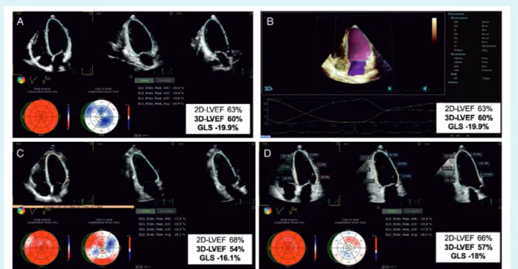
Figure 4 The case of a 66-year-old female with invasive breast ductal carcinoma (ER+ HER2+) treated by the combination of doxorubicin, cyclophosphamide, paclitaxel, radiotherapy (35 Gy + 10) and trastuzumab. (A) Baseline apical two-dimensional (2D) echocardiographic four-, two- and three-chamber views, showing normal left ventricular ejection fraction (LVEF), with speckle tracking-derived bull’s eye and normal global longitudinal strain (GLS). (B) Baseline three-dimensional (3D) volumetric analysis of the left ventricle and left atrium; measurements are normal. (C) At 3-month follow-up, 2D LVEF remains normal, while 3D LVEF drops by 10% and GLS by 19%. This entailed the initiation of anti-remodelling treatment with no interruption of oncologic drugs. (D) At 6-month follow-up, while continuing cancer and cardiac medications, the 3D LVEF reversed by 5%, and GLS recovered by 10%. LA, left atrium; LV, left ventricle.
reported limited utility for serial echocardiographic screening as a risk mitigation tool in unselected patients receiving carfilzomib. However, the evaluation was limited to four parameters [LVEF, estimated pulmonary artery pressure, tricuspid annular plane sys-tolic excursion (TAPSE) and right ventricular (RV) fractional area change] and less than 50% of patients completed the echocardio-gram surveillance protocol limiting its validity.68
Echocardiography during immune
checkpoint inhibitor treatment
Immune checkpoint inhibitors have improved clinical outcome and overall survival in cancer patients with various metastatic malignancies. CV toxicity associated with ICI (e.g. ipilimumab, nivolumab, pembrolizumab, atezolizumab, avelumab, durvalumab), including myocarditis sometimes causing cardiogenic shock69 and/or malignant ventricular tachyarrhythmias, pericarditis (includ-ing effusion and tamponade), arrhythmias, and non-inflammatory LV systolic dysfunction, was initially considered rare (<1%) but with expanding use its incidence is increasing.70,71 ICI-mediated fulminant myocarditis is relatively rare but has been associated with a high mortality rate (25–50%).24 The echocardiographic findings may vary from a normal examination to reduced wall ...
thickening, reduced GLS, regional and global wall motion abnor-malities and/or diastolic dysfunction.72–74Serial echocardiographic screening may be considered in patients at high risk (combina-tion ICI, ICI in combina(combina-tion with a second oncology drug with known cardiotoxicity, significant pre-existing heart disease, e.g. HF, cardiomyopathy). A recent study suggests a reduction in GLS is an early sign of ICI-induced myocarditis.23 The timing and duration of surveillance remains to be determined as severe myocarditis and pericarditis usually appear early (within the first four cycles) whereas non-inflammatory LV dysfunction emerges later.24
Cardiac magnetic resonance
imaging during cancer therapy:
why and when?
The routine use of CMR in cardio-oncology for surveillance is not feasible due to the lack of widespread accessibility and relatively high cost. However, when available, it is a very useful tool to iden-tify changes in ventricular volumes and ejection fraction, especially in patients with poor quality echocardiographic images if a discrep-ancy between measurements of LV function exists, or if myocardial perfusion assessment for ischaemia is simultaneously planned.39,43
Table 5 Echocardiographic surveillance during and after HER2-targeted therapies
Baseline risk of cardiotoxicity During HER2-targeted therapies Following completion
of HER2-targeted therapy
. . . .
Early invasive HER2+ breast cancer with neoadjuvant or adjuvant trastuzumaba
Low
• Baseline • Every 4 cycles
• Optional 6–12 months after final cycle
Medium
• Baseline
• Every 3 cycles, then reduce to every 4 if stable at 4 monthsc
• 6 months after final cycle
• Optional 12 months after final cycle High
• Baseline
• Every 2 cycles, then reduce to every 3 if stable at 3 monthsd
• 3 and 12 months after final cycle • Optional 6 months after final cycle Metastatic HER2+ breast cancer or gastric cancer
with long-term HER2-targeted therapiesb
Low
• Baseline
• Every 4 cycles in year 1 andevery 6 cycles in year 2, then reduce frequency to 6 monthly
Not indicated unless symptomatic
Medium
• Baseline
• Every 3 cycles, then if stable reduce to 6 monthlyc
Not indicated unless symptomatic
High
• Baseline
• Every 2 or 3 cycles for 3 months, then reduce to every 4 cycles in year 1, then reduce frequencyd
Not indicated unless symptomatic
cycle, chemotherapy infusion; HER2, human epidermal growth factor receptor 2.
NB. All low and medium cardiovascular risk cancer patients who develop new cardiac symptoms or new left ventricular dysfunction during HER2-targeted therapy are reclassified as high cardiovascular risk, and if HER2-targeted therapy continues they should follow the high-risk surveillance.
aNeoadjuvant trastuzumab or trastuzumab and pertuzumab.
bLong-term trastuzumab, trastuzumab and pertuzumab, or trastuzumab emtansine.
cChoice of 2 or 3 depends upon variables including baseline left ventricular function, cardiovascular history, baseline troponin and previous anthracycline chemotherapy. In
patients starting with surveillance after the first 2 cycles, reducing to every 3 and then every 4 from 6–12 months (and thereafter in metastatic patients) if asymptomatic and left ventricular function stable is recommended.
dIn high-risk patients close surveillance every 2 cycles is recommended for the first 4 cycles and then reducing to every 3 cycles for the remainder of the first year of treatment.
For high-risk patients with metastatic HER2+ breast cancer requiring long-term treatment, we recommend a reassessment at 12 months to then guide long-term frequency of surveillance depending upon symptoms, new left ventricular dysfunction and prognosis.
CMR also offers helpful information regarding the presence of prior myocardial infarction scar, diffuse fibrosis and intracellular or interstitial oedema (T1 mapping with extracellular volume frac-tion quantificafrac-tion and T2-STIR) during cancer treatment, facil-itating our understanding of the pathogenesis of cardiotoxicity from the different cancer drug classes and radiation.75–77 Recent data suggest that novel CMR indices may be potentially the ear-liest markers of AC-induced damage: an intracellular water life time 𝜏ic, related to the size of cardiomyocyte,47 and a prolonga-tion of T2 relaxation time, correlated with intra-cardiomyocyte oedema.78
Cardiac magnetic resonance is particularly important for can-cer patients receiving ICI with new cardiac symptoms, arrhythmias or cardiac troponin elevation when ICI-mediated myocarditis is suspected.79 Additionally, CMR is an excellent test for the com-prehensive evaluation of pericardial diseases, cardiac masses, infil-trative (amyloidosis) as well as storage diseases.80,81
...
Cardiac nuclear imaging during
cancer treatment
In a retrospective study of Hodgkin’s lymphoma patients receiv-ing AC-containreceiv-ing chemotherapy, serial [18F]fluorodeoxyglucose (18F-FDG) positron emission tomography-CT scans showed an increase in cardiac FDG uptake, which was associated with a decline in LVEF.82Increased myocardial glucose utilization has also been observed after trastuzumab and radiation therapy, probably linked to myocardial inflammation and cell damage.11 Given the common use of18F-FDG PET to monitor cancer progression, this phenomenon of elevated18F-FDG uptake might be exploited for cardiotoxicity surveillance. If echocardiography and CMR are not available, then SPECT MUGA may be used to measure LVEF.
Cardiac FDG-PET can be used to assess for ICI-mediated myocarditis in cases where CMR is not available, contraindi-cated, or provides equivocal results. There are also indications
Figure 5 A surveillance pathway using biomarkers and echocardiography for patients receiving neoadjuvant anthracycline (AC) chemotherapy (doxorubicin or epirubicin) and trastuzumab followed by 12 months of adjuvant trastuzumab for HER2+ early breast cancer with timing based upon baseline cardiovascular risk. Pathways for low risk, medium risk and high risk are presented. B, baseline pre-treatment; C, cycle of chemotherapy or adjuvant trastuzumab; Cn, neoadjuvant cycle of trastuzumab; M, months post-final cycle; PAPT, post-anthracycline chemotherapy pre-trastuzumab. *, **Optional additional assessment timepoints.
for nuclear imaging studies where a specific tracer can evaluate for the presence of cardiac metastases, for example, radiolabelled octreotide for cardiac carcinoid metastases.
Cardiovascular imaging in first
year after completing cancer
treatment
Echocardiography is recommended during follow-up in cancer patients who developed new CTRCD or other CV toxicities requiring initiation of CV therapy during cancer therapy. The timing will depend upon several variables including the type of treatment (AC chemotherapy, HER2-targeted therapy, PI, VEGFi, second- and third-generation Bcr-Abl TKI, ICI), nature and severity of the CV toxicity and underlying status of their cancer and overall progno-sis. All patients started on CV therapies (angiotensin-converting enzyme inhibitor, beta-blocker, angiotensin receptor blocker, min-eralocorticoid receptor antagonist) for new LV dysfunction should have an echocardiogram 3–6 months after completing cancer treatment, whilst continuing cardiac medication before weaning CV medication. CMR may be indicated to assess response to treatment following systemic therapy, RT and/or surgery to cardiac tumours.
Cardiovascular imaging during
and after radiation therapy
Radiotherapy including the heart in the radiation field (mediasti-nal, left breast or left chest) can affect the heart structures and ...
induce the excess of CV morbidity and mortality in cancer sur-vivors. The prevalence of CTRCD increases linearly with the mean heart radiation dose; the risk can be potentiated by the adjunctive AC and interaction with pre-existing CVD.83Long-term CTRCD include valvular heart disease, constrictive pericarditis, cardiomy-opathy, coronary artery disease, arrhythmias, autonomic dysfunc-tion, carotid artery disease and other vascular disease.
Echocardiography
Echocardiography can assess left and right ventricular function, pericardial constriction and effusion and valvular disease.84 Pericar-dial changes are the most frequent RT-induced CV abnormality and can develop months to years after completion of RT.85,86 Echocar-diography is useful for evaluation of the presence and quantification of pericardial effusion and the presence of constrictive physiology.87 Cardiomyopathy with a decrease in left and right ventricular function is the result of cell loss and myocardial fibrosis induced by high doses of RT. RT exposure to the heart of ≥15 Gy is associated with an increased risk of cardiotoxicity in comparison with non-irradiated survivors, especially in combination with AC (Figure 6, online supplementary Video S4).88Even lower doses of radiation to the heart in left breast cancer patients can interact with pre-existing CVD increasing the risk of HF including cases with preserved ejection fraction.3
Valvular disease can be caused by a fibrotic process within the valvular apparatus, which can result in leaflet thickening, fibrotic changes, shortening and calcifications, predominantly in left-sided valves with subsequent development of stenosis or insufficiency.
Figure 6 The case of a 44-year-old male in New York Heart Association functional class III. He had a history of Hodgkin’s lymphoma at the age of 19 treated with doxorubicin, bleomycin, vinblastine, dacarbazine (ABVD) and mediastinal radiation. (A) Two-dimensional (2D) echocardiography with speckle tracking showed severe systolic dysfunction: low left ventricular ejection fraction (LVEF) and global longitudinal strain (GLS). Medical heart failure treatment (sacubitril/valsartan, bisoprolol, eplerenone, furosemide) and cardiac rehabilitation were administered. (B) Three-dimensional (3D) LVEF was equal to 30%. (C) After 6 months, a significant improvement of 2D, 3D LVEF and GLS is observed in parallel with a shift to New York Heart Association functional class I. LA, left atrium; LV, left ventricle.
Typically, alterations involve the base and mid-portions of the mitral valve leaflets, sparing tips and commissures. The incidence of valve disease increases significantly after 20 years following RT, and linearly with the RT dose, therefore careful evaluation of valve structure and function in serial echocardiography should be considered. The reasonable time of examination in asymptomatic cases may be at 5 years in high-risk patients and at 10 years in the rest of the patients followed by 5 yearly echocardiography.
Computed tomography coronary
angiography and calcium score
Radiation-related coronary artery disease is observed 5 years and beyond after RT.89 Cancer survivors have a more rapid progression of pre-existing atherosclerosis,90,91 indicating a potential need for earlier and more aggressive approach in older patients with known coronary artery disease or risk factors (online supplementary Figure S3). Conversely, in younger cancer survivors, a specific radiation-induced coronary disease, which is different from atherosclerosis, may develop following expo-sure to high radiation doses. Therefore, the role of surveillance CTCA to detect subclinical coronary artery disease has been proposed.
As in the general population, in RT survivors, the accuracy of CTCA and calcium score in the diagnosis of significant coro-nary artery disease is high and demonstrates excellent nega-tive predicnega-tive value.92–94 Moreover, recent data show that the inclusion of CTCA in the diagnostic workup of stable patients improves long-term prognosis by reducing the incidence of myocar-dial infarction.94However, the timing of CTCA for surveillance in asymptomatic cancer survivors following high-dose radiation to the chest is unknown and requires further study.
Incidental coronary calcium in thoracic CT for staging and/or RT planning, subsequent follow-up CT and/or PET-CT scans should be reported and quantified according to recent recommenda-tions from the Society of Cardiovascular Computed Tomography.95 Coronary artery calcification obtained from non-gated chest CT scans correlates well with a 3 mm coronary calcium scan and is incrementally associated with worse CV outcomes in cancer patients96 implicating timely prescription of preventive therapies.
Cardiovascular imaging in specific
cancer populations
Cancer patients with pulmonary arterial
hypertension and/or right ventricular
dysfunction
Data on RV remodelling and dysfunction in oncology patients remain scarce. There are particular cardiotoxic cancer treatments that may specifically cause pulmonary arterial hypertension (dasatinib97) and/or RV dysfunction (AC,98 trastuzumab,99 cyclophosphamide100 and dasatinib97). A significant reduction of RV longitudinal strain has been shown within 3 months of ...
...
...
the commencement of AC therapy.101 RV circumferential strain, assessed by CMR, decreased after 6 months of trastuzumab use in a cohort of HER2-positive breast cancer patients.102
Right ventricular function and pulmonary artery pressure should be assessed at pre-treatment baseline and subsequently during echocardiographic surveillance (Table 3). The frequency of scanning depends upon the severity of the pre-existing pulmonary arterial hypertension or RV dysfunction and the risk of cardiotoxicity analogously to the monitoring of LV systolic dysfunction (Tables 4 and 5). Conventional 2D echocardiographic measurements such as RV fractional area change or TAPSE are recommended.101 The EACVI suggests routine measurement of RV free wall strain, which is more representative of RV longitudinal deformation than septal strain103; recent advances in 3D quantification makes the estimation of RV ejection fraction possible not only by CMR but also by 3D echocardiography.104
Cardiac masses
Echocardiography as initial imaging modality for the diagnosis of cardiac tumours provides important information regarding their location, size, attachment, mobility, echogenicity, calcification and potential mechanical complications, for example, valve obstruc-tion (online supplementary Video S5A).105 Nonbacterial throm-botic endocarditis is one of the findings, frequently associated with adenocarcinomas of the lung, ovary, gastrointestinal system.106 Real-time 3D echocardiography by transthoracic or transoe-sophageal approach provides more accurate assessment of tumour mass (volume), homogeneity, vascularity or necrosis (online supple-mentary Video S5B).107Contrast echocardiography improves def-inition of intra-cavity structures and may help distinguish between vascular and perfused tumour vs. non-perfused thrombus, including chemotherapy infusion line-related right atrial thrombus.108,109
Cardiac magnetic resonance and CT are excellent tools for mass tissue characterization and evaluation of perfusion. A CMR protocol includes black-blood T1- and T2-weighted imaging with or without fat tissue suppression before and after injection of gadolinium.110 Cardiac metastases appear as single or multiple masses with associated oedema in a patient with a known primary malignancy elsewhere. Compared with benign, malignant primary cardiac tumours are rare, larger, more frequently located in the right heart and pericardium, typically hyperintense on T2-weighted images, demonstrate vascularity on first-pass perfusion and are more likely to have positive late gadolinium enhancement.111,112 Primary cardiac lymphoma may show features of diffuse infiltra-tion into the myocardium on contrast images and sign of ‘float-ing artery’, when epicardial vessels are encased by tumour but remain patent.113 Advanced CMR techniques such as parametric mapping or fat-water separation may help in differentiation from benign conditions such as lipomatous hypertrophy of the interatrial septum.114,115
Computed tomography scanning can distinguish fat and calcium components and detect the relationship of a mass to adjacent structures including the coronary and pulmonary vessels.116,117 PET with18F-FDG isotope can also be used to characterize cardiac
3Ch 4Ch 2Ch Longitudinal Strain Circumferential Strain 81% 89% 89% 86% 84% 86% 10% 20% 30% 40% 50% 60% 70% 10% 20% 30% 40% 50% 60% 70% 80% 90% 100% Baseline Follow Up #1 56 days Follow Up #2 76 days Follow Up #3 154 days Follow Up #4 204 day Follow Up #5 390 day E jec tion Fra ction Percentag e o f Normal F unctioning S egments (< -17%) Healthy At-Risk Pre-Heart Failure Heart Failure Epirubicin – 270 mg/m2 Epirubicin – 360 mg/m2 Epirubicin – 360 mg/m2, Paclitaxel – 960 mg/m2, 3 x Trastuzumab Epirubicin – 360 mg/m2, Paclitaxel – 960 mg/m2, 3 x Trastuzumab Epirubicin – 360 mg/m2, Paclitaxel – 960 mg/m2, 14 x Trastuzumab 17 x Perjeta 3Ch 4Ch 2Ch Longitudinal Strain Circumferential Strain 3Ch 4Ch 2Ch Longitudinal Strain Circumferential Strain 3Ch 4Ch 2Ch Longitudinal Strain Circumferential Strain 3Ch 4Ch 2Ch Longitudinal Strain Circumferential Strain 3Ch 4Ch 2Ch Longitudinal Strain Circumferential Strain
Figure 7 The case of a 58-year-old female suffering from HER2+ right breast cancer with a high baseline risk of cardiotoxicity. Cardiac magnetic resonance exams including Fast-SENC MyoStrain testing were performed at baseline and five follow-up intervals through 390 days after initiation of chemotherapy with no signs of cardiac damage. The graph shows % normal MyoStrain (≤−17%) in black with cardiac magnetic resonance left ventricular ejection fraction in green and echocardiography left ventricular ejection fraction in red. MyoStrain segmental reports are shown below the graph of % normal MyoStrain (blue colour codes normal deformation, green codes strain in the range between −17% and −10%, yellow codes strain less than −10%). HF, heart failure.
masses or detect metastases if diagnostic uncertainty exists or if CMR is contraindicated.118
Cardiac amyloidosis
Cardiac amyloidosis is an infiltrative disease in which the extra-cellular space of the myocardium is expanded by the deposition of abnormal protein known as amyloid.119 Most cases of amyloid involvement of the heart are either transthyretin (ATTR) type or immunoglobulin-derived light-chains (AL) from an underlying MM or lymphoproliferative malignancy.120
Standard echocardiography typically shows all or some of the well-known characteristic features including LV wall thickening with normal or reduced LV volumes, enlarged atria, increased thickness of RV wall and cardiac valves, loss of drop of interatrial septum and pericardial or pleural effusion. Symmetric hypertrophy is generally related to AL amyloidosis whereas asymmetric patterns are found in 80% of ATTR amyloidosis.121Due to extensive amyloid deposits, myocardial texture may develop a ‘sparkling’ appearance, although this is hard to recognize during harmonic imaging and more read-ily appreciated during fundamental imaging. Functional assessment may reveal normal or impaired LV systolic function, left or bi-atrial dilatation and restrictive LV filling pattern.40,122 Myocardial defor-mation analysis using speckle tracking echocardiography or CMR tissue tracking imaging shows significantly reduced global LV lon-gitudinal strain, with more evident decrease of segmental strain in the basal and mid-ventricular zones compared to the apical area—a feature known as ‘apical sparing’.42,123– 127 ...
...
Cardiac magnetic resonance typically demonstrates a combina-tion of global subendocardial, diffuse transmural or patchy late enhancement in a non-coronary distribution with a dark blood pool. Difficulties in nulling the myocardium when defining correct inversion time is another characteristic finding.128,129 Both types of cardiac amyloidosis significantly increase native T1 relaxation time and extracellular volume, which can be estimated using CMR parametric mapping.130,131
99mTechnetium labelled pyrophosphate (99mTc-PYP) and 3,3-diphosphono-1,2-propanodicarboxic acid (99mTc-DPD) accu-mulate in the myocardium infiltrated by transthyretin amyloid, whereas hearts with AL deposits demonstrate 18F-florbetapir uptake,132,133 with no or minimal 99mTc-DPD uptake. Positive 99mTc-PYP or99mTc-DPD scan is specific for ATTR diagnosis and in combination with CMR and absence of monoclonal protein band may be sufficient to confirm ATTR cardiac amyloidosis without the need for cardiac biopsy.134
Carcinoid cardiac disease
Carcinoid tumours can secrete vasoactive substances causing a ‘carcinoid syndrome’ in the setting of liver or pulmonary metastases.135 Carcinoid-related serotonin is deposited in the right heart endocardium and both tricuspid and pulmonary valves causing fibrosis.136 Typical echocardiographic features in more than 50% of patients of carcinoid include retracted, shortened and thickened leaflets of both tricuspid and pulmonic valves.137 The leaflets appear fixed and usually there is a significant coaptation
70% 46% 59% 54% 59% 78% 65% 10% 20% 30% 40% 50% 60% 70% 10% 20% 30% 40% 50% 60% 70% 80% 90% 100% Baseline Follow Up #1 59 days Follow Up #2 80 days Follow Up #3 170 days Follow Up #4 185 days Follow Up #5 275 days Follow Up #6 371 days Ejection Fract io n % Norm a l MyoStrain (< -17%) Healthy At-Risk Pre-Heart Failure Heart Failure Epirubicin – 270 mg/m2 Epirubicin – 360 mg/m2, Candesartan 4 mg (1-0-1) Bisoprolol 1.25 mg (0-0-1) Inadequate HF Therapy Clinical Cardiotoxicity Epirubicin – 360 mg/m2, Paclitaxel – 960 mg/m2, Trastuzumab Candesartan 4 mg (1-0-1) Bisoprolol 1.25 mg (0-0-1) Epirubicin – 360 mg/m2, Paclitaxel – 960 mg/m2, Trastuzumab Candesartan 4 mg (1-0-1) ↑ Bisoprolol 2.5 mg (1-0-½) Adjusted HF Therapy Epirubicin – 360 mg/m2, Paclitaxel – 960 mg/m2, Trastuzumab Candesartan 4 mg (1-0-1) Bisoprolol 2.5 mg (1-0-½) HF Therapy HF Therapy 3Ch 4Ch 2Ch Longitudinal Strain Circumferential Strain 3Ch 4Ch 2Ch Longitudinal Strain Circumferential Strain 3Ch 4Ch 2Ch Longitudinal Strain Circumferential Strain 3Ch 4Ch 2Ch Longitudinal Strain Circumferential Strain 3Ch 4Ch 2Ch Longitudinal Strain Circumferential Strain 3Ch 4Ch 2Ch Longitudinal Strain Circumferential Strain Epirubicin – 360 mg/m2, Paclitaxel – 960 mg/m2, Trastuzumab Radiation Therapy Candesartan 4 mg (1-0-1) Bisoprolol 2.5 mg (1-0-½) 3Ch 4Ch 2Ch Longitudinal Strain Circumferential Strain
Figure 8 The case of a 52-year-old female suffering from HER2+ right breast cancer with a high risk of cardiotoxicity. Cardiac magnetic resonance (CMR) exams including Fast-SENC MyoStrain testing were performed at baseline and six follow-up intervals through 371 days after initiation of chemotherapy. The graph shows % normal MyoStrain (≤−17%) in black with cardiac magnetic resonance left ventricular ejection fraction in green and echocardiography left ventricular ejection fraction in red. Upon administration of 270 mg/m2epirubicin at 59 days of follow-up, the patient exhibited clinical cardiotoxicity, with MyoStrain % normal left ventricular myocardium worsened from 70% to 46%. Echocardiography left ventricular ejection fraction (60% to 67%) and global longitudinal strain (−19.7%) did not identify the cardiotoxic response. The dynamics of imaging parameters in response of titration of cardioprotective therapy is shown. MyoStrain segmental reports are shown below the graph of % normal MyoStrain (blue colour codes normal deformation, green codes strain in the range between −17% and −10%, yellow codes strain less than −10%).
gap leading to severe or torrential tricuspid and pulmonary regur-gitation. Subsequently, volume and pressure overload develop causing hypertrophy and dilatation of the right chambers. Less commonly, there may be a tricuspid or pulmonary stenosis.138 Further cardiac imaging with high sensitivity and specificity include SPECT-CT with111Indium-labelled octreotide and PET-CT with 68Gallium-labelled octreotide to examine for myocardial carcinoid metastases which are present in ∼4% of carcinoid patients.139,140In a minority of cases (∼15%) in patients with pulmonary metastases, an intracardiac shunt can be detected138; in the presence of high levels of vasoactive substances, left-sided heart valves may also be affected. Expert opinion regarding surveillance for development and progression of carcinoid valvular heart disease recommends 6 monthly echocardiography in asymptomatic patients with metastatic carcinoid syndrome and elevated N-terminal pro B-type natriuretic peptide levels.141
Future directions and imaging
technologies
The important question is how to alter the management of can-cer patients in whom new abnormalities of cardiac function are ...
detected with imaging. This is complex and will depend upon many variables including pre-existing CVD, pre-existing cardiac medica-tion, current CV physiological parameters, the cause and sever-ity of cardiotoxicsever-ity, the planned duration of ongoing treatment and patient preferences. Some guidance has been provided fol-lowing new changes in GLS and/or biomarkers in a real-world cardio-oncology clinic.32 This topic will be addressed in a future HFA cardio-oncology position statement.
The main challenge in creating CV imaging surveillance recom-mendations is the lack of scientific evidence from randomized clin-ical trials. The ongoing SUCCOUR study will provide crucial data on the value of strain imaging for early detection of cardiotoxic-ity comparing to the conventional measurement of LVEF for timely guidance of cardioprotective treatment.19Among the endpoints of the study are not only the risk of cardiac dysfunction and HF devel-opment, but also the completion rate of the planned chemotherapy. An advanced strain-encoded (SENC and fast-SENC) CMR tag-ging technology provides high accuracy and reproducibility during single heartbeat acquisitions without contrast and may be helpful in the future to detect early cardiotoxicity.142The PROACT study with mixed blinded and unblinded design will include breast can-cer, lymphoma and sarcoma patients receiving AC chemotherapy,