Clinicopathologic challenge
Chronic annular lesions of the cheeks
Anne Sophia Lother, Jorge E. Arrese,
MD, PhD, and Arjen F. Nikkels
MD, PhDDepartments of Dermatopathology and Dermatology, University Hospital Center, Liege, Belgium
Correspondence Arjen F. Nikkels,MD,PhD
Department of Dermatology University Hospital of Lie`ge B-4000 Liege
Belgium
E-mail: [email protected]
What is your diagnosis?
History
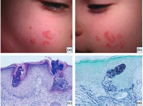
One year previously an otherwise healthy 10-year-old girl presented with asymptomatic symmetrical annular lesions involving both cheeks (Fig. 1a,b). Progressively, she devel-oped slightly erythematous and hyperkeratotic papules with small nodular elements. The pattern was linear, annular, and serpiginous. Some lesions revealed central atrophy. No other skin lesions were present. There was no
signifi-cant medical, allergic, or surgical history, and the family history was unremarkable. An extensive work-up excluded the association with other genetic disorders. There was no oral drug intake. The patient was previously treated with emollients, topical corticosteroids, antimycotics and kerato-lytics without success. This clinical appearance suggested a diagnosis of granuloma annulare or post-traumatic epider-mal inclusion cysts. A 2-mm punch-biopsy was performed under local anesthesia (Fig. 1c,d).
(a) (b)
(c) (d)
Figure 1 Well-circumscribed erythematous and hyperkeratotic serpiginous papules of the left (a) and right (b) cheeks. (c) Moderately acanthotic epidermis and a transepidermal perforating channel containing elastic fibers (H/E stain, 1009). (d) Orcein staining revealing the transepidermal elimination of altered elastic fibers (Orcein stain, 1009)
ª 2013 The International Society of Dermatology International Journal of Dermatology 2013, 52, 649–650 649
What is your diagnosis?
Elastosis perforans serpiginosa (EPS).
Discussion
EPS is one of the primary perforating dermatoses of der-mal origin.1 Histologically, EPS reveals a moderately
acanthotic epidermis and a transepidermal perforating channel containing basophilic material. An inflammatory reaction may be present in the superficial dermis with multinucleated giant cells. Orcein staining illustrates the transepidermal elimination of the elastic fibers and the increased quantity of elastic fibers in the upper dermis.2 Focal dermal elastosis is also observed.3 Histopathology is also capable of eliminating other reactive perforating collagenoses.
EPS usually begins in childhood or early adulthood. Most commonly, the lateral aspects of the neck are involved, followed by the arms and flexural areas. This young girl presented with bilateral EPS lesions on the cheeks, a rather unusual localization for EPS.
The etiology of EPS is unknown,3 but in 30–40% of
the cases, it is associated with systemic diseases, such as Down syndrome, Ehlers–Danlos syndrome, acrogeria, osteogenesis imperfecta, pseudoxanthoma elasticum, Rothmund–Thomson syndrome, or Marfan syndrome.4,5
A few reports cite familial occurrence.6Besides the idio-pathic type, a pencillamine-induced form is described.7A thorough work-up is recommended to search for these associations.
Clinically, multiple small, firm keratotic papules and nodules are arranged in annular plaques over the neck, axillae, antecubital fossae, and forearms. EPS is fre-quently misdiagnosed because of its rarity.8 The clinical
differential diagnosis includes tinea, granuloma annulare, sarcoidosis, actinic granuloma, perforating pseudoxantho-ma elasticum, porokeratosis, and discoid lupus erythepseudoxantho-mat- erythemat-osus.
There is no standard therapy for EPS. Successful treat-ments have been reported with liquid nitrogen cryother-apy, isotretinoin, tazarotene, imiquimod, and CO2,
Erbium-YAG, and pulsed dye lasers.9,10 More anecdotal reports mention menthol, sulfur, and benzoyl peroxide.
Surgical options include local excisions. Electrodessication or cellophane tape-stripping are other treatment alterna-tives. Koebner effects should be avoided. Long-term results are variable. Treatment may cause atrophy and scarring. Spontaneous resolution of EPS has occasionally been reported, often after several years.1
Topical retinoids were prescribed without success. CO2
laser resurfacing will be proposed in the future. EPS should be included in the differential diagnosis of annular lesions of the face during childhood.
References
1 Pereira AC, Baeta IG, Costa Júnior SR, et al. Elastosis perforans serpiginosa in a patient with Downs syndrome. An Bras Dermatol 2010; 85: 691–694.
2 Lewis KG, Bercovitch L, Dill SW, Robinson-Bostom L. Acquired disorders of elastic tissue: part II. Decreased elastic tissue. J Am Acad Dermatol 2004; 51: 165–185. 3 Gregersen PA, Stausbøl-Grøn B, Ramsing M, Sommerlund
M. Elastosis perforans serpiginosa in a patient with Down syndrome treated with imiquimod 5% cream. Dermatol Reports 2010; 2: e15, 42–43.
4 Mehta RK, Burrows NP, Payne CM, et al. Elastosis perforans serpiginosa and associated disorders. Clin Exp Dermatol 2001; 26: 521–524.
5 Vearrier D, Buka RL, Roberts B, et al. What is standard of care in the evaluation of elastosis perforans
serpiginosa? A survey of pediatric dermatologists. Pediatr Dermatol 2006; 23: 219–224.
6 Langeveld-Wildschut EG, Toonstra J, van Vloten WA, Beemer FA. Familial elastosis perforans serpiginosa. Arch Dermatol 1993; 129: 205–207.
7 Lewis BK, Chern PL, Stone MS. Penicillamine-induced elastosis of the mucosal lip. J Am Acad Dermatol 2009; 60: 700–703.
8 Atzori L, Pinna AL, Pau M, Aste N. D-penicillamine elastosis perforans serpiginosa: description of two cases and review of the literature. Dermatol Online J 2011; 15: 3. 9 Kelly SC, Purcell SM. Imiquimod therapy for elastosis perforans serpiginosa. Arch Dermatol 2006; 142: 829– 830.
10 Outland JD, Brown TS, Callen JP. Tazarotene is an effective therapy for elastosis perforans serpiginosa. Arch Dermatol 2002; 138: 169–171.
International Journal of Dermatology 2013, 52, 649–650 ª 2013 The International Society of Dermatology Clinicopathologic challenge Chronic annular lesions of the cheeks Lother, Arrese, and Nikkels 650