A N N A L S O F T H E N E W Y O R K A C A D E M Y O F S C I E N C E S
Issue: Neuroimmunomodulation in Health and Disease
Somatotrope GHRH/GH/IGF-1 axis at the crossroads
between immunosenescence and frailty
Gwennaelle Bodart,1Lindsay Goffinet,1Gabriel Morrhaye,1Khalil Farhat,1Marie de Saint-Hubert,2Florence Debacq-Chainiaux,3Christian Swine,2Vincent Geenen,1 and Henri J. Martens1
1GIGA Research Institute, University of Li `ege, Li `ege, Belgium.2Department of Geriatrics, University Hospital of
Mont-Godinne, NARILIS-Namur Research Institute for Life Sciences, Universit ´e Catholique de Louvain, Louvain-la-Neuve, Belgium.3Unit of Research on Cellular Biology, NARILIS-Namur Research Institute for Life Sciences, University of Namur
(FUNDP), Namur, Belgium
Address for correspondence: Henri J. Martens, Ph.D., GIGA-I3 Immunendocrinology CHU-B34, B-4000 Li `ege-Sart Tilman, Belgium. hmartens@ulg.ac.be
Immunosenescence, characterized by complex modifications of immunity with age, could be related to frailty syndrome in elderly individuals, leading to an inadequate response to minimal aggression. Functional decline (i.e., the loss of ability to perform activities of daily living) is related to frailty and decreased physiological reserves and is a frequent outcome of hospitalization in older patients. Links between immunosenescence and frailty have been explored and 20 immunological parameters, including insulin-like growth factor-1 (IGF-1), thymopoeisis, and telomere length, were shown to be affected in elderly patients with functional decline. A strong relationship between IGF-1 and thymic ouput was evidenced. IGF-1, a mediator of growth hormone (GH), was subsequently shown to induce interleukin-7 secretion in cultured primary human thymic epithelial cells. We are exploring the stress hypothesis in which an acute stressor is used as the discriminator of frailty susceptibility. GH can counteract the deleterious immunosuppressive effects of stress-induced steroids. Under nonstress conditions, the immunosenescent system preserves physiological responses, while under stress conditions, the combination of immunosenescence and a defect in the somatotrope axis might lead to functional decline.
Keywords: immunosenescence; aging; frailty; growth-hormone-releasing hormone; GHRH; growth hormone;
insulin-like growth factor-1; IGF-1
Immunosenescence
Immunosenescence is characterized by a panel of changes in immune function associated with aging and has been shown to contribute to the higher incidence of infectious diseases, inflammatory dis-orders, cancer, and poorer responses to vaccines in older compared to younger adults.1–4 Most studies on human immunosenescence are cross-sectional and face the challenge of comparing different young and aged populations, and there are very few lon-gitudinal observations. Moreover, investigation of the specificity of immune alterations with age in humans is susceptible to several biases, such as the selection bias of healthy elderly individuals in the SENIEUR protocol and supercentenarians,
who reflect a survivor selection, or biases related to the changes in life conditions between the mid-20th century and the present day.5,6 However, some consensual observations have emerged about aging immunity that are especially relevant for the T cell compartment. Cell-mediated immunity is predominantly affected by aging and a decline in cell-mediated immunity has been attributed to thymic involution.3,7 While thymopoiesis persists until 60 years of age, its progressive decline and the decreased generation of T cell receptor (TCR) diversity are not sufficient to renew the total per-centage of naive T cells in the periphery.8Hallmarks of immunosenescence include expansion of the T cells bearing a memory phenotype,9 together
with an attrition of TCR diversity.10 These two events have been attributed to nonexclusive phe-nomena of recurrent and consecutive stimulation of T cell clones filling the immune space and a loss of T cell renewal from the shrinking aged thy-mus. Common features include a decrease in the CD4:CD8 ratio, with CD4 lymphopenia describ-ing the immune risk profile.11Immunosenescence is not only characterized by changes in immune cell proportion, but also by functional alterations, such as, notably, the increase in CD28– cells; CD28 is the ligand of CD80/86 expressed by antigen pre-senting cells, and the loss of CD28 makes T cells unable to respond to antigen presentation.12In very old age, there is a shift in the T helper (TH)1/TH2 cytokine profile toward a TH2 dominance.13,14 Nev-ertheless, a proinflammatory cytokine profile in old age has also been reported, which initiates the inflammaging paradigm.15–17 Since the con-cept emerged in the late 20th century, at least five causes for this low-grade chronic inflammation have been identified: (1) self-debris, (2) harmful products from gut microbiota and mitochondria, (3) senes-cent cells producing proinflammatory cytokines, (4) increased activation of the coagulation system, and (5) an increase in innate immunity.18 Interest-ingly, the inflammatory cytokine interleukin (IL)-6—used as a biomarker of inflammation, found at high levels in the elderly, and associated with risk of morbidity—has been shown to decrease circulating insulin-like growth factor-1 (IGF-1) levels, which has anti-inflammatory effects.19,20
Immunological parameters as biomarkers of frailty
Functional decline (FD) frequently occurs in older patients after hospitalization and is associated with not only illness severity but also the patient’s premorbid frailty status.21FD is defined as a loss of autonomy in carrying out activities of daily living (ADL; e.g., walking, dressing). Several authors have suggested that the extent of FD after acute stress reflects the level of frailty, defined as “the inability to withstand acute illness without loss of function.”22,23Identification of patients at risk for FD is important as geriatric intervention may pre-vent or limit these losses. Several clinical tools have therefore been developed to assess this risk,24–26 but their predictive performance is limited.27 Combining clinical and biological markers has been
suggested as a way to improve this prediction.28 Nevertheless, little is known about the precise biological mechanisms underlying frailty and FD. Immunosenescence and telomere shortening have been proposed as significant candidates.29
On the basis of the assumption that immuno-logical parameters delineating immunosenescence could be reliable biomarkers to diagnose and predict frailty status, a previous research program investigated over 600 potential immune-related markers in four well-defined elderly cohorts: community dwelling (as an example of robust subjects) and three acutely stressed clinic popu-lations with hip fracture, acute heart failure, or documented infection.30–34These studies examined the relationship between immune parameters measured at emergency admission and FD, defined as a loss of at least one point on the ADL scale35 between baseline level (2 weeks before admission) and 3-month post-discharge functional status. Among 20 statistically significant associations iden-tified were high plasma IL-6, low plasma IGF-1, shorter peripheral blood mononuclear cell (PBMC) telomere length, and lower levels of PBMC TCR rearrangement excision circles (TRECs) levels. Immunological biomarkers of frailty
Ongoing thymopoiesis provides new T cells with stochastic TCR gene segment rearrangements. During the rearrangement process, episomal DNA fragments (TRECs) are produced.36 TRECs are stable molecules that persist in peripheral T cells until mitosis, but they are not replicated and therefore dilute with cell proliferation. It has been demonstrated that TREC frequency decreases with age, while the ratio of late-to-early TRECs (sj/D TRECs) reflects intrathymic pre-T cell proliferation.36–39
Antigen encounter in the periphery leads to massive T cell proliferation, and the number of possible T cell divisions is tightly correlated to the length of telomeric DNA. Given the repeated stimulation of antigen-specific T cells through-out life, there is risk of telomere erosion affect-ing global immune function.40,41 An association between telomere length and functional level has been suggested,42since telomere attrition is associ-ated with various age-relassoci-ated diseases,43,44as well as with physical and emotional burdens45that predis-pose to frailty.
IGF-1 is a growth-promoting factor regulating cellular survival, proliferation, and differentiation.46 Mainly produced by the liver under the control of growth hormone (GH), IGF-1 and GH are involved in several immune functions, especially T cell proliferation and thymic function.47 Interestingly, GH has been shown to increase IL-6 production in aging animals,48 while high levels of IL-6 and low levels of IGF-1 both appear in the frail population of subjects reported in previous studies.
GH and thymic function in adults with GH deficiency
The concomitant low levels of TRECs and IGF-1 in frail subjects strongly support the hypothesis that the somatotrope axis could be involved in decreased thymic function, thereby leading to T cell replace-ment under a threshold where adaptive immune sys-tem homeostasis is compromised. As a consequence, physiological stress encounters insufficient immune responses and the subject is unable to deliver an optimal response, which is a definition of frailty. To examine this issue, a study in patients with adult growth hormone deficiency (AGHD) assessed plas-matic sjTREC frequency, sj/b TREC ratio, and IGF-1 concentrations.37All subjects were asked to stop GH treatment for 1 month before resuming it, and samples were collected before the arrest, 1 month after withdrawal, and 1 month after resumption. It was found that plasma sjTREC frequency decreased after 1 month without GH and was restored after resumption. Moreover, plasma sjTREC frequency
was highly correlated with IGF-1 concentration (Fig. 1). Intrathymic T cell proliferation was also reduced after GH withdrawal, as indicated by the reduced sj/D TREC ratio. From this study, we concluded that the somatotrope GH/IGF-1 axis is involved in the maintenance of normal thy-mus function in human adults.
Somatotrope axis and thymic function Hypophysectomy was already shown in 1930 to induce thymus involution.49Forty years later, GH antiserum was shown to induce thymus atrophy.50 If somatotrope axis impairment seemed to drive thymic dysfunction, GH supplementation also appeared to restore normal thymus function, as implantation of GH-secreting cells reverses thymic aging in rats,51 and GH administration improves thymic cellularity and thymic T cell proliferation in dwarf DW/J mice that lack GH and prolactin.52 IGF-1, the main mediator of GH, intimately modulates the thymic homing of T cell precursors, thymopoiesis, and trafficking of thymocytes in the thymus microenvironment, and is also implicated in several peripheral immune functions.53–56More recently, ghrelin, a GH secretagogue, was shown to significantly improve thymopoiesis in old mice, as revealed by the increased number of recent thymic emigrants and TCR diversity of the peripheral T cell repertoire.57
Among others, these important observations promoted clinical studies using GH supplementa-tion to improve thymopoeisis in immunodeficient
Figure 1. Plasma IGF-1 concentration and sjTREC frequency in PBMCs from patients with GH deficiency and following GH treatment. (A) The interruption of GH treatment for 1 month induced a significant decrease in blood IGF-1 and sjTREC levels. (B) Both parameters were restored to initial levels 1 month after GH resumption. ***P< 0.001 (by Wilcoxon’s signed rank test, N= 22). As shown in C, there is a significant positive correlation between blood IGF-1 levels and sjTREC frequencies (R = 0.61, P< 0.01 by Spearman’s analysis). Adapted, with permission, from Morrhaye et al.37
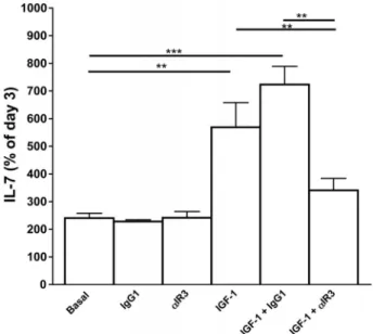
Figure 2. IL-7 secretion by human TECs under IGF-1 stimulation. Results are expressed as percentage of day-3 culture IL-7 supernatant concentration considered as basal levels. IGF-1 (10 nM) treatment significantly increased IL-7 secretion by cultured human TECs on day 8 of culture versus control medium, IgG1 (650 ng/mL) and␣IR3 (650 ng/mL). This effect was inhibited by anti-IGF-1R␣IR3 (650 ng/mL). **P < 0.01; ***P < 0.001 (by Wilcoxon’s signed rank test, N = 6). Adapted, with permission, from Goffinet et al.60
subjects. For example, GH treatment was evalu-ated to enhance immune reconstitution in HIV patients under highly active antiretroviral therapy (HAART). A first pilot study showed that GH supplementation reverses thymic atrophy of
HAART-treated HIV-infected patients and
enhances circulating naive CD4 T cells.58 A prospective randomized study confirmed these data and further evidenced that GH strongly increases the number of circulating sjTRECs in PBMCs.59
Given that TREC formation is due to
recombinase-activating gene (RAG) activity and that RAG is strongly induced by IL-7, the relation-ship between IGF-1 and IL-7 production by thymic epithelial cells (TECs) was explored in vitro.60IGF-1 induces strong expression and release of IL-7, which can be completely counteracted by blocking anti-body to IGF-1R (Fig. 2). Interestingly, physiological concentrations of GH do not induce any signifi-cant production of IGF-1 by TECs, suggesting that the increase of IL-7 production by human TECs is essentially mediated by peripheral IGF-1.61 Limits of immunological biomarkers in the prognosis of frailty and FD
Using a combination of proinflammatory and hormonal biomarkers (IL-6 and IGF-1) with a
clinical screening tool has been shown to improve the accuracy of FD prediction 3 months after hospitalization.34 The association of IL-6, IGF-1, TREC frequency, and telomere length with clinical frailty scoring using the predictive tool SHERPA26 first led to an improvement in the area under the receiver operating characteristic (ROC) curve from 70% to more than 84%. However, relaxing the inclu-sion criteria to include more individuals in larger cohorts (i.e., hospitalized instead of emergency patients) did not confirm that the selected biomark-ers improved the clinical SHERPA score. Several biomarkers still correlated with the SHERPA score without improving evaluation of the prognosis.
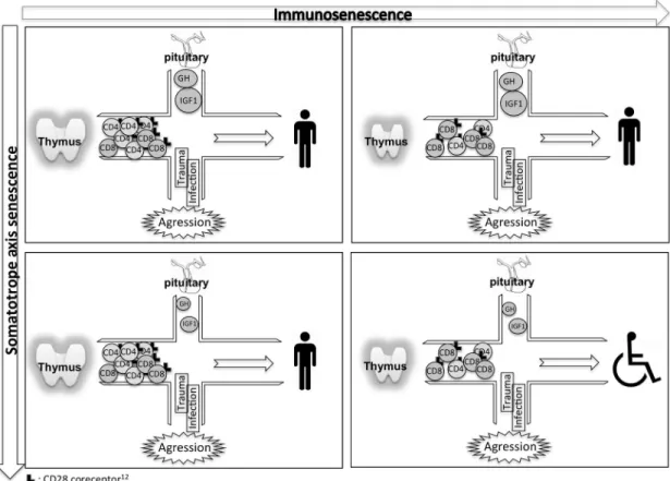
In reexamining the difference between old and new cohorts, a hypothesis emerged: an acute stress might be the crucial factor for immune biomark-ers of frailty to show their significance. Indeed, the first cohort of patients in earlier studies30–34 was recruited in the emergency department at admis-sion, while the second cohort stayed in the more comfortable geriatric hospital department. This hypothesis is closely related to the stress hypothesis (Fig. 3) proposed by Dorshkind and Horseman,62 who suggest that GH exerts a minimal effect on thymic function and the T cell system under basal conditions, but is able to successfully counteract the
Figure 3. Combined senescence of the immune system and somatotrope axis causes frailty. Upper left: young and mature subjects have functional thymuses producing abundant new CD28+T cells and normal levels of GH and its mediator IGF-1. External aggression, such as infection and trauma, is overthrown and subjects fully recover. Upper right and lower left: either thymus or somatotrope axis is senescent. Loss of CD28+T cells and a decreased CD4:CD8 ratio result from an involutive thymus (upper right) or low GH and IGF-1 concentrations (lower left). Nevertheless, aggression is still defeated without harmful consequences. Lower right: both the somatotrope axis and immune responses are deficient. Stress from aggression leads to a functional decline in these frail individuals.
deleterious effects of stress mediated by corticoids on thymic and effective T cells.63
Ongoing studies
We are currently exploring this frailty-related stress hypothesis both in animals and humans. In these ongoing studies, a model of transgenic mice defi-cient in hypothalamic growth hormone–releasing hormone (GHRH)64 is used to assess the effect of somatotrope axis hormones on immune function and responses. Preliminary results showed no evi-dent defects in parameters of thymus and immune system function under basal conditions, except for a limited B cell lymphopenia.65Moreover, the dwarf phenotype of this model has been shown to be par-tially reverted by GH supplementation,66which can be tested for the effect of induced stress on immune
system and thymic function. Recently, this model of Ghr–/–mice was shown to be resistant to exper-imental allergic encephalomyelitis while becoming sensible after GH supplementation.67
We are also beginning new studies in a pop-ulation of aged family caregivers intended to explore the immunological biomarkers identified in previous studies. The subjects are potentially stressed or nonstressed, depending on, for example, the social situation, and will be tested for psycho-logical and physiopsycho-logical parameters. This specific population has already been noted for showing telomere erosion and compromised immunity,45 and telomere shortening was one of our potential frailty biomarkers. Examining aged caregivers for physiological status and selected biomarkers with regard to their stress situation might help to solve
the contradiction related to the pertinent frailty markers seen in previous studies without extension to a larger elderly population.
Acknowledgments
We thank all the patients included in this study for their participation. This work is supported by Wallonia (DGTRE Reseaux 2-SENEGENE No. 05/1/6192 SPW, Belgium). We are also grateful to the Fund Leon Fredericq for biomedical research at the University Hospital of Liege and CAREGIVER program of the Germaine Tillion project funded by Wallonia (No. 1318184). V. Geenen, O. Toussaint, F. Debacq-Chainiaux, and G. Bodart are, respec-tively, Research Director, Senior Research Associate, Research Associate, and Research Assistant of the F.R.S.-FNRS Belgium.
Conflict of interest
The authors declare no conflicts of interest. References
1. Kovaiou, R.D., D. Herndler-Brandstetter & B. Grubeck-Loebenstein. 2007. Age-related changes in immunity: impli-cations for vaccination in the elderly. Expert Rev. Mol. Med.
9: 1–17.
2. Hakim, F.T. & R.E. Gress. 2007. Immunosenescence: deficits in adaptive immunity in the elderly. Tissue Antigens 70: 179– 189.
3. Gruver, A.L., L.L. Hudson & G.D. Sempowski. 2007. Immunosenescence of ageing. J. Pathol. 211: 144–156. 4. Goronzy, J.J. et al. 2001. Value of immunological markers in
predicting responsiveness to influenza vaccination in elderly individuals. J. Virol. 75: 12182–12187.
5. Vacante, M. et al. Centenarians and supercentenarians: a black swan. Emerging social, medical and surgical problems.
BMC Surg. 12(Suppl 1): S36.
6. Castle, S.C., K. Uyemura & T. Makinodan. 2001. The SENIEUR Protocol after 16 years: a need for a paradigm shift? Mech. Ageing Dev. 122: 127–130.
7. Haynes, B.F. et al. 2000. The human thymus during aging.
Immunol. Res. 22: 253–261.
8. Naylor, K. et al. 2005. The influence of age on T cell genera-tion and TCR diversity. J. Immunol. 174: 7446–7452. 9. Lazuardi, L. et al. 2005. Age-related loss of naive T cells and
dysregulation of T-cell/B-cell interactions in human lymph nodes. Immunology 114: 37–43.
10. Pawelec, G. et al. 2005. Human immunosenescence: is it infectious? Immunol. Rev. 205: 257–268.
11. Wikby, A. et al. 1998. Changes in CD8 and CD4 lympho-cyte subsets, T cell proliferation responses and non-survival in the very old: the Swedish longitudinal OCTO-immune study. Mech. Ageing Dev. 102: 187–198.
12. Vallejo, A.N. 2005. CD28 extinction in human T cells: altered functions and the program of T-cell senescence. Immunol.
Rev. 205: 158–169.
13. Sandmand, M. et al. 2002. Is ageing associated with a shift in the balance between Type 1 and Type 2 cytokines in humans?
Clin. Exp. Immunol. 127: 107–114.
14. Rink, L., I. Cakman & H. Kirchner. 1998. Altered cytokine production in the elderly. Mech. Ageing Dev. 102: 199– 209.
15. Roubenoff, R. et al. 1998. Monocyte cytokine production in an elderly population: effect of age and inflammation.
J. Gerontol. A Biol. Sci. Med. Sci. 53: M20–26.
16. Bruunsgaard, H. & B.K. Pedersen. 2003. Age-related inflam-matory cytokines and disease. Immunol. Allergy Clin. North
Am. 23: 15–39.
17. Franceschi, C. et al. 2000. Inflamm-aging. An evolutionary perspective on immunosenescence. Ann. N.Y. Acad. Sci. 908: 244–254.
18. Franceschi, C. & J. Campisi. 2014. Chronic inflamma-tion (inflammaging) and its potential contribuinflamma-tion to age-associated diseases. J. Gerontol. A Biol. Sci. Med. Sci.
69(Suppl 1): S4–S9.
19. DeBenedetti, F. et al. 1997. Interleukin 6 causes growth impairment in transgenic mice through a decrease in insulin-like growth factor-I. A model for stunted growth in children with chronic inflammation. J. Clin. Invest. 99: 643–650. 20. Sukhanov, S. et al. 2007. IGF-1 reduces
inflamma-tory responses, suppresses oxidative stress, and decreases atherosclerosis progression in ApoE-deficient mice.
Arte-rioscler. Thromb. Vasc. Biol. 27: 2684–2690.
21. Inouye, S.K. et al. 1993. A predictive index for functional decline in hospitalized elderly medical patients. J. Gen.
Intern. Med. 8: 645–652.
22. Sager, M.A. et al. 1996. Functional outcomes of acute medical illness and hospitalization in older persons. Arch. Intern.
Med. 156: 645–652.
23. Carlson, J.E. et al. 1998. Measuring frailty in the hospitalized elderly: concept of functional homeostasis. Am. J. Phys. Med.
Rehabil. 77: 252–257.
24. Sager, M.A. et al. 1996. Hospital admission risk profile (HARP): identifying older patients at risk for functional decline following acute medical illness and hospitalization.
J. Am. Geriatr. Soc. 44: 251–257.
25. Fried, L.P. et al. 2001. Frailty in older adults: evidence for a phenotype. J. Gerontol. A Biol. Sci. Med. Sci. 56: M146–M156.
26. Cornette, P. et al. 2006. Early evaluation of the risk of func-tional decline following hospitalization of older patients: development of a predictive tool. Eur. J. Public Health 16: 203–208.
27. deSaint-Hubert, M. et al. 2010. Comparison of three tools predicting functional decline after hospitalization of older patients. J. Am. Geriatr. Soc. 58: 1003–1005.
28. Wu, A.W. et al. 2000. Predicting functional status outcomes in hospitalized patients aged 80 years and older. J. Am.
Geri-atr. Soc. 48: S6–S15.
29. Walston, J. et al. 2006. Research agenda for frailty in older adults: toward a better understanding of physiology and etiology: summary from the American Geriatrics Soci-ety/National Institute on Aging Research Conference on Frailty in Older Adults. J. Am. Geriatr. Soc. 54: 991–1001. 30. Vo, T.K. et al. 2010. Transcriptomic biomarkers of the
response of hospitalized geriatric patients with infectious diseases. Immun. Ageing 7: 9.
31. Vo, T.K. et al. 2010. Differentially abundant transcripts in PBMC of hospitalized geriatric patients with hip fracture compared to healthy aged controls. Exp. Gerontol. 46: 257– 264.
32. Vo, T.K. et al. 2009. Transcriptomic biomarkers of human ageing in peripheral blood mononuclear cell total RNA. Exp.
Gerontol. 45: 188–194.
33. Vo, T.K. et al. 2011. Transcriptomic biomarkers of the response of hospitalized geriatric patients admitted with heart failure. Comparison to hospitalized geriatric patients with infectious diseases or hip fracture. Mech. Ageing Dev.
132: 131–139.
34. de Saint-Hubert, M. et al. 2010. Serum IL-6 and IGF-1 improve clinical prediction of functional decline after hospi-talization in older patients. Aging Clin. Exp. Res. 23: 106–111. 35. Katz, S. et al. 1963. Studies of illness in the aged. The index of Adl: a standardized measure of biological and psychosocial function. JAMA 185: 914–919.
36. Douek, D.C. et al. 1998. Changes in thymic function with age and during the treatment of HIV infection. Nature 396: 690–695.
37. Morrhaye, G. et al. 2009. Impact of growth hormone (GH) deficiency and GH replacement upon thymus function in adult patients. PLoS One 4: e5668.
38. Geenen, V. et al. 2003. Quantification of T cell receptor rearrangement excision circles to estimate thymic function: an important new tool for endocrine-immune physiology.
J. Endocrinol. 176: 305–311.
39. Dion, M.L., R.P. Sekaly & R. Cheynier. 2007. Estimating thymic function through quantification of T-cell receptor excision circles. Methods Mol. Biol. 380: 197–213. 40. Lord, J.M., A.N. Akbar & D. Kipling. 2002. Telomere-based
therapy for immunosenescence. Trends Immunol. 23: 175– 176.
41. Hodes, R.J., K.S. Hathcock & N.P. Weng. 2002. Telomeres in T and B cells. Nat. Rev. Immunol. 2: 699–706.
42. Woo, J. et al. 2008. Telomeres and frailty. Mech. Ageing Dev.
129: 642–648.
43. Valdes, A.M. et al. 2008. Leukocyte telomere length is asso-ciated with cognitive performance in healthy women.
Neu-robiol. Aging 31: 986–992.
44. Benetos, A. et al. 2004. Short telomeres are associated with increased carotid atherosclerosis in hypertensive subjects.
Hypertension 43: 182–185.
45. Damjanovic, A.K. et al. 2007. Accelerated telomere erosion is associated with a declining immune function of caregivers of Alzheimer’s disease patients. J. Immunol. 179: 4249–4254. 46. Stewart, C.E. & P. Rotwein. 1996. Growth, differentiation, and survival: multiple physiological functions for insulin-like growth factors. Physiol. Rev. 76: 1005–1026.
47. Goffinet, L. et al. 2011. Impact of the somatotrope growth hormone (GH)/insuline-like growth factor 1 axis upon thy-mus function: pharmacological implications in regeneration of immune functions. Immun. Endocr. Metab. Agents Med.
Chem. 11: 10–20.
48. Goya, R.G. et al. 1992. In vivo effects of growth hormone on thymus function in aging mice. Brain Behav. Immun. 6: 341–354.
49. Smith, P. 1930. Effects of hypophysectomy upon involution of the thymus in the rat. Anat. Rec. 47: 119–129.
50. Pierpaoli, W. & E. Sorkin. 1968. Hormones and immuno-logic capacity. I. Effect of heterologous anti-growth hormone (ASTH) antiserum on thymus and peripheral lymphatic tis-sue in mice. Induction of a wasting syndrome. J. Immunol.
101: 1036–1043.
51. Kelley, K.W. et al. 1986. GH3 pituitary adenoma cells can reverse thymic aging in rats. Proc. Natl. Acad. Sci. USA 83: 5663–5667.
52. Murphy, W.J., S.K. Durum & D.L. Longo. 1993. Differential effects of growth hormone and prolactin on murine T cell development and function. J. Exp. Med. 178: 231–236. 53. Savino, W. et al. 2002. The thymus gland: a target organ for
growth hormone. Scand. J. Immunol. 55: 442–452. 54. Savino, W. & M. Dardenne. 2000. Neuroendocrine control
of thymus physiology. Endocr. Rev. 21: 412–443.
55. Montecino-Rodriguez, E., R. Clark & K. Dorshkind. 1998. Effects of insulin-like growth factor administration and bone marrow transplantation on thymopoiesis in aged mice.
Endocrinology 139: 4120–4126.
56. Clark, R. 1997. The somatogenic hormones and insulin-like growth factor-1: stimulators of lymphopoiesis and immune function. Endocr. Rev. 18: 157–179.
57. Dixit, V.D. et al. 2007. Ghrelin promotes thymopoiesis dur-ing agdur-ing. J. Clin. Invest. 117: 2778–2790.
58. Napolitano, L.A. et al. 2002. Increased thymic mass and cir-culating naive CD4 T cells in HIV-1-infected adults treated with growth hormone. AIDS 16: 1103–1111.
59. Napolitano, L.A. et al. 2008. Growth hormone enhances thymic function in HIV-1-infected adults. J. Clin. Invest.
118: 1085–1098.
60. Goffinet, L. et al. 2011. IGF-1 promotes interleukin 7 (IL-7) synthesis and secretion by primary cultures of human thymic epithelial cells. Scand. J. Immunol. 73: 351. 61. Kermani, H. et al. 2012. Expression of the growth
hormone/insulin-like growth factor axis during Balb/c thy-mus ontogeny and effects of growth hormone upon ex vivo T cell differentiation. Neuroimmunomodulation 19: 137– 147.
62. Dorshkind, K. & N.D. Horseman. 2001. Anterior pituitary hormones, stress, and immune system homeostasis.
Bioes-says 23: 288–294.
63. Dorshkind, K. et al. 2003. Effects of housing on the thymic deficiency in dwarf mice and its reversal by growth hormone administration. Clin. Immunol. 109: 197–202.
64. Alba, M. & R. Salvatori. 2004. A mouse with targeted abla-tion of the growth hormone-releasing hormone gene: a new model of isolated growth hormone deficiency. Endocrinology
145: 4134–4143.
65. Bodart, G. et al. 2014. Thymic and immunological phe-notype of growth hormone releasing hormone (GHRH)-deficient mice in basal conditions. Presented at 9th Congress of the International Society for NeuroImmunoModulation, Liege, Belgium.
66. Alba, M., D. Fintini & R. Salvatori. 2005. Effects of recom-binant mouse growth hormone treatment on growth and body composition in GHRH knock out mice. Growth Horm.
IGF Res. 15: 275–282.
67. Shohreh, R. et al. 2011. GH, but not GHRH, plays a role in the development of experimental autoimmune encephalomyeli-tis. Endocrinology 152: 3803–3810.


