HAL Id: dumas-02958092
https://dumas.ccsd.cnrs.fr/dumas-02958092
Submitted on 5 Oct 2020HAL is a multi-disciplinary open access
archive for the deposit and dissemination of sci-entific research documents, whether they are pub-lished or not. The documents may come from teaching and research institutions in France or abroad, or from public or private research centers.
L’archive ouverte pluridisciplinaire HAL, est destinée au dépôt et à la diffusion de documents scientifiques de niveau recherche, publiés ou non, émanant des établissements d’enseignement et de recherche français ou étrangers, des laboratoires publics ou privés.
Distributed under a Creative Commons Attribution - NonCommercial - ShareAlike| 4.0 International License
étude rétrospective monocentrique de 312 cas
Élise Dandache
To cite this version:
Élise Dandache. Profils cliniques évolutifs de la pemphigoïde bulleuse : étude rétrospective monocen-trique de 312 cas. Médecine humaine et pathologie. 2018. �dumas-02958092�
UNIVERSITE DE MONTPELLIER
FACULTE DE MEDECINE MONTPELLIER-NIMES
THESE
Pour obtenir le titre de
DOCTEUR EN MEDECINE
Présentée et soutenue publiquement
Par
Elise DANDACHE
Le 23 novembre 2018
PROFILS CLINIQUES EVOLUTIFS DE LA
PEMPHIGOÏDE BULLEUSE : ETUDE RETROSPECTIVE
MONOCENTRIQUE DE 312 CAS
Directeur de thèse : Pr Olivier DEREURE
JURY
Président : Pr Olivier DEREURE
Assesseurs : Pr Bernard GUILLOT
Dr Aurélie DU THANH
Dr Céline GIRARD
ANNEE UNIVERSITAIRE 2017 - 2018 PERSONNEL ENSEIGNANT Professeurs Honoraires ALLIEU Yves ALRIC Robert ARNAUD Bernard ASTRUC Jacques AUSSILLOUX Charles AVEROUS Michel AYRAL Guy BAILLAT Xavier BALDET Pierre BALDY-MOULINIER Michel BALMES Jean-Louis BALMES Pierre BANSARD Nicole BAYLET René BILLIARD Michel BLARD Jean-Marie BLAYAC Jean Pierre BLOTMAN Francis BONNEL François BOUDET Charles
BOURGEOIS Jean-Marie BRUEL Jean Michel BUREAU Jean-Paul BRUNEL Michel CALLIS Albert CANAUD Bernard CASTELNAU Didier CHAPTAL Paul-André CIURANA Albert-Jean CLOT Jacques D’ATHIS Françoise DEMAILLE Jacques DESCOMPS Bernard DIMEGLIO Alain DU CAILAR Jacques DUBOIS Jean Bernard DUMAS Robert DUMAZER Romain ECHENNE Bernard FABRE Serge
FREREBEAU Philippe GALIFER René Benoît GODLEWSKI Guilhem GRASSET Daniel GROLLEAU-RAOUX Robert GUILHOU Jean-Jacques HERTAULT Jean HUMEAU Claude JAFFIOL Claude JANBON Charles JANBON François JARRY Daniel JOYEUX Henri LAFFARGUE François LALLEMANT Jean Gabriel LAMARQUE Jean-Louis LAPEYRIE Henri LESBROS Daniel LOPEZ François Michel LORIOT Jean
LOUBATIERES Marie Madeleine MAGNAN DE BORNIER Bernard MARY Henri MATHIEU-DAUDE Pierre MEYNADIER Jean MICHEL François-Bernard MICHEL Henri MION Charles MION Henri MIRO Luis NAVARRO Maurice NAVRATIL Henri OTHONIEL Jacques PAGES Michel PEGURET Claude POUGET Régis PUECH Paul PUJOL Henri PUJOL Rémy RABISCHONG Pierre RAMUZ Michel RIEU Daniel RIOUX Jean-Antoine ROCHEFORT Henri
ROUANET DE VIGNE LAVIT Jean Pierre
SAINT AUBERT Bernard SANCHO-GARNIER Hélène SANY Jacques SENAC Jean-Paul SERRE Arlette SIMON Lucien SOLASSOL Claude THEVENET André VIDAL Jacques VISIER Jean Pierre
Professeurs Emérites ARTUS Jean-Claude BLANC François BOULENGER Jean-Philippe BOURREL Gérard BRINGER Jacques CLAUSTRES Mireille DAURES Jean-Pierre DAUZAT Michel DEDET Jean-Pierre ELEDJAM Jean-Jacques GUERRIER Bernard JOURDAN Jacques MAURY Michèle MILLAT Bertrand MARES Pierre MONNIER Louis PRAT Dominique PRATLONG Francine PREFAUT Christian PUJOL Rémy ROSSI Michel SULTAN Charles TOUCHON Jacques VOISIN Michel ZANCA Michel
Professeurs des Universités - Praticiens Hospitaliers PU-PH de classe exceptionnelle
ALBAT Bernard - Chirurgie thoracique et cardiovasculaire
ALRIC Pierre - Chirurgie vasculaire ; médecine vasculaire (option chirurgie vasculaire) BACCINO Eric - Médecine légale et droit de la santé
BASTIEN Patrick - Parasitologie et mycologie BONAFE Alain - Radiologie et imagerie médicale CAPDEVILA Xavier - Anesthésiologie-réanimation COMBE Bernard - Rhumatologie
COSTA Pierre - Urologie
COTTALORDA Jérôme - Chirurgie infantile COUBES Philippe - Neurochirurgie
CRAMPETTE Louis - Oto-rhino-laryngologie
CRISTOL Jean Paul - Biochimie et biologie moléculaire DAVY Jean Marc - Cardiologie
DE LA COUSSAYE Jean Emmanuel - Anesthésiologie-réanimation DELAPORTE Eric - Maladies infectieuses ; maladies tropicales
DE WAZIERES Benoît - Médecine interne ; gériatrie et biologie du vieillissement, médecine générale, addictologie
DOMERGUE Jacques - Chirurgie générale DUFFAU Hugues - Neurochirurgie
DUJOLS Pierre - Biostatistiques, informatique médicale et technologies de la communication ELIAOU Jean François - Immunologie
FABRE Jean Michel - Chirurgie générale GUILLOT Bernard - Dermato-vénéréologie
HAMAMAH Samir-Biologie et Médecine du développement et de la reproduction ; gynécologie médicale HEDON Bernard-Gynécologie-obstétrique ; gynécologie médicale
HERISSON Christian-Médecine physique et de réadaptation JABER Samir-Anesthésiologie-réanimation
JEANDEL Claude-Médecine interne ; gériatrie et biologie du vieillissement, médecine générale, addictologie
JONQUET Olivier-Réanimation ; médecine d’urgence
JORGENSEN Christian-Thérapeutique ; médecine d’urgence ; addictologie KOTZKI Pierre Olivier-Biophysique et médecine nucléaire
LANDAIS Paul-Epidémiologie, Economie de la santé et Prévention LARREY Dominique-Gastroentérologie ; hépatologie ; addictologie LEFRANT Jean-Yves-Anesthésiologie-réanimation
LE QUELLEC Alain-Médecine interne ; gériatrie et biologie du vieillissement, médecine générale, addictologie
MARTY-ANE Charles - Chirurgie thoracique et cardiovasculaire MAUDELONDE Thierry - Biologie cellulaire
MERCIER Jacques - Physiologie MESSNER Patrick - Cardiologie MOURAD Georges-Néphrologie
PELISSIER Jacques-Médecine physique et de réadaptation
RENARD Eric-Endocrinologie, diabète et maladies métaboliques ; gynécologie médicale REYNES Jacques-Maladies infectieuses, maladies tropicales
RIBSTEIN Jean-Médecine interne ; gériatrie et biologie du vieillissement, médecine générale, addictologie
RIPART Jacques-Anesthésiologie-réanimation ROUANET Philippe-Cancérologie ; radiothérapie SCHVED Jean François-Hématologie; Transfusion TAOUREL Patrice-Radiologie et imagerie médicale UZIEL Alain -Oto-rhino-laryngologie
VANDE PERRE Philippe-Bactériologie-virologie ; hygiène hospitalière YCHOU Marc-Cancérologie ; radiothérapie
PU-PH de 1re classe
AVIGNON Antoine-Nutrition
AZRIA David -Cancérologie ; radiothérapie
BAGHDADLI Amaria-Pédopsychiatrie ; addictologie BEREGI Jean-Paul-Radiologie et imagerie médicale
BLAIN Hubert-Médecine interne ; gériatrie et biologie du vieillissement, médecine générale, addictologie
BLANC Pierre-Gastroentérologie ; hépatologie ; addictologie BORIE Frédéric-Chirurgie digestive
BOULOT Pierre-Gynécologie-obstétrique ; gynécologie médicale CAMBONIE Gilles -Pédiatrie
CAMU William-Neurologie CANOVAS François-Anatomie
CARTRON Guillaume-Hématologie ; transfusion
CHAMMAS Michel-Chirurgie orthopédique et traumatologique COLSON Pascal-Anesthésiologie-réanimation
CORBEAU Pierre-Immunologie
COSTES Valérie-Anatomie et cytologie pathologiques COURTET Philippe-Psychiatrie d’adultes ; addictologie CYTEVAL Catherine-Radiologie et imagerie médicale DADURE Christophe-Anesthésiologie-réanimation DAUVILLIERS Yves-Physiologie
DE TAYRAC Renaud-Gynécologie-obstétrique, gynécologie médicale DEMARIA Roland-Chirurgie thoracique et cardio-vasculaire
DEMOLY Pascal-Pneumologie ; addictologie DEREURE Olivier-Dermatologie - vénéréologie DROUPY Stéphane -Urologie
DUCROS Anne-Neurologie -
FRAPIER Jean-Marc-Chirurgie thoracique et cardiovasculaire KLOUCHE Kada-Réanimation ; médecine d’urgence
KOENIG Michel-Génétique moléculaire LABAUGE Pierre- Neurologie
LAFFONT Isabelle-Médecine physique et de réadaptation LAVABRE-BERTRAND Thierry-Cytologie et histologie LECLERCQ Florence-Cardiologie
LEHMANN Sylvain-Biochimie et biologie moléculaire LUMBROSO Serge-Biochimie et Biologie moléculaire
MARIANO-GOULART Denis-Biophysique et médecine nucléaire MATECKI Stéfan -Physiologie
MEUNIER Laurent-Dermato-vénéréologie MONDAIN Michel-Oto-rhino-laryngologie MORIN Denis-Pédiatrie
NAVARRO Francis-Chirurgie générale
PAGEAUX Georges-Philippe-Gastroentérologie ; hépatologie ; addictologie PETIT Pierre-Pharmacologie fondamentale ; pharmacologie clinique ; addictologie
PERNEY Pascal-Médecine interne ; gériatrie et biologie du vieillissement, médecine générale, addictologie
PUJOL Jean Louis-Pneumologie ; addictologie PUJOL Pascal-Biologie cellulaire
PURPER-OUAKIL Diane-Pédopsychiatrie ; addictologie
QUERE Isabelle-Chirurgie vasculaire ; médecine vasculaire (option médecine vasculaire) SOTTO Albert-Maladies infectieuses ; maladies tropicales
TOUITOU Isabelle-Génétique TRAN Tu-Anh-Pédiatrie
VERNHET Hélène-Radiologie et imagerie médicale
PU-PH de 2ème classe
ASSENAT Éric-Gastroentérologie ; hépatologie ; addictologie BERTHET Jean-Philippe-Chirurgie thoracique et cardiovasculaire BOURDIN Arnaud-Pneumologie ; addictologie
CAPDEVIELLE Delphine-Psychiatrie d'Adultes ; addictologie CAPTIER Guillaume-Anatomie
CAYLA Guillaume-Cardiologie
CHANQUES Gérald-Anesthésiologie-réanimation
COLOMBO Pierre-Emmanuel-Cancérologie ; radiothérapie COSTALAT Vincent-Radiologie et imagerie médicale
COULET Bertrand-Chirurgie orthopédique et traumatologique CUVILLON Philippe-Anesthésiologie-réanimation
DAIEN Vincent-Ophtalmologie
DE VOS John-Cytologie et histologie DORANDEU Anne-Médecine légale -
DUPEYRON Arnaud-Médecine physique et de réadaptation
FESLER Pierre-Médecine interne ; gériatrie et biologie du vieillissement, médecine générale, addictologie
GARREL Renaud -Oto-rhino-laryngologie GAUJOUX Viala Cécile-Rhumatologie GENEVIEVE David-Génétique
GODREUIL Sylvain-Bactériologie-virologie ; hygiène hospitalière GUILLAUME Sébastien-Urgences et Post urgences psychiatriques -
GUILPAIN Philippe-Médecine Interne, gériatrie et biologie du vieillissement; addictologie GUIU Boris-Radiologie et imagerie médicale
HAYOT Maurice-Physiologie
HOUEDE Nadine-Cancérologie ; radiothérapie JACOT William-Cancérologie ; Radiothérapie JUNG Boris-Réanimation ; médecine d'urgence KALFA Nicolas-Chirurgie infantile
KOUYOUMDJIAN Pascal-Chirurgie orthopédique et traumatologique LACHAUD Laurence-Parasitologie et mycologie
LALLEMANT Benjamin-Oto-rhino-laryngologie
LAVIGNE Jean-Philippe-Bactériologie-virologie ; hygiène hospitalière LE MOING Vincent-Maladies infectieuses ; maladies tropicales LETOUZEY Vincent-Gynécologie-obstétrique ; gynécologie médicale LOPEZ CASTROMAN Jorge-Psychiatrie d'Adultes ; addictologie LUKAS Cédric-Rhumatologie
MAURY Philippe-Chirurgie orthopédique et traumatologique MILLET Ingrid-Radiologie et imagerie médicale
MORANNE Olvier-Néphrologie MOREL Jacques -Rhumatologie
NAGOT Nicolas-Biostatistiques, informatique médicale et technologies de la communication NOCCA David-Chirurgie digestive
PANARO Fabrizio-Chirurgie générale
PARIS Françoise-Biologie et médecine du développement et de la reproduction ; gynécologie médicale PASQUIE Jean-Luc-Cardiologie
PEREZ MARTIN Antonia-Physiologie
POUDEROUX Philippe-Gastroentérologie ; hépatologie ; addictologie PRUDHOMME Michel-Anatomie
RIGAU Valérie-Anatomie et cytologie pathologiques RIVIER François-Pédiatrie
ROGER Pascal-Anatomie et cytologie pathologiques ROSSI Jean François-Hématologie ; transfusion ROUBILLE François-Cardiologie
SEBBANE Mustapha-Anesthésiologie-réanimation SEGNARBIEUX François-Neurochirurgie
SIRVENT Nicolas-Pédiatrie
SOLASSOL Jérôme-Biologie cellulaire SULTAN Ariane-Nutrition
THOUVENOT Éric-Neurologie THURET Rodolphe-Urologie
VENAIL Frédéric-Oto-rhino-laryngologie VILLAIN Max-Ophtalmologie
VINCENT Denis -Médecine interne ; gériatrie et biologie du vieillissement, médecine générale, addictologie
VINCENT Thierry-Immunologie
WOJTUSCISZYN Anne-Endocrinologie-diabétologie-nutrition
PROFESSEURS DES UNIVERSITES
1re classe :
COLINGE Jacques - Cancérologie, Signalisation cellulaire et systèmes complexes
2ème classe :
LAOUDJ CHENIVESSE Dalila - Biochimie et biologie moléculaire VISIER Laurent - Sociologie, démographie
PROFESSEURS DES UNIVERSITES - Médecine générale
1re classe :
LAMBERT Philippe
2ème classe : AMOUYAL Michel
PROFESSEURS ASSOCIES - Médecine Générale
DAVID Michel RAMBAUD Jacques
PROFESSEUR ASSOCIE - Médecine
BESSIS Didier - Dermato-vénéréologie)
PERRIGAULT Pierre-François - Anesthésiologie-réanimation ; médecine d'urgence ROUBERTIE Agathe – Pédiatrie
Maîtres de Conférences des Universités - Praticiens Hospitaliers
MCU-PH Hors classe
CACHEUX-RATABOUL Valère-Génétique
CARRIERE Christian-Bactériologie-virologie ; hygiène hospitalière CHARACHON Sylvie-Bactériologie-virologie ; hygiène hospitalière
FABBRO-PERAY Pascale-Epidémiologie, économie de la santé et prévention
HILLAIRE-BUYS Dominique-Pharmacologie fondamentale ; pharmacologie clinique ; addictologie PELLESTOR Franck-Cytologie et histologie
PUJOL Joseph-Anatomie
RAMOS Jeanne-Anatomie et cytologie pathologiques RICHARD Bruno-Thérapeutique ; addictologie RISPAIL Philippe-Parasitologie et mycologie
SEGONDY Michel-Bactériologie-virologie ; hygiène hospitalière STOEBNER Pierre -Dermato-vénéréologie
MCU-PH de 1re classe
ALLARDET-SERVENT Annick-Bactériologie-virologie ; hygiène hospitalière BADIOU Stéphanie-Biochimie et biologie moléculaire
BOUDOUSQ Vincent-Biophysique et médecine nucléaire BOULLE Nathalie-Biologie cellulaire
BOURGIER Céline-Cancérologie ; Radiothérapie BRET Caroline -Hématologie biologique COSSEE Mireille-Génétique Moléculaire GABELLE DELOUSTAL Audrey-Neurologie
GIANSILY-BLAIZOT Muriel-Hématologie ; transfusion GIRARDET-BESSIS Anne-Biochimie et biologie moléculaire LAVIGNE Géraldine-Hématologie ; transfusion
LE QUINTREC Moglie-Néphrologie
MATHIEU Olivier-Pharmacologie fondamentale ; pharmacologie clinique ; addictologie MENJOT de CHAMPFLEUR Nicolas-Neuroradiologie
MOUZAT Kévin-Biochimie et biologie moléculaire PANABIERES Catherine-Biologie cellulaire
PHILIBERT Pascal-Biologie et médecine du développement et de la reproduction RAVEL Christophe - Parasitologie et mycologie
SCHUSTER-BECK Iris-Physiologie
STERKERS Yvon-Parasitologie et mycologie
TUAILLON Edouard-Bactériologie-virologie ; hygiène hospitalière YACHOUH Jacques-Chirurgie maxillo-faciale et stomatologie
MCU-PH de 2éme classe
BERTRAND Martin-Anatomie
BRUN Michel-Bactériologie-virologie ; hygiène hospitalière DU THANH Aurélie-Dermato-vénéréologie
GALANAUD Jean Philippe-Médecine Vasculaire GOUZI Farès-Physiologie
JEZIORSKI Éric-Pédiatrie
KUSTER Nils-Biochimie et biologie moléculaire
LESAGE François-Xavier-Médecine et Santé au Travail MAKINSON Alain-Maladies infectieuses, Maladies tropicales
MURA Thibault-Biostatistiques, informatique médicale et technologies de la communication OLIE Emilie-Psychiatrie d'adultes ; addictologie
THEVENIN-RENE Céline-Immunologie
MAITRES DE CONFERENCES DES UNIVERSITES - Médecine Générale
COSTA David
FOLCO-LOGNOS Béatrice
MAITRES DE CONFERENCES ASSOCIES - Médecine Générale
CLARY Bernard GARCIA Marc MILLION Elodie PAVAGEAU Sylvain REBOUL Marie-Catherine SEGURET Pierre
MAITRES DE CONFERENCES DES UNIVERSITES Maîtres de Conférences hors classe
BADIA Eric - Sciences biologiques fondamentales et cliniques
Maîtres de Conférences de classe normale
BECAMEL Carine - Neurosciences BERNEX Florence - Physiologie
CHAUMONT-DUBEL Séverine - Sciences du médicament et des autres produits de santé CHAZAL Nathalie - Biologie cellulaire
DELABY Constance - Biochimie et biologie moléculaire
GUGLIELMI Laurence - Sciences biologiques fondamentales et cliniques HENRY Laurent - Sciences biologiques fondamentales et cliniques
LADRET Véronique - Mathématiques appliquées et applications des mathématiques LAINE Sébastien - Sciences du Médicament et autres produits de santé
LE GALLIC Lionel - Sciences du médicament et autres produits de santé
LOZZA Catherine - Sciences physico-chimiques et technologies pharmaceutiques MAIMOUN Laurent - Sciences physico-chimiques et ingénierie appliquée à la santé MOREAUX Jérôme - Science biologiques, fondamentales et cliniques
MORITZ-GASSER Sylvie - Neurosciences MOUTOT Gilles - Philosophie
PASSERIEUX Emilie - Physiologie RAMIREZ Jean-Marie - Histologie TAULAN Magali - Biologie Cellulaire
PRATICIENS HOSPITALIERS UNIVERSITAIRES CLAIRE DAIEN-Rhumatologie
BASTIDE Sophie-Epidémiologie, économie de la santé et prévention FAILLIE Jean-Luc-
Pharmacologie fondamentale ; pharmacologie clinique ; addictologie GATINOIS Vincent-Histologie, embryologie et cytogénétique
HERLIN Christian -Chirurgie plastique ; reconstructrice et esthétique ; brûlologie HERRERO Astrid-Chirurgie générale
PANTEL Alix-Bactériologie-virologie ; hygiène hospitalière
PERS Yves-Marie-Thérapeutique, médecine d’urgence ; addictologie
PINETON DE CHAMBRUN Guillaume-Gastroentérologie ; hépatologie ; addictologie TORRE Antoine-Gynécologie-obstétrique ; gynécologie médicale
REMERCIEMENTS
A l’ensemble des membres du jury, qui m’ont fait l’honneur d’accepter
cette fonction et de juger mon travail.
A Monsieur le Professeur Olivier DEREURE
Je vous remercie de m’avoir proposé ce sujet très intéressant et d’avoir
été mon directeur de thèse. Je vous remercie également pour votre
perspicacité inaltérable, vos connaissances impressionnantes, votre
bienveillance. Cela a été pour moi un honneur de travailler avec vous.
A Monsieur le Professeur Bernard GUILLOT
Je vous remercie pour votre accompagnement et pour l’intérêt que vous
portez à notre formation depuis le début de mon internat. Votre
dévouement envers les patients, votre sens clinique et votre sérénité sont
un modèle pour moi.
A Madame le Docteur Aurélie DU THANH
Je te remercie pour tes compétences médicales étendues, rigoureuses et
infaillibles ainsi que pour ta disponibilité et ta gentillesse. Les
consultations à tes côtés sont toujours d’agréables moments, riches en
apprentissage.
A Madame le Docteur Céline GIRARD
Je te remercie de m’avoir aidée à aboutir à ce sujet de thèse. Tu as
toujours été disponible et un modèle pour moi tant sur le plan humain
que médical. Ta sympathie, ta polyvalence et ton aide précieuses m’ont
A tous les chefs qui ont contribué à ma formation tout au long de mon
cursus :
A Mesdames et Messieurs Professeurs et Docteurs :
Pr Laurent MEUNIER, Pr Pierre STOEBNER, Pr Didier BESSIS, Dr
Michel DANDURAND, Dr Candice LESAGE, Dr Bernadette
OVTCHINNKOFF, Dr Myriam MARQUE, Dr Nadia
RAISON-PEYRON, Dr Valérie PALLURE, Dr Jean-Luc BOURRAIN, Dr Luc
DURAND, Dr Christophe DELFOUR
Merci de tout ce que vous m’avez transmis durant ces 4 dernières
années. J’espère avoir été digne de la confiance que vous m’avez
accordée.
A tout le personnel des services de Dermatologie des CHU de
Montpellier et de Nimes, d’Anatomopathologie du CHU de Montpellier,
de Rhumatologie du CHU de Montpellier, de Médecine interne générale
du CH de Perpignan
A mes chers collègues et anciens internes ou assistants : Agathe,
Alexandre, Alicia, Aline, Alizée, Amandine, Annelies, Anne-Sophie,
Anouck, Astrid, Aurélie C., Blanche, Camille, Chloé, Coline, Eleonore,
Elsa, Emilie, Emma, Eric, Fawaz, Hugo, Julie, Justine, Laura, Léo-Paul,
Louisa, Lucie, Marie D., Marie F, Marion B. et Marion B., Marion P.,
Mathilde, Mehdi, Melissa, Ondine, Quentin, Sarah, Séverin, Valentin,
Valérie R., Vincent
A mes chers amis qui répondent toujours présent : Alice et Bastien,
Camille, Claude et Chantal, Clémentine et Paul, Dorian, Marion et
Julien, Nico, Pierrick, Sophie et Ben
A mes parents: pour votre amour sans faille, vous m’avez porté tout au
long de ma vie. J’espère pouvoir vous le rendre.
A mon frère et à ma soeur, Jad et Célia : unis pour la vie, merci pour
tous ces moments passés ensemble et à venir. Que des souvenirs
mémorables.
Et à leur moitié, Laura et Julien
A Jesse, pour ton soutien indéfectible. Tu me rends meilleure chaque
jour. Ton humour, ton intelligence remplissent ma vie de bonheur et de
surprises.
SOMMAIRE
Liste des abréviations françaises
Contexte général
Article
“TIME-RELATED EVOLVING CLINICAL PATTERN OF
BULLOUS PEMPHIGOID: A MONOCENTER RETROSPECTIVE
SURVEY OF 312 CASES”
Abstract
Abbreviations and acronyms list
Introduction
Patients and Methods
Discussion
Conclusion
Tables
References
Serment
Permis d’imprimer
Abstract
LISTE DES ABREVIATIONS FRANCAISES
BPAG1 bullous pemphigoid antigen 1 BPAG2 bullous pemphigoid antigen 2
ELISA Enzyme-Linked ImmunoSorbent Assay IFD immunofluorescence directe
Ig immunoglobuline
JDE jonction dermo-épidermique kD kiloDalton
PB Pemphigoïde Bulleuse
PDM pemphigoïde des muqueuses PNE polynucléaire éosinophile
CONTEXTE GENERAL
La pemphigoïde bulleuse (PB) est la maladie bulleuse auto-immune sous-épidermique la plus fréquente. La PB fait partie d’un groupe de 8 maladies bulleuses auto-immunes sous-épidermiques qui comprend également pemphigoïde gestationnelle, pemphigoïde des muqueuses (PDM), pemphigoïde des enfants, dermatite herpétiforme, dermatose à IgA linéaire, pemphigoide anti-p200/laminin g1 et épidermolyse bulleuse acquise (1).
Dans la PB, les autoanticorps déposés à la jonction dermo-épidermique sont dirigés contre les hémi-desmosomes et entraînent la formation d’une bulle sous-épidermique se développant au sein de la lamina lucida, dans la région supérieure de la membrane basale. Ces autoanticorps sont dirigés contre des protéines de structure des hémi-desmosomes, principalement BPAg1 (protéine de 230kD) et BPAg2 (ou collagène XVII, protéine de 180 kD). Le domaine NC16a de l’ectodomaine de la protéine BP180, épitope proche du pôle basal des kératinocytes, a été identifié comme domaine immunodominant dans la PB et est différent de l’épitope-cible des autoanticorps anti-180kd des PDM avec autoanticorps anti-BPAg2 (2,3).
L’incidence dans le monde est estimée à 4,5 à 14 nouveaux cas par million d’habitants (4–9) et par an et en France à 21,7 nouveau cas par million d’habitants et par an, soit 3 fois supérieure à l’incidence estimée il y a 15 ans (10). Il n’y a pas de prédominance de sexe, d’ethnie ou géographique. Le taux de survie cumulée à 1 an est de 62% (10). Le risque de décès est 3 à 6 fois plus important que dans la population générale du même âge et de même sexe, plus en lien avec l’âge, l’état général du patient, les comorbidités neurologiques et les traitements spécifiques de la pemphigoïde bulleuse tels que la prise prolongée de corticostéroïdes systémiques qu’avec la sévérité de la maladie elle-même (10).
La PB est significativement associée à des affections neurologiques, principalement neurodégénératives mais également vasculaires. L’affection neurologique précède la PB dans 50 à 72% des cas, en moyenne 5,5 ans avant le diagnostic de PB. Le
lien physiopathologique entre BP et maladie neurologique n’est pas bien établi, peut-être par autoimmunité croisée vis-à-vis de l’antigène BPAG1 également présents dans le système nerveux central et démasquée par la maladie neurologique (11–14).
Cliniquement la PB se manifeste par un prurit souvent isolé au départ puis associé à une éruption bulleuse généralisée ou localisée accompagnée de lésions inflammatoires pseudo-urticariennes ou eczématiformes. La lésion élémentaire est une bulle reposant classiquement sur des plaques inflammatoires pseudo-urticariennes. Il existe des formes non-bulleuses où les symptômes sont un prurit isolé, à type de prurigo nodulaire, des plaques pseudo-urticariennes ou eczématiformes sans bulle, voire une érythrodermie. Contrairement à d’autres maladies bulleuses auto-immunes sous-épidermiques, les lésions cicatricielles sont classiquement absentes et la PB épargne la tête et cou ainsi que les muqueuses.
Le diagnostic de PB repose sur l’aspect clinique décrit ci-dessus, la présence d’une bulle sous épidermique avec un infiltrat dermique à PNE en histologie, la présence de dépôts linéaires d’IgG et/ou C3 à la jonction dermo-épidermique en IFD et la mise en évidence fréquente d’anticorps circulants anti BPAG1 et surtout anti BPAG2 par ELISA et Western Blot.
En 1998 le Groupe Bulle (Centre de référence national sur les maladies bulleuses auto-immunes) a proposé et validé un ensemble de 4 critères cliniques fortement associés au diagnostic de PB : âge supérieur à 70 ans, absence de cicatrices atrophiques, absence d’atteinte muqueuse, absence d’atteinte prédominante de la tête et du cou. Si au moins 3 critères sur 4 étaient présents, le diagnostic de PB pouvait être affirmé avec une sensibilité de 90%, une spécificité de 83% et une valeur prédictive positive de 95%. En 2004, le Groupe Bulle a confirmé la validité de ces critères clinique pour le diagnostic de PB en utilisant l’analyse du sérum des patients en immunoblot comme critère diagnostique principal, avec une sensibilité de 86%, une spécificité de 90% et une excellente valeur prédictive positive supérieure à 95%, si au moins 3 des 4 critères étaient présents, confirmant ainsi l’intérêt de cet ensemble de caractéristiques cliniques pour le diagnostic rapide de PB.
Cependant, le spectre clinique de la PB se modifie avec le temps et récemment un certain nombre de caractéristiques cliniques inhabituelles semblent émerger chez certains patients atteints de PB, notamment des atteintes palmo-plantaires prédominantes, des érosions extensives et une plus grande fréquence de lésions intra-buccales et de la tête et du cou. Cette étude monocentrique rétrospective portant sur une cohorte de grande taille de PB bien documentées a été menée pour mettre en évidence un changement possible des caractéristiques cliniques au cours du temps avec 2 objectifs principaux complémentaires : (i) décrire la fréquence des caractéristiques cliniques atypiques, dichotomisées en topographies atypiques et lésions cutanées élémentaires atypiques, et leur possible émergence au cours des 2 dernières décennies (ii) vérifier la validité au cours du temps des 4 critères cliniques diagnostiques mentionnés ci-dessus étant donné la possible augmentation de fréquence de caractéristiques cliniques atypiques.
REFERENCES
1. Schmidt E, della Torre R, Borradori L. Clinical features and practical diagnosis of bullous pemphigoid. Immunol Allergy Clin North Am. mai 2012;32(2):217-32, v.
2. Zillikens D, Mascaro JM, Rose PA, Liu Z, Ewing SM, Caux F, et al. A highly sensitive
enzyme-linked immunosorbent assay for the detection of circulating anti-BP180 autoantibodies in patients with bullous pemphigoid. J Invest Dermatol. nov 1997;109(5):679-83.
3. Di Zenzo G, Thoma-Uszynski S, Fontao L, Calabresi V, Hofmann SC, Hellmark T, et al.
Multicenter prospective study of the humoral autoimmune response in bullous pemphigoid. Clin Immunol Orlando Fla. sept 2008;128(3):415-26.
4. Bernard P, Vaillant L, Labeille B, Bedane C, Arbeille B, Denoeux JP, et al. Incidence and distribution of subepidermal autoimmune bullous skin diseases in three French regions. Bullous Diseases French Study Group. Arch Dermatol. janv 1995;131(1):48-52.
5. Increased risk of bullous pemphigoid in male and very old patients: A population-based study on incidence. - PubMed - NCBI [Internet]. [cité 24 juill 2018]. Disponible sur:
https://www.ncbi.nlm.nih.gov/pubmed/10426901
6. Langan SM, Smeeth L, Hubbard R, Fleming KM, Smith CJP, West J. Bullous
pemphigoid and pemphigus vulgaris--incidence and mortality in the UK: population based cohort study. BMJ. 9 juill 2008;337:a180.
7. Gudi VS, White MI, Cruickshank N, Herriot R, Edwards SL, Nimmo F, et al. Annual
incidence and mortality of bullous pemphigoid in the Grampian Region of North-east Scotland. Br J Dermatol. août 2005;153(2):424-7.
8. Cozzani E, Parodi A, Rebora A, Delmonte S, Barile M, Nigro A, et al. Bullous pemphigoid in Liguria: a 2-year survey. J Eur Acad Dermatol Venereol JEADV. juill 2001;15(4):317-9.
9. Serwin AB, Bokiniec E, Piascik M, Masny D, Chodynicka B. Epidemiological and
clinical analysis of pemphigoid patients in northeastern Poland in 2000-2005. Med Sci Monit Int Med J Exp Clin Res. août 2007;13(8):CR360-364.
10. Joly P, Baricault S, Sparsa A, Bernard P, Bédane C, Duvert-Lehembre S, et al. Incidence and mortality of bullous pemphigoid in France. J Invest Dermatol. août 2012;132(8):1998-2004.
11. Lai YC, Yew YW, Lambert WC. Bullous pemphigoid and its association with
neurological diseases: a systematic review and meta-analysis. J Eur Acad Dermatol Venereol JEADV. déc 2016;30(12):2007-15.
12. Cordel N, Chosidow O, Hellot M-F, Delaporte E, Lok C, Vaillant L, et al. Neurological disorders in patients with bullous pemphigoid. Dermatol Basel Switz. 2007;215(3):187-91.
13. Gambichler T, Segert H, Höxtermann S, Schmitz L, Altmeyer P, Teegen B. Neurological
disorders in patients with bullous pemphigoid: clinical and experimental investigations. J Eur Acad Dermatol Venereol JEADV. sept 2015;29(9):1758-62.
14. Milani-Nejad N, Zhang M, Kaffenberger J. The association between bullous pemphigoid
and neurological disorders: a systematic review. Eur J Dermatol EJD. 1 oct 2017;27(5):472-81. 15. de Quatrebarbes J, Joly P. [Bullous pemphigoid]. Rev Prat. 15 juin 2005;55(11):1165-8. 16. della Torre R, Combescure C, Cortés B, Marazza G, Beltraminelli H, Naldi L, et al. Clinical presentation and diagnostic delay in bullous pemphigoid: a prospective nationwide cohort. Br J Dermatol. nov 2012;167(5):1111-7.
17. Bakker CV, Terra JB, Pas HH, Jonkman MF. Bullous Pemphigoid as Pruritus in the
Elderly: A Common Presentation. JAMA Dermatol. 1 août 2013;149(8):950-3.
18. Joly P, Courville P, Lok C, Bernard P, Saiag P, Dreno B, et al. Clinical criteria for the diagnosis of bullous pemphigoid: a reevaluation according to immunoblot analysis of patient sera. Dermatol Basel Switz. 2004;208(1):16-20.
criteria for diagnosis of bullous pemphigoid. French Bullous Study Group. Arch Dermatol. sept 1998;134(9):1075-80.
20. Esmaili N, Hallaji Z, Soori T, Chams Davatchi C. Bullous pemphigoid in Iranian patients: a descriptive study on 122 cases. Acta Med Iran. 2012;50(5):335-8.
21. Chang YT, Liu HN, Wong CK. Bullous pemphigoid--a report of 86 cases from Taiwan.
Clin Exp Dermatol. janv 1996;21(1):20-2.
22. Kridin K, Bergman R. Association of Bullous Pemphigoid With Dipeptidyl-Peptidase 4
Inhibitors in Patients With Diabetes: Estimating the Risk of the New Agents and Characterizing the Patients. JAMA Dermatol [Internet]. 8 août 2018 [cité 14 août 2018]; Disponible sur: https://jamanetwork.com/journals/jamadermatology/fullarticle/2695085
23. Di Zenzo G, Marazza G, Borradori L. Bullous pemphigoid: physiopathology, clinical
features and management. Adv Dermatol. 2007;23:257-88.
24. Di Zenzo G, Thoma-Uszynski S, Fontao L, Calabresi V, Hofmann SC, Hellmark T, et al.
Multicenter prospective study of the humoral autoimmune response in bullous pemphigoid. Clin Immunol Orlando Fla. sept 2008;128(3):415-26.
25. Di Zenzo G, Thoma-Uszynski S, Calabresi V, Fontao L, Hofmann SC, Lacour J-P, et al.
Demonstration of epitope-spreading phenomena in bullous pemphigoid: results of a prospective multicenter study. J Invest Dermatol. nov 2011;131(11):2271-80.
26. Heymann WR. Bullae for You: The Increasing Importance and Implications of
Drug-induced Bullous Pemphigoid. J Am Acad Dermatol. 1 oct 2018;
27. Chijiwa C, Takeoka S, Kamata M, Tateishi M, Fukaya S, Hayashi K, et al. Decrease in eosinophils infiltrating into the skin of patients with dipeptidyl peptidase-4 inhibitor-related bullous pemphigoid. J Dermatol. mai 2018;45(5):596-9.
28. Kawaguchi Y, Shimauchi R, Nishibori N, Kawashima K, Oshitani S, Fujiya A, et al.
Dipeptidyl peptidase-4 inhibitors-associated bullous pemphigoid: A retrospective study of 168 pemphigoid and 9,304 diabetes mellitus patients. J Diabetes Investig. 19 juin 2018;
TIME-RELATED EVOLVING CLINICAL PATTERN OF
BULLOUS PEMPHIGOID: A MONOCENTER
RETROSPECTIVE SURVEY OF 312 CASES
Elise Dandache1, Aurélie Du Thanh1, Bernard Guillot1, Céline Girard1, Didier Bessis1, Olivier Dereure1
1: Department of Dermatology, University of Montpellier and INSERM U1058, Montpellier, France
Correspondence to:
Olivier Dereure
Department of Dermatology Hôpital Saint-Eloi
University of Montpellier and INSERM U1058 80 avenue Augustin Fliche
Montpellier Cedex 5 France Tel : (33) 467 33 69 06 Fax : (33) 467 33 69 58
Email: o-dereure@chu-montpellier.fr
Key words: bullous pemphigoid, atypical presentation, clinical patterns Word count: 5923
References: 28 Figures: 0 Table: 5
ABSTRACT
Background : The clinical spectrum of Bullous Pemphigoid (BP) may have changed over time as
suggested by the recently growing occurrence of unusual clinical features reported in daily practice. Accordingly a retrospective, large-scale time-related analysis was conducted to reappraise BP clinical profile during the last 2 decades and to reassess the validity of a set of 4 previously established clinical criteria.
Patients and methods : All well-documented BP patients diagnosed between January 2001 and April
2017 in a tertiary referral center were included and the following data were collected : baseline characteristics of patients, extent of the disease, distribution and type of cutaneous lesions, presence of mucous lesions, presence of at least one atypical clinical (topographical or lesional) feature, presence or absence of at least 3 of previously validated diagnostic clinical criteria. In order to identify a possible time-related shift of clinical features, data from 4 chronological groups of 4 years were statistically compared.
Results : 312 patients were finally included and analyzed. At least 3 of the 4 classical criteria were
present in 278 patients (89.1%), but with a trend toward a steady decrease of relevance over time (G1 93.0% vs G4 83.8%, p 0.067). The most frequent missing criteria were absence of mucosal involvement followed by absence of head and neck involvement. 206 patients (66.0%) displayed at least one atypical clinical feature and chronological analysis confirmed a significant time-related increase of atypical pattern frequency (G1 57.9%, G4 73.7%, p 0.041). Head and neck, palmoplantar, mucosae involvement were observed in 56 (17.9%), 151 (48.4%) and 58 (18.6%) patients respectively, with an upward, although non significant trend between G1 and G4 (p 0.071, p 0.088, p 0.094 for these 3 items respectively).
Discussion : Clinical presentation of BP seems to have evolved over time with a progressively higher
proportion of atypical topographical and/or lesional characteristics, mainly regarding mucous membrane, head and neck and palmo-plantar involvement. This shift probably underlies the progressive reduction of the percentage of BP patients fulfilling at least 3 of the 4 BP previously validated diagnostic criteria even though these criteria remain robust in our study. Environnemental causes, mainly drugs might affect clinical presentation of BP since a higher frequency of mucosal lesions has been recently reported in dipeptidyl peptidase 4 inhibitors (DPP4is)-induced BP compared to other BP patients.
Conclusion : BP clinical spectrum seems to have changed with the emergence of more atypical forms,
ABBREVIATIONS AND ACRONYMS LIST
BP: bullous pemphigoid
BPag1: Bullous Pemphigoid antigen 1 Bpag2: Bullous Pemphigoid antigen 2 DIBP : Drug-induced bullous pemphigoid DIF: direct immunofluorescence
DPP4is: dipeptidyl peptidase 4 inhibitors
ELISA: Enzyme-Linked Immunosorbent Assay G1-G4: group 1-group 4
IgG: immunoglobulin G
PD-1/PD-L1 inhibitors: programmed cell death 1/ programmed death ligand 1 PMSI: programme de médicalisation des systèmes d’information
INTRODUCTION
Bullous pemphigoid (BP) is the most frequent autoimmune bullous disease and mainly affects the elderly population. (2,3). Causative autoantibodies target structural proteins of hemidesmosomes, mainly 230 kD BPag1 and 180 kD BPag2 at the dermo-epidermal junction, resulting in subepidermal blistering within the upper part of lamina lucida. The annual world incidence has been estimated between 4.5 and 14 new cases per million per year (4–9) and may be higher in France with 21.7 new cases per million per year. This latter value is about 3 times higher than the estimated incidence 15 years ago, according to a recent survey (10). The overall 1-year survival rate is 62% (10) with a death hazard three to six times higher than in the age- and gender-matched general population, mainly related to general health status, comorbidities with a significant association with degenerative and vascular neurological disorders and to treatment complications especially of systemic steroids rather than to the severity of the blistering disease itself (10).
Clinically, BP is mainly characterized by pruritus, often isolated at onset of the disease, associated with inflammatory, urticaria-like lesions and a widespread or more localized bullous eruption characteristically developing on inflammatory skin and predominant on some body areas. (15). Contrasting with other subepithelial auto-immune blistering diseases, scarring is theoretically absent and the disease usually spares head and neck regions and mucous membranes (1,16)(17). BP diagnosis is based on a combination of consistent clinical features, subepidermal cleavage with an with eosinophil-rich infiltrate on histopathological examination and IgG and/or C3 deposits at the dermoepidermal junction on direct immunofluorescence (IF) microscopy. Circulating anti-BPAg1 and mainly anti-BPAg2 autoantibodies are usually detected on immunoblot or by ELISA (1,18).
In 1998, The French Auto-immune Bullous Diseases taskforce proposed and validated a set of 4 clinical criteria highly consistent with BP diagnosis that consisted of age greater than 70 years, absence of atrophic scars, absence of mucosal involvement and absence of predominant bullous lesions on the neck and head. If 3 of these 4 characteristics were present, a clinical diagnosis of BP could be made with a sensitivity of 90%, a specificity of 83% and a positive predictive value of 95% (19). In 2004 the same taskforce reappraised the validity of these clinical criteria for BP diagnosis using immunoblot analysis of patients’ sera as the main diagnostic criterion (18) which confirmed a sensitivity of 86%, a specificity of 90% and an excellent predictive diagnostic value of more than 95% if at least 3 of these 4 clinical criteria were present, therefore confirming the interest of this set of clinical features for the rapid
diagnosis of BP (18).
However, clinical spectrum of diseases may change over time and recently a number of unusual clinical features appear to have emerged in some BP patients including merely or predominant palmo-plantar blistering, extensive erosions and a higher than expected occurrence of intrabuccal and head and neck lesions. Accordingly a monocenter retrospective study based on a large cohort of well-documented BP was designed to document this possible shift of BP clinical pattern over time with two main complementary objectives: (i) to describe the possible occurrence of unusual/atypical clinical patterns of BP with respect to lesions’ topography and/or clinical subtype and their possible time-related emergence over the last two decades (ii) to reassess the validity of the four aforementioned clinical diagnostic criteria over time in this setting of possibly increasing frequency of atypical features.
PATIENTS AND METHODS Patients’ selection procedure
All patients with a well-established diagnosis of BP and treated in the department of dermatology of the University of Montpellier, a tertiary reference center, from January 2001 to April 2017 were considered for inclusion. The identification of patients was based on a list of individuals obtained from the computerized epidemiological database in use in French healthcare establishments through a specific coding (PMSI) with bullous pemphigoid selected as principal, related or associated diagnosis. Data from selected patients were then retrospectively collected using electronic medical records from the local medical software and anonymized before analysis.
In a second step, BP diagnosis was verified in all patients selected through this procedure using consistent histological (subepidermal blister with eosinophil-rich dermal infiltrate) and direct immunofluorescence (IgG and/or C3 linear deposits at the dermoepidermal junction) data obtained on initial skin biopsies. BP180 ELISA was often performed on patients’ sera, particularly in case of unclear diagnosis mainly owing to inconclusive and/or discrepant histological and DIF data. Patients with diagnostic or coding errors (most often other auto-immune bullous diseases or skin infections) or patients for whom clinical details were missing were excluded of final analysis.
Collected data
The following data were systematically collected from selected patients’ files:
- Baseline characteristics of patients: age at diagnosis, gender, presence and type of associated neurological disorder.
- Initial extent of the disease: multibullous (at least 10 new blisters daily), paucibullous (less than 10 blisters daily) or nonbullous subset (solitary pruritus or associated with prurigo-like lesions or excoriations or merely inflammatory lesions); localized disease (one affected body area only).
- Initial distribution of cutaneous lesions: limbs, trunk, head and neck, palmo-plantar regions
plaques, atrophic scars, erythematosquamous lesions, extensive skin erosions. - Presence, clinical pattern and topography of mucous membranes lesions. - Histological, DIF and if applicable ELISA findings
- Presence or absence of at least 3 of the clinical criteria of BP validated in prior reports by the French taskforce (19).
Chronological group and time-related comparative analysis of data
In order to identify a possible time-related shift of clinical features, patients were chronologically divided in four groups of 4 years each:
- Group 1 (G1) diagnosed between January 2001 and December 2004 - Group 2 (G2) diagnosed between January 2005 and December 2008 - Group 3 (G3) diagnosed between January 2009 and December 2012 - Group 4 (G4) diagnosed between January 2013 and April 2017
The following data were described as figures and percentages, and Pearson's Chi-squared test or Fisher’s exact test according to samples’ size were used to compare these data between the 4 chronological groups (G1, G2, G3, G4) and more specifically between G1 and G4 to search for a possible time-related shift of clinical pattern:
- presence of at least one atypical clinical element, either topographic or lesional, formally defined as involvement of palmoplantar regions, head and neck or mucous membranes (topographic atypical features) and extensive erosions, erythematosquamous or eczematous lesions, atrophic scars (lesional atypical features)
- presence/absence of at least 3 of 4 of the above-mentioned validated clinical BP diagnostic criteria
RESULTS
Baseline characteristics of patients
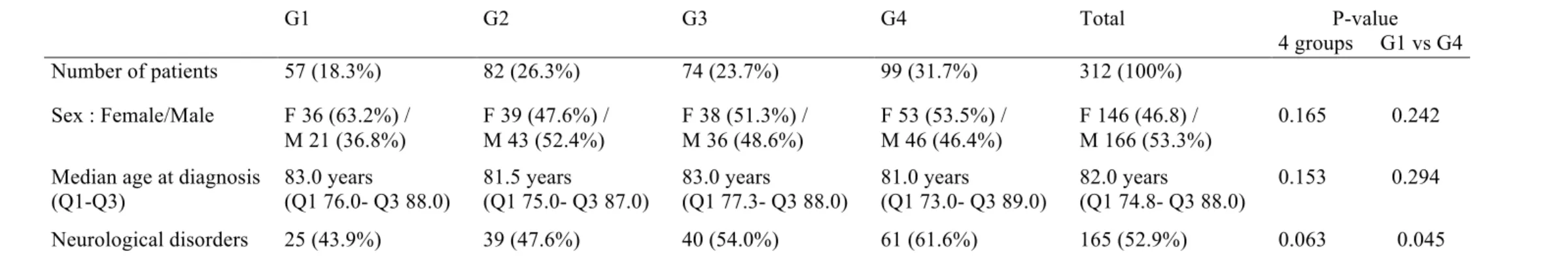
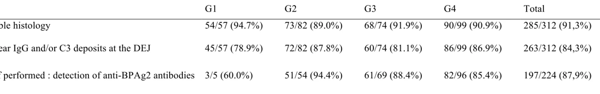
360 files were initially selected through PMSI database survey but only 312 patients (86.67%) were retained for final analysis. 48 patients were excluded: 17 for diagnostic errors (3 prurigo or isolated pruritus, 1 eczema, 3 mucous membrane pemphigoid, 1 cutaneous adverse drug reaction, 1 bullous impetigo, 2 epidermolysis bullosa acquisita, 2 pemphigoid gestationis, 1 linear IgA disease, 2 pemphigus, 1 eosinophilic cellulitis) and 31 for missing clinical details. 146 (46.8%) were female and 166 (53,3%) male patients. Age ranged from 49 to 106 years (median 82.0 years, Q1 74.8 – Q3 88.0 years). 165/313 (52.9%) patients had a medical history of neurological disease (degenerative, vascular or neuropsychiatric) (Table A). Histological and DIF findings were consistent with BP in 285 (91.3%) and 263 (84.3%) patients respectively but all patients had at least one consistent criteria. BP180 ELISA was performed in 224 patients and was positive for 197 (87.9%) patients (Table B).
Overall descriptive analysis of initial clinical pattern (Table C).
Topographic distribution of lesions
Initially non bullous, paucibullous, multibullous, or localized BP was diagnosed in 12 (3.8%), 137 (43.9%), 158 (50.6%) and 5 (1.6%) patients respectively.
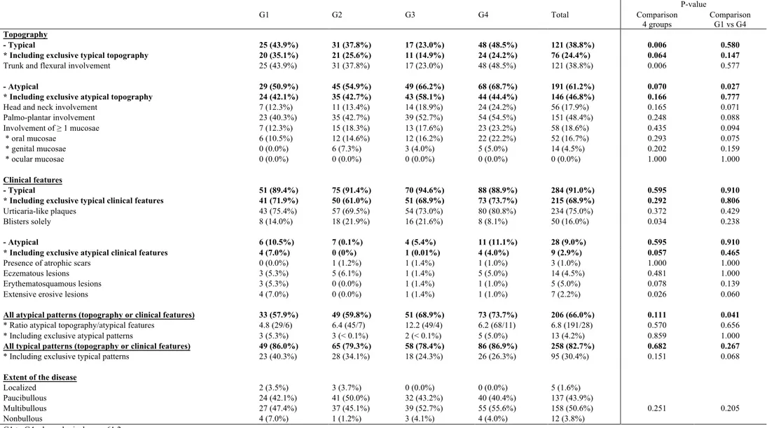
A typical topographic distribution of lesions (limbs and abdomen) was solely or mainly observed in a minority of patients (121; 38.8%) while head and neck, palmo-plantar regions and at least one mucosae were affected in 56 (17.9%), 151 (48.4%) and 58 (18.6%) patients respectively regardless of the relative importance of these latter lesions regarding overall location of skin and mucosae changes. Conversely, a predominant atypical distribution including head and neck, palmo-plantar and/or mucosal involvement was observed in 191 (61.2%) cases including 146 (46.8%) cases with exclusive atypical topography. As regards mucosal involvement, oral mucosae was mainly affected in 52 (16.67%) cases followed by genital mucosae in 14 (4.49%) cases including 6 cases with exclusive genital involvment; no ocular involvement was reported.
Clinical pattern of lesions
A typical clinical pattern of urticaria-like plaques and blisters was exclusively present in 234 (75%) cases. 50 (16.0%) patients presented with blisters with no urticaria-like plaques nor atypical clinical features. Conversely, at least one atypical/unusual clinical feature was observed in 28 (9.0%) patients overall including 9 (2.9%) patients where atypical features were solely present: atrophic scars in 3 (0.96%), eczematous lesions in 14 (4%), erythematosquamous lesions in 5 (2%) and extensive erosions in 7 (2%) patients respectively.
Overall, 206/312 (66.0%) patients displayed at least one atypical element, either lesional or topographic with a ratio atypical topography / atypical clinical features of 6.8 (191/28). 13 (4.2%) patients displayed a totally atypical form (both lesional and topographic) while 95 (30.4%) showed no atypical element whatsoever.
Presence/absence of formerly validated clinical criteria for BP diagnosis
(Table D)
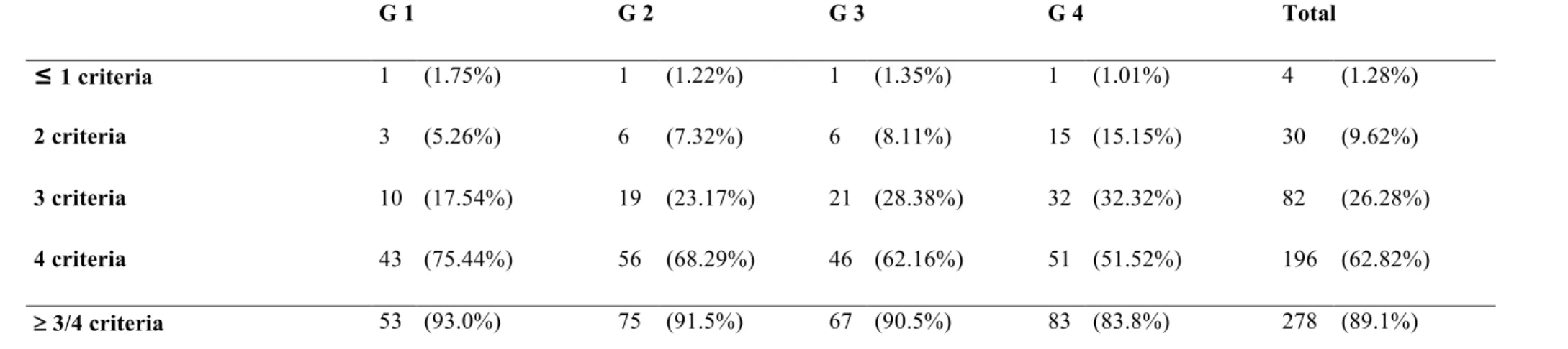
Overall, the 4 criteria were present at diagnostic in 196 (62.82%) patients while 3, 2, 1 or 0 criteria were present in 82 (26.28%), 30 (9.61%), and 4 (1.28%) patients respectively. Accordingly, at least 3 of 4 criteria were present in 278 (89.10%) patients. By order of frequency, the missing criteria were the absence of mucosal involvement (58 patients, 18.59%), of predominant bullous lesions on the neck and head (56 patients, 17.95%), an age greater than 70 years (41 patients, 13.14%) and the absence of atrophic scars (3 patients, 0.96%).
Comparative data analysis in chronological groups (Table C and D)
Proportion of woman and median age were similar across the 4 groups (G1 63%, G2 48%, G3 51%, G4 53%, p = 0.165; G1 83.0 years, G2 81.5 years, G3 83.0 years, G4 81.0 years, p = 0.153 respectively) and between G1 and G4 (P = 0?242 and 0.294 respectively). The association with neurological comorbidities, irrespective of their subtype, increased over time, but with no significant difference between the 4 groups (G1 43.8%, G2 47.6%, G3 54.0%, G4 61.6%, p = 0.063) nor between G1 and G4 (p = 0.045). The proportion of multibullous BP was similar in the 4 groups (G1 47.4%, G2 45.1%, G3
52.7%, G4 55.6%, p= 0.508) and between G1 and G4 (p= 0.324).
The percentage of patients displaying at least one atypical, either lesional or topographic clinical feature steadily increased over time in the 4 chronological groups: G1 57.9%, G2 59.8%, G3 68.9%, G4 73.7%, with no overall difference (p = 0.111) between the 4 groups but a significant difference (p = 0.041) was observed between G1 and G4. No difference was identified between the 4 groups regarding the proportion of exclusively atypical disease regarding both topographic and lesional pattern (G1 5.3%, G2 <0.1%, G3 <0.1%, G4 5.0%) (p 0.859) nor between G1 and G4 (p = 1.000). Regarding the proportion of exclusively typical diseases as to both topographic and lesional features, a steady but non significant decrease was identified over time when comparing the four chronological groups (G1 40.3%, G2 34.1%, G3 24.3%, G4 26.3%, p = 0.151) but a more marked trend (although still non significant) was observed when comparing G1 and G4 (p = 0.068).
The presence of a mere or predominant typical topographic pattern (mainly involving limbs and abdomen) was statistically different in the 4 groups (G1 to G4: 43.8%, 37.8%, 23.0%, 48.5% respectively; p= 0.006) but with no clear chronological shift toward progressive decrease over time as no significant difference between G1 and G4 (p= 0.577) was found. As to the initial presence of typical urticaria-like plaques, no significant difference was identified between the 4 groups (75.4%, 69.5%, 73.0%, 80.8% respectively; p= 0.372) nor between G1 and G4 (p 0.126).
Comparative analysis of data regarding patients with predominant atypical distribution of lesions revealed a trend to steady increase over time between the 4 groups (G1 50.9%, G2 54.9%, G3 66.2%, G4 68.7%) (p= 0.070) and a statistically significant difference between G1 and G4 (p= 0.027). Conversely, the specific analysis of patients with exclusive atypical topography failed to identify significant difference between the 4 groups (G1 42.1%, G2 42.7%, G3 58.1%, G4 44.4%) (p= 0.166) nor between G1 and G4 (p= 0.777).
The percentage of patients with at least one atypical lesional subtype was not different between the 4 groups (G1 10.5%, G2 0.1%, G3 5.4%, G4 11.1%) (p= 0.595) nor between G1 and G4 (p= 0.910). The restricted number of patients with exclusively
atypical lesions (9) precluded any significant statistical analysis between chronological groups.
The main atypical features identified from files’ review among predefined atypical elements (involvement of palmoplantar regions, of head and neck or of mucous membranes, extensive erosions, erythematosquamous or eczematous lesions, atrophic scars) were individually submitted to a specific time-related analysis and in most cases no significant difference was identified between the 4 chronological groups nor between G1 and G4 more particularly. However, a trend toward a significant difference was observed between G1 and G4 for oral lesions (10.5% vs 22.2%; p= 0.075) and for head and neck lesions (12.3% vs 24.2%; p= 0.071) with a steady increase in frequency over time for both features, highly consistent with overall clinical feeling. Additionally, a significant difference between the 4 groups (p= 0.026) and a trend to significant difference between G1 and G4 (p= 0.060) were observed for extensive erosive lesions but the limited size of the sample must be pointed out (G1 7.0%, G2 0%, G3 1.4%, G4 1.0%).
Finally, the percentage of patients fulfilling at least 3 of 4 clinical criteria of BP diagnosis was no statistically significant difference between the 4 groups (93% G1, 91.5% G2, 90.5% G3, 83.8% G4; p= 0.227) despite a steady decrease over time but a trend toward significance was observed between G1 and G4 (p = 0.067). Conversely, a significant decrease of the percentage of patients fulfilling the 4 clinical criteria was observed over time (75.4% G1, 68.3% G2, 62.2% G3, 51.5% G4 : p=0.016) and between G1 and G4 (p=0.003).
DISCUSSION
This retrospective analysis based on a large cohort of 312 cases of BP initially diagnosed over a long period of time (approximatively 2 decades) is the first study ever specifically designed to evaluate a possible shift over time of BP clinical profile and of the relevance of the previously established set of 4 clinical criteria for BP diagnosis, with a specific emphasis on the frequency and spectrum of atypical clinical features related to either topographic or lesional features.
This large-scale descriptive, monocenter series mainly shows a significant increase over time of the proportion of patients displaying at least 1 atypical feature, either topographic or lesional or both, with head and neck, palmoplantar and mucous membranes involvement being by far the more frequently observed atypical features compared to atypical clinical subsets of lesions. In parallel, the proportion of patients with an exclusively typical pattern regarding both clinical subtype and topography of lesions tended to decrease over time even though only a trend was observed. Likewise, the proportion of patients fulfilling at least 3 of the 4 previously validated diagnostic criteria steadily decreased over the past two decades but with no significant difference, meaning that these criteria are probably still robust. Conversely, the proportion of patients fulfilling the 4 criteria significantly decreased during the period of the study.
BP severity regarding disease extension was similar across the 4 chronological groups and the rare localized form cases were all pretibial. Baseline characteristics of patients reflected previous data regarding age, sex ratio and association to neurological disorders (1,12,13,20-22).
Only few past studies aimed to record atypical clinical features in BP, although based on a lower number of patients compared to our series and none of them were specifically designed to appraise a possible shift of clinical profile over time. Della Torre carried out a prospective nationwide cohort in Switzerland between 1 January 2001 and 31 December 2002 including 117 patients: 17.1% patients had only excoriations, eczematous and⁄or urticarial infiltrated lesions, head⁄neck as well as palmo-plantar involvement were found in up to 20% of patients, while mucosal lesions were present in 14.5% of the cases (16). Esmaili carried out a descriptive study in 2012 in Iran on 122
patients with documented bullous pemphigoid and found oral lesions in 27% cases, non-specific rash in 19.7%, eczema in 7.4%, genital lesions in 4.1%, face in 25.4%, scalp in 17.2%, palm and sole in 8.2% (20). Chang reported in 1996 in 86 cases of Taiwan 3 cases of dishydrosiform BP and mucous involvement in 12.8% cases (21). Di Zenzo reported 10 to 20% involvement of oral mucosae in a prospective study of 49 cases in 2008 (3). Eventually, Kridin included 82 patients with BP and diabetes in 2018 and revealed that mucosal involvement was present in 22.2% patients taking dipeptidyl peptidase 4 inhibitor (DPP4is) vs 6.5% in other patients (22).
Proportion of head and neck involvement was the same in our series as previous study. A bigger proportion of mucosal and palmoplantar involvement was observed in our series compared to previous study. However, the proportion of patients displaying an atypical pattern was smaller than in some previous other studies but such features are poorly described in literature. Literature data regarding the presence of clinical atypical features and our main results are summarized and compared in Table E.
In 1998, the the French Taskforce for Bullous disorders established a set of 4 clinical BP diagnosis criteria (age greater than 70 years, absence of atrophic scars, absence of mucosal involvement and absence of predominant bullous lesions on the neck and head) ; if 3 of these 4 characteristics were present, a diagnosis of BP could be made with a sensitivity of 90%, a specificity of 83% and a positive predictive value of 95% (19). Some years later, the validity of these clinical criteria were reassessed using immunoblot analysis of patient sera as the main diagnostic element, confirming a high degree of sensitivity (86%) and of specificity (90%) and an excellent prognostic positive value over 95% among patients with various subepidermal auto-immune bullous disorders if 3 of these 4 clinical criteria were fulfilled (18). If these results confirmed the interest of this set of clinical criteria for BP rapid diagnosis, no further study reappraised the validity of these criteria nor specifically investigated a possible shift of their relevance over time. In our study the overall percentage of patients fulfilling at least 3 criteria was still high (89.1%) favouring a persistent robustness of this set of criteria but a trend toward a steady decrease of this percentage over time must be pointed out especially when taking in account the two extreme chronological groups (93.0% vs 83.8% respectively; p = 0.067). The most frequent missing criteria were the absence of
mucosal involvement (missing in 18.6% of patients) and of predominant bullous lesions on the head and neck (missing in 17.9% of patients), results that match quite well the data regarding the steady increase over time of the frequency of these atypical features.
A number of hypothesis underlying this possible shift to a higher percentage of atypical features in BP over time may be raised. First, it cannot be ruled out that higher attention was recently paid to atypical features in PB with a more frequent recording of such unusual elements in clinical files as a consequence; in this setting, the observed time-related increase of atypical features might be at least partially artifactual. A second hypothesis could be related to etiological factors and more particularly to recently-introduced drugs that can act as antigenic haptens binding to and/or modifying basement membrane proteins (23–25). More specifically, dipeptidyl peptidase 4 inhibitors (DPP4is) currently largely used in diabetic patients has been recently repeatedly involved in BP occurrence and associated with a threefold increased risk for BP. Patients with DPP4is-induced BP display a higher mucosal involvement compared to non DPP4is-related BP wi (22,26–28). However, our study was not designed to evaluate the impact of medication on clinical presentation of BP. Further study are needed to more adequately investigate a possible relationship between medication uptake and the occurrence of atypical forms of BP.
CONCLUSION
A shift in the clinical spectrum of BP seems to have occurred during the past two decades with the emergence of more atypical forms mainly regarding the distribution of lesions. These changes in clinical pattern might at least partially be related to environmental causes, such as newly introduced drugs but further, large-scale prospective investigations are warranted to support this hypothesis.
Table A. Baseline characteristics of patients G1 G2 G3 G4 Total P-value 4 groups G1 vs G4 Number of patients 57 (18.3%) 82 (26.3%) 74 (23.7%) 99 (31.7%) 312 (100%) Sex : Female/Male F 36 (63.2%) / M 21 (36.8%) F 39 (47.6%) / M 43 (52.4%) F 38 (51.3%) / M 36 (48.6%) F 53 (53.5%) / M 46 (46.4%) F 146 (46.8) / M 166 (53.3%) 0.165 0.242
Median age at diagnosis (Q1-Q3) 83.0 years (Q1 76.0- Q3 88.0) 81.5 years (Q1 75.0- Q3 87.0) 83.0 years (Q1 77.3- Q3 88.0) 81.0 years (Q1 73.0- Q3 89.0) 82.0 years (Q1 74.8- Q3 88.0) 0.153 0.294 Neurological disorders 25 (43.9%) 39 (47.6%) 40 (54.0%) 61 (61.6%) 165 (52.9%) 0.063 0.045
Table B. Histological, direct immunofluorescence and ELISA findings
G1 G2 G3 G4 Total
Compatible histology 54/57 (94.7%) 73/82 (89.0%) 68/74 (91.9%) 90/99 (90.9%) 285/312 (91,3%) DIF : linear IgG and/or C3 deposits at the DEJ 45/57 (78.9%) 72/82 (87.8%) 60/74 (81.1%) 86/99 (86.9%) 263/312 (84,3%)
ELISA if performed : detection of anti-BPAg2 antibodies 3/5 (60.0%) 51/54 (94.4%) 61/69 (88.4%) 82/96 (85.4%) 197/224 (87,9%)
Table C. Clinical patterns of bullous pemphigoid P-value G1 G2 G3 G4 Total Comparison 4 groups Comparison G1 vs G4 Topography - Typical 25 (43.9%) 31 (37.8%) 17 (23.0%) 48 (48.5%) 121 (38.8%) 0.006 0.580 * Including exclusive typical topography 20 (35.1%) 21 (25.6%) 11 (14.9%) 24 (24.2%) 76 (24.4%) 0.064 0.147
Trunk and flexural involvement 25 (43.9%) 31 (37.8%) 17 (23.0%) 48 (48.5%) 121 (38.8%) 0.006 0.577
- Atypical 29 (50.9%) 45 (54.9%) 49 (66.2%) 68 (68.7%) 191 (61.2%) 0.070 0.027 * Including exclusive atypical topography 24 (42.1%) 35 (42.7%) 43 (58.1%) 44 (44.4%) 146 (46.8%) 0.166 0.777
Head and neck involvement 7 (12.3%) 11 (13.4%) 14 (18.9%) 24 (24.2%) 56 (17.9%) 0.165 0.071 Palmo-plantar involvement 23 (40.3%) 35 (42.7%) 39 (52.7%) 54 (54.5%) 151 (48.4%) 0.248 0.088 Involvement of ≥ 1 mucosae 7 (12.3%) 15 (18.3%) 13 (17.6%) 23 (23.2%) 58 (18.6%) 0.435 0.094 * oral mucosae 6 (10.5%) 12 (14.6%) 12 (16.2%) 22 (22.2%) 52 (16.7%) 0.293 0.075 * genital mucosae 0 (0.0%) 6 (7.3%) 3 (4.0%) 5 (5.0%) 14 (4.5%) 0.202 0.159 * ocular mucosae 0 (0.0%) 0 (0.0%) 0 (0.0%) 0 (0.0%) 0 (0.0%) 1.000 1.000 Clinical features - Typical 51 (89.4%) 75 (91.4%) 70 (94.6%) 88 (88.9%) 284 (91.0%) 0.595 0.910 * Including exclusive typical clinical features 41 (71.9%) 50 (61.0%) 51 (68.9%) 73 (73.7%) 215 (68.9%) 0.292 0.806
Urticaria-like plaques 43 (75.4%) 57 (69.5%) 54 (73.0%) 80 (80.8%) 234 (75.0%) 0.372 0.429 Blisters solely 8 (14.0%) 18 (21.9%) 16 (21.6%) 8 (8.1%) 50 (16.0%) 0.034 0.238
- Atypical 6 (10.5%) 7 (0.1%) 4 (5.4%) 11 (11.1%) 28 (9.0%) 0.595 0.910 * Including exclusive atypical clinical features 4 (7.0%) 0 (0%) 1 (0.01%) 4 (4.0%) 9 (2.9%) 0.057 0.465
Presence of atrophic scars 0 (0.0%) 1 (1.2%) 1 (1.4%) 1 (1.0%) 3 (1.0%) 1.000 1.000 Eczematous lesions 3 (5.3%) 5 (6.1%) 1 (1.4%) 5 (5.0%) 14 (4.5%) 0.481 1.000 Erythematosquamous lesions 3 (5.3%) 0 (0.0%) 1 (1.4%) 1 (1.0%) 5 (5.0%) 0.078 0.139 Extensive erosive lesions 4 (7.0%) 0 (0.0%) 1 (1.4%) 1 (1.0%) 7 (2.2%) 0.026 0.060
All atypical patterns (topography or clinical features) 33 (57.9%) 49 (59.8%) 51 (68.9%) 73 (73.7%) 206 (66.0%) 0.111 0.041
* Ratio atypical topography/atypical features 4.8 (29/6) 6.4 (45/7) 12.2 (49/4) 6.2 (68/11) 6.8 (191/28) 0.570 0.656 * Including exclusive atypical patterns 3 (5.3%) 3 (< 0.1%) 2 (< 0.1%) 5 (5.0%) 13 (4.2%) 0.859 1.000
All typical patterns (topography or clinical features) 49 (86.0%) 65 (79.3%) 58 (78.4%) 86 (86.9%) 258 (82.7%) 0.682 0.267
* Including exclusive typical patterns 23 (40.3%) 28 (34.1%) 18 (24.3%) 26 (26.3%) 95 (30.4%) 0.151 0.068
Extent of the disease
Localized 2 (3.5%) 3 (3.7%) 0 (0.0%) 0 (0.0%) 5 (1.6%) Paucibullous 24 (42.1%) 41 (50.0%) 32 (43.2%) 40 (40.4%) 137 (43.9%)
Multibullous 27 (47.4%) 37 (45.1%) 39 (52.7%) 55 (55.6%) 158 (50.6%) 0.251 0.205 Nonbullous 4 (7.0%) 1 (1.2%) 3 (4.1%) 4 (4.0%) 12 (3.8%)
Table D. Presence of clinical criteria of Vaillant and al. (absence of atrophic scars, absence of head and neck involvement, absence of mucosal involvement, and age greater than 70 years)
G 1 G 2 G 3 G 4 Total ≤ 1 criteria 1 (1.75%) 1 (1.22%) 1 (1.35%) 1 (1.01%) 4 (1.28%) 2 criteria 3 (5.26%) 6 (7.32%) 6 (8.11%) 15 (15.15%) 30 (9.62%) 3 criteria 10 (17.54%) 19 (23.17%) 21 (28.38%) 32 (32.32%) 82 (26.28%) 4 criteria 43 (75.44%) 56 (68.29%) 46 (62.16%) 51 (51.52%) 196 (62.82%) ³ 3/4 criteria 53 (93.0%) 75 (91.5%) 67 (90.5%) 83 (83.8%) 278 (89.1%)
P-value ³ 3/4 criteria : comparison /4 groups : p 0.227 ; comparison/G1 vs G4 : p 0.067 P-value 4/4 criteria : comparison/4 groups : p 0.016 ; comparison/G1 vs G4 : p 0.003
Table E. Atypical clinical features in BP patients as reported in the literature
Study Number of patients Atypical topography Atypical lesional features
Chang 1996 (28) 86 12.8% mucosae 3.5% eczematous lesions
Di Zenzo 2008 (3) 49 10 to 20% oral mucosae
Della Torre 2012 (16) 117 20% head and neck
17.1% exclusive atypical lesions (excoriations, eczematous lesions) 14.5% mucosae
Esmaili 2012 (28) 122 17.2% scalp 7.4% eczematous lesions
25.4% face 19.7% non specific rash
8.2% palm ans sole 27% oral mucosae 4.1% genital mucosae
Kridin 2018 (22) 82 22.2% mucosae with DPP4is, 6.5% mucosae without DPP4is
This study, 2018 312 61.2% atypical topography 9.0% atypical lesions
17.9% head and neck 1.0% atrophic scars 48.4% palm and sole 4.5% eczematous lesions
18.6% mucosae 5.0% erythematosquamous lesions
2.2% extensive erosive lesions 3 .7% non bullous form DPP4is, dipeptidyl peptidase 4 inhibitors