Bacillus licheniformis
BlaR1 L3 Loop Is a Zinc
Metalloprotease Activated by Self-Proteolysis
Ste´phanie Berzigotti., Kamal Benlafya., Je´re´my Se´pulchre, Ana Amoroso, Bernard Joris*Centre for Protein Engineering, Department of Life Sciences, University of Lie`ge, Lie`ge, Belgium
Abstract
In Bacillus licheniformis 749/I, BlaP b-lactamase is induced by the presence of a b-lactam antibiotic outside the cell. The first step in the induction mechanism is the detection of the antibiotic by the membrane-bound penicillin receptor BlaR1 that is composed of two functional domains: a carboxy-terminal domain exposed outside the cell, which acts as a penicillin sensor, and an amino-terminal domain anchored to the cytoplasmic membrane, which works as a transducer-transmitter. The acylation of BlaR1 sensor domain by the antibiotic generates an intramolecular signal that leads to the activation of the L3 cytoplasmic loop of the transmitter by a single-point cleavage. The exact mechanism of L3 activation and the nature of the secondary cytoplasmic signal launched by the activated transmitter remain unknown. However, these two events seem to be linked to the presence of a HEXXH zinc binding motif of neutral zinc metallopeptidases. By different experimental approaches, we demonstrated that the L3 loop binds zinc ion, belongs to Gluzincin metallopeptidase superfamily and is activated by self-proteolysis.
Citation: Berzigotti S, Benlafya K, Se´pulchre J, Amoroso A, Joris B (2012) Bacillus licheniformis BlaR1 L3 Loop Is a Zinc Metalloprotease Activated by Self-Proteolysis. PLoS ONE 7(5): e36400. doi:10.1371/journal.pone.0036400
Editor: Andreas Hofmann, Griffith University, Australia
Received March 23, 2011; Accepted April 4, 2012; Published May 18, 2012
Copyright: ! 2012 Berzigotti et al. This is an open-access article distributed under the terms of the Creative Commons Attribution License, which permits unrestricted use, distribution, and reproduction in any medium, provided the original author and source are credited.
Funding: This research was supported in part by Belgian Program on Interuniversity Poles of Attraction initiated by the Belgian State (Grant P6/19), the Actions de Recherche Concerte´es (Grant 03/08-297), the Fonds de la Recherche Fondamentale Collective (Grant 6 2.4511.06 and 2.4524.03), the Fonds de la recherche en Sciences Medicales (Grant 7 3.4586.05) and the Fonds pour la formation a` la Recherche dans l’Industrie et dans l’Agriculture (FRIA). Ste´phanie Berzigotti is a fellow of FRIA. The funders had no role in study design, data collection and analysis, decision to publish, or preparation of the manuscript.
Competing Interests: The authors have declared that no competing interests exist. * E-mail: [email protected]
. These authors contributed equally to this work.
Introduction
During evolution, the most common resistance mechanism acquired by eubacteria to resist b-lactam antibiotic action is the production of a b-lactamase that degrades these antibiotics by opening their b-lactam ring [1,2]. The cleaved antibiotic is then unable to acylate its membrane-bound D,D-peptidase targets, which are involved in the peptidoglycan cross-linking or remod-eling. These enzymes inhibited by b-lactam antibiotics are named penicillin-binding proteins (PBPs).
In Bacillus licheniformis and Staphylococcus aureus, the production of an inducible class A b-lactamase, BlaP and BlaZ respectively, is regulated by the BlaI repressor that maintains b-lactamase production at a low level in the absence of a b-lactam antibiotic outside the cell. In presence of a b-lactam antibiotic at sublethal concentration, the BlaI repressor is inactivated by a protein relay including the membrane-bound penicillin-sensory transducer BlaR1 and the BlaR2 protein, yet to be identified [3,4,5]. The regulatory genes, blaI and blaR1 are located downstream the blaP/ blaZ genes and are divergently transcribed as a polycistronic mRNA [6].
In S. aureus, another related mechanism that mediates resistance to b-lactam antibiotics has been described. It involves the induction of the low-affinity PBP2a or MecA capable to replace the inhibited constitutive PBPs [7]. The mecA expression is under the control of MecI and MecR proteins that are homologous to S. aureus and B. licheniformis BlaI and BlaR1, and play the same
role. Furthermore, MecI and BlaI from S. aureus are interchange-able, but BlaR1 and MecR are not [8,9].
The BlaR1 and MecR membrane-bound penicillin-sensory transducers are composed of two domains, an amino-terminal domain (BlaR/MecR-NTD) anchored into the cytoplasmic membrane and a carboxy-terminal domain (BlaR/MecR-CTD) exposed outside the cell. This latter domain contains in its primary structure, the conserved motifs of the serine-penicillin recognizing enzymes (SPRE) and acts as a penicillin sensor [10,11]. The BlaR-CTD from B. licheniformis and from S. aureus can be produced as soluble domains and their crystal structures exhibit the conserved SPRE 3D-fold [12,13,14]. The membrane topology of B. licheniformis BlaR-NTD (Met1
-Pro339) has been experimentally determined: it contains four transmembrane segments (TM1, TM2, TM3, TM4) connected by three loops named L1 (Lys27 -Thr35), L2 (Pro53-Ser115) and L3 (Tyr134-Arg322). L1 and L3 are cytoplasmic, whereas L2 is exposed outside the cell (Figure 1) [15]. Although very little is known concerning the mechanism of signal transduction by BlaR1/MecR, the first step is the acylation of BlaR/MecR-CTD by the b-lactam antibiotic outside the cell. The acylation of this domain by the antibiotic causes a modification of the interaction between BlaR-CTD and L2 and gives rise to the activation of the L3 cytoplasmic loop via a conformation change of the four transmembrane segments. The activated L3 would launch a cytoplasmic signal whose target is BlaI/MecI [10,15,16,17,18]. In this signaling pathway, L2 and the four transmembrane segments would act as a signal transducer and L3 as a cytoplasmic signal amplifier. In S. aureus, the BlaR1 L3 activation results in its
superfamily) such as thermolysin (Figure 1) [23]. In S. aureus BlaR1, the mutation of the conserved glutamic acid residue to alanine gives rise to a non-inducible b-lactamase phenotype [19]. In this paper, we performed site-directed mutagenesis experi-ments combined with zinc-blot, Western blot and b-lactamase induction analyses to demonstrate that the cytoplasmic BlaR1 L3 loop belongs to Gluzincin metallopeptidase superfamily and that its cleavage during BlaR1 activation occurs by self-proteolysis.
Results
L3 Conserved Residues
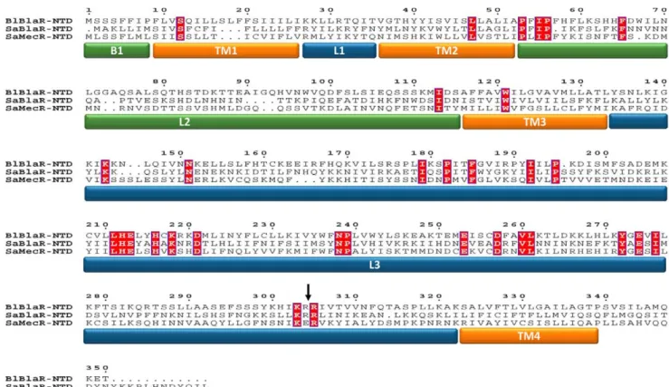
A multiple sequence alignment of BlaR1/MecR N-terminal domains of B. licheniformis and S. aureus (Figure 2) revealed 32 strictly conserved residues in the three aligned sequences corresponding to 9.4% of identity when the length of B. licheniformis BlaR-NTD sequence is used as reference ([34/339]6100 = 9.4%). Out of these 32 conserved residues, 24 are located within the L3 loops ([24/187]6100 = 12.8%) highlighting the importance of this
loop. The best candidate for this role is a conserved tyrosine residue (Y272in B. licheniformis BlaR-NTD).
Conserved L3 Residues Important for Signal Transduction or Zn Binding
To highlight if the conserved E, D, H and Y residues are important for the zinc binding or for the catalytic activity in the activated L3 loop, the mutants listed in Table 1 were constructed. The resulting plasmids (pDML1268-78, Table 2) were introduced into B. subtilis 168 and the recombinant resulting strains tested for their ability to induce BlaP b-lactamase in presence of the inducer. In all cases, the mutated receptor expression in non-induced or induced membranes has been detected by Western blotting analysis (Figure 3) showing that the introduced mutations do not lead to the production of an unstable, proteolysis susceptible receptor.
In order to determine the importance of the HEXXH motif in L3 activity, alanine scanning mutagenesis was used to generate the following mutants: H212A/E213A/H216A, E213A, H212A/H216A
Figure 1. Membrane topology of the penicillin-sensory transducer, BlaR1. This receptor contains two domains, an extracellular domain: BlaR-CTD and a transmembrane domain: BlaR-NTD. BlaR-CTD exhibits the three motifs of the penicillin binding protein family (S*402TYK, Y476GN,
K539TG, where S402is the active serine). BlaR-NTD includes four transmembrane segments (TM1, TM2, TM3, TM4) connected by three loops (L1, L2, L3). The cytoplasmic L3 loop contains the H212EXXH motif, characteristic to zinc-metalloproteases.
and H216A. Analyses of these mutants indicate that all of these substitutions give rise to a non-inducible phenotype (Table 1) with a b-lactamase production similar to that obtained for a non-induced wild type strain (data not shown).
Neutral Zinc proteases catalyse cleavage of peptide bonds via a general-base type mechanism in which the glutamate residue of the conserved motif acts as catalytic base. The mutation of this glutamate residue to aspartate (E213D) or to glutamine
Figure 2. Sequence alignment of the amino-terminal domain of the proteinsB. licheniformis749/I BlaR1 (BlBlaR-NTD) andS. aureus
RN4220 BlaR1 (SaBlaR-NTD) andS. aureusMRSA252 MecR (SaMecR-NTD). The conserved residues are highlighted in red and the loop and transmembrane segments are marked: the transmembrane segments (TM1 to 4) in orange; the extracellular loops B1 (M1-P8) and L2 (P53-S115) in
green and the cytoplasmic loops L1 (K27-T35) and L3 (Y134-K322) in blue. The site of cleavage, situated between the R293and R294in S. aureus BlaR1, is
indicated by an arrow.
doi:10.1371/journal.pone.0036400.g002
Table 1. Induction of BlaPb-lactamase of the BlaR1 L3 loop mutants.
Mutation Plasmids Induction factor* (3h)
Wild type pDML995 37.861.5
Mutants in H212EXXH motif H212A/E213A/H216A pDML1268 1.660.1
E213A pDML1269 1.260.1
H212A/H216A pDML1270 1.260.1
H216A pDML1271 1.2760.01
E213D pDML1279 1.0760.02
E213Q pDML1280 1.660.2
Mutants in putative Zn++ligands D221A pDML1277 1.260.1
E253A pDML1273 1.260.3
D257A pDML1274 1.960.1
E274A pDML1275 33.960.9
Mutants in putative general base Y272A pDML1278 1.160.1
Mutants in R304R cleavage site R304A/R305A pDML3045 7.661.7
*The induction factor corresponds to the ratio between the b-lactamase quantity/A600for induced culture and the b-lactamase quantity/A600for uninduced culture. doi:10.1371/journal.pone.0036400.t001
(E213Q) prevents b-lactamase induction (Table 1, Figure 3). These results demonstrate that all the residues of this conserved motif play an essential role in the induction process.
The sequence alignment of the protein BlaR1 from B. licheniformis with the proteins BlaR1 and MecR from S. aureus, highlights four conserved residues that could act as the third
pDML1251 pUCBM20 derivative carrying the SstI/EcoRIblaR1 fragment This study
pDML1255 pDML1251 derivative carrying BlaR1H212A/E213A/H216A mutation This study
pDML1256 pDML1251 derivative carrying BlaR1 E213A mutation This study
pDML1257 pDML1251 derivative carrying BlaR1 H212A/H216A mutation This study
pDML1258 pDML1251 derivative carrying BlaR1 H216A mutation This study
pDML1260 pDML1251 derivative carrying BlaR1 E253A mutation This study
pDML1261 pDML1251 derivative carrying BlaR1 D257A mutation This study
pDML1262 pDML1251 derivative carrying BlaR1 E274A mutation This study
pDML1264 pDML1251 derivative carrying BlaR1 D221A mutation This study
pDML1265 pDML1251 derivative carrying BlaR1 Y272A mutation This study
pDML1266 pDML1251 derivative carrying BlaR1 E213D mutation This study
pDML1267 pDML1251 derivative carrying BlaR1 E213Q mutation This study
pDML1268 pDML995 derivative carrying BlaR1 H212A/E213A/H216A mutation This study
pDML1269 pDML995 derivative carrying BlaR1 E213A mutation This study
pDML1270 pDML995 derivative carrying BlaR1 H212A/H216A mutation This study
pDML1271 pDML995 derivative carrying BlaR1 H216A mutation This study
pDML1273 pDML995 derivative carrying BlaR1 E253A mutation This study
pDML1274 pDML995 derivative carrying BlaR1 D257Amutation This study
pDML1275 pDML995 derivative carrying BlaR1 E274A mutation This study
pDML1277 pDML995 derivative carrying BlaR1 D221A mutation This study
pDML1278 pDML995 derivative carrying BlaR1 Y272A mutation This study
pDML1279 pDML995 derivative carrying BlaR1 E213D mutation This study
pDML1280 pDML995 derivative carrying BlaR1 E213Q mutation This study
pDML3045 pDML995 derivative carrying BlaR1 R304A/R305A mutation This study
pDML1283 pCR-ScriptTMSK(+) derivative carrying BlaR1 L3 loop coding sequence This study
pDML1284 pDML1283 derivative carrying BlaR1 L3 E213A mutation This study
pDML1285 pDML1283 derivative carrying BlaR1 L3 H212A/H216A mutation This study
pDML1286 pDML1283 derivative carrying BlaR1 L3 D257A mutation This study
pDML1287 pDML1283 derivative carrying BlaR1 L3 D221A mutation This study
pDML1293 pDML1283 derivative carrying BlaR1 L3 E253A mutation This study
pDML1288 pLEX derivative allowing the expression of BlaR1 L3 loop This study
pDML1289 pLEX derivative allowing the expression of BlaR1 L3 carrying E213A mutation This study pDML1290 pLEX derivative allowing the expression of BlaR1 L3 carrying H212A/H216A mutation This study pDML1291 pLEX derivative allowing the expression of BlaR1 L3 carrying D257A mutation This study pDML1292 pLEX derivative allowing the expression of BlaR1 L3 carrying D221A mutation This study pDML1294 pLEX derivative allowing the expression of BlaR1 L3 carrying E253A mutation This study Apr: ampicillin resistance.
Cmr: chloramphenicol resistance. doi:10.1371/journal.pone.0036400.t002
zinc ligand (D221, E253, D257and E274; see above and Figure 2). Site-directed mutagenesis of these residues in alanine has been done and the resulting plasmids carrying these mutations (pDML1273-74-75-77) were introduced in B. subtilis 168 to probe the b-lactamase induction (Table 2). The D221, E253and
D257A mutants show a non-inducible phenotype while the
E274A mutant retains an inducible phenotype similar to the wild type receptor (Table 1, Figure 3). This result indicates that E274 cannot be the third zinc ligand. The three other conserved residues could play an important role either in the enzymatic activity, in the Zinc coordination and/or in the folding of BlaR1 L3 loop. Finally, the mutation of the conserved tyrosine residue in alanine (Y272A) also leads to the loss of the b-lactamase induction (Table 1, Figure 3) suggesting that this residue could be part of the relay system involved in the acid-base catalysis.
L3 Overexpression and Zinc-binding Analysis
The DNA sequence coding for the B. licheniformis L3 loop was introduced into the pLex vector in which l3 was under the control of the inducible PL promoter from lambda phage. The resulting plasmid, pDML1288 (Table 2) was introduced in E. coli GI724. In the induced recombinant strain, the L3 loop was overproduced as inclusions bodies. After partial purification and SDS solubilization of inclusion bodies, proteins were separated by SDS-PAGE (Figure 4 A) and transferred onto a nitrocellu-lose membrane. The blotted proteins were refolded in presence of radioactive zinc chloride solution and a specific radioactive band of 25 kDa was highlighted by autoradiography (Figure 4 B, lane 2). In the utilized technique, it is well accepted that, after protein refolding, a fraction of enzyme macromolecules are correctly folded and active. This band is compatible with the expected molecular mass of L3 (22.116 kDa). The recombinant strains harboring plasmids with mutated l3 gene encoding for H212A/H216A, E213A, D221A, E253A or D257A (Table 2) has been also analyzed by Zinc blot analysis after solubilization of the inclusion bodies (Figure 4 B). As shown in the Figure 4 B, the H212A/H216A, E253A and D257A have lost their ability to bind Zn++ whereas E213A and D221A could be detected as zinc-binding proteins, highlighting the role of H212, H216, E253A and D257 residues in Zn binding.
B. licheniformis L3 is Activated by Autocleavage
To avoid inclusion bodies and/or misfolded proteins due to overexpression of recombinant membrane protein, the fate of BlaR1 in presence or not of b-lactam (respectively induced or uninduced) was carried out by Western blot analysis of B. subtilis-pDML995 membrane fractions with purified anti-BlaR-CTD antibodies. These experiments revealed the presence of two induced bands: a major band of 33 kDa and a minor one of 68 kDa (Figure 5). The band of low molecular mass is compatible with the cleaved and activated BlaR1 C-terminal domain whereas the other of high molecular mass should be the full-length receptor. These results are in agreement with a cleavage of B. licheniformis BlaR1 between the R304 and R305residues as in the case of S. aureus BlaR1 cleavage between R294and R295(Figure 2, [19]). To confirm these results in B. licheniformis BlaR1, the putative residues of the cleavage site has been mutated to R304A/R305A and the modified receptor was assayed for its ability to induce BlaP production. In presence of b-lactam antibiotic, R304A/R305A mutants lightly induces BlaP b-lactamase (IF = 7.6, Table 1 and Figure 5). Furthermore, Western blot analysis of membrane fractions from non- and induced cells with purified anti-BlaR-CTD antibodies did not show any L3 cleavage (Figure 6). To explore if the cleavage of BlaR1 observed in induced B. subtilis/ pDML995 could be the result of L3 proteolytic activity, we analyzed the fate of BlaR1 HA213XXH in induced B. subtilis/ pDML1269 membrane fractions by Western blotting as described above. In this mutant, the catalytic activity of L3 loop is abolished due to the mutation of the conserved glutamic residue of the HEXXH motif involved in the neutral Zinc metallopeptidase family acid-base relay. As shown in Figure 6, no cleavage of BlaR1
HA213XXH could be detected in presence of the inducer
suggesting that the receptor should be activated via self-proteolysis.
Discussion
Structural studies on thermolysin and homologous enzymes (Thermolysin-like proteinases, TLPs) have revealed two short conserved motifs in their active sites: HEXXH and EXXXD [25]. The two histidine residues of the HEXXH sequence serve as ligands to zinc, whereas the glutamic acid is involved in the catalytic mechanism by transferring hydrogen atoms and polar-izing a zinc-bound water molecule for nucleophilic attack on the
Figure 3. Western blot analysis of membrane-bound BlaR1 produced by B. subtilisstrains carrying plasmids harboring blaR1 mutants. Membrane proteins from induced (+) or uninduced (-) cultures were separatedon on SDS-PAGE. Purified BlaR-CTD antibodies were used for the Western blotting. Pre-stained protein molecular weight markers were used (M). The « » pinpoints an intense and non-specific band (for details see Figure 6). BlaR1 and BlaR1* point out, respectively, the full size and the activated B. licheniformis BlaR1 receptor. Except for mutant E274A, all other mutants exhibit non-inducible b-lactamase phenotype. The E274A mutant has the same profile as the wild-type (to compare see Figure 6). For details
see Tables 1 and 2 and Experimental procedures. doi:10.1371/journal.pone.0036400.g003
Figure 4. Coomassie Blue-stained SDS-PAGE of partially purified inclusion bodies of wild-type and L3 loop mutants (A) and Zinc blot analysis (B). In A and B: 1: bovine carbonic anhydrase (AC) (,30 kDa; 5 mg), was used as positive control and 2 to 6 are, respectively, wild type (WT), E213A, H212A/H216A, D257A, D221A and E253A L3 loop mutants. For each gel 40mg of inclusion bodies were loaded. The fluorographies were
exposed at -70uC for 72 hours. M: molecular weight marker. The arrow indicates L3 loops. doi:10.1371/journal.pone.0036400.g004
Figure 5. Induction of b-lactamase synthesis in presence (induced) or not (non-induced) of inducer (2.5mg ml21cephalosporin C)
for theB. subtilisstrains transformed with pDML995 (wild-type divergeon;
N
) or pDML3045 (mutation R304A/R305A;#). The level ofthe induction is expressed as an induction factor (IF, the ratio between the b-lactamase quantity/A600for induced culture and the b-lactamase
quantity/A600for uninduced culture).
scissile peptide bond of the bound substrate. The third zinc ligand is the glutamic acid residue of the sequence motif EXXXD located 20–60 residues downstream of the second histidine of the first motif. For this reason, members of thermolysin enzyme family are named gluzincins [26,27]. The second histidine residue of the HEXXH motif was found to bridge both the zinc ion and the carboxylate side chain of the aspartic acid residue within the second conserved motif. The exact role of this conserved triad, Zn-His-Asp, is not well understood but is crucial for the catalytic activity [26,27]. In the absence of a substrate, a water molecule acts as an additional fourth ligand to zinc and is clamped between the catalytic glutamic acid residue of the HEXXH motif and the metal. During TLP catalysis, the catalytic glutamic acid residue accepts a proton from the zinc-bound water nucleophile and transfers it to the leaving group of the substrate. Further stabilization of the transition state is provided by the side-chain of a conserved histidine residue (H231in thermolysin) [28].
Site-directed mutagenesis of selected conserved residues in BlaR1/ MecR L3 loops postulated to be involved in catalysis or zinc chelation, combined with zinc-blot experiments and b-lactamase induction assay have confirmed all the features present in the active sites of TLPs. We show that the two histidine residues of the motif H212EXXH (B. licheniformis BlaR1 numbering) effectively coordinate a Zinc ion and are essential for cytoplasmic signalling. The observed non-inducible b-lactamase phenotype, the conservation of the Zinc binding capacity and the loss of self-proteolysis exhibited by
H(E213A)XXH mutant are in agreement with a glutamic acid
residue acting as catalytic residue and a peptidase activity of L3 loop. The E253A and D257A mutants, which exhibited the same non-inducible phenotype (Table 1), showed that these residues are crucial for the L3 enzymatic activity and that they are good candidates to be included in the second conserved motif of TLPs. In BlaR1/MecR L3 loops, the EXXXD motif is located 37 residues downstream HEXXH motif, a distance similar to that observed in TLPs, which varies from 20 residues in thermolysin to 59 in mammalian neutral endopeptidase. These new observations allow us to classify BlaR1/ MecR L3 loops as new members of TLPs or gluzincins. According to the properties of this enzyme family, the glutamic residue of the EXXXD motif is the third zinc ligand and the aspartic acid residue forms a carboxylate-histidine-zinc interaction crucial for TLP activity (see above). Indeed, as shown by 3D structures from several different enzymes included in the TLP family, this hydrogen bonding
stabilizes the coordination of the zinc ion by one of the histidine residues of the HEXXH sequence [29]. In rat dipepeptidyl peptidase, the mutation of the EXXXD motif to EXXXA gives rise to the loss of the zinc ion and concomitantly the loss of enzyme activity [26] as it is the case for L3 EXXXA257mutant (figure 4 and Table 1). By analogy, we postulate that the D257residue of L3 loop is involved in zinc binding by orienting the imidazole ring of the H212 residue and/or by enhancing the basicity of the histidine-zinc ligand. To complete the zinc-coordination polyhedron, a water molecule, in interaction with the glutamic acid residue of the HEXXH motif, should be the fourth zinc ligand.
The conserved residues, D221 and Y272 (B. licheniformisBlaR1 numbering) are crucial for the penicillin receptor signalling. The most likely role for Y272 is to be involved in the stabilization of the tetrahedral intermediate complex during the catalysis of the activated BlaR1/MecR L3 loop. Indeed, carboxypeptidase A, which shares a common topology with thermolysin for its active site, possesses anY248positioned at the same spatial position of H231in thermolysin [30]. As D221 side chain is negatively charged, it is difficult to postulate that this side chain could directly interact with the negatively charged tetrahedral intermediate. Another hypoth-esis is that this conserved aspartic acid could be essential for substrate binding. As our results are consistent with a self-activation and as the cleavage site includes at least the positively charged side chain of one arginine residue, this hypothesis is plausible.
Similar to many other proteolytic enzymes, TLPs are synthe-sized as precursors with a propeptide. Up to 70% of known TLPs possess a propeptide containing two distinguished domains: the first one is an intramolecular chaperone (FTP domain) essential for the folding of the catalytic domain whereas the second one (PepSY domain) acts as an inhibitor to prevent detrimental TLP activity [31]. The cleavage of the propeptide occurs through autoproces-sing and is mediated by the propeptide [26]. In some TLPs, C-terminal extensions, fused to the Zinc catalytic domain, mediate their binding to insoluble substrates. These C-terminal extensions can be autoprocessed and their cleavage modulates the enzyme specificity and activity [32].
BlaR1/MecR L3 loop activation results in a cascade of events originated by the acylation of the C-teminal sensor by the penicillin and finishing by selfproteolysis of the TLP receptor domain. It has been postulated that the cleavage of the L3 domain is triggered by a conformational change of the transmembrane
Figure 6. Western blot analysis of membranes of B. subtilistransformed with pMK4 (negative control), pDML995 (wild type), pDML1269 (E213A mutant) or pDML3045 (R304A/R305A mutant). Membrane proteins from induced (+) or uninduced (2) cultures were separatedon on SDS-PAGE. Purified BlaR-CTD antibodies were used for the Western blotting. Pre-stained protein molecular weight markers were used (M). The « » indicates an intense and non-specific band. BlaR1 and BlaR1* highlight, respectively, the full size and the activated B. licheniformis BlaR1 receptor.
presence of b-lactam antibiotic, the acylation of the mutated BlaR1 receptor results in a slight induction of BlaP b-lactamase production (about 20% of the wild type) but without L3 cleavage. In the uncleaved protein, the C-terminal portion of the L3 loop acts as an inhibitor of its TLP domain. Cleavage of the KRQR (both BlaR1 proteins) or KEQR (S. aureusMecR) results in an activation of the peptidase activity, but it is not known whether the C-terminal peptide remains associated or not with the active domain. In fact, in this mutant, we have decoupled the L3 activation due to BlaR-CTD acylation from that caused by L3 selfproteolysis. The BlaP induction results indicate that in this case, the uncleaved L3 loop exhibits a residual activity which seems unable to perform a successful autocleavage but is able to produce a sufficient amount of coactivator to partially inactivate BlaI. This finding is in agreement with a selfproteolysis mechanism in which it is necessary that BlaR-CTD acylation slightly activates L3 loop to allow its own proteolysis and its full activation.
In conclusion BlaR1/MecR L3 loop is a gluzincin and a new member of the TLP superfamily that has diverged very early from canonical TLPs, since no significant identity can be detected between BlaR1/MecR L3 loops and TLPs by using comparison algorithms such as BLAST. The L3 signalling activity of BlaR1/ MecR receptor would be repressed by the combination of two intramolecular interactions: one between L3 and its C-terminal propeptide and the second between L2 and BlaR-CTD. Similarly the full L3 activation triggered BlaR-CTD acylation would be the consequence of two successive events described above.
Materials and Methods
Bacterial Strains, Plasmids and DNA Manipulations
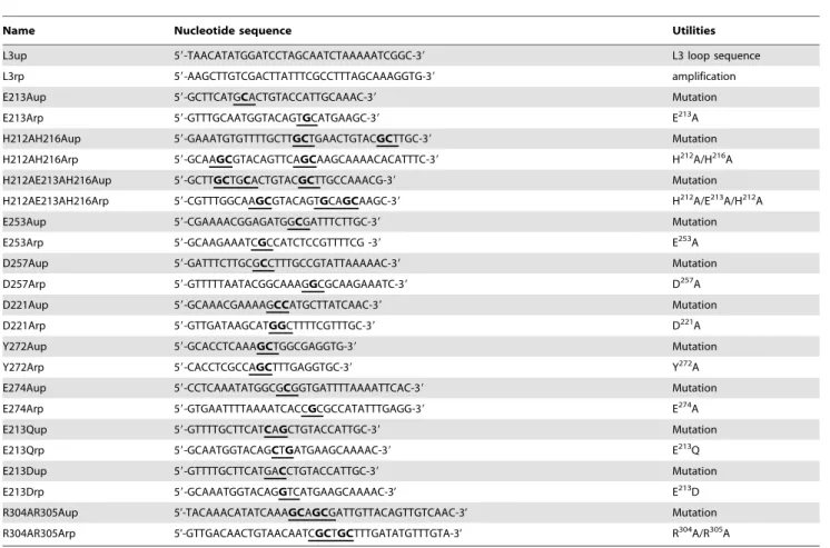
The strains and plasmids used in this study are described in Table 2. To introduce point mutations into BlaR1 or L3 loop, mutagenesis was carried out using the Quick Change Site-Directed mutagenesis procedure (Stratagene). For all mutations, except BlaR1 R304A/R305A mutation, the plasmids pDML1251 and 1283 were used as the templates in polymerase chain reactions (PCR) for blaR1 and l3 loop site-directed mutagenesis, respectively.
For BlaR1 R304A/R305A mutation, pDML995 was used as
template. Synthetic oligonucleotides 23–35 bases long containing a mutated codon in the middle of their sequences (Table 3) were employed to mutagenize the original codons. Following the verification of mutations by DNA sequencing (GIGA-DNA sequencing plateform, University of Lie`ge), the corresponding restricted fragments (blaR1: 672 bp SstI-EcoRI; l3 loop: 567 bp NdeI-SalI) were cloned into pDML995 and pLex, respectively.
All routine DNA isolation and manipulation were performed as described by Sambrook et al. [33] or following instructions of the manufacturer. Restriction enzymes and mini-prep kit to prepare small-scale plasmid DNA were purchased from Promega and Fermentas respectively.
derivatives carrying respectively the mutation E A, H H/
H216H, E253A, D257A or D221A (Table 2). These mutations were introduced as described above.
The E. coli GI724/pDML1288-94 strains were grown over
night at 30uC in RM medium (40 mM Na2HPO4, 20 mM
KH2PO4,10 mM NaCl, 200 mM NH4Cl, 2% casamino acids,
1% glycerol, 1 mM MgCl2, 100mg ml21ampicillin). The culture
was then diluted 100 times in IM medium (40 mM Na2HPO4,
20 mM KH2PO4, 10 mMNaCl, 200 mM NH4Cl, 2% casamino
acids, 0.5% glucose, 1 mM MgCl2, 100mg ml21ampicilline) and cultivated at 30uC. When the absorbance at 550 nm reached 0.6, L3 loop expression was induced by both the addition of tryptophan (final concentration: 100mg ml21) and a temperature shift to 37uC. After 3 hours of induction, the cells were harvested by centrifugation, washed with 50 mM HEPES-NaOH (pH 7.5) and suspended in 50 mM Tris-HCl (pH 7.5). Bacterial cells were disrupted using a Basic Z Model disintegrator (Warwick, UK). After the addition of Benzonase (final concentration: 20mg ml21) and an incubation of 30 min at 4uC, the insoluble cell fraction containing inclusion bodies, was separated from the soluble fraction by centrifugation at 18000 g for 30 min at 4uC. The insoluble fraction was suspended in 50 mM HEPES-NaOH (pH 7.5), 0.5 mM NaCl, 5 mM DTT. After addition of Triton X100 (final concentration: 0.1%), the suspension was incubated under agitation at room temperature for 1 hour. The inclusion bodies containing the L3 loop were recovered by centrifugation at 18000 g, for 30 min and at 4uC.
Zinc Binding Assay
Soluble or insoluble Zinc binding proteins were detected by Zinc blot assay [34,35,36]. Purified inclusion bodies were solubilized by sodium dodecyl sulfate (SDS, solubilization buffer: Tris-HCl 0.5M pH 6.8) and separated by SDS-PAGE. After electrophoresis, the gel was soaked for one hour at 37uC in 25 mM Tris-HCl (pH 8.3), 190 mM glycine, 0.1% SDS, 5% 2-b-Mercaptoe´thanol. The proteins were electrophoretically trans-ferred to a nitrocellulose membrane in 25 mM Tris-HCl (pH 8.3), 190 mM glycine, 0.1% SDS, 20% methanol.
The nitrocellulose membrane was first incubated for 1 hour at room temperature in 10 mM Tris-HCl (pH 7.5) to partially renature blotted proteins. The membrane was then soaked two times, 15 min per soaking, in 50 ml of binding buffer (10 mM Tris-HCl (pH 7.5), 0.1 M KCl) containing 1mCi 65ZnCl and washed 30 minutes with two changes of binding buffer. The blot was dried one hour at room temperature and analyzed by
autoradiography using X-OMATTMfilm (Eastman Kodak
Com-pany) and an exposure time of 72 hours at 270uC.
b-lactamase Induction Assay
Bacillus subtilis 168 transformed by pDML995 or derivative plasmids was grown at 37uC in Luria-Bertani (LB) medium
containing 7mg ml21of chloramphenicol until A600reached 0.2. Then, the culture is divided in two portions, one as a control (non induced cells) and the other for the addition of 5mg ml21 cephalosporin C (induced cells). In these conditions the concen-tration of inducer is below its minimal inhibitory concenconcen-tration: 10 and 20mg ml21, respectively, for B. subtilis recombinant strains exhibiting non- and inducible b-lactamase phenotype (Ana Amoroso, unpublished data).
The incubation was continued and samples were withdrawn at hourly intervals for 6 hours. For each sample, the A600 was measured to monitor the cell density as well as the b-lactamase activity. This latter activity was spectrophotometrically determined by using nitrocefin as previously describred [17]. The b-lactamase quantity by cell [Et] was calculated by using the following equation: v0/(cellular density) = DA
482
/(Dt. e. A600) = (kcat6 [Et]6 [S])/ (Km+ [S]) were v0: the initial velocity; DA482and Dt are absorbance and time variations, respectively; kcat: catalytic constant (470 s21) [37]; [S]: substrate concentration (100mM); Km: 40mM [37] and e:
nitrocefin coefficient of molar extinction at 482 nm
(15000 M21cm21). The b-lactamase induction level was expressed as ‘‘induction factor’’ (IF), determined as: FI = ([Et] of induced culture)/([Et] of uninduced culture).
Anti-BlaR-CTD Antibodies Production and Western Blotting
A polyclonal antiserum directed against the BlaR1 sensor (BlaR-CTD), was generated by immunizing New Zealand rabbits with purified BlaR-CTD [17]. Polyclonal antibodies were purified
against BlaR-CTD immobilized on Sepharose 4B as described in reference [38]. The purified primary antibodies were used in immunoblotting at a final dilution of 1:1000.
Ten milliliters of induced or uninduced recombinant B. subtilis strains were harvested by centrifugation and resuspended in 200ml of 50 mM Tris-HCl (pH 7.5). Cells were disrupted by sonication in a Branson ultrasonic disintegrator at amplitude of 6mm for tree 30 s bursts. Membranes fraction, containing the BlaR1 receptor were obtained as insoluble fraction by centrifugation of the lysates. Membrane proteins were solubilized by SDS (solubilization buffer: Tris-HCl 0.5 M pH 6.8) and separated by SDS-PAGE (15% acrylamide) and transferred onto a nitrocellulose membrane (Millipore). Incubation of the membrane with primary antibodies was followed by incubation with alkaline phosphatase-conjugated anti-rabbit antibodies. The immune complexes were detected by a color reaction with nitroblue tetrazolium and 5-bromo-4-chloro-3-indoyl phosphate, as recommended by the manufacturer (Roche Applied Science).
Acknowledgments
We warmly thank Dr Jean-Marie Fre`re for fruitful discussions. Author Contributions
Conceived and designed the experiments: SB KB AA BJ. Performed the experiments: SB KB JS. Analyzed the data: SB KB JS AA BJ. Contributed reagents/materials/analysis tools: SB KB AA BJ. Wrote the paper: SB AA BJ.
Table 3. Oligonucleotides used in this study.
Name Nucleotide sequence Utilities
L3up 59-TAACATATGGATCCTAGCAATCTAAAAATCGGC-39 L3 loop sequence
L3rp 59-AAGCTTGTCGACTTATTTCGCCTTTAGCAAAGGTG-39 amplification
E213Aup 59-GCTTCATGCACTGTACCATTGCAAAC-39 Mutation
E213Arp 59-GTTTGCAATGGTACAGTGCATGAAGC-39 E213A
H212AH216Aup 59-GAAATGTGTTTTGCTTGCTGAACTGTACGCTTGC-39 Mutation
H212AH216Arp 59-GCAAGCGTACAGTTCAGCAAGCAAAACACATTTC-39 H212A/H216A
H212AE213AH216Aup 59-GCTTGCTGCACTGTACGCTTGCCAAACG-39 Mutation
H212AE213AH216Arp 59-CGTTTGGCAAGCGTACAGTGCAGCAAGC-39 H212A/E213A/H212A
E253Aup 59-CGAAAACGGAGATGGCGATTTCTTGC-39 Mutation
E253Arp 59-GCAAGAAATCGCCATCTCCGTTTTCG -39 E253A
D257Aup 59-GATTTCTTGCGCCTTTGCCGTATTAAAAAC-39 Mutation
D257Arp 59-GTTTTTAATACGGCAAAGGCGCAAGAAATC-39 D257A
D221Aup 59-GCAAACGAAAAGCCATGCTTATCAAC-39 Mutation
D221Arp 59-GTTGATAAGCATGGCTTTTCGTTTGC-39 D221A
Y272Aup 59-GCACCTCAAAGCTGGCGAGGTG-39 Mutation
Y272Arp 59-CACCTCGCCAGCTTTGAGGTGC-39 Y272A
E274Aup 59-CCTCAAATATGGCGCGGTGATTTTAAAATTCAC-39 Mutation
E274Arp 59-GTGAATTTTAAAATCACCGCGCCATATTTGAGG-39 E274A
E213Qup 59-GTTTTGCTTCATCAGCTGTACCATTGC-39 Mutation
E213Qrp 59-GCAATGGTACAGCTGATGAAGCAAAAC-39 E213Q
E213Dup 59-GTTTTGCTTCATGACCTGTACCATTGC-39 Mutation
E213Drp 59-GCAAATGGTACAGGTCATGAAGCAAAAC-3’ E213D
R304AR305Aup 5’-TACAAACATATCAAAGCAGCGATTGTTACAGTTGTCAAC-3’ Mutation
R304AR305Arp 5’-GTTGACAACTGTAACAATCGCTGCTTTGATATGTTTGTA-3’ R304A/R305A
Modified codons are underlined and mutagenised bases are highlighted. doi:10.1371/journal.pone.0036400.t003
61–65.
7. Stapleton PD, Taylor PW (2002) Methicillin resistance in Staphylococcus aureus: mechanisms and modulation. Sci Prog 85: 57–72.
8. Lewis RA, Dyke KG (2000) MecI represses synthesis from the beta-lactamase operon of Staphylococcus aureus. J Antimicrob Chemother 45: 139–144. 9. McKinney TK, Sharma VK, Craig WA, Archer GL (2001) Transcription of the
gene mediating methicillin resistance in Staphylococcus aureus (mecA) is corepressed but not coinduced by cognate mecA and beta-lactamase regulators. J Bacteriol 183: 6862–6868.
10. Zhu Y, Englebert S, Joris B, Ghuysen JM, Kobayashi T, et al. (1992) Structure, function, and fate of the BlaR signal transducer involved in induction of beta-lactamase in Bacillus licheniformis. J Bacteriol 174: 6171–6178.
11. Joris B, Ledent P, Kobayashi T, Lampen JO, Ghuysen JM (1990) Expression in Escherichia coli of the carboxy terminal domain of the BLAR sensory-transducer protein of Bacillus licheniformis as a water-soluble Mr 26,000 penicillin-binding protein. FEMS Microbiol Lett 58: 107–113.
12. Kerff F, Charlier P, Colombo ML, Sauvage E, Brans A, et al. (2003) Crystal structure of the sensor domain of the BlaR penicillin receptor from Bacillus licheniformis. Biochemistry 42: 12835–12843.
13. Wilke MS, Hills TL, Zhang HZ, Chambers HF, Strynadka NC (2004) Crystal structures of the Apo and penicillin-acylated forms of the BlaR1 beta-lactam sensor of Staphylococcus aureus. J Biol Chem 279: 47278–47287.
14. Birck C, Cha JY, Cross J, Schulze-Briese C, Meroueh SO, et al. (2004) X-ray crystal structure of the acylated beta-lactam sensor domain of BlaR1 from Staphylococcus aureus and the mechanism of receptor activation for signal transduction. Journal of the American Chemical Society 126: 13945–13947. 15. Hardt K, Joris B, Lepage S, Brasseur R, Lampen JO, et al. (1997) The penicillin
sensory transducer, BlaR, involved in the inducibility of beta-lactamase synthesis in Bacillus licheniformis is embedded in the plasma membrane via a four-alpha-helix bundle. Mol Microbiol 23: 935–944.
16. Hanique S, Colombo ML, Goormaghtigh E, Soumillion P, Fre`re JM, et al. (2004) Evidence of an intramolecular interaction between the two domains of the BlaR1 penicillin receptor during the signal transduction. J Biol Chem 279: 14264–14272.
17. Duval V, Swinnen M, Lepage S, Brans A, Granier B, et al. (2003) The kinetic properties of the carboxy terminal domain of the Bacillus licheniformis 749/I BlaR penicillin-receptor shed a new light on the derepression of beta-lactamase synthesis. Mol Microbiol 48: 1553–1564.
18. Llarrull LI, Toth M, Champion MM, Mobashery S (2011) Activation of BlaR1 protein of methicillin-resistant Staphylococcus aureus, its proteolytic processing, and recovery from induction of resistance. The Journal of biological chemistry 286: 38148–38158.
24. Pelmenschikov V, Blomberg MR, Siegbahn PE (2002) A theoretical study of the mechanism for peptide hydrolysis by thermolysin. J Biol Inorg Chem 7: 284–298.
25. Hooper NM (1994) Families of zinc metalloproteases. FEBS letters 354: 1–6. 26. Gao X, Wang J, Yu DQ, Bian F, Xie BB, et al. (2010) Structural basis for the
autoprocessing of zinc metalloproteases in the thermolysin family. Proc Natl Acad Sci U S A 107: 17569–17574.
27. Le Moual H, Devault A, Roques BP, Crine P, Boileau G (1991) Identification of glutamic acid 646 as a zinc-coordinating residue in endopeptidase-24.11. J Biol Chem 266: 15670–15674.
28. Lipscomb WN, Strater N (1996) Recent Advances in Zinc Enzymology. Chemical reviews 96: 2375–2434.
29. Fukasawa KM, Hata T, Ono Y, Hirose J (2011) Metal preferences of zinc-binding motif on metalloproteases. Journal of amino acids 2011: 574816. 574816 p.
30. Hausrath AC, Matthews BW (1994) Redetermination and refinement of the complex of benzylsuccinic acid with thermolysin and its relation to the complex with carboxypeptidase A. The Journal of biological chemistry 269: 18839–18842.
31. Demidyuk IV, Gasanov EV, Safina DR, Kostrov SV (2008) Structural organization of precursors of thermolysin-like proteinases. The protein journal 27: 343–354.
32. Del Papa MF, Hancock LE, Thomas VC, Perego M (2007) Full activation of Enterococcus faecalis gelatinase by a C-terminal proteolytic cleavage. Journal of bacteriology 189: 8835–8843.
33. Sambrook J, EF Fritsch, T Maniatis (2001) Molecular cloning: a laboratory manual. NY: Cold Spring Harbor Laboratory Press.
34. Gong Z, Hew CL (1994) Zinc and DNA binding properties of a novel LIM homeodomain protein Isl-2. Biochemistry 33: 15149–15158.
35. Mazen A, Gradwohl G, de Murcia G (1988) Zinc-binding proteins detected by protein blotting. Anal Biochem 172: 39–42.
36. McEuen AR, Edwards B, Koepke KA, Ball AE, Jennings BA, et al. (1992) Zinc binding by retroviral integrase. Biochemical and biophysical research commu-nications 189: 813–818.
37. Matagne A, Misselyn-Bauduin AM, Joris B, Erpicum T, Granier B, et al. (1990) The diversity of the catalytic properties of class A beta-lactamases. Biochem J 265: 131–146.
38. Sambrook J, EF Fritsch, T Maniatis (2001) Purification of antibodies. Molecular cloning: a laboratory manual. 2008/02/12 ed. NY: Cold Spring Harbor Laboratory Press. pp 18.11–18.18.
39. Sullivan MA, Yasbin RE, Young FE (1984) New shuttle vectors for Bacillus subtilis and Escherichia coli which allow rapid detection of inserted fragments. Gene 29: 21–26.